molecular cloning and characterization of mutant and wild-type
TRANSCRIPT

Vol. 4, No. 10MOLECULAR AND CELLULAR BIOLOGY, Oct. 1984, p. 1%1-19690270-7306/84/101961-09$02.00/0Copyright © 1984, American Society for Microbiology
Molecular Cloning and Characterization of Mutant and Wild-TypeHuman 13-Actin Genes
JOHN LEAVITT,'* PETER GUNNING,2 PATRICIA PORRECA,' SUN-YU NG,2t CHING-SHWUN LIN,1 ANDLARRY KEDES2
Armand Hammer Cancer Research Center, Linus Pauling Institute of Science and Medicine, Palo Alto, California 94306,1and MEDIGEN Project, Department of Medicine, Stanford University School of Medicine and Veterans Administration
Medical Center, Palo Alto, California 943042
Received 23 April 1984/Accepted 19 June 1984
There are more than 20 P-actin-specific sequences in the human genome, many of which are pseudogenes. Tofacilitate the isolation of potentially functional f-actin genes, we used the new method of B. Seed (Nucleic AcidsRes. 11:2427-2446, 1983) for selecting genomic clones by homologous recombination. A derivative of the nVXminiplasmid, wAN7pl, was constructed by insertion of the 600-base-pair 3' untranslated region of the l-actinmRNA expressed in human fibroblasts. Five clones containing i-actin sequences were selected from anamplified human fetal gene library by homologous recombination between library phage and the miniplasmid.One of these clones contained a complete P-actin gene with a coding sequence identical to that determined forthe mRNA of human fibroblasts. A DNA fragment consisting of mostly intervening sequences from this genewas then used to identify 13 independent recombinant copies of the analogous gene from two speciallyconstructed gene libraries, each containing one of the two types of mutant l-actin genes found in a line ofneoplastic human fibroblasts. The amino acid and nucleotide sequences encoded by the unmutated gene predictthat a guanine-to-adenine transition is responsible for the glycine-to-aspartic acid mutation at codon 244 andwould also result in the loss of a HaeII site. Detection of this HaeIII polymorphism among the fibroblast-derived clones verified the identity of the i-actin gene expressed in human fibroblasts.
Two actin isoforms, I and y, are coexpressed in dividingnonmuscle cells and undifferentiated myoblasts (26-28, 30).These two nearly identical polypeptides polymerize intomicrofilaments which form highly polymorphic aggregates(bundles and cables) within the soluble and cytoskeletalportions of the cytoplasm (5, 21). Although the specificstructural and functional roles of cytoskeletal actins have notbeen determined, it seems certain that polymeric actin is amajor determinant of cytoarchitecture, cytoskeletal rear-rangement, movement of organelles, and cell motility.
Electrophoretic variants of 1-actin have been found inneoplastically transformed human and mouse fibroblasts (2,13-15, 26). These variants appear to have arisen as aconsequence of somatic mutations in the structural ,-actingene (13, 26). Two human variants were induced or selectedin succession after mutagenesis in a fibroblast cell culture(13, 15), and their expression was accompanied by enhancedtumorigenic potential (13). The first of the two mutationswas presumably induced by the carcinogen 4-nitroquinolin-1-oxide (11) and has been defined as an exchange of glycinefor aspartic acid at residue 244 in the polypeptide (26). Ourknowledge of the nucleotide sequence for the mRNA de-rived from a P-actin cDNA clone (23) and the amino acidexchange that occurred (26) predicts that a guanine-to-adenine transition has occurred in the second base of thecodon, which in turn should result in the loss of a HaeIIIrestriction site spanning that codon. A mutation at a differentsite, superimposed upon the same mutant gene, is predictedby the occurrence of a second charge alteration of themutant ,-actin found in a subclonal derivative (HUT-14T) ofthe original mutant cell line HUT-14 (13).
* Corresponding author.t Present address: Linus Pauling Institute of Science and Medi-
cine, Palo Alto, CA 94306.
To define both mutations and assess their role in thechanging phenotypes of the human fibroblast transformants,we cloned the expressed f-actin gene of human fibroblastsand copies of mutant and wild-type genes from the HUT-14and HUT-14T cell lines. The cloning and identification ofthese genes were complicated by the presence of a largemulti-pseudogene subfamily for ,-actin (22). A 600-base-pair(bp) sequence which is the 3' untranslated region (3' UTR) ofthe P-actin mRNA, although 0-actin specific in sequence(22), did not distinguish the expressed gene from many of thepseudogenes in hybridization and duplex melting experi-ments (22). We therefore turned to a method of cloning (25)that involves homologous recombinaiton between X phagecontaining ,B-actin genomic sequences and the 3' UTRsequence of the 3-actin cDNA carried by a small 7rVX-likeplasmid (25), 7rAN7 (H. Huang, personal communication).This method preferentially selected, by recombination, theclones with the least divergent sequence, i.e., the functionalgene and closely related pseudogenes.
MATERIALS AND METHODSGeneral methods. Growth and transformation of Esche-
richia coli, colony hybridization (8), and purification ofplasmid DNA were done as described previously (3). Prepa-ration of Charon 4A and XgtWES phage recombinant DNA,agarose gels, and hybridization blots and the conditions usedfor hybridization were as described previously (22). Geno-mic DNA preparation from mammalian cells, DNA digestionwith restriction enzymes, and hybridizations on nitrocellu-lose blots with dextran sulfate were done as described byPonte et al. (24). The human cell strains were grown andmaintained as previously described (15).
Construction of the KD, HUT-14, and HUT-14T humangene libraries. Purified X Charon 4A (1) vector DNA (EcoRIarms), XgtWES.XB' (16) vector DNA (full-length phage
1961
on February 15, 2018 by guest
http://mcb.asm
.org/D
ownloaded from
1962 LEAVITT ET AL.
genome), and packaging extracts prepared from E. coliBHB2688 and BHB2690 were purchased from AmershamCorp., Arlington Heights, Ill. Two size classes of fully orpartially EcoRI-digested fragments from genomic DNA, 2 to14 or 10 to 23 kilobases (kb), were purified from 0.5%agarose gels [Seakem HGT(P)] by adsorption to glass pow-der (31). The EcoRI DNA fragments 2 to 14 kb long wereligated to AgtWES DNA arms that were generated by EcoRIand SacI digestion of XgtWES.XB' DNA (16). The EcoRIfragments (full or partial digests) 10 or 12 to 23 kb long wereligated into A Charon 4A EcoRI arms. The ligation reactionmixture consisted of one part human insert DNA and threeparts vector DNA, 66 mM Tris-hydrochloride (pH 7.4), 5mM MgC92, 1 mM ATP, 5 mM dithiothreitol, 100 ,ug ofbovine serum albumin (fraction 5), and T4 ligase (a gift fromR. Simoni, Stanford University). Ligation reactions (13°C,overnight) were always tested for completion by agarose gelanalysis of samples taken at the beginning and end of thereaction. A 4-,u sample of the ligation reaction products wasmixed with the two packaging extracts, and phage assemblywas allowed to occur for 2 h at room temperature. Thepackaging reaction mixtures were then diluted with 0.5 ml ofphage dilution buffer (10 mM Tris-hydrochloride [pH 7.4], 10mM MgSO4, 0.01% gelatin), followed immediately by 10 ,ulof chloroform, and stored at 4°C. Packaging titers weredetermined by infection of E. coli LE392.
Construction of the 'nAN7,1 miniplasmid. A 600-bpEcoRI-BamHI fragment of the cDNA (13-actin 3' UTRsequence) insert in pHF,BA-3' UT (22) was purified by gelelectrophoresis and adsorption to glass powder (31) and thenligated to the EcoRI-BamHI large fragment (alkaline phos-phatase treated) of plasmid 7rAN7. E. coli W3110(p3) wastransformed with the ligation mixture (4), and plasmid DNAfrom individual ampicillin-resistant (Amp') and tetracycline-resistant (Tet') colonies was amplified. The structure of7nAN7P1 (see Fig. 1) was confirmed by restriction analysisand DNA blotting experiments.
Selection of inAN7IH recombinant A phage. We first per-formed a recombination screen (26, 4) to isolate phagecontaining DNA homologous to the 3' UTR sequence mini-plasmid rrAN7p1 from a highly amplified gene library (17)prepared by ligating partial EcoRI digests of DNA derivedfrom a human fetus to the Charon 4A vector. Phage stockswere prepared by infecting bacteria carrying rrAN7P1 with106 PFU of the Charon 4A library (17). Phage able to formplaques on E. coli W3110 (Su-) bacteria were present in thelysate at frequencies between 10-7 and 10-9 (Table 1). Thepresence of actin coding sequences as well as the 3' UTRand plasmid vector sequence in these rare clones wasconfirmed by blotting experiments on Southern transfers of
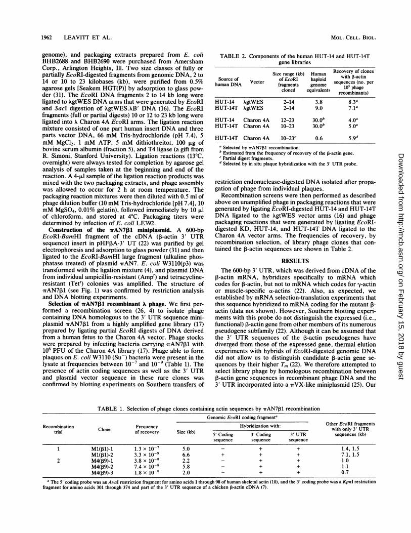
TABLE 2. Components of the human HUT-14 and HUT-14Tgene libraries
Size range (kb) Human Recovery of clonesSource of of EcoRI haploid with P-actin
human DNA Vector fragments genome sequences (no. percloned equivalents 10i phage
recombinants)
HUT-14 XgtWES 2-14 3.8 8.3aHUT-14T XgtWES 2-14 9.0 7. la
HUT-14 Charon 4A 12-23 30 0b 4.0aHUT-14T Charon 4A 10-23 30.0b 5.0a
HUT-14T Charon 4A 10-23C 0.6 5.9da Selected by rAN7l31 recombination.b Estimated from the frequency of recovery of the P-actin gene.c Partial digest fragments.d Selected by in situ plaque hybridization with the 3' UTR probe.
restriction endonuclease-digested DNA isolated after propa-gation of phage from individual plaques.Recombination screens were then performed as described
above on unamplified phage in packaging reactions that weregenerated by ligating EcoRI-digested HUT-14 and HUT-14TDNA ligated to the XgtWES vector arms (16) and phagepackaging reactions that were generated by ligating EcoRI-digested KD, HUT-14, and HUT-14T DNA ligated to theCharon 4A vector arms. The frequencies of recovery, byrecombination selection, of library phage clones that con-tained the ,-actin sequences are shown in Table 2.
RESULTSThe 600-bp 3' UTR, which was derived from cDNA of the
P-actin mRNA, hybridizes specifically to mRNA whichcodes for ,-actin, but not to mRNA which codes for -y-actinor muscle-specific a-actins (22). Also, as expected, weestablished by mRNA selection-translation experiments thatthis sequence hybridized to mRNA coding for the mutant 1-
actin (data not shown). However, Southern blotting experi-ments with this probe do not distinguish the expressed (i.e.,functional) ,B-actin gene from other members of its numerouspseudogene subfamily (22). Although it can be assumed thatthe 3' UTR sequences of the 1-actin pseudogenes havediverged from those of the expressed gene, thermal elutionexperiments with hybrids of EcoRI-digested genomic DNAdid not allow us to distinguish candidate 1-actin gene se-quences by their higher Tm (22). We therefore attempted toselect library phage by homologous recombination between13-actin gene sequences in recombinant phage DNA and the3' UTR incorporated into a TVX-like miniplasmid (25). Our
TABLE 1. Selection of phage clones containing actin sequences by 'TrAN7P1 recombination
Genomic EcoRl coding fragmenta
Recombination Frequency Hybridization with: Other EcoRI fragmentstrial Clone of_recovery_Sizekb_. with only 3' UTRtrial Of recovery Size (kb) 5' Coding 3' Coding 3' UTR sequences (kb)
sequence sequence sequence
1 Ml(pi)-1 1.3 x 10⁻⁷ 5.0 - + + 1.4, 1.5Ml(pl)-2 3.3 x 10⁻⁹ 6.6 + + + 7.1, 1.5
2 M4(P9)-1 3.8 x 10⁻⁸ 2.2 - + + 1.0M4(39)-2 7.4 x 10⁻⁸ 5.8 - + + 1.1M4(09)-3 1.8 x 10⁻⁸ 2.0 - + + 0.7
a The 5' coding probe was an AvaI restriction fragment for amino acids 1 through 98 of human skeletal actin (10), and the 3' coding probe was a KpnI restrictionfragment for amino acids 301 through 374 and part of the 3' UTR sequence of a chicken 0-actin cDNA (7).
MOL. CELL. BIOL.
on February 15, 2018 by guest
http://mcb.asm
.org/D
ownloaded from

HUMAN 1-ACTIN GENE 1%3
expectation was that the P-actin gene would be selectedpreferentially from 3-actin pseudogenes by the greater simi-larity of the 3' UTR sequence.
Isolation of the 1-actin gene from a human fetal DNAlibrary. The structure of the 1,500-bp miniplasmid fAN701,which was constructed by inserting an EcoRI-BamHI frag-ment containing the 600-bp 3' UTR of human P-actin cDNAinto sites within the polylinker of rrAN7, is shown in Fig. 1.The 3' UTR sequence is oriented so that the SalI site in theminiplasmid is placed near the junction of the 3' terminus ofthe 3' UTR and the miniplasmid. Recombinant insertion ofthe miniplasmid into a X phage carrying the human 1-actingene should thereby insert a reference Sall site at the end ofthat 3' UTR which is adjacent to the coding portion of theclone. The only Sall site within the coding region of thehuman ,B-actin cDNA is located at codon 10 (22). Therefore,after selection of a recombinant clone, determination of thesize of the SalI fragment bearing the actin coding sequenceshould help distinguish candidate genes with introns frompseudogenes without introns.Our first attempt to clone the ,-actin gene made use of a X
phage library of human fetal DNA (12). The phage in thislibrary had been amplified on numerous occasions. We werealready aware that EcoRI restriction fragments carrying ,3-actin 3' UTR sequences were present in the library in greatlydifferent amounts from their equimolar representation in thegenome (unpublished data). The recovery frequencies ofphage isolated after aTAN7p1 recombination would thereforebe the product of their skewed concentrations in the libraryand their independent recombination frequencies. We never-theless proceeded to isolate P-actin-carrying clones selectedby fTAN711 recombination.Two separate recombination trials were performed in
which 106 PFU of library phage were amplified by infectionof either the recombination-proficient E. coli WoP3('rAN7,1) or the nonproficient E. coli WoP3 (7rAN7). Lyticprogeny phage from each amplification were then used toinfect a host strain (WoP3SupO) in which Charon 4A phagedo not propagate; consequently, no lytic plaques wereproduced with the phage derived from the WoP3 (rrAN7)infection. Lytic phage derived from infection of strain WoP3(TrAN7,B1) produced plaques at a consistent frequency,between 10-7 and 10-9 of the true titer [in the WoP3(nAN7p1) and LE392 hosts]. All phage that were isolated
ECoR /
AvaI-
FIG. 1. rrAN7,1 plasmid. Construction of this chimeric plasmidis described in the text.
contained actin coding sequences and had undergone recom-bination with the 7rAN7P1 plasmid.
Five distinct phage clones were selected (Table 1). Eachcontained ,-actin sequences, and they are described in termsof the sizes of EcoRI fragments that contained either codingor noncoding 3' UTR sequences. In the first recombinationtrial, 51 plaques were isolated and the properties of thehuman DNA insert were examined with three actin-specificprobes. Of the 51 plaques, 50 were identical and weredesignated Ml(p1)-1. In addition to three EcoRI fragmentsthat contained actin sequences (5.0, 1.4, and 1.5 kb), oneEcoRI fragment (3.5 kb) which lacked an actin sequence wascommon to all 50 isolates. This first trial consisted of threeindependent infections. Thus, the 50 identical phage werederived from a minimum of three separate recombinationevents but not necessarily from more than three events. Asingle additional plaque [M1(1l)-2] contained a differentphage with a different set of EcoRI fragments: three frag-ments (6.6, 7.1, and 1.5 kb), contained actin sequences, andtwo fragments (2.0 and 1.2 kb) lacked actin sequences. Asecond recombination trial produced three more differentrecombinant clones (Table 1). We interpreted the variablerecovery of different plaque types during independent trialsas a reflection of the skewed nature of the human X library aswell as the degree of sequence similarity between the 7rAN7,-actin insert and the various genomic P-actin sequences.Recombination with the 3' UTR sequence leads to inser-
tion of the entire nTAN7P1 plasmid, beginning at the 3' side ofthe crossover site (25). Thus, two chimeric 3' UTR se-quences that are separated by the 7TAN7 sequence werecreated by the recombination event. Two of the clones,M1(,1)-1 and M1(,1l)-2, had an additional EcoRI fragmentprecisely the size of a unit-length rAN7p1 plasmid (1.5 kb),which was inserted into the phage genome as a tandemrepeat of the rAN7P1 plasmid. This explanation was veri-fied by demonstrating that the same unit-length plasmidfragment containing a 3' UTR sequence could be generatedby digestion with two additional restriction enzymes (SalIand AvaI) whose restriction sites occurred only once withinthe plasmid sequence.
M1(,1)-2 was further distinguished from Ml(ql)-1 and thethree isolates of the second recombination trial in that it wasthe only clone that hybridized to a probe that contained the5' actin coding sequence (codons 1 through 98). BecauseM1(pl)-2 contained both 5' and 3' sequences of ,-actin, itwas likely to contain the entire coding region of the ,B-actingene. A map of key restriction sites (Fig. 2) disclosed thatSall digestion of M1(,1l)-2 generated a 2,500-bp fragmentthat contained most of the coding sequences for ,-actin plusthe 3' UTR sequence. If indeed this fragment contained1,098 bp of coding sequence (codons 10 to 375) and 600 bp ofthe 3' UTR sequence, then the additional 800 bp might beaccounted for by the four predicted intron sequences (20)within the coding region. The nucleotide sequence of thisgene (S.-Y. Ng et al., manuscript in preparation) confirmedthe position of the Sall site at codon 10 and the existence offour intron regions, the sum of whose lengths was 731 bp.Furthermore, the nucleotide sequences of the coding regionsof M1(,1l)-2 were identical to the ,B-actin cDNA sequence.
Isolation of a I-actin gene-specific probe and assessment ofthe frequency of 13-actin genes and pseudogenes. A probederived from intron I of the 1-actin gene (Fig. 2) hybridizesto only two discrete bands (ca. 12 to 14 and 6.4 kb) in blottingexperiments with EcoRI digests of human genomic DNA(Ng et al., in preparation). By contrast, the 3' UTR probehybridizes to more than 20 bands (22). Restriction mapping
VOL. 4, 1984
on February 15, 2018 by guest
http://mcb.asm
.org/D
ownloaded from

1964 LEAVITT ET AL.
showed that the A clone M1(1l)-2 contained the 1-actinsequence corresponding to a 12.2-kb genomic fragmentwhich was divided into two EcoRI fragments of 6.6 and 7.1kb by xrAN7 recombination.
Detection of the ,B-actin mutation at codon 244 (26) in a
genomic clone would verify the identity of the functional(expressed) 1-actin gene in human fibroblasts. Therefore, weconstructed libraries of EcoRI fragments derived from KD,HUT-14, and HUT-14T cell DNA. The construction of theselibraries is described above and shown in Table 2. Thecritical features of these libraries are as follows. (i) SmallerEcoRI restriction endonuclease digest fragments (2 to 12 kb)were found exclusively when the XgtWES vector was used,and larger EcoRI restriction digest fragments were foundalmost exclusively when the Charon 4A vector was used.This method of dividing the human genome proved advanta-geous in that scrambling of larger fragments with smallerfragments was minimized and most clones derived fromeither library component contained only a single humanEcoRI fragment. This feature helped to ensure that after7rAN7P1 recombination the resulting recombinant phagewould not become less viable or resist packaging. (ii) A totalof 104 recombinant phage generated by the packaging reac-
tion were amplified as a unit within sup+ bacteria on a singleagar plate, but the progeny phage of this amplification stepwere never combined with other amplification aliquots; thus,isolated clones of genes and pseudogenes could be consid-ered the products of independent cloning events when
isolated from different amplification aliquots. Accordingly,we could test for the presence of the ,B-actin gene homologsin each aliquot by nAN7,B1 selection. If the ,-actin genewere present, we could use the intron probe to recover, fromthe original aliquot, genomic clones without rAN7 insertion.(iii) The frequency of gene and pseudogene isolation fromsuch libraries is an assessment of the complexity of 1-actin-related sequences within the human genome. Conversely,the ratio serves as a criterion for the randomness andcompleteness of the library itself.
,-Actin clones from human 10- to 23-kb EcoRI fragments.When size-fractionated EcoRI fragments ranging from 10 or12 to 23 kb from HUT-14 and HUT-14T cell DNA were usedto prepare recombinant phage, the frequency of recovery of1-actin clones per total recombinant phage screened rangedfrom 4 x 10⁻⁵ to 5 x 10⁻⁵ for the KD, HUT-14, and HUT-14T libraries (Table 2). Amplification aliquots (104 packagingevents) were first screend by 7rAN7f31 recombinant selectionto determine which library aliquots contained ,B-actin genesor pseudogenes. Library aliquots that contained ,B-actin 3'UTR sequences were rescreened by conventional in situplaque hybridization to select clones that hybridized to the 3'UTR probe. This probe was used to collect a spectrum of thepseudogene family in addition to multiple isolates of theputative functional gene. After purification, each 1-actinclone was recombined with 7rAN7131. Both the nonrecombi-nant and 7rAN7131 recombinant forms of each clone were
then examined by EcoRI and Sall restriction endonuclease
TABLE 3. Clones containing 3-actin sequences isolated from libraries of 10- to 23-kb genomic EcoRI fragments
Genomic EcoRI Distance (kb) from 5' 2.5-kb Sall HybridizationLibrary source' Clone fragment size EcoRI site to 3' end of fragment to intron I
(kb) the 3' UTR (Sall site)b present' probe
HUT-14T, fully 14T13-15 13.8 6.6 + +digested, Charon 4A 14T,B-16 13.8 6.6 + +
14T1-17 13.8 6.6 + +14T,B-18 11.0 4.3 - +14T13-19 13.8 6.6 + +14Tf-20 13.8 6.6 + +14T,B-21 13.8 6.6 + +14T,B-22 14.2 8.5 - +14T1-23 13.8 6.6 + +14T,B-24 13.8 6.6 + +
HUT-14, fully digested 14,B-25 18.5 14.6Charon 4A 14,B-26 14.4 6.0 + +
1413-27 13.8 6.6 + +141-28 NDd 14.1 - -14P-29 13.8 6.6 + +1413-30 13.8 6.6 + +14a-31 13.8 6.6 + +14a-32 13.8 6.6 + +
KD, fully KD1-1 13.8 6.6 + +digested, Charon 4A
HUT-14T, partially 14T,B-1 5.3 4.3 - -
digested, Charon 4A 14T13-2 10.5 7.9 - -
14T,B-4 4.3 3.8 - -14T1-5 8.1 ND - -14T13-12 2.9 ND - -
a Cell line, degree of EcoRI fragment digestion, and vector.b Plasmid 1TAN701 recombinant phage clones were constructed with r,laque-pure clones selected by in situ plaque hybridization with the 3' UTR probe. Before
recombination, each clone contained a single human EcoRI fragment, whereas after recombination two EcoRI fragments were generated by insertion of rrAN7j13into the genomic EcoRI fragment; the sizes of the two EcoRi fragments generated and identification of the fragment containing the coding and intron I sequencesshowed the position of the ,-actin sequence within the genomic EcoRI fragment.cThe 2.5-kb SalI fragment is generated by irAN7l31 insertion during recombination and is characteristic of the P-actin gene (Fig. 1).d ND, Not determined.
MOL. CELL. BIOL.
on February 15, 2018 by guest
http://mcb.asm
.org/D
ownloaded from

HUMAN 1-ACTIN GENE 1965
digestion, and the resulting DNA fragments were hybridizedwith the intron I, 3' UTR, and coding probes to fully assesstheir identity and relatedness (Table 3). In total, 8 of 10isolates from HUT-14T DNA and 5 of 8 isolates from HUT-14 DNA contained a 1-actin gene similar to that found inM1(P1l)-2, because each of these separate clones hybridizedstrongly to the intron probe. In addition, their rAN7P1recombinants contained the characteristic 2.5-kb Sall frag-ment that carries the ,B-actin coding, intron, and 3' UTRsequences. The size of the uninterrupted genomic fragmentfor these clones was about 13.8 kb. Three other cloneshybridized only weakly to the intron probe. The TrAN7P1recombinants of two of these, 14T1-18 and 14T1-22, hadunique EcoRI and Sall digestion products as well as anuninterrupted EcoRI fragment of a different size. It thereforeseems likely that these two clones were unrelated and weredissimilar to the 13 common clones. Another clone, 141-26,had a 14.4-kb EcoRI fragment and gave only weak hybridiza-tion to the intron probe, but did yield a 2.5-kb Sall fragmentafter wrAN71l recombination.The results with libraries constructed with genomic EcoRI
DNA fragments of 10 to 23 kb are in contrast to the resultsobtained when we constructed libraries of 2- to 14-kbgenomic DNA EcoRI fragments cloned in the XgtWESvector (Table 2). This library yielded no 1-actin clonescarrying either a 12.2- or a 13.8-kb EcoRI restriction frag-ment as well as the characteristic 2.5-kb Sall fragment of the,B-actin--TnAN7 recombinants. Instead, we isolated 61 clonescarrying 1-actin sequences at frequences of 7 x 10⁻⁵ to 8 x10 (Table 2). All of these clones probably carry pseudo-genes, as determined by their lack of hybridization to the 1-
actin intron I probe and comparison of detailed restrictionmaps (data not shown). In addition, we performed a limitedscreening of another library, constructed from partiallyEcoRI-digested genomic DNA fragments (Table 2). Theclones derived from this library all contained pseudogenessince they lacked key restriction sites indicative of theexpressed ,B-actin gene and failed to hybridize with theintron I probe (Table 3).EcoRI polymorphism at the 5' flank of the Il-actin gene. The
EcoRI fragment carrying the ,B-actin gene, including itsintrons, in the wrAN71l KD, HUT-14, and HUT-14T recom-binants (Table 3) was 8.2 kb long. By contrast, the EcoRI
* coding region
3 5' UTR
ES 3' UTR
7rAN 7
introns and flanking region
0C.)
L0
l(2kb 3kb 4kb
fragment bearing the 1-actin gene in M1(,11)-2, derived froma human fetal DNA library, appeared to be only 6.6 kb long(Fig. 2). Both clone types contained a common 7.1-kb EcoRIfragment in addition to a fragment of either 8.2 or 6.6 kbwhich hybridized to the intron I probe. The 7.1-kb fragmentappeared to hybridize only with the 3' UTR probe. Weconclude that this 7.1-kb fragment was generated by recom-bination of ITAN7131 with either a 12.2- or 13.8-kb genomicEcoRI DNA fragment.We attempted to determine whether this difference in
fragment length (8.2 versus 6.6 kb) was due to a simple 5'EcoRI restriction site polymorphism neighboring ortholo-gous genes or whether the different fragment sizes represent-ed paralogous alleles. Accordingly, the EcoRI digestionfragments of three of the 7rAN7P1 recombinant 1-actinclones from HUT-14 DNA [141-27(11), 141-29(11), and 1413-30(,13)] and the fetal gene clone M1(1l)-2 were subclonedinto plasmid pBR322. We digested these subclones withEcoRI and separated the resulting fragments by agarose gelelectrophoresis. Each blot was hybridized first to the 1-actinintron I probe and then to the ,B-actin 3' UTR probe (Fig. 3).The intron probe hybridized to the 8.2-kb EcoRI fragment of141-27(p13), 141-29(p13), and 141-30(13) and to the 6.6-kbEcoRI fragment of M1(,11)-2. By contrast, the 3' UTR probehybridized to the 7.1-kb EcoRI DNA fragment common toall four clones as well as to the 8.2- or 6.6-kb EcoRIfragments containing the intron I sequences. This resultindicates that the genes isolated from HUT-14 and HUT-14Tcell DNA differ from the fetus-derived gene in M1(1l)-2 inthe location of an EcoRI site in the genomic DNA flankingthe 5'-region of the actin gene. All 13 independent wAN7p1lrecombinant clones derived from both HUT libraries andone additional clone derived from the KD cell DNA library(Table 3) had an identical arrangement with respect to thepositions of flanking EcoRI sites. Furthermore, these cloneswere derived from both alleles (see below). The uninterrupt-ed EcoRI fragment in the corresponding non-iiAN7 recombi-nant clones was 13.8 kb long (Table 3). We thereforeconclude that the 1-actin gene resides on a 13.8-kb genomicEcoRI fragment. The different length of the restrictionfragment in the genomic DNA of clone M1(,11)-2 most likelyarises from an EcoRI polymorphism in human genomes,although, the possibility that it is an artifact incurred during
co Im
-~~~~~~.1a.a. 244
l I .-.-100bp,'
,/
5kb 6kbFIG. 2. Restriction endonuclease map and structure of the human ,-actin gene--TAN7,31 recombinant clone M1(31)-2. Inset, 366-bp
BstEII fragment.
'/ I E1 E FN,, vI I f I .........
xI M
2~~~~~~~~~~~~~~~~~~~~~~~~1II I ~ ~~~~II I
VOL. 4, 1984
on February 15, 2018 by guest
http://mcb.asm
.org/D
ownloaded from

1966 LEAVITT ET AL.
NTRONPROBE
A B C D E
A B C D E
3'UTRPROBE w S7.1
FIG. 3. Southern blot of EcoRI-digested pBR322 subclones ofEcoRI fragments derived from separate bacteriophage clones ofhuman 1-actin genes. Lanes: A, 14P-27(ol), 8.2-kb fragment; B,14P-29(p11), 8.2-kb fragment; C, 14P-30(p11), 8.2-kb fragment; D,M1(1l)-2, 6.6-kb fragment; E, M1(,11)-2, 7.1-kb fragment. A singlenitrocellulose blot from an 0.8% agarose gel was first hybridized tothe 32P-labeled intron I probe (top) and then rehybridized to the 32P-labeled 3' UTR probe (bottom).
A phage clone construction or propagation cannot be exclud-ed.
Discrimination between mutant and wild-type allelic clonesof the j3-actin gene by a HaeIII cleavage test for the mutationat codon 244. The sequences derived from the gene inM1(1l)-2 (Ng et al., in preparation) and from a cDNA clone(23) show that codons 243, 244, and 245 (Asp-Gly-Gln) wereencoded by GAC GGC CAA. Since the first 1-actin mutationin HUT-14 cells resulted in an exchange of the glycine(codon 244) for an aspartic acid residue, the predictedsequence for codon 244 after the mutation is GAC. Theunmutated sequence GGCC (codons 244 and 245) is arestriction site for the endonuclease HaeIII, a site whichshould be absent in mutant copies of the gene from HUT-14and HUT-14T cells. BstEII sites flank the mutation site,cleaving between codons 158 and 159 and at a site 38 bp intointron IV, respectively (Fig. 2). This BstEII fragment (366bp) was isolated from the DNA of three plasmid subclones ofthe HUT-14 rrAN711-derived 1-actin genes [the 8.2-kbEcoRI fragment from 1413-27(11), 1413-29(11), and 1413-30(,11)] and three additional plasmid subclones from non-irAN7-derived HUT-14T 1-actin genes (the 13.8-kb EcoRIfragment from 14T,B-17, 14T1-21, and 14T,B-24). Within thisBstEII fragment, there were HaeIII sites at codons 182, 204,and 228 and at codon 244, the site of the mutation (Fig. 2).Digestion of the BstEII fragment from the wild-type ,B-actingene with HaeIII generated five restriction fragments, of 71,.65, 72, 52, and 106 bp, whereas the mutated gene missing theHaeIII site at codon 244 should be cleaved into only fourrestriction fragments, of 71, 65, 72, and 158 bp. The 158-bp
HaeIII fragment diagnostic of the HUT-14 mutation atcodon 244 was predicted from the sum of the 52- and 106-bpsegments. Figure 4 shows the HaeIII digestion products forthis 366-bp BstEII fragment from the six clones tested. Fourof six clones from HUT-14 [clones 14,B-27(p1) and 143-29(,11)] and HUT-14T (clones 14T1-21 and 14T1-24) had the158-bp HaeIII-BstEII fragment indicative of copies of thegene mutated at codon 244. The two remaining clones, 14,B-30(11) and 14T1-17, showed the wild-type digestion patternindicative of the normal, unmutated gene. The 1-actin genescloned from the HUT-14 and HUT-14T cell DNA librariestherefore represented both the wild-type and mutant alleles.Furthermore, the presence of the predicted mutation in oneof the alleles formally proves that these genes, and not theother EcoRI 1-actin coding fragments, are the expressed 1-actin genes in these human fibroblast strains. Sequenceanalysis of the genes carrying the mutations confirmed thatthese genes contain the predicted point mutation and there-fore are expressed (C.-S. Lin et al., manuscript in prepara-tion).
Recombination frequencies within 3' UTR sequences ofclones of (8-actin genes and pseudogenes. Our original premisewas that TAN7A31 would preferentially recombine with thereal ,B-actin gene, since its sequence should have the highestdegree of base matching compared with that of pseudogenes.Accordingly, we measured the recombination frequencybetween rAN7A13 and 13 of the nonallelic 1-actin clonesisolated by in situ plaque hybridization (Table 4). A true ,B-actin gene clone, 14T1-16 (Table 3), gave the highest recom-bination frequency, as expected. Clones HRL25 (22) and14T,B-13 (HRL25-like), both of which show weak intron-specific hybridization and seem to correspond to the 6.4-kbEcoRI fragment detected in genomic blots (Ng et al., manu-script in preparation), were lower in recombination frequen-cy by 1 order of magnitude, indicating sequence divergencewithin the 3' UTR.The 10 remaining clones listed in Table 4 showed further
184123104 -80-64 --51
- 158_*- 106-.*- - 72 71* 65-- 52
FIG. 4. A 10% polyacrylamide gel of ethidium bromide-stainedHaeIII restriction digest fragments of a 366-bp BstEII fragmentspanning the ,B-actin codon 244. Lanes: A, HaeIII pBR322 DNAdigest markers; B, M1(p11)-2; C, 14P-30; D, 14P-29; E, 14,B-27; F,14T1-24; G, 14T1-21; H, 14T1-17. Sizes (in bp) of marker fragmentsare indicated to the left. The precise sizes of the ,B-actin DNAfragments as determined by DNA sequencing are indicated to theright.
MOL. CELL. BIOL.
on February 15, 2018 by guest
http://mcb.asm
.org/D
ownloaded from

HUMAN 1-ACTIN GENE 1%7
TABLE 4. Frequency of recombination between X clonescontaining 1-actin or pseudogene sequences and 7rAN7l3l
FrequencyClonea of TrAN71
recombinantsb
14Tp-16C ........................................ 3.3 x 1O-314T-5 ........................................ 9.4 x 10⁻⁴HRL25 ......... ................................ 2.4 x 1-4
14TP-13d ........................................ 1.5 x 10-414TP-4........................................ 1.4 x 1-4
HRL51 ......... ................................ 1.0 X 10-414TP-1 ........................................ >7.6 x 10-514T-2 ........................................ 3.6 x 1i-14TP-12 ........................................ >1.6 x 10-HRL24 ........................................ 1.5 x 10-HRL35 ........................................ 7.1 x 10⁻⁶HRL21 ........................................ 6.2 x 10⁻⁶
a Clones 14TP-16, 14TP-1, 14TP-2, 14TP4, 14TP-5, 14T,-12, and 14TP-13were isolated from libraries described in Table 2, and the properties of theseclones are summarized in Table 3; clones HRL21, -24, -25, -35, and -51 aredescribed by Ponte et al. (22).
b Large plaques of each clone were produced by infection of the recombina-tion-proficient strain E. coli W3110 (p3 rrAN7p1); bacteriophage were theneluted from the plaques and titrated on sup' and supo hosts to determine thefrequency of wrAN7P1 recombination.
c -Actin gene.d HRL25-like.
reductions in recombination frequency with nrAN7p1. Theseclones have been analyzed by restriction site mapping (22;unpublished data), by limited sequence analysis (J. Engel,Ph.D. thesis, Stanford University, Stanford, Calif., 1982),and by blotting experiments with probes of intron se-quences, 5' and 3' coding sequences, and 3' UTR sequences
(6, 7, 22; J. Engel, Ph.D. thesis; unpublished data). From theresults of these studies, all 10 of these clones appear tocontain P-actin pseudogenes. The 7rAN7 recombination sys-tem therefore seems to be a way to preferentially select truegenes from among divergent pseudogene members of amultigene family.
DISCUSSIONThe nTVX-ITAN7 recombination method for selecting
recombinant library clones allowed first-trial selection fromthe ,-actin multigene family of a clone that eventuallyproved to contain the expressed gene. This gene was clonedfrom a highly amplified human fetal gene library previouslyscreened at random for the P-actin gene by in situ plaquehybridization without success (7, 22; J. Engel, Ph.D. thesis).In retrospect, the reason for the previous failures (18, 19, 22)must be related to the skewed representation of the P-actingene in the human fetal library DNA relative to that of othermembers of the ,-actin gene and pseudogene families. TheP-actin gene recombines between threefold and ca. 2,000-fold more frequently than any other member of the ,B-actinfamily of sequences (Table 4).We identified recombinant phage M1(pl)-2 as containing
the likely ,-actin gene from among more than 100 otherindependent recombinant clones that contained ,-actin se-
quences. We determined that it carried both a SalI site atcodon 10, as predicted from the cDNA sequence (22), andenough extra bp to accommodate the several predictedintrons. As a consequence of the homologous recombinationevent, a reference Sall restriction site in the plasmid poly-linker was inserted into the recombinant phage genomeadjacent to the 3' UTR of the P-actin gene. The sizes ofliberated Sall fragments of clones indicated the coding
potential of the clone as well as the cumulative size of theintrons between codon 10 and the 3' UTR sequence. Sincemost of the 1-actin clones lacked additional sequencesbetween the SaiI sites and appeared to contain processedpseudogenes (18, 19), and since more than half lacked a SalIsite within the entire human DNA insert, many clones wereeasily disqualified as candidates.The arrangement of EcoRI fragments in nrAN7 recombi-
nant clones was easily deduced by identifying the fragmentsthat contained a coding sequence adjacent to the 3' UTRsequence and those that contained only the 3' UTR se-quence. This information allowed us to predict the positionof the gene sequence within a map of the genomic EcoRIfragment uninterrupted by the plasmid insertion. Once thismap was constructed, the likelihood that the clone containedthe complete gene could be evaluated on the basis ofreasonable assumptions about the size of the introns.Clones containing the correct ,-actin gene in their rAN7
recombinant form had a characteristic 2.5-kb Sall fragmentwhich accounted for 365 codons, a 600-bp 3' UTR, and ca.800 extra bp, the cumulative intron size predicted from thesizes of introns of the ,B-actin gene from rats (20). Thisfinding led us to begin sequencing M1(p1)-2 as the primecandidate over many other clones that had been isolated.Determination of the nucleotide sequence of the ,-actin genein clone M1(pl)-2 (Ng et al., in preparation) has shown thatthis gene contains a coding sequence identical to the se-quence of a full-length ,-actin cDNA isolated from a cDNAlibrary of human fibroblast mRNA (10, 23). We concludethat M1(,1)-2 contains the human ,B-actin gene expressed inhuman fibroblasts.Because of the complexity of the human P-actin gene-
pseudogene family, however, there remained a formal possi-bility that the gene cloned from the fetal gene library was notthe gene that had undergone mutation in HUT-14 cells andtheir progeny. Since a mutation in codon 244 is expressed inone of two functional alleles in the human fibroblast cell lineHUT-14 (26), the presence of this mutation in approximatelyhalf the HUT-14 DNA segments analogous to M1(pl)-2would confirm the identity of the fetus-derived ,-actin geneas the gene expressed in human fibroblasts. Because thepredicted size of the genomic EcoRI fragment containing theP-actin gene was 12.2 kb, based on the fetus-derived gene,we initally assumed that this clone could be isolated by usingthe XgtWES vector that can produce viable clones withinserts of up to 14 kb (16). Repeated screening of XgtWESlibraries (derived from both HUT strains) by in situ plaquehybridization with the intron-I ,-actin-specific probe orselection of ffAN7p1 recombinants failed to produce thegene analogous to the fetus-derived 3-actin gene (data notshown). Instead, 61 pseudogenes were isolated from theXgtWES libraries, including two distinct copies of a pseudo-gene on a 6.4-kb EcoRI genomic fragment that gave weakhybridization to the intron-specific probe. On the otherhand, Charon 4A libraries containing EcoRI DNA fragmentsbetween 10 and 23 kb long were found to be enriched for a13.8-kb EcoRI fragment which showed strong hybridizationreactivity with the intron-specific probe. Clones bearing thisfragment had identical restriction maps and urAN7 recombi-nant structure with the fetus-derived gene with but oneexception: the 5' genomic EcoRI site flanking the HUT-derived genes was ca. 1.6 kb more distal from the ,-actingene than was the EcoRI site in the fetus-derived gene. Thisrestriction fragment length difference is probably due to anEcoRI polymorphism in the human population or, less likely,may have been introduced as an artifact during the construc-
VOL. 4, 1984
on February 15, 2018 by guest
http://mcb.asm
.org/D
ownloaded from

1968 LEAVITT ET AL.
tion or propagation of the fetal DNA library. Since identicalEcoRI fragments were isolated from the three related librar-ies (KD, HUT-14, and HUT-14T) and reproduced in 14discrete recombinants derived from separate alleles, it seemsreasonable to conclude that the 13.8-kb EcoRI fragment is anaturally existing form. This conclusion is further supportedby our isolation of two copies of the 1-actin gene from anunrelated cell line that also have a 13.8-kb EcoRI fragment(unpublished data). The codon 244 mutation was found insome, but not all, of the clones from the HUT-14 and HUT-14T cell lines, and these clones also had a 2.5-kb Sallfragment (after wTAN7p1l recombination). Since these sameclones share equal specificity with the fetus-derived gene forthe intron-specific probe, we conclude that these genes fromthe HUT cell lines are alleles and are the homologs of thegene in M1(p1l)-2, which was cloned from the human fetalDNA library.The fetus-derived clone HRL25 and the two clones from
the HUT-14 and HUT-14T libraries (14,B-13 and 14T,B-14)appear to be independent isolates of the same ,B-actinpseudogene because ITAN7 recombinant derivatives pro-duced identical restriction maps. These clones also showuniformly weak hybridization with the intron-specific probe.The f-actin sequence in this clone type, which lacks a Sailsite at codon 10 and therefore contains a pseudogene, mayhave diverged from that of the expressed ,B-actin gene after agene duplication event. It may therefore be representative ofa class of pseudogenes with introns which are distinct fromthe majority of ,B-actin pseudogenes that appear to be of theprocessed type.The human ,B- and -y-actins of the cytoskeleton differ from
each other by only 4 amino acids (26, 28) and by only 17 or 18amino acids from the abundant actin isoform of the lowereucaryotic unicellular amoeba Physarum polycephalum (28,29). Such strong selective pressure is probably related tocritical functions of the actins as major components of thecytoskeleton. We have now isolated cloned copies of thewild-type and two mutant ,B-actin genes, one with a codon244 mutation and the other with a currently undefinedsecond mutation (13). Additional mutant ,B-actin genes canbe constructed by site-directed mutagenesis. Since normalactin genes are expressed after gene transfer (9), we shouldnow be able to examine the relationship between mutationaldefects in 13-actin, cytoskeletal dysfunction, and alteredcellular phenotype.
ACKNOWLEDGMENTSWe thank Tom Maniatis for providing bacterial strains and
technical advice and Henry Huang for providing the nrAN7 plasmidand their unpublished data.
This work was supported by an American Cancer Society grant toJ.L. and by Public Health Service grants from the National Insti-tutes of Health and the Veterans Administration to L.K.
LITERATURE CITED
1. Blattner, F., B. G. Williams, A. E. Blechl, K. Denniston-Thompson, H. E. Faber, L. A. Furlong, D. J. Grunwald, D. 0.Kiefer, D. D. Moore, J. W. Schumm, E. L. Sheldon, and 0.Smithies. 1977. Charon phages: safer derivatives of bacterio-phage X for DNA cloning. Science 196:161-169.
2. Bravo, R., S. J. Fey, J. V. Small, P. M. Larsen, and J. E. Celis.1981. Coexistence of three major isoactins in a single sarcoma180 cell. Cell 25:195-202.
3. Childs, G., R. Maxson, and L. H. Kedes. 1979. Histone geneexpression during sea urchin embryogenesis: isolation andcharacterization of early and late messenger RNAs of Strongy-locentrotus purpuratus by gene specific hybridization and tem-plate activity. Dev. Biol. 73:153-173.
4. DiMaio, D., V. Corbin, E. Sibley, and T. Maniatis. 1984. High-level expression of a cloned HLA heavy chain gene introducedinto mouse cells on a bovine papillomavirus vector. Mol. Cell.Biol. 4:340-350.
5. Egelnan, E. H., N. Francis, and D. DeRosier. 1982. F-actin is ahelix with a random variable twist. Nature (London) 298:131-135.
6. Engel, J., P. Gunning, and L. Kedes. 1982. Human cytoplasmicactin proteins are encoded by a multigene family. Mol. Cell.Biol. 2:674-684.
7. Engel, J. N., P. W. Gunning, and L. H. Kedes. 1981. Isolationand characterization of human actin genes. Proc. Natl. Acad.Sci. U.S.A. 78:4674-4678.
8. Grunstein, M., and D. S. Hogness. 1975. Colony hybridization: amethod for isolation of cloned DNAs that contain a specificgene. Proc. Natl. Acad. Sci. U.S.A. 72:3961-3965.
9. Gunning, P., P. Ponte, L. Kedes, R. Hickey, and A. Skoultchi.1984. Expression of human cardiac actin in mouse L-cells: asarcometric actin associates with the Triton-X-100 non-musclecytoskeleton. Cell 36:709-715.
10. Gunning, P., P. Ponte, H. Okayama, J. Engel, H. Blau, and L.Kedes. 1983. Isolation and characterization of full-length cDNAclones for human a-, P-, and y-actin mRNAs: skeletal but notcytoplasmic actins have an amino-terminal cysteine that issubsequently removed. Mol. Cell. Biol. 3:787-795.
11. Kakunaga, T. 1978. Neoplastic transformation of human diploidfibroblast cells by chemical carcinogens. Proc. Natl. Acad. Sci.U.S.A. 75:1334-1338.
12. Lawn, R. M., E. F. Fritsch, R. C. Parker, G. Blake, and T.Maniatis. 1978. The isolation and characterization of linked 8and 1-globin genes from a cloned library of human DNA. Cell15:1157-1174.
13. Leavitt, J., G. Bushar, T. Kakunaga, H. Hamada, T. Hirakawa,D. Goldman, and C. Merril. 1982. Variations in mutant ,B-actinexpression accompanying incremental increases in human fibro-blast tumorigenicity. Cell 28:259-268.
14. Leavitt, J., D. Goldman, C. Merril, and T. Kakunaga. 1982.Changes in gene expression accompanying chemically-inducedmalignant transformation of human fibroblasts. Carcinogenesis3:61-70.
15. Leavitt, J., and T. Kakunaga. 1980. Expression of a variant formof actin and additional polypeptide changes following chemical-induced in vitro neoplastic transformation of human fibroblasts.J. Biol. Chem. 255:1650-1661.
16. Leder, P., D. Tiemeier, and L. Enquist. 1977. EK2 derivatives ofbacteriophage A useful in the cloning of DNA from higherorganisms: the AgtWES system. Science 196:175-178.
17. Maniatis, T., R. Hardison, E. Lacy, J. Lauer, C. O'Connell, D.Quan, G. Sim, and A. Efstratiadis. 1978. The isolation ofstructural genes from libraries of eukaryotic DNA. Cell 15:687-701.
18. Moos, M., and D. Gallwitz. 1982. Structure of a human ,B-actinrelated pseudogene which lacks intervening sequences. NucleicAcids Res. 10:7843-7849.
19. Moos, M., and D. Gallwitz. 1983. Structure of two human -actin related processed genes one of which is located next to asimple repetitive sequence. Eu. Mol. Biol. Org. J. 2:757-761.
20. Nudel, U., R. Zakut, M. Shani, S. Neuman, Z. Levy, and D.Yaffe. 1983. The nucleotide sequence of the rat cytoplasmic -actin gene. Nucleic Acids Res. 11:1759-1771.
21. Pollack, R., M. Osborn, and K. Weber. 1975. Patterns oforganization of actin and myosin in normal and transformednon-muscle cells. Proc. Natl. Acad. Sci. U.S.A. 72:994-998.
22. Ponte, P., P. Gunning, H. Blau, and L. Kedes. 1983. Humanactin genes are single copy for a-skeletal and a-cardiac actin butmulticopy for ,B- and -y-cytoskeletal genes: 3'-untranslated re-gions are isotype specific but are conserved in evolution. Mol.Cell. Biol. 3:1783-1791.
23. Ponte, P. A., S.-Y. Ng, J. Engel, P. Gunning, and L. Kedes. 1984.Evolutionary conservation in the untranslated regions of actinmRNAs: DNA sequence of a human beta-actin cDNA. NucleicAcids Res. 12:1687-1696.
24. Ponte, P. A., M. Siekevitz, R. C. Schwartz, M. L. Gefter, and
MOL. CELL. BIOL.
on February 15, 2018 by guest
http://mcb.asm
.org/D
ownloaded from

HUMAN 3-ACTIN GENE 1969
G. E. Sonenshein. 1981. Transcription of immunoglobulinheavy-chain sequences from the excluded allele. Nature (Lon-don) 291:594-596.
25. Seed, B. 1983. Purification of genomic sequences from bacterio-phage libraries by recombination and selection in vivo. NucleicAcids Res. 11:2427-2446.
26. Vandekerckhove, J., J. Leavitt, T. Kakunaga, and K. Weber.1980. Coexpression of a mutant 13-actin and the two normal I-
and y-cytoplasmic actins in a stably transformed human cellline. Cell 22:893-899.
27. Vandekerckhove, J., and K. Weber. 1978. Actin amino acidsequences: comparisons of actins from calf thymus, bovinebrain, and SV-40-transformed mouse 3T3 cells with rabbit
skeletal muscle actin. Eur. J. Biochem. 90:451-462.28. Vandekerckhove, J., and K. Weber. 1978. Mammalian cytoplas-
mic actins are the products of at least two genes and differ inprimary structure in at least 25 identified positions from skeletalmuscle actins. Proc. Natl. Acad. Sci. U.S.A. 75:1106-1110.
29. Vandekerckhove, J., and K. Weber. 1978. The amino acidsequence of Physarum actin. Nature (London) 276:720-721.
30. Vandekerckhove, J., and K. Weber. 1979. The amino acidsequence of actin from chicken skeletal muscle actin andchicken gizzard smooth muscle actin. FEBS Lett. 102:219-222.
31. Vogelstein, B., and D. Gillespie. 1979. Preparative and analyticalpurification of DNA from agarose. Proc. Natl. Acad. Sci.U.S.A. 76:615-619.
VOL. 4, 1984
on February 15, 2018 by guest
http://mcb.asm
.org/D
ownloaded from