molecular investigation of artificial and natural
TRANSCRIPT

Full Terms & Conditions of access and use can be found athttps://www.tandfonline.com/action/journalInformation?journalCode=tbsd20
Journal of Biomolecular Structure and Dynamics
ISSN: (Print) (Online) Journal homepage: https://www.tandfonline.com/loi/tbsd20
Molecular investigation of artificial and naturalsweeteners as potential anti-inflammatory agents
Eleni Chontzopoulou, Christina D. Papaemmanouil, Maria V.Chatziathanasiadou, Dimitrios Kolokouris, Sofia Kiriakidi, AthinaKonstantinidi, Ioanna Gerogianni, Theodore Tselios, Ioannis K. Kostakis,Evangelia D. Chrysina, Dimitra Hadjipavlou-Litina, Demeter Tzeli, Andreas G.Tzakos & Thomas Mavromoustakos
To cite this article: Eleni Chontzopoulou, Christina D. Papaemmanouil, Maria V.Chatziathanasiadou, Dimitrios Kolokouris, Sofia Kiriakidi, Athina Konstantinidi, Ioanna Gerogianni,Theodore Tselios, Ioannis K. Kostakis, Evangelia D. Chrysina, Dimitra Hadjipavlou-Litina, DemeterTzeli, Andreas G. Tzakos & Thomas Mavromoustakos (2021): Molecular investigation of artificialand natural sweeteners as potential anti-inflammatory agents, Journal of Biomolecular Structureand Dynamics, DOI: 10.1080/07391102.2021.1973565
To link to this article: https://doi.org/10.1080/07391102.2021.1973565
View supplementary material
Published online: 09 Sep 2021.
Submit your article to this journal
View related articles
View Crossmark data

Molecular investigation of artificial and natural sweeteners as potentialanti-inflammatory agents
Eleni Chontzopouloua, Christina D. Papaemmanouilb, Maria V. Chatziathanasiadoub, Dimitrios Kolokourisc, SofiaKiriakidia, Athina Konstantinidic, Ioanna Gerogiannid,e, Theodore Tseliosf, Ioannis K. Kostakisc, Evangelia D.Chrysinad,e, Dimitra Hadjipavlou-Litinaa, Demeter Tzelia, Andreas G. Tzakosb,g and Thomas Mavromoustakosa
aDepartment of Chemistry, National and Kapodistrian University of Athens, Athens, Greece; bDepartment of Chemistry, Section of OrganicChemistry and Biochemistry, University of Ioannina, Ioannina, Greece; cDepartment of Pharmacy, National and Kapodistrian, University ofAthens, Athens, Greece; dInstitute of Biology, Medicinal Chemistry and Biotechnology, Department of Pharmaceutical Chemistry, School ofPharmacy, Faculty of Health Sciences, National Hellenic Research Foundation, Athens, Greece; eDepartment of Pharmacy, Aristotle Universityof Thessaloniki, Thessaloniki, Greece; fDepartment of Chemistry, University of Patras, Rion, Greece; gInstitute of Materials Science andComputing, University Research Center of Ioannina (URCI), Ioannina, Greece
Communicated by Ramaswamy H. Sarma
ABSTRACTRepurposing existing drugs, as well as natural and artificial sweeteners for novel therapeutic indica-tions could speed up the drug discovery process since numerous associated risks and costs for drugdevelopment can be surpassed. In this study, natural and artificial sweeteners have been evaluated byin silico and experimental studies for their potency to inhibit lipoxygenase enzyme, an enzyme partici-pating in the inflammation pathway. A variety of different methods pinpointed that aspartame inhibitsthe lipoxygenase isoform 1 (LOX-1). In particular, “LOX-aspartame” complex, that was predicted bydocking studies, was further evaluated by Molecular Dynamics (MD) simulations in order to assess thestability of the complex. The binding energy of the complex has been calculated after MD simulationsusing Molecular Mechanics/Generalized Born Surface Area (MM/GBSA) method. Furthermore, QuantumMechanics/Molecular Mechanics (QM/MM) calculations have been applied for geometry optimizationof the “enzyme-ligand” complex. After having fully characterized the “LOX-aspartame” complex in sil-ico, followed in vitro biological assays confirmed that aspartame inhibits LOX-1 (IC50¼50±3.0 lM) andblocks its biological response. The atomic details of aspartame’s interaction profile with LOX-1 wererevealed through Saturation Transfer Difference (STD) NMR (Nuclear Magnetic Resonance). Finally,aspartame was also tested with Molecular Docking and Molecular Dynamics studies for its potentbinding to a number of different LOX isoforms of many organisms, including human. The in silicomethods indicated that aspartame could serve as a novel starting point for drug design against LOXenzyme.
ARTICLE HISTORYReceived 18 May 2021Accepted 23 August 2021
KEYWORDSAspartame; lipoxygenase;molecular dynamics; STDNMR; in vitro assays
1. Introduction
Inflammation is the biological response of human’s tissuesagainst any type of harmful stimulus. The biochemical path-way of inflammation is characterized by the presence ofthree enzymes responsible for the creation of inflammatoryagents in the cell: human cytosolic phospholipase A2(cPLA2), cyclooxygenase (COX) and lipoxygenase (LOX)(Khanapure & Gordon Letts, 2004). Cycloxygenase superfam-ily has been thoroughly investigated and many COX-1 and
COX-2 inhibitors are commercially available, such as aspirin(Flower, 2003) and NSAIDs (Rouzer & Marnett, 2009), whilethere is only one commercially available inhibitor targetinglipoxygenase enzyme. Despite the fact that LOXs’ crystalstructures have been revealed since decades and the associ-ation of their metabolites with a variety of diseases has beenestablished, there is only one commercially available inhibitortargeting human 5-LOX (zileuton/ABT-671) which is adminis-trated for the treatment of asthma (Carter et al., 1991).Zileuton presents high liver toxicity and short half-life time,
CONTACT Thomas Mavromoustakos [email protected] data for this article can be accessed online at https://doi.org/10.1080/07391102.2021.1973565.
� 2021 Informa UK Limited, trading as Taylor & Francis Group
JOURNAL OF BIOMOLECULAR STRUCTURE AND DYNAMICShttps://doi.org/10.1080/07391102.2021.1973565

hence the need to discover novel LOX inhibitors withimproved pharmacokinetics and pharmacodymamic profile isa significant priority (Valentovic, 2007).
Lipoxygenase metabolizes arachidonic acid (AA) and pro-duces an array of compounds, such as leukotrienes (A4, B4,C4, etc.) and lipoxins (Murphy & Gijon, 2007). In particular,LOX superfamily catalyzes the regio- and stereospecific diox-ygenation of polyunsaturated fatty acids (PUFAs), containinga (1Z,4Z)- penta-1,4-diene system, leading to the productionof hydroperoxy derivatives (Brash, 1999). The implication ofthese metabolites in inflammatory pathways is unambiqu-ously related to the development of numerous differentpathological diseases, such as asthma (Mashima & Okuyama,2015), atherosclerosis (Feinmark & Cornicelli, 1997; Rioux &Castonguay, 1998), rheumatoid arthritis (Gheorghe et al., 2009),psoriasis (Dobrian et al., 2011), brain disorders (Karatas & Cakir-Aktas, 2019; Pratic�o et al., 2004) and cancer (Avis et al., 1996;Catalano & Procopio, 2005; Claria & Romano, 2005; Haeggstr€om& Funk, 2011; Rioux & Castonguay, 1998). Moreover, LOXs havebeen reported to possess tumor promoting properties, whiletheir products, leukotriene LTB4, can lead to the activation ofthe NF-Kb, a master regulator of inflammation processes in thetumor microenvironment (Zhao et al., 2012). Furthermore,human 5-LOX has been found overexpressed in pancreatic neo-plastic lesions and thus targeting lipoxygenases has beenemerged as an alternative method to suppress inflammationand tumor development.
LOXs’ main structure is monomeric and it consists of a C2b-barrel domain (region connecting to the lipid bilayers ofthe cell) and an a-helix catalytic domain containing theactive site of the protein (Boyington et al., 1990). The archi-tecture of active site is characterized by the Fe atom coordi-nated by 3 histidine residues and one C-terminal isoleucine,while the other ligands coordinating Fe cation vary amongLOX isoforms (Choi et al., 2008; Gilbert et al., 2020;Offenbacher et al., 2017; Xu et al., 2012).
Recently, the concept of “drug repurposing” has gainedground since it promotes the discovery of novel uses forapproved drugs as a mean to deliver the quickest possibletransition from bench to bedside (Oprea et al., 2011; Rana etal., 2019; Sharma et al., 2020). Food supplements and naturalproducts have been also explored for their potent pharma-ceutical properties. The discovery of special properties tosuch compounds may accelerate their commercial circulation,as they have already been assessed for their safety and tox-icity. Various artificial high-intensity sweeteners such as ace-sulfame potassium, sucralose, neotame and advantame havebeen approved by the FDA as food or beverage additives(US Food & Drug Administration., n.d). Moreover, naturalsweeteners have been extensively used in the last decadesas sugar substitutes to a variety of foods and beverages,while they are also utilized as additives in pharmaceutics formasking drugs’ taste (Gupta et al., 2017).
Until now, these molecules have been thoroughly studiedin human and animal studies mainly for their impact inhuman health (Lim, 2016) and the results led virtually totheir approval as safe for consumption (Magnuson et al.,2007). Recent studies reported that sweeteners such as
sucralose or stevia affect positively the increase of flavonoidsconcentration in human plasma. It is of great concern thatnumerous flavonoids, phenolic acids, anthocyanidins and alltheir structural derivatives have been implicated in the treat-ment against various diseases, including cancer, and, thus,their presence in human plasma is vital (Agull�o et al., 2021).In addition, it has also been found that natural sweetenerscould act as protecting agents against oxidative stress, a cru-cial leader of DNA damage and cytotoxicity (Li et al., 2021).These widely-used sweeteners could be the pillars for theinvestigation concerning the health benefits that may pro-vide and their implication in molecular mechanisms and sub-sequently their potential use as lead compounds. In thisdirection, acesulfame potassium and saccharin have beenproved to inhibit selectively the carbonic anhydrase IX, anenzyme playing crucial role in the survival of tumor cells(Lomelino et al., 2018; Mahon et al., 2015; Murray et al.,1986). Moreover, sucralose reduces the VEGF-induced vascu-logenesis in human retinal microvascular endothelial cells byactivating the GPCR receptor T1R3 (Lizunkova et al., 2019).
Aspartame (Figure 1b), a methyl ester of the aspartic acid-phenylalanine dipeptide, is an artificial sweetener used as asugar substitute in many edible products. Aspartame binds inthe closed form of Venus Flytrap Module (VFTM) (Maillet et al.,2015) which is the active sweet human receptor TIR2 (hTIR2)and is approximately 200 times sweeter than sucrose (US Food& Drug Administration., n.d). Despite the controversy concern-ing the effects of aspartame in human health that had beenraised in the past, the majority of the scientific studies havesupported that the artificial sweetener is safe to consume whilethe acceptable daily uptake has been set up to 50mg/kg bythe FDA (US Food & Drug Administration., n.d).
In this study, all natural and artificial sweeteners havebeen evaluated in silico for their potency to exhibit anti-inflammatory properties and serve as LOX inhibitors. Themost prominent “LOX-1-sweetener” complex that was gener-ated by docking studies, was further assessed for its stabilitythrough Molecular Dynamics and Quantum Mechanics/Molecular Mechanics calculations. All the in silico and experi-mental studies of natural and artificial sweeteners have beenperformed to soybean LOX-1 isoform, thus this enzyme couldplay the role-model for drug design against all isoforms ofLOX superfamily. After having established compounds’ inhibi-tory activity against LOX-1, in silico studies have been per-fomed to other LOX isoforms in order to evaluate thecompounds’ activity towards different LOXs and investigate ifLOX-1 could be the prototype for anti-LOX drug design pro-cess. Finally, in vitro assays verified the in silico findings andthey indicated that aspartame is able to inhibit LOX-1, whilethe molecular interaction profile of the inhibition wasunveiled with STD NMR experiments.
Currently, other lipoxygenase inhibitors have been alsoevaluated, such as atreleuton (Tardif et al., 2010) and diethyl-carbamazine (Zuo et al., 2004) that have been used for thetreatment of atherosclerosis and filariasis, while licofelone(Cicero & Laghi, 2007) and masoprocol (Luo et al., 1998) actas dual COX/LOX inhibitors for the treatment of osteoarthritisand prostate cancer respectively (West et al., 2004). Even if
2 E. CHONTZOPOULOU ET AL.

these LOX inhibitors are currently under investigation, thereare no commercially available drugs targeting LOX, thus it iscrucial to intensify the studies concerning the discovery ofnovel anti-LOX compounds and understand their linkage tosevere diseases like cancer.
2. Materials and methods
2.1. Induced fit docking
The crystal structures used for the different LOX isoforms forthe in silico studies were PDB IDs: 5T5V (Offenbacher et al.,2017), 3RDE (Xu et al., 2012), 3O8Y (Gilbert et al., 2011),1LOX (Gillmor et al., 1997), 1RRH (Skrzypczak-Jankun et al.,2006), 3D3L, 4G32 (Garreta et al., 2013), 4NRE (Kobe et al.,2014), 5IR4 (Banthiya et al., 2016) and they were retrievedfrom Protein Data Bank (Protein Data Bank, 2021). Proteinpreparation wizard, module available in Schr€odinger Suites(Schr€odinger Maestro, 2018), was used to prepare the crystalstructures for the in silico calculations. Since LOX is a metal-loprotein, it has been taken into account that there are sev-eral computational challenges that need to be addressed. Inorder to account for the quantum effects associated with thepresence of a Fe3þ cation in the protein’s active site, weused the “create zero-order bonds to metals” module of theSchr€odinger’s Maestro molecular modeling platform. Thismodule breaks existing bonds to metals -since they cannotbe examined through the oversimplified model of a springattached to a hard sphere- adds new zero-order bondsbetween metals and nearby atoms and corrects their formalcharges accordingly, in order to constrain the X-ray acquiredcoordination geometry.
The natural and artificial sweeteners’ structures were pre-pared with LigPrep (Schr€odinger LigPrep, 2018) module, whileduring the ligand preparation the “add metal binding states”option was chosen The geometries were optimized withMacroModel (Schrodinger, L.L.C, 2013) and the force field usedwas OPLS2005 (Jorgensen et al., 1996). Ligands were subjectedto proper treatment of their protonation states at physiologicalpH (�7.4). The three-dimensional ligands’ structures were fur-ther minimized, more rigorously, by MacroModel with water assolvent .The minimized structures were further used as input to
a mixed-torsional/low-sampling conformational search forced tokeep the input chiralities. The 10 most favored conformationswere used as input for the following docking calculations(Schrodinger, 2015; Schr€odinger LLC, 2011; Schr€odinger LLCPrime, 2014; Sherman et al., 2006).
Docking calculations were performed with the Induced FitDocking (IFD) method (Schrodinger, 2015). Protein prepar-ation constrained refinement was applied in the Glide dock-ing stage. Trimming side chains automatically (based on B –factor) and Prime (Schr€odinger LLC Prime, 2014) refinement ofthe protein side chains were applied and the docking processwas accomplished by Glide/XP (Schr€odinger LLC, 2011). Finally,the binding energy for each ligand was calculated. The activesite was described using a dielectric constant of 80 and all thecrystallographic waters of the active site were preserved sincethe LOX’s cavity should be hydrated. The same protocol wasapplied for all LOX isoforms.
2.2. Molecular dynamics
The system for the MD studies was setup with SPC/E mod-eled waters surrounding the drug-protein complex and neu-tralized with Naþ and Cl- ions until the experimental saltconcentration of 0.150M NaCl was reached. The N- terminusof the protein was capped by an acetyl group, whereas theC-terminus remained uncapped since it is part of the pro-tein’s active site. The OPLS2005 force field was used tomodel all protein-ligand interactions and the long-rangeelectrostatics were treated with the particle mesh Ewaldmethod (PME) (Essmann et al., 1995; Martyna et al., 1994)and a grid spacing of 0.8 Å. Van der Waals and short-rangeelectrostatic interactions were smoothly truncated at 9.0 Å.Temperature was kept constant using the Nos�e-Hooverthermostat (Humphreys et al., 1994) while the Martyna-Tobias-Klein method (Martyna et al., 1994) was used to con-trol the pressure. Periodic boundary conditions were appliedand the dimensions of the simulation box were(10.0� 10.0� 10.0) Å. The equations of motion were inte-grated using the multistep RESPA integrator (Lyman &Zuckerman, 2006) with an inner time step of 2 fs for bondedinteractions and non-bonded interactions within a cutoff of9 Å. An outer time step of 6.0 fs was used for non-bonded
Figure 1. (a) Biochemical pathway of inflammation in human tissues. (b) Chemical structure of aspartame.
JOURNAL OF BIOMOLECULAR STRUCTURE AND DYNAMICS 3

interactions beyond the cut-off. Each system was equilibratedusing the default protocol provided by Desmond (Version,2021). The system was relaxed initially with Brownian dynam-ics simulation in the NVT ensemble at T¼ 310 K withrestraints on solute heavy atoms. Before commencing withthe production phase, the system was left to relax in theNPT ensemble with no restraints for 1.0 ns. The productionphase of the MD simulation was set to 200 ns, which pro-vides with an adequate sample size in order to analyze thebinding mode of the molecule to protein’s cavity.
The MD simulations were run in workstations using the GPUimplementation of the MD simulations codes and the statisticsof our simulation were evaluated based on the RMSD conver-gence of the protein backbone Ca atoms and the RMSD ofthe ligand.
All MD simulations have been performed three times toverify the reproducibility of the results and the trajectory hasbeen analyzed with the Desmond and VMD TrajectoryAnalysis Tools.
2.3. Molecular Mechanics/generalized born surface area(MM/GBSA)
MM/GBSA was used to examine protein-ligand complexes tocalculate free binding energy. MM/GBSA equations wereextended to complex structures using the Prime module ofMaestro. The three statistically predominant ligand-proteincomplexes that derived from MD trajectory cluster analysiswere subjected to MM/GBSA calculations. VSGB solvationmodel (Schr€odinger LigPrep, 2018) which is realistic paramet-rization of the solvation and OPLS-2005 forcefield were usedfor protein flexibility (Pattar et al., 2020).
2.4. Quantum mechanics/molecular mechanicscalculations
The LOX-inhibitor complex was optimized at the hybrid QM/MM level using the QSite program which couples the Jaguarand Impact programs of the Schr€odinger package. The QMpart of the model consisted of 137 atoms from enzyme’s res-idues Ile839, His504, His499, Asn694, His690, H2O1045 andFeþ3 ion, and of atoms of the bound inhibitor. For the QMpart, the DFT methodology was used, applying the metahybrid Minnesota functional with double the amount of non-local exchange (M06-2X) with the Los Alamos nationallaboratory effective core potential (LACVP�) basis set. TheM06-2X functional was chosen based on previous studies onmetalloproteins carrying a metal in the active site. The MMpart of the system was treated with the OPLS2005 force fieldwith no cut-offs introduced for nonbonding interactions, andthe energy was minimized using the Truncated NewtonConjugate Gradient (TNCG) method. Convergence criterion isbased on energy change (1.0� 10�7 kcal/mol) and gradient(0.01 kcal/mol Å). The nonbonded interactions (electrostaticand van der Waals) cutoff is set to 10 Å while the dielectricconstant e in the gas phase is set to 79. Continuum solvationwas implemented with Poisson Boltzmann Solver (PBF) andthe PBF resolution was set to low. Single-point energy
calculations were performed separately to determine theinteraction energy (DE) between the ligand and residue inthe QM layer using the same level of theory as applied forthe geometry optimization (Sladek et al., 2017).
2.5. In vitro biological assays
For the in vitro biological assays, a stock solution (10mM) ofthe tested compound was prepared in DMSO. Six different con-centrations (0.01-100mM) were used in order to determine theIC50 values of the tested compounds. The experiment wasrepeated following the same experimental conditions in six rep-licates and each time a duplicate was performed. The resultsare calculated from the mean of six different experiments,where the standard deviation did not exceed 10% (a¼ 0.01).
Inhibition of Soybean LipoxygenaseThe tested compound as stock solution (10mM) was dis-
solved in DMSO. Aspartame and NDGA in a final concentrationof 0.01-100 lM were incubated at room temperature withsodium linoleate (0.100mL) and 0.2mL of enzyme solution (1/9� 10�4 w/v in saline) in buffer pH 9 (Tris) at room tempera-ture (final volume 1ml). The conversion of sodium linoleate to13-hydroperoxylinoleic acid at 234nm was recorded. The resultswere compared with the appropriate standard inhibitor NDGA.A blank control with the use of DMSO under the same experi-mental conditions was performed.
2.6. Saturation transfer difference (STD) NMR
NMR samples for STD experiments were prepared using 20mMTris buffer, pH ¼ 7.2 in 99.9% D2O. Firstly, aspartame was dis-solved in 10lL DMSO, and then Tris buffer pH ¼ 7.2 in D2Owas added, in a total volume of 600lL. The concentration ofthe ligand aspartame in the NMR tube (600lL) was 1mM,whereas the concentration of the protein (soybean LOX-1) inthe NMR tube was 0.02mM, resulting in protein-ligand ratio of1:50. Samples were subjected to STD experiments at 25 �C.
STD NMR experiments were recorded on Bruker AV 500MHzspectrometer (Bruker Biospin, Rheinstetten, Germany) using theTopspin 2.1 suite. The spectral width was 6009.615Hz. Pulsesequences provided in Bruker libraries of pulse programs wereused. Relaxation delay was set to 1.5 s. Selective on-resonanceirradiation frequency was set to 1.6 parts per million (ppm)with saturation time of 2 s. Selective saturation was achieved bya train of 50-ms Gauss-shaped pulses separated by a 2-msdelay. The duration of the presaturation of 2 s was adjustedusing n¼ 16 cycles. Off-resonance irradiation frequency for thereference spectrum was applied at 20ppm. Water suppressionwas achieved with excitation sculpting. Spectra were zero filledtwice and the line broadening function of 1Hz was applied. Asfar as the epitope mapping is concerned, the STD amplificationfactor was determined (Supporting Information, Section 3).
To investigate the interaction between soybean LOX-1and aspartame, firstly, we recorded a proton 1H NMR of theligand in Tris buffer in D2O, pH ¼ 7.2, so as to have a com-plete assignment for the picture of aspartame. In a secondstep, the proper concentration of soybean LOX-1, which was
4 E. CHONTZOPOULOU ET AL.

estimated as 1:50 towards aspartame, was added to theaspartame solution and a proton 1H NMR spectrum wasrecorded, so as to select the desirable frequency related tothe protein which will be saturated. In the next step, the STDNMR experiments were recorded, by saturating the proteinat 804.314Hz. The STD NMR experiment consists of twospectra: (a) an off-resonance spectrum where the frequencyof saturation is moved away from the signals of the protein(20 ppm), so it has no effect to the binding affinity betweenthe ligand and the protein; (b) an on-resonance spectrumwhere the protein is saturated in a specific frequency. Thesetwo spectra are automatically subtracted by the pulsesequence, to provide the STD spectrum (the difference spec-trum), which is a result of the subtraction.
The intensity of the interaction between the aspartameprotons and the protein, was determined through the STDamplification factor (Supporting Information, Section 3).
3. Results
3.1. In silico identification of potent LOX-1 binders
3.1.1. Natural sweeteners’ docking studies to LOX-1Induced Fit docking calculations have been applied tonumerous available natural sweeteners so as to be assesedas potential anti-inflammatory agents. The docking results ofall compounds to soybean LOX-1 is presented in Table 1where they are ranked with the scoring function XP GScorethat indicates the strength of ligand’ s binding to the activesite of the enzyme and takes into account the type of bond,the electrostatic, van der Waals and hydrophobic interactionscreated between the enzyme and the ligand during the for-mation of the complex. As illustrated in Table 1, numerousnatural sweeteners bind strongly to LOX-1’s active site (sorb-itol, lactitol, isomalt), while others produce no poses in LOX’sactive site (maltodextrin, monatin, steviol). All natural
sweeteners were further evaluated by SwissADME tool(http://swissadme.ch./) (Daina et al., 2017) in order to predicttheir pharmacokinetics and drug-likeness properties. In thetable below, the binding affinity of each natural sweetener inthe active site of LOX-1 is presented, while the structural,pharmacokinetic and drug-likeness violations are pinpointed.It is evident that all natural sweeteners present importantpharmacokinetic violations (mainly Lipinki and Ghose viola-tions) (Ghose et al., 1999) and despite their high bindingaffinity to LOX enzyme, they are not compatible with estab-lish drug-likeness criteria.
3.1.2. Artificial sweeteners’ docking studiesThe eight commercially available artificial sweeteners werescreened as potent LOX-1 inhibitors. In silico docking studiesindicated that among the tested compounds, aspartame andsucralose presented the strongest binding in the active siteof the LOX-1 enzyme. The docking results of all eight com-pounds presented in Table 2 were ranked with the scoringfunction XP GScore. Sucralose and aspartame were furtherassessed by the SwissADME tool in order to predict theirpharmacokinetic properties. From the two structures investi-gated, aspartame was superior in several pharmacokineticand druglikeness properties in comparison to sucralose as itis demonstrated in Table S1.
3.2. Investigation of the stability of “LOX-1-aspartame”interaction
3.2.1. Docking calculations of aspartame to LOX-1Induced fit docking (IFD), using Glide/XP algorithm, gener-ated 17 conformers for aspartame docked in LOX-1 thatwere sorted based on decreasing IFDScore scoring function(Table S2) results, that takes into account not only thestrength of ligand’s binding to the protein’s cavity, but alsothe Prime energy of the protein in all the protein-lig-and complexes.
Conformer number 1 was chosen as the best dockingpose in order to proceed to MD simulations. This conform-ation was chosen since it combines ligand’s direct interactionwith the Fe cation of the active site, while its low value ofIFDScore (-1687.4 kcal/mol) indicates that the protein – ligandcomplex is the most stable compared to the others pre-sented in Table S2. Moreover, both energetically and struc-turally, this cluster of conformers does not representsignificant differences in their binding mode and any slightdifference will converge within the process of MD simulation
Table 1. Docking results of the natural sweeteners to LOX-1 (PDB ID: 5T5V).
Natural SweetenersBinding affinity
(kcal/mol) Violations
Sorbitol �8.6 1 Lipinski violationXylitol �7.4 3 Ghose violationsErythritol �7.0 4 Ghose violationGlycyrrhizin – 3 Lipinski violationsGlycerol �4.6 4 Ghose violationsMannitol �8.8 1 Lipinski violationDextrose �6.9 1 Lipinski violationMaltitol �11.5 2 Lipinski violationsIsomalt �13.0 2 Lipinski violationsTagatose �7.6 Low gastrointestinal absorptionLactitol �11.7 2 Lipinski violationsMaltose �9.4 2 Lipinski violationsGalactose �7.9 Low gastrointestinal absorptionTrehalose – 2 Lipinski violationsRebodioside – 3 Lipinski violationsMaltodextrin – 2 Lipinski violationsMonatin – Egan violationOsladin – 3 Lipinski violationsD-psycose �7.5 2 Ghose violationsThaumatin – Lead-compound incompetenceFructose �6.8 Low gastrointestinal absorptionSteviol – Lead-compound incompetenceIsomaltulose �13.3 2 Lipinski violationsLactulose �11.4 2 Lipinski violations
Table 2. Docking results of commercially available artificial sweeteners toLOX-1 (PDB ID: 5T5V).
Artificial sweetener XP GScore (kcal/mol)
Saccharin �4.7Acesulfame potassium �3.0Sucralose �9.9Neohesperidindihydrochalcone –Sodium cyclamate �5.1Mogroside_VV –Aspartame �7.6Neotame –
JOURNAL OF BIOMOLECULAR STRUCTURE AND DYNAMICS 5

and sampling to a mean protein-ligand structure. Emodelvalues that account for the various conformers were within arange of 7.85 as well as the XP GScore values differed onlyby 0.64 kcal/mol.
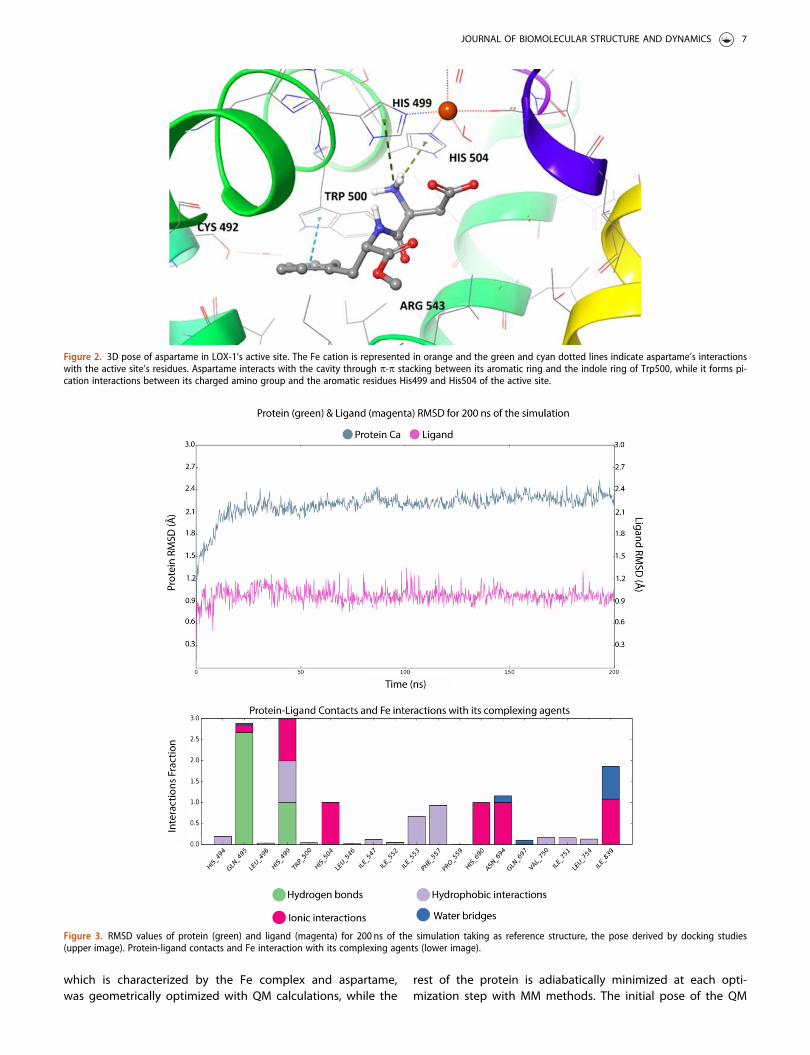
Aspartame in its best pose conformation (Figure 2) inter-acts strongly with the binding site of the protein. Thestrength of this binding is also related to the distances ofthe atoms participating in each interaction. These interac-tions, leading to the stabilization of the complex, include p –p stacking between the indole group of Trp500 and the aro-matic ring of aspartame (3.14 Å), the formation of two pi-cations between the amino group of aspartame and the twohistidine residues of the active site, His499 and His504(4.76 Å and 3.61 Å respectively) and the formation of a saltbridge between the Fe cation and the carboxyl group of themolecule (3.84 Å), which is better illustrated in Figure 2. Thelow binding energy in the cavity is also a result of the orien-tation of the lipophilic part of the molecule (aromatic ringand methyl ester) towards the hydrophobic residues of thecavity (i.e. Met497, Leu496, Cys492, Pro559, Phe557).
3.2.2. Molecular dynamics of the “LOX-1-aspartame”interaction
Molecular Dynamics simulations were conducted in order toevaluate the stability of aspartame’s pose proposed by thedocking studies. The MD simulation of the protein-ligandcomplex (DGbind=-7.6 kcal/mol) indicated aspartame’s stablebinding to LOX’s cavity. To quantify this indication, theRMSD of the ligand was measured with respect to its dock-ing pose coordinates. Throughout the simulation time(200 ns), aspartame is bound to LOX’s binding site, whileadopting a slightly different, more favorable, conformationthan the one proposed by docking studies. Aspartame’s sta-ble binding in the cavity during the simulation time verifiesour docking predictions and indicates that aspartame isindeed a strong LOX binder and a promising LOX inhibitor.
In Figure 3, the low RMSD value of protein’s Ca atoms(<2.4Å) indicates the good convergence of the system while theligand RMSD is also presented for 200ns of MD simulation time.The docking pose 1 was taken as reference-pose and the RMSDof ligand’s heavy atoms was measured. The ligand’s binding isconsidered quite stable since the RMSD value for the ligand is�0.9Å during the whole simulation time as depicted in Figure 3.Further evaluation was conducted by computing the ligand’satoms’ Root Mean Square Fluctuation (RMSF) presented in FigureS1. For most atoms, the RMSF values are as low as below 0.4Åwhereas all ligand’s atoms have small fluctuations (lower than0.4Å). Molecular dynamics simulations were performed 3 timesin order to investigate the reproducibility of the results statedabove. The RMSD and RMSF values of both the protein and theligand in all three simulations indicated the stability of the com-plex. The standard deviation of the RMSD values of the proteinamong the three simulations operated is 2.25±0.2Å, while thesame value concerning the ligand is 0.85±0.3Å. The standarddeviation of the RMSF value of the ligand fit on the proteinbetween the three simulations is 1.0±0.3Å.
In order to further assess the stability of the binding pose,we performed cluster analysis on our MD trajectory based on
the available ligand’s conformations. Cluster analysis of thetrajectory is a useful method for identifying, via statisticalanalysis, the most favored conformation that aspartameadopts during the simulation inside LOX’s cavity. The trajec-tory was divided in 10 clusters of similar geometry based onligand’s conformations, using Desmond’s algorithms and thecluster that was predominant for most of the simulationtime, i.e. 27%, was chosen as the most relevant. Cluster ana-lysis revealed important insights for aspartame’s binding toLOX’s active site. During the MD simulations, aspartameadopts a more favorable pose inside LOX’s cavity and it isfound to reside closer to the catalytic Fe cation in compari-son with the docking pose (Figure S2). In particular, MDsimulation is initiated with aspartame in spatial vicinity withcatalytic Fe (3.84 Å), while we observe that in the predomin-ant pose aspartame has approached the catalytic site andthe distance between Fe and the carboxyl group of aspar-tame is measured 2.2 Å, as illustrated in Figure 4a.Furthermore, after the superposition of the two poses (dock-ing pose and predominant cluster), we could observe thatnot only the carboxyl group of the molecule has beenslightly displaced during MD simulation, but also the wholeskeleton of aspartame has been displaced by approximately3.01 Å (Figure 4a). Moreover, the conformation of aspartamedoes not heavily change during the whole time of the simu-lation, as illustrated by the superposition of all clustersderived by trajectory analysis (Figure 4b). These results cor-roborates the initial assumption that aspartame binds favor-ably to the active site of LOX-1 and remains stable inside theenzymatic cavity. Finally, in Figure 5c, we could observe thatthe active site of the protein has slightly changed during theMD simulation. In particular, the conformations of the resi-dues in the active site present reduced but noticeable con-formational changes, as well as the residues in the firstcoordination sphere of the catalytic iron. Hence, it is manda-tory to perform further Quantum Mechanics/MolecularMechanics (QM/MM) studies for the metalloprotein, in orderto predict the most accurate configuration of the active site.
3.2.3. MM/GBSA calculationsMM/GBSA calculations have been applied to the statisticallypredominant “LOX-1-aspartame” complexes that derivedfrom Desmond trajectory clustering. In the table below(Table 3), the binding energies of aspartame to soybeanLOX-1 are illustrated. According to the DGbind values that arepresented in the table, aspartame binds favourably to theactive site of soybean LOX-1 and in all the predominant clus-ters, the DG value is quite low. For the most prominent clus-ter (27% of the simulation time), DGbind is calculated at�41.7 kcal/mol, while for cluster number 1 (21% of the simu-lation time) DGbind value is quite lower (-54.8 Kcal/mol).
3.2.4. Qm/MM calculations on the LOX-aspartame interactionIn order to further evaluate the accuracy of the MD simula-tions and the credibility of the pose derived by classicalmechanics calculations, we performed QM/MM studies forthe “LOX-aspartame” complex. The active site of the protein
6 E. CHONTZOPOULOU ET AL.
which is characterized by the Fe complex and aspartame,was geometrically optimized with QM calculations, while the
rest of the protein is adiabatically minimized at each opti-mization step with MM methods. The initial pose of the QM
Figure 2. 3D pose of aspartame in LOX-1’s active site. The Fe cation is represented in orange and the green and cyan dotted lines indicate aspartame’s interactionswith the active site’s residues. Aspartame interacts with the cavity through p-p stacking between its aromatic ring and the indole ring of Trp500, while it forms pi-cation interactions between its charged amino group and the aromatic residues His499 and His504 of the active site.
Figure 3. RMSD values of protein (green) and ligand (magenta) for 200 ns of the simulation taking as reference structure, the pose derived by docking studies(upper image). Protein-ligand contacts and Fe interaction with its complexing agents (lower image).
JOURNAL OF BIOMOLECULAR STRUCTURE AND DYNAMICS 7
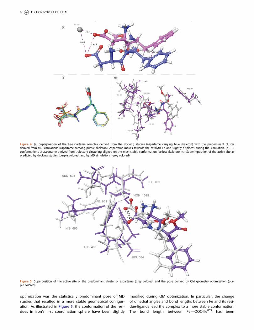
optimization was the statistically predominant pose of MDstudies that resulted in a more stable geometrical configur-ation. As illustrated in Figure 5, the conformation of the resi-dues in iron’s first coordination sphere have been slightly
modified during QM optimization. In particular, the changeof dihedral angles and bond lengths between Fe and its resi-due-ligands lead the complex to a more stable conformation.The bond length between Fe—OOC-Ile839 has been
Figure 4. (a) Superposition of the Fe-aspartame complex derived from the docking studies (aspartame carrying blue skeleton) with the predominant clusterderived from MD simulations (aspartame carrying purple skeleton). Aspartame moves towards the catalytic Fe and slightly displaces during the simulation. (b). 10conformations of aspartame derived from trajectory clustering aligned on the most stable conformation (yellow skeleton). (c). Superimposition of the active site aspredicted by docking studies (purple colored) and by MD simulations (grey colored).
Figure 5. Superposition of the active site of the predominant cluster of aspartame (grey colored) and the pose derived by QM geometry optimization (pur-ple colored).
8 E. CHONTZOPOULOU ET AL.

decreased from 2.28Å to 1.95 Å, as well as the bondbetween Fe—HN-His690 from 2.33 Å to 1.96 Å. The sameeffect is observed for the following bonds: Fe— HN-His499
(from 2.36 Å to 1.99 Å), Fe— HN-His504 (from 2.26Å to1.97 Å), Fe— HN-His504 (from 2.26Å to 1.97 Å), Fe— H2O
1045
(from 2.13 Å to 1.93 Å) and Fe— OC-Asn694 (from 2.16 Å to1.93 Å). Besides the variations of the bond lengths, importantconformational changes concerning the dihedral anglesoccur during QM optimization to the aforementioned histi-dine residues. In particular, His690 flips its imidazole ring byapproximately 60.6 �, His499 by 53.5 � and His504 by 52.2 �
with respect to their original conformations. Furthermore,aspartame moves slightly from its original position and theonly conformational difference from the initial pose is thedisplacement of its carboxyl group 2.33 Å away from Fe cat-ion (Figure 5). Consequently, the “LOX-aspartame” interactionpredicted by all the implemented in silico studies proposedthat aspartame enters LOX’s cavity and adopts the sameorientation in docking, MD and QM calculations. Hence, carb-oxyl group of the molecule is oriented towards catalytic Feof the protein in all the different calculations performed. Theaccordance of QM calculations of the active site with thepreviously performed docking and MD studies reveals thatall the parameters set to simulate the LOX-aspartame inter-action potential are accurate and especially underlines thefact that OPLS2005 is a suitable forcefield for simulating thesystem under study.
3.2.5. In vitro biological evaluation of the interaction ofaspartame to LOX-1
Having identified in silico the potential of aspartame to bindto LOX-1 we conducted further studies in order to verify itsinhibition potential using relevant enzymatic assay. We foundthat aspartame presents high inhibitory activity compared tothe reference compound nor-dihydroguaeretic acid (NDGA),a well-known lipoxygenase inhibitor, under the same experi-mental conditions (Table 4). The calculated IC50 of aspartamedue to its binding towards LOX-1 is quite high compared toother known strong LOX inhibitors (i.e. flavonoids, zileuton,BW 755 C) (Sostres et al., 2010).
3.2.6. Charting aspartame - LOX-1 interaction through sat-uration transfer difference (STD) NMR experiments
Having determined the potency of aspartame to inhibit LOX-1 we aimed to further probe its direct interaction as also tochart in atomic detail its epitope implicated in the recogni-tion with the easily accessible soybean LOX-1. To evaluatethe potential interaction of aspartame with LOX-1 we utilizedSaturation Transfer Difference (STD) NMR (Mayer & Meyer,2001). STD NMR not only probes the potential of a ligand to
interact with a potential pharmaceutical target, but alsohighlights the protons of the ligand that are implicated inthe molecular interaction (Chatzikonstantinou et al., 2018).Recently, we successfully applied in cell STD NMR to probeintracellularly the interaction of a ligand with a cellular tar-get. It is thus, a quite valuable tool to explore ligand-proteininteractions.
The interaction between LOX-1 and aspartame, throughSTD NMR, is presented in Figure 6. As it can be seen fromthe difference spectrum, all the peaks which are related tothe aromatic and aliphatic protons of aspartame, illustratelower intensity than the 1H-NMR reference spectrum. Thisresult further confirms the interaction of aspartame withLOX-1.
The peak intensities can be integrated, in order to deter-mine how close each proton is to the binding site of thereceptor. According to calculations, the aromatic protons H-2and H-4 interact with a percentage value of 38.26% withLOX-1, whereas the aromatic proton H-3 interacts with avalue of 34.84%. Among all the aromatic protons, the highestpercentage of interaction belongs to protons H-1 and H-5with 44.34%, implying that these protons are closer to thebinding site of the receptor.
In addition, for the aliphatic regions, the comparisonbetween the 1H NMR spectrum of the complex aspartame-soybean LOX-1 and the STD difference spectrum shows thatall the aliphatic protons, as well as the aspartyl –CH3 groupinteract with the binding site of the protein (Figure S3). Thelower intensity of the peaks at the STD difference spectrum,compared to the reference Khanapure & Gordon Letts,2004H proton spectrum, confirm that there is a bindinginteraction. According to calculations, the a-protons H-8, H-12a and H-12b near the two carboxylic groups interact suc-cessfully with the binding site of soybean LOX-1 with STDamplification factor values to be 20%. In addition, protons H-7a and H-7b, placed near the aromatic moiety interact withthe protein with values of 12.72% and 30.44%, respectively.On the other hand, protons H-10a and H-10b placedbetween the two carbons bridging the amidic group bindsufficiently to the protein with values of 11.45% and 100%respectively, whereas proton H-11, which is located near theaminic group interacts at a percentage of 40.48%. H-11, thus,is closer to the binding site of the protein. Lastly, the CH3
group which is part of the aspartyl moiety, also shows weakinteraction with the binding site of the protein, with STDamplification factor value of 9.21% (Figure S4). The differentSTD amplification factors of the aspartame protons allow tochart its pharmacophore groups implicated in the binding.Previous studies concerning the inhibition of LOX-1 from fla-vonoids (Ribeiro et al., 2014), have shown that a catecholicmoiety in the molecule could cause potent interaction withthe active site of the enzyme. The number of the –OH
Table 3. DGbind of LOX-1-aspartame complexes as calculated afterMD studies.
Clusternumber
MM/GBSA DGbind(kcal/mol)
Percentage of timeduring the simulation
1 �54.8 21%2 �54.6 19%3 �41.7 27%
Table 4. IC50 values against LOX-1.
CompoundsIC50 (lM)±SD�
Aspartame 50 ± 3.0NDGA 0.45 ± 0.013
The values are the mean of six replicates; �SD standard deviation.
JOURNAL OF BIOMOLECULAR STRUCTURE AND DYNAMICS 9

groups in the aromatic ring influences the potency of theinhibitory activity of a ligand. Flavonoids like luteolin, quer-cetin or taxifolin have been proven to be potent inhibitors ofLOX-1, as they interact successfully with the active center ofLOX-1. In addition, other flavonoids containing aromatic ringslike curcumin and protocatechuic acid, have also been foundto interact very efficiently with the active center of LOX-1,and, as a result they are potent LOX-1 inhibitors(Borbulevych et al., 2004). These data are in accordance toour findings indicating that the aromatic ring of the phenyl-alanine amino acid of aspartame interacts with LOX-1,through protons H-1 and H-5 pinpointing enhanced STDamplification factors. In addition, among other protons,which are part of the carbon chain, proton H-11 which isplaced on the same carbon atom bearing the NH group,seems to be affected by the presence of hydrogen bonds,and, thus, it binds with efficient value to the binding site ofLOX-1. Furthermore, aspartame seems to be planar in space,as its structure can be affected by the presence of the aro-matic ring of phenylalanine, as well as the esteric moietyand the carboxylic group of the aspartic acid. This character-istic can also influence the strength of interaction betweenthe enzyme and a possible ligand (Borbulevych et al., 2004).
The molecular profile of aspartame inside LOX-1’s cavityproposed by STD NMR experiments is in accordance with theresults derived from the in silico studies. As illustrated inFigure S2, depicting aspartame in LOX’s cavity (predominantMD cluster), the aromatic ring (H1-H5) forms a p-p stackinginteraction with Phe557, while the alkyl chain of the mol-ecule (H10, H11, H12) carrying the carboxyl- and the amino-functional groups resides deep inside the cavity and interactsin the residues of the Fe-complex and Gln495.
3.3. In silico studies of aspartame to differentLOX isoforms
After having identified aspartame as a promising binder tosoybean LOX-1 using in silico, in vitro and STD NMR studies,
aspartame has been tested in silico for its potency to bind toother LOX isoforms. In order to conclude if aspartame couldserve as a potential hit compound for designing novel anti-inflammatory drugs against LOX, it needs to be assessed forits binding to other organisms expressing the enzyme andmostly to mammals. Docking calculations and MD simula-tions have been implemented to different LOX isoforms.Table 5 presents the docking affinities of aspartame to LOXs’active sites, as well as it reports the compound’s stability ineach cavity as derived from the 200 ns of MD simulations.
According to the results derived from docking studies,aspartame presents high affinity to the active site of all LOXisoforms, as illustrated in the Table 5 (DG�-7.6 kcal/mol).However, these results need to be further evaluated in orderto assess if aspartame’s strong binding to LOX cavity is anactual fact or just an artifact depicting only one snapshot ofaspartame forced into the cavity. Herein, long MD simula-tions (�200 ns) were conducted so as to predict aspartame’sstability in each cavity, and the results are demonstrated inthe right column of Table 5. RMSD values of the stable pro-teins’ skeletons are presented in Figure S5, indicating theproper converge of the system and thus the accuracy of theresults. LOX-3 exerts strong binding to aspartame and MDsimulations indicated the stability of the formed complex.Furthermore, aspartame remains stable in human 5-LOX’scavity, which is an important result pointing aspartame as apromising anti-inflammatory drug targeting 5-LOX. Another“LOX-aspartame” complex in mammals that also remainsquite stable is the one of rabbit organism (Oryctolagus cuni-culus). In addition, aspartame forms unstable interactions inthe rest of the LOX complexes, as it abandons the activesites of the enzymes and eventually the whole protein, andends up in the aquatic environment of the solvent for mostof the simulation time. These differences observed amongLOX isoforms (formation of stable and unstable complexes)may be attributed to the structural differences among theenzyme’s isoforms, including slight differences in the activesite. Some of these proteins carry quite accessible to thesolvent cavities, where aspartame could easily unbind from
Figure 6. (a). 1H-NMR reference spectrum of aspartame (1mM) containing the protein LOX-1 (0.02mM), at 1:50 ratio towards aspartame, in buffer Tris pH ¼ 7.2and 600 lL D2O at 500MHz and 25 �C. (b). STD NMR difference spectrum of the complex aspartame-LOX-1, saturated in a frequency related to the protein(804.314 Hz) at 500MHz and 25 �C. The strength of the interaction between the protons of aspartame and the binding site of LOX-1 is expressed through therespective percentages. The structure of aspartame is embedded in the top of the figure.
10 E. CHONTZOPOULOU ET AL.

the active site and end up in the solvent area. Finally, allLOX isoforms derived from microorganisms do not seem tostabilize aspartame in their cavities in opposition to soybeanisoforms that carry aspartame bound to their active sitesthroughout the whole simulation time. As far as humanLOXs, two out of three different human isoforms could formstable aspartame’s complexes. This finding could play animportant role in the selectivity of aspartame as a potentialdrug molecule.
4. Discussion
Drug discovery is a very costly, tedious and time-consumingprocess. Along these lines we have initiated in our labs aprocess to discover potent bioactive compounds from foodand beverage additives. This process could be of capitalimportance since such discovery could lead to orally bioavail-able and safe drugs. Driven by our original hypothesis thatpotent LOX inhibitors could be identified from such sources,we screened natural and artificial sweeteners with the aid ofin silico calculations. We discovered that aspartame is anorthosteric binder of LOX-1, which is a result verified withdifferent computational and experimental methods. MD andQM/MM calculations predicted the most prominent conform-ation that aspartame adopts into the active site of LOX-1, aswell as all the interactions aspartame forms with the residuesin the cavity have been thoroughly evaluated. Electroniceffects of the catalytic metallic active site were also takeninto account, in order to improve the accuracy of our results.In vitro biological assays confirmed aspartame’s binding toLOX enzyme and precisely calculated that aspartame exhibitsIC50 value of 50± 3.0 lM. This IC50 value reveals that aspar-tame binds to LOX-1, although the strength of the bindingneeds to be further intensified. The molecular and atomicdetails of the binding were experimentally unveiled by STDNMR experiments and the results were in accordance withthe proposed aspartame’s conformation derived from the insilico studies. After having fully mapped the molecular detailsof aspartame’s binding to LOX-1, further evaluation of theinhibitory properties of aspartame to other LOX isoforms hasbeen conducted. Aspartame presents strong binding in 2 outof 3 different LOX isoforms of human organism, as unveiledby docking and MD studies. Thus, these discoveries revealaspartame’s inhibitory activity towards LOX enzyme andcould pave the way to the structure derivatization of aspar-tame in order to produce stronger LOX binders and hencefind numerous therapeutic applications against inflammation,
cardiovascular diseases and cancer. From all the informationacquired by this study, we conclude that aspartame is animportant binder of LOX enzyme and it could serve as a hitfor the design of novel, therapeutic agents. Furthermore,many compounds that have been identified as competentLOX-1 inhibitors, exhibit similar IC50 values with this of aspar-tame. For instance, an investigation conducted by Katsori etal., revealed curcumin derivatives that exhibit inhibitory activ-ity against LOX-1 (IC50¼47lM) (Katsori et al., 2011), whileYar’s research group identified indolic compounds as LOXinhibitors with that exhibit IC50¼53.1 lM (Hu & Ma, 2018). Inorder to further evaluate the aspartame as a potent drug,the compound should be tested in vitro with human 15-LOXand 5-LOX isoforms. Herein, the results deriving from ourcurrent investigation indicate that since LOX-1 is implicatedin the development of various inflammatory conditions, deri-vatization of aspartame will lead to important biologicallyactive molecules targeting lipoxygenase isoforms and serveas potent drugs for the treatment of numerous inflammatorydisorders and crucial diseases.
Acknowledgements
Materials were supported by Special Account for Research Grants(SARG), National Kapodistrian University of Athens (NKUA).
Disclosure statement
The authors declare no conflict of interest.
Author contributions
Conceptualization, T.M. T.T, D.T, I.K and E.C.; methodology, A.K.; software,E.C.; S.K. D.K, I.G; validation, T.M.; E.C; C.D.P; M.V.C, S.K.; writing—originaldraft preparation, T.M.; E.C; C.D.P; M.V.C writing—review and editing,T.M; A.G.T visualization, A.G., T.M; supervision, M.T.; A.G.T; D.H.-L.
Funding
This work was financially supported by Greece and the European Union(European Social Fund- ESF) through the Operational Programme«Human Resources Development, Education and Lifelong Learning» inthe context of the project “Strengthening Human Resources ResearchPotential via Doctorate Research” under Grant MIS-5000432, imple-mented by the State Scholarships Foundation (IKY).
ORCID
Thomas Mavromoustakos http://orcid.org/0000-0001-5309-992X
References
Agull�o, V., Dom�ınguez-Perles, R., & Garc�ıa-Viguera, C. (2021). Sweetenerinfluences plasma concentration of flavonoids in humans after anacute intake of a new (poly)phenol-rich beverage. Nutrition,Metabolism, and Cardiovascular Diseases: NMCD, 31(3), 930–938.https://doi.org/10.1016/j.numecd.2020.11.016
Avis, I. M., Jett, M., Boyle, T., Vos, M. D., Moody, T., Treston, A. M.,Mart�ınez, A., & Mulshine, J. L. (1996). Growth control of lung cancerby interruption of 5-lipoxygenase-mediated growth factor signaling.
Table 5. In silico studies of aspartame to different LOX isoforms.
Enzyme PDB ID OrganismDGbind
(kcal/mol)Complexstability
13S-LOX 5T5V Glycine max �7.6 Stable5-LOX 3O8Y Homo sapiens �8.5 Stable15S-LOX 1LOX Oryctolagus cuniculus �9.8 Stable9S-LOX-3 1RRH Glycine max �7.9 Stable12S-LOX 3D3L Homo sapiens �10.0 Unstable15-LOX 4G32 Pseudomonas Aeruginosa �9.3 Unstable12-LOX 3RDE Sus scrofa �8.3 Stable15-LOX-2 4NRE Homo sapiens �10.3 Unstable13R-LOX 5IR4 Pseudomonas Aeruginosa �9.4 Unstable
JOURNAL OF BIOMOLECULAR STRUCTURE AND DYNAMICS 11
The Journal of Clinical Investigation, 97(3), 806–813. https://doi.org/10.1172/JCI118480
Banthiya, S., Kalms, J., Galemou Yoga, E., Ivanov, I., Carpena, X.,Hamberg, M., Kuhn, H., & Scheerer, P. (2016). Structural and functionalbasis of phospholipid oxygenase activity of bacterial lipoxygenasefrom Pseudomonas aeruginosa. Biochimica et Biophysica Acta,1861(11), 1681–1692. https://doi.org/10.1016/j.bbalip08.002.
Borbulevych, O. Y., Jankun, J., Selman, S. H., & Skrzypczak-Jankun, E.(2004). Lipoxygenase interactions with natural flavonoid, quercetin,reveal a complex with protocatechuic acid in its X-ray structure at 2.1A resolution. Proteins, Structure, Function, and genetics, 54(1), 13–19.https://doi.org/10.1002/prot.10579
Boyington, J. C., Gaffney, B. J., & Amzel, L. M. (1990). Crystallization andpreliminary X-ray analysis of soybean lipoxygenase-1, a non-hemeiron-containing dioxygenase. The Journal of Biological Chemistry,265(22), 12771–12773.
Brash, A. R. (1999). Lipoxygenases: Occurrence, functions, catalysis, andacquisition of substrate. The Journal of Biological Chemistry, 274(34),23679–23682. https://doi.org/10.1074/jbc.274.34.23679
Carter, G. W., Young, P. R., Albert, D. H., Bouska, J., Dyer, R., Bell, R. L.,Summers, J. B., & Brooks, D. W. (1991). 5-lipoxygenase inhibitory activ-ity of zileuton. The Journal of Pharmacology and ExperimentalTherapeutics, 256(3), 929–937.
Catalano, A., & Procopio, A. (2005). New aspects on the role of lipoxyge-nases in cancer progression. Histology and Histopathology, 20(3),969–975. https://doi.org/10.14670/HH-20.969
Chatzikonstantinou, A. V., Chatziathanasiadou, M. V., Ravera, E., Fragai, M.,Parigi, G., Gerothanassis, I. P., Luchinat, C., Stamatis, H., & Tzakos, A. G.(2018). Enriching the biological space of natural products and chartingdrug metabolites, through real time biotransformation monitoring: TheNMR tube bioreactor. Biochimica et Biophysica Acta. General Subjects,1862(1), 1–8. https://doi.org/10.1016/j.bbagen.2017.09.021
Choi, J., Jae, K. C., Kim, S., & Shin, W. (2008). Conformational flexibility inmammalian 15S-lipoxygenase: Reinterpretation of the crystallographicdata. Proteins Struct Proteins, 70(3), 1023–1032. https://doi.org/10.1002/prot.21590
Cicero, A. F. G., & Laghi, L. (2007). Activity and potential role of licofelonein the management of osteoarthritis. Clinical Interventions in Aging,2(1), 73–79. https://doi.org/10.2147/ciia.2007.2.1.73
Claria, J., & Romano, M. (2005). Pharmacological intervention of cyclooxy-genase-2 and 5-lipoxygenase pathways. Impact on inflammation andcancer. Current Pharmaceutical Design, 11(26), 3431–3447. https://doi.org/10.2174/138161205774370753
Daina, A., Michielin, O., & Zoete, V. (2017). SwissADME: A free web toolto evaluate pharmacokinetics, drug-likeness and medicinal chemistryfriendliness of small molecules. Scientific Reports, 7, 42717. https://doi.org/10.1038/srep42717
Dobrian, A. D., Lieb, D. C., Cole, B. K., Taylor-Fishwick, D. A., Chakrabarti,S. K., & Nadler, J. L. (2011). Functional and pathological roles of the12- and 15-lipoxygenases. Progress in Lipid Research, 50(1), 115–131.https://doi.org/10.1016/j.plipres.2010.10.005
Essmann, U., Perera, L., Berkowitz, M. L., Darden, T., Lee, H., & Pedersen,L. G. (1995). A smooth particle mesh Ewald method. Journal ofChemical Physics, 103(19), 8577–8593. https://doi.org/10.1063/1.470117
Feinmark, S. J., & Cornicelli, J. A. (1997). Is there a role for 15-lipoxyge-nase in atherogenesis? Biochemical Pharmacology, 54(9), 953–959.https://doi.org/10.1016/S0006-2952(97)00135-4
Flower, R. (2003). All the things that aspirin does. BMJ (Clinical Researched.), 327(7415), 572–573.
Garreta, A., Val-Moraes, S. P., Garc�ıa-Fern�andez, Q., Busquets, M., Juan, C.,Oliver, A., Ortiz, A., Gaffney, B. J., Fita, I., Manresa, �A., & Carpena, X.(2013). Structure and interaction with phospholipids of a prokaryoticlipoxygenase from Pseudomonas aeruginosa. FASEB Journal: OfficialPublication of the Federation of American Societies for ExperimentalBiology, 27(12), 4811–4821. https://doi.org/10.1096/fj.13-235952
Gheorghe, K. R., Korotkova, M., Catrina, A. I., Backman, L., af Klint, E.,Claesson, H.-E., Rådmark, O., & Jakobsson, P.-J. (2009). Expression of 5-lipoxygenase and 15-lipoxygenase in rheumatoid arthritis synoviumand effects of intraarticular glucocorticoids. Arthritis Research &Therapy, 11(3), R83. https://doi.org/10.1186/ar2717
Ghose, A. K., Viswanadhan, V. N., & Wendoloski, J. J. (1999). A know-ledge-based approach in designing combinatorial or medicinal chem-istry libraries for drug discovery. 1. A qualitative and quantitativecharacterization of known drug databases. Journal of CombinatorialChemistry, 1(1), 55–68. https://doi.org/10.1021/cc9800071
Gilbert, N. C., Bartlett, S. G., Waight, M. T., Neau, D. B., Boeglin, W. E.,Brash, A. R., & Newcomer, M. E. (2011). The structure of human 5-lip-oxygenase. Science (New York, N.Y.), 331(6014), 217–219. https://doi.org/10.1126/science.1197203
Gilbert, N. C., Gerstmeier, J., Schexnaydre, E. E., B€orner, F., Garscha, U.,Neau, D. B., Werz, O., & Newcomer, M. E. (2020). Structural and mech-anistic insights into 5-lipoxygenase inhibition by natural products.Nature Chemical Biology, 16(7), 783–790. https://doi.org/10.1038/s41589-020-0544-7
Gillmor, S. A., Villase~nor, A., Fletterick, R., Sigal, E., & Browner, M. F. (1997).The structure of mammalian 15-lipoxygenase reveals similarity to thelipases and the determinants of substrate specificity. Nature StructuralBiology, 4(12), 1003–1009. https://doi.org/10.1038/nsb1297-1003
Gupta, P., Tiwari, A., & Mishra, M. K. (2017). Taste masking of drugs: Anextended approach. International Journal of Current Advanced Research,6(3), 2571–2578. https://doi.org/10.24327/ijcar.2017.2578.0051
Haeggstr€om, J. Z., & Funk, C. D. (2011). Lipoxygenase and leukotrienepathways: Biochemistry, biology, and roles in disease. ChemicalReviews, 111(10), 5866–5898. https://doi.org/10.1021/cr200246d
Hu, C., & Ma, S. (2018). Recent development of lipoxygenase inhibitorsas anti-inflammatory agents. Medchemcomm, 9(2), 212–225. https://doi.org/10.1039/c7md00390k
Humphreys, D. D., Friesner, R. A., & Berne, B. J. (1994). A multiple-time-stepMolecular Dynamics algorithm for macromolecules. The Journal ofPhysical Chemistry, 98(27), 6885–6892. https://doi.org/10.1021/j100078a035
Jorgensen, W. L., Maxwell, D. S., & Tirado-Rives, J. (1996). Development andtesting of the OPLS all-atom force field on conformational energetics andproperties of organic liquids. Journal of the American Chemical Society,118(45), 11225–11236. https://doi.org/10.1021/ja9621760
Karatas, H., & Cakir-Aktas, C. (2019). 12/15 lipoxygenase as a therapeutictarget in brain disorders. Noro Psikiyatri Arsivi, 56(4), 288–291. https://doi.org/10.29399/npa.23646
Katsori, A. M., Chatzopoulou, M., Dimas, K., Kontogiorgis, C., Patsilinakos,A., Trangas, T., & Hadjipavlou-Litina, D. (2011). Curcumin analogues aspossible anti-proliferative & anti-inflammatory agents. EuropeanJournal of Medicinal Chemistry, 46(7), 2722–2735. https://doi.org/10.1016/j.ejmech.2011.03.060
Khanapure, S. P., & Gordon Letts, L. (2004). Perspectives and clinical sig-nificance of the biochemical and molecular pharmacology of eicosa-noids. In The Eicosanoids (pp. 129–162). John Wiley &Sons.
Kobe, M. J., Neau, D. B., Mitchell, C. E., Bartlett, S. G., & Newcomer, M. E.(2014). The structure of human 15-lipoxygenase-2 with a substratemimic. The Journal of Biological Chemistry, 289(12), 8562–8569. https://doi.org/10.1074/jbc.M113.543777
Lim, U. (2016). Artificial sweeteners and cancer - national cancerinstitute.
Lizunkova, P., Enuwosa, E., & Chichger, H. (2019). Activation of the sweettaste receptor T1R3 by sucralose attenuates VEGF-induced vasculo-genesis in a cell model of the retinal microvascular endothelium.Graefe’s Archive for Clinical and Experimental Ophthalmology¼AlbrechtVon Graefes Archiv Fur Klinische Und Experimentelle Ophthalmologie,257(1), 71–81. https://doi.org/10.1007/s00417-018-4157-8
Lomelino, C. L., Murray, A. B., Supuran, C. T., & McKenna, R. (2018). SweetBinders: Carbonic Anhydrase IX in Complex with Sucralose. ACSMedicinal Chemistry Letters, 9(7), 657–661. https://doi.org/10.1021/acs-medchemlett.8b00100
Luo, J., Chuang, T., Cheung, J., Quan, J., Tsai, J., Sullivan, C., Hector, R. F.,Reed, M. J., Meszaros, K., King, S. R., Carlson, T. J., & Reaven, G. M.(1998). Masoprocol (nordihydroguaiaretic acid): A new antihyperglyce-mic agent isolated from the creosote bush (Larrea tridentata).European Journal of Pharmacology, 346(1), 77–79. https://doi.org/10.1016/S0014-2999(98)00139-3
Lyman, E., & Zuckerman, D. M. (2006). Ensemble-based convergence ana-lysis of biomolecular trajectories. Biophysical Journal, 91(1), 164–172.https://doi.org/10.1529/biophysj.106.082941
12 E. CHONTZOPOULOU ET AL.

Magnuson, B. A., Burdock, G. A., Doull, J., Kroes, R. M., Marsh, G. M.,Pariza, M. W., Spencer, P. S., Waddell, W. J., Walker, R., & Williams,G. M. (2007). Aspartame: A safety evaluation based on current use lev-els, regulations, and toxicological and epidemiological studies. CriticalReviews in Toxicology, 37(8), 629–727. https://doi.org/10.1080/10408440701516184
Mahon, B. P., Hendon, A. M., Driscoll, J. M., Rankin, G. M., Poulsen, S. A.,Supuran, C. T., & McKenna, R. (2015). Saccharin: A lead compound forstructure-based drug design of carbonic anhydrase IX inhibitors.Bioorganic & Medicinal Chemistry, 23(4), 849–854. https://doi.org/10.1016/j.bmc.2014.12.030
Maillet, E. L., Cui, M., Jiang, P., Mezei, M., Hecht, E., Quijada, J., Margolskee,R. F., Osman, R., & Max, M. (2015). Characterization of the binding site ofaspartame in the human sweet taste receptor. Chemical Senses, 40(8),577–586. https://doi.org/10.1093/chemse/bjv045
Martyna, G. J., Tobias, D. J., & Klein, M. L. (1994). Constant pressuremolecular dynamics algorithms. Journal of Chemical Physics., 101(5),4177–4189. https://doi.org/10.1063/1.467468
Mashima, R., & Okuyama, T. (2015). The role of lipoxygenases in patho-physiology; new insights and future perspectives. Redox Biology, 6,297–310. https://doi.org/10.1016/j.redox.2015.08.006
Mayer, M., & Meyer, B. (2001). Group epitope mapping by saturationtransfer difference NMR to identify segments of a ligand in directcontact with a protein receptor. Journal of the American ChemicalSociety, 123(25), 6108–6117. https://doi.org/10.1021/ja0100120
Murphy, R. C., & Gijon, M. A. (2007). Biosynthesis and metabolism of leu-kotrienes. The Biochemical Journal, 405(3), 379–395. https://doi.org/10.1042/BJ20070289
Murray, J. J., Tonnel, A. B., Brash, A. R., Roberts, L. J., Gosset, P., Workman, R.,Capron, A., & Oates, J. A. (1986). Release of prostaglandin D2 into humanairways during acute antigen challenge. The New England Journal ofMedicine, 315(13), 800–804. https://doi.org/10.1056/nejm09253151304.
Offenbacher, A. R., Hu, S., Poss, E. M., Carr, C. A. M., Scouras, A. D.,Prigozhin, D. M., Iavarone, A. T., Palla, A., Alber, T., Fraser, J. S., &Klinman, J. P. (2017). Hydrogen-Deuterium exchange of lipoxygenaseuncovers a relationship between distal, solvent exposed proteinmotions and the thermal activation barrier for catalytic proton-coupled electron tunneling. ACS Central Science, 3(6), 570–579.https://doi.org/10.1021/acscentsci.7b00142
Oprea, T. I., Bauman, J. E., Bologa, C. G., Buranda, T., Chigaev, A., Edwards,B. S., Jarvik, J. W., Gresham, H. D., Haynes, M. K., Hjelle, B., Hromas, R.,Hudson, L., Mackenzie, D. A., Muller, C. Y., Reed, J. C., Simons, P. C.,Smagley, Y., Strouse, J., Surviladze, Z., … Sklar, L. A. (2011). Drug repur-posing from an academic perspective. Drug Discovery Today. TherapeuticStrategies, 8(3-4), 61–69. https://doi.org/10.1016/j.ddstr.2011.10.002
Li, P., Wang, Z., Lam, S. M., & Shui, G. (2021). Rebaudioside a enhancesresistance to oxidative stress and extends lifespan and healthspan incaenorhabditis elegans. Antioxidants, 10, 262. https://doi.org/10.3390/antiox10020262
Pattar, S. V., Adhoni, S. A., Kamanavalli, C. M., & Kumbar, S. S. (2020). Insilico molecular docking studies and MM/GBSA analysis of coumarin-carbonodithioate hybrid derivatives divulge the anticancer potentialagainst breast cancer. Beni-Suef University Journal of Basic and AppliedSciences, 9, 36-46. https://doi.org/10.1186/s43088-020-00059-7
Pratic�o, D., Zhukareva, V., Yao, Y., Uryu, K., Funk, C. D., Lawson, J. A.,Trojanowski, J. Q., & Lee, V. M. Y. (2004). 12/15-Lipoxygenase isincreased in Alzheimer’s disease: possible involvement in brain oxida-tive stress. The American Journal of Pathology, 164(5), 1655–1662.https://doi.org/10.1016/S0002-9440(10)63724-8
Protein Data Bank. (2021). Protein Data Bank RCSB PDB: Homepage.Rana, R., Sharma, R., & Kumar, A. (2019). Repurposing of fluvastatin
against candida albicans CYP450 lanosterol 14 a-demethylase, a tar-get enzyme for antifungal therapy: An in silico and in vitro study.Current Molecular Medicine, 19(7), 506–524. https://doi.org/10.2174/1566524019666190520094644
Ribeiro, D., Freitas, M., Tom�e, S. M., Silva, A. M. S., Porto, G., Cabrita, E. J.,Marques, M. M. B., & Fernandes, E. (2014). Inhibition of LOX by flavo-noids: A structure-activity relationship study. European Journal ofMedicinal Chemistry, 72, 137–145. https://doi.org/10.1016/j.ejmech.2013.11.030
Rioux, N., & Castonguay, A. (1998). Inhibitors of lipoxygenase: A newclass of cancer chemopreventive agents. Carcinogenesis, 19(8),1393–1400. https://doi.org/10.1093/carcin/19.8.1393
Rouzer, C. A., & Marnett, L. J. (2009). Cyclooxygenases: Structural andfunctional insights. Journal of Lipid Research, 50 Suppl, S29–S34.https://doi.org/10.1194/jlr.r800042-jlr200
Schr€odinger LigPrep. (2018). Schr€odinger. Schr€odinger Release 2018–2.Schr€odinger LLC Prime. (2014). Schr€odinger LLC Prime, version 3.5. New
York.Schr€odinger LLC. (2011). Schr€odinger Llc New York Ny Glide, version 5.7.
Glid. Schr€odinger LLC.Schr€odinger Maestro. (2018). Schr€odinger. Schr€odinger Release 2018–2.Schrodinger, L.L.C. (2013). MacroModel, Version 10.2. New York.Schrodinger. (2015). Schrodinger software release 2015-2 induced fit dock-
ing. Schrodinger Press.Sharma, N., Singh, A., & Sharma, R. (2020). Repurposing of auranofin
against bacterial infections: An In silico and In vitro study. Curr ComputAided Drug Des.
Sherman, W., Day, T., Jacobson, M. P., Friesner, R. A., & Farid, R. (2006).Novel procedure for modeling ligand/receptor induced fit effects.Journal of Medicinal Chemistry, 49(2), 534–553. https://doi.org/10.1021/jm050540c
Skrzypczak-Jankun, E., Borbulevych, O. Y., Zavodszky, M. I., Baranski,M. R., Padmanabhan, K., Petricek, V., & Jankun, J. (2006). Effect of crys-tal freezing and small-molecule binding on internal cavity size in alarge protein: X-ray and docking studies of lipoxygenase at ambientand low temperature at 2.0 A resolution. Acta Crystallographica.Section D, Biological Crystallography, 62(Pt 7), 766–775. https://doi.org/10.1107/S0907444906016982
Sladek, V., K�o�na, J., & Tokiwa, H. (2017). In silico analysis of interaction patternswitching in ligandreceptor binding in Golgi a-mannosidase II induced bythe protonated states of inhibitors . Physical Chemistry Chemical Physics :PCCP, 19(19), 12527–12537. https://doi.org/10.1039/c7cp01200d
Sostres, C., Gargallo, C. J., Arroyo, M. T., & Lanas, A. (2010). Adverse effects ofnon-steroidal anti-inflammatory drugs (NSAIDs, aspirin and coxibs) onupper gastrointestinal tract. Best Practice & Research. ClinicalGastroenterology, 24(2), 121–132. https://doi.org/10.1016/j.bpg.2009.11.005
Tardif, J.-C., L’Allier, P. L., Ibrahim, R., Gr�egoire, J. C., Nozza, A., Cossette,M., Kouz, S., Lavoie, M.-A., Paquin, J., Brotz, T. M., Taub, R., &Pressacco, J. (2010). Treatment with 5-lipoxygenase inhibitor VIA-2291(atreleuton) in patients with recent acute coronary syn-drome.110.937169. Circulation: Cardiovascular Imaging, 3(3), 298–307.https://doi.org/10.1161/CIRCIMAGING
US Food & Drug Administration. (n.d.). US food & drug administrationadditional information about high-intensity sweeteners permitted foruse in food in the United States.
Valentovic, M. Z. (2007). In xPharm: The comprehensive pharmacologyreference.
Version, D.D. (2021). Version, D.D. Desmond Tutorial. Schroedinger.,https://doi.org/10.1162/rest_a_00790
West, M., Mhatre, M., Ceballos, A., Floyd, R. A., Grammas, P., Gabbita,S. P., Hamdheydari, L., Mai, T., Mou, S., Pye, Q. N., Stewart, C., West, S.,Williamson, K. S., Zemlan, F., & Hensley, K. (2004). The arachidonicacid 5-lipoxygenase inhibitor nordihydroguaiaretic acid inhibits tumornecrosis factor alpha activation of microglia and extends survival ofG93A-SOD1 transgenic mice . Journal of Neurochemistry, 91(1),133–143. https://doi.org/10.1111/j.1471-415902700.x.
Xu, S., Mueser, T. C., Marnett, L. J., & Funk, M. O. (2012). Crystal structureof 12-Lipoxygenase catalytic-domain-inhibitor complex identifies asubstrate-binding channel for catalysis. Structure (London, England :1993), 20(9), 1490–1497. https://doi.org/10.1016/j.str.2012.06.003
Zhao, Y., Wang, W., Wang, Q., Zhang, X., & Ye, L. (2012). Lipid metabol-ism enzyme 5-LOX and its metabolite LTB4 are capable of activatingtranscription factor NF-jB in hepatoma cells. Biochemical andBiophysical Research Communications, 418(4), 647–651. https://doi.org/10.1016/j.bbrc.2012.01.068
Zuo, L., Christofi, F. L., Wright, V. P., Bao, S., & Clanton, T. L. (2004).Lipoxygenase-dependent superoxide release in skeletal muscle.Journal of Applied Physiology (Bethesda, Md. : 1985), 97(2), 661–668.https://doi.org/10.1152/japplphysiol.00096.2004
JOURNAL OF BIOMOLECULAR STRUCTURE AND DYNAMICS 13