molecular investigation of the microbial community ... · molecular investigation of the microbial...
TRANSCRIPT

Molecular investigation of the microbial communityassociated with the fire sponge, Tedania ignis, in BermudaNicholas J Jouett, Meredith E Bibbings, Clarisse E Sullivan, Rachel J Parsons
The complex, phylogenetically diverse, and specific microbial communities associated withmarine sponges are a key aspect of the ecology and evolution of the metazoan host andthe endosymbiotic microbes. Using fluorescence in situ hybridization (FISH) methods,terminal restriction fragment length polymorphism (T RFLP), and functional gene probingvia PCR, the current study investigates the microbial associations in the commonCaribbean fire sponge, Tedania ignis. Sponge and water samples were collected fromdifferent sites around Bermuda from 2012 to 2014 in order to assess their respectivemicrobial communities. Using FISH, SAR202 (Chloroflexi) (5.82% ± 0.59%) andCrenarchaea (7.97% ± 1.08%) were identified as the most abundant contributors to themicrobial assemblage of T. ignis while the Alphaproteobacterium SAR11 (30.68% ± 1.68%)was identified as the most dominant species in the surrounding seawater. Due to thepresence of Crenarchaea, the Archaeal gene for ammonia oxidation (amoA) was probedvia PCR and found to be present. T RFLP identified the most abundant fragment lengthpresent in the sponge as 336 bp (>60% of T RFLP peak abundance). The spongecommunity was consistent and markedly distinct from that of the ambient seawater asidentified by both FISH and T RFLP. Epifluorescent microscopy with DAPI staining alsoidentified T. ignis as a high microbial abundance (HMA) sponge, in contrast to previousstudies. Together, these data characterize the microbiome of T. ignis in much further detailthan has previously been described.
PeerJ PrePrints | https://dx.doi.org/10.7287/peerj.preprints.905v1 | CC-BY 4.0 Open Access | rec: 17 Mar 2015, publ: 17 Mar 2015
PrePrin
ts

2 1, 2*Nicholas J. Jouett, 1,3Meredith E. Bibbings, 1, 2Clarisse E. Sullivan and 1Rachel J. Parsons34 1Bermuda Institute of Ocean Sciences (BIOS), St. George’s, Bermuda5 2University of Rhode Island, Kingston, RI, USA6 3McGill University, Montreal, Quebec, Canada78 *For correspondence. Email [email protected]
101112131415161718192021222324252627282930313233343536373839404142434445464748495051
PeerJ PrePrints | https://dx.doi.org/10.7287/peerj.preprints.905v1 | CC-BY 4.0 Open Access | rec: 17 Mar 2015, publ: 17 Mar 2015
PrePrin
ts
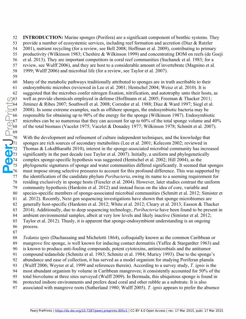
52 INTRODUCTION: Marine sponges (Porifera) are a significant component of benthic systems. They 53 provide a number of ecosystemic services, including reef formation and accretion (Diaz & Rutzler 54 2001), nutrient recycling (for a review, see Bell 2008; Hoffman et al. 2009), contributing to primary 55 productivity (Wilkinson 1983; Cheshire & Wilkinson 1999) and concentrating DOM on reefs (de Goeji 56 et al. 2013). They are important competitors in coral reef communities (Suchanek et al. 1983; for a 57 review, see Wulff 2006), and they are host to a considerable amount of invertebrate (Magnino et al. 58 1999; Wulff 2006) and microbial life (for a review, see Taylor et al. 2007). 5960 Many of the metabolic pathways traditionally attributed to sponges are in truth ascribable to their 61 endosymbiotic microbes (reviewed in Lee et al. 2001; Hentschel 2004; Weisz et al. 2010). It is 62 suggested that the microbes confer nitrogen fixation, nitrification, and autotrophy unto their hosts, as 63 well as provide chemicals employed in defense (Hoffmann et al. 2005; Freeman & Thacker 2011; 64 Jiminez & Ribes 2007; Southwell et al. 2008; Corredor et al. 1988; Diaz & Ward 1997; Siegl et al. 65 2008). In some extreme examples, such as offshore sponges, the endosymbiotic bacteria may be 66 responsible for obtaining up to 90% of the energy for the sponge (Wilkinson 1987). Endosymbiotic 67 microbes can be so numerous that they can account for up to 60% of the total sponge volume and 40% 68 of the total biomass (Vacelet 1975; Vacelet & Donadey 1977; Wilkinson 1978; Schmitt et al. 2007). 6970 With the development and refinement of culture independent techniques, and the knowledge that 71 sponges are rich sources of secondary metabolites (Lee et al. 2001; Kelecom 2002; reviewed in 72 Thomas & LokaBharathi 2010), interest in the sponge-associated microbial community has increased 73 significantly in the past decade (see Taylor et al. 2007). Initially, a uniform and phylogenetically 74 complex sponge-specific hypothesis was suggested (Hentschel et al. 2002; Hill 2004), as the 75 phylogenetic signatures of sponge and water communities differed significantly. It seemed that sponges 76 must impose strong selective pressures to account for this profound difference. This was supported by 77 the identification of the candidate phylum Poribacteria, owing its name to a seeming requirement for 78 residing exclusively in sponge hosts (Fieseler et al. 2004). However, later studies contrast the uniform 79 community hypothesis (Hardoim et al. 2012) and instead focus on the idea of core, variable and 80 species-specific members of sponge-associated microbial communities (Schmitt et al. 2012; Simister et 81 al. 2012). Recently, Next gen sequencing investigations have shown that sponge microbiomes are 82 generally host-specific (Hardoim et al. 2012; White et al. 2012; Cleary et al. 2013; Easson & Thacker 83 2014). Additionally, due to deep sequencing technology, Poribacteria have been found to be present in 84 ambient environmental samples, albeit at very low levels and likely inactive (Simister et al. 2012; 85 Taylor et al. 2012). Thusly, it is apparent that sponge-endosymbiont understanding is an ongoing 86 process.8788 Tedania ignis (Duchassaing and Michelotti 1864), colloquially known as the common Caribbean or 89 mangrove fire sponge, is well known for inducing contact dermatitis (Yaffee & Stargardter 1963) and 90 is known to produce anti-fouling compounds, potent cytotoxins, antimicrobials and the antitumor 91 compound tedanolide (Schmitz et al. 1983; Schmitz et al. 1984; Muricy 1993). Due to the sponge’s 92 abundance and ease of collection, it has served as a model organism for studying Poriferan planula 93 (Wulff 2006; Weyrer et al. 1999 and references therein). According to a survey study, T. ignis is the 94 most abundant organism by volume in Caribbean mangroves; it consistently accounted for 50% of the 95 total biovolume at three sites surveyed (Wulff 2009). In Bermuda, this ubiquitous sponge is found in 96 protected inshore environments and prefers dead coral and other rubble as a substrate. It is also 97 associated with mangrove roots (Sutherland 1980; Wulff 2005). T. ignis appears to prefer the absence
PeerJ PrePrints | https://dx.doi.org/10.7287/peerj.preprints.905v1 | CC-BY 4.0 Open Access | rec: 17 Mar 2015, publ: 17 Mar 2015
PrePrin
ts
98 of predatory fish (family Scaridae) within mangrove environments (pers. obs. Jouett) and coral reef 99 systems (Pawlik et al. 2013).
100101 This study investigated the microbial consortium of T. ignis. Sponge and water samples were collected 102 from around Bermuda and the microbial community was analyzed using FISH, T RFLP and PCR 103 targeting the ammonia monooxygenase gene in order to assess T. ignis’ putative role in the 104 environment. Our findings show that, in Bermuda, T. ignis is a high microbial abundance sponge with 105 the potential to contribute to nitrification processes due to an abundance of Crenarchaea and the 106 ammonia monooxygenase gene. FISH and T RFLP analysis confirm that the microbial consortium 107 within this sponge is markedly different from that of the surrounding seawater. T RFLP has also 108 identified the most abundant fragment length (336 bp at >60%) in the sponge consortium, which is 109 currently not ascribable to a known bacterium.110111 MATERIALS & METHODS: 112113 Sample collection and preparation114 Samples were collected from 5 different sites located around Bermuda during the period of November 115 2012 to July 2014. Sponges are only protected in Bermuda when they reside within a marine sanctuary, 116 and none of the sites were located within protected areas. The coordinates and details for each site can 117 be found in Table 1. At each site, four T. ignis specimens were retrieved by a gloved hand and 118 transferred to a plastic bag while still at depth. Two 1 liter water samples were taken from depth a few 119 meters from the sponge. Sponges and water were never taken from more than 2 meters deep, as T. ignis 120 is a shallowly residing sponge. Samples were immediately put on ice, transferred to the lab, and fixed 121 with formalin for all subsequent analyses. Sponge samples were stored at 4°C until further use. 122123 For microscopy, approximately 1 dry gram of sponge tissue was cut, using sterile scissors, from the 124 main sponge sample. The sponge was then transferred to a mortar and mixed with 10 ml of 0.1 µm 125 filtered sterile seawater (SSW) from the corresponding sample site. The sponge was homogenized and 126 transferred to a centrifuge and spun at 6000 rpm for 5 min, in order to create a pellet of sponge and a 127 supernatant of microbes suspended in solution. Previous studies (Friedrich et al. 1999; Thoms et al. 128 2003) show through TEM that bacteria are equally distributed throughout HMA sponge mesohyl, and 129 therefore this was the justification of the ‘slurry’ approach (Friedrich et al. 2001). After centrifugation, 130 the supernatant was poured off and the pellet discarded. The supernatant was then filtered through a 131 Nitex mesh (500 µm) and then through a 3.0 µm filter to remove residual sponge tissue. This stock 132 solution was then fixed with 10% formalin.133134 In order to allow for appropriate loading densities during microscopy, this twice filtered supernatant 135 was diluted again. To make the appropriate loading dilution, 0.1 µm filtered SSW (10 ml) was mixed 136 with sponge stock solution (200-600 µl). This created a reduction where 0.002-0.006 grams of sponge 137 tissue was used. These dilutions constitute our sponge microbial samples and were fixed with 10% 138 formalin. Water samples (40 ml) were also fixed with 10% formalin and stored at -80°C until sample 139 processing.140141 Samples were not collected in 2012 for DNA analysis. In 2013 and 2014, the remainder of the sponge 142 stock solution was filtered onto a 0.2 µm filter and stored in sucrose lysis buffer (1 ml) at -80°C until 143 DNA sample processing. In 2013 and 2014, the remaining water samples (1 l) were filtered onto a 0.2 144 µm filter and stored in sucrose lysis buffer (1 ml) at -80°C until DNA sample processing.
PeerJ PrePrints | https://dx.doi.org/10.7287/peerj.preprints.905v1 | CC-BY 4.0 Open Access | rec: 17 Mar 2015, publ: 17 Mar 2015
PrePrin
ts

145146 Microbial cell abundance147 Microbial samples were thawed (5 ml for water and 1 ml of the diluted sponge) and were filtered 148 through a 0.2 µm filter pre-stained with Irgalan Black (0.2 g in 2% acetic acid) under gentle vacuum 149 (~100mm Hg). The filters were post-stained with 0, 6-diamidino-2-phenyl dihydrochloride (DAPI; 5µg 150 ml-1, SIGMA-Aldrich, St. Louis, MO, USA) (Porter & Feig 1980) and then were enumerated with an 151 AX70 epifluorescent microscope (Olympus, Tokyo, Japan) under UV excitation at 100x magnification. 152 Ten fields were averaged per sample representing at least 250 cells. 153154 Enumeration by FISH and CARD FISH155 The FISH probes used for this study are detailed in Parsons et al. (2014) and Morris et al. (2004). 156 Probes were validated in silico for specificity using Probe Match on the Ribosomal Database Project 157 (Cole et al. 2009), and TestProbe and Probebase on the SILVA website (Loy et al. 2007). Bacterial 158 abundance water samples (5 ml) and sponge dilutions (1-3 ml) were filtered onto 0.2 μm polycarbonate 159 filters under gentle vacuum (~100 mmHg) and stored at -20ºC with desiccant. Quarter filters were 160 washed in 95% ethanol for 2 min and then probed according to Morris et al. (2002) and Parsons et al. 161 (2014). Because fluorescent signals are typically low for Archaea, Archaeal enumeration was 162 performed using catalyzed reporter deposit (CARD) FISH (Teira et al. 2004; Herndl et al. 2005). 163 Permeabilization of the cell membrane was conducted using 0.1N HCl with no prior embedding in 164 agarose. The hybridization and wash conditions with all probe sequences are described in Parsons et al. 165 (2014). The resulting filters from FISH and CARD FISH were mounted with 20 μl of 1.67 μg ml-1 166 DAPI in citifluor solution (Ted Pella, Inc., Reading, CA, USA), sealed with nail polish and stored 167 frozen in the dark (Parsons et al. 2011). Image analysis coupled with epifluorescence microscopy 168 (Olympus AX70 microscope) was used to process FISH and CARD FISH slides excited with Cy3 (550 169 nm) and UV wavelengths as previously described. Image acquisition was performed using a Toshiba 170 (Irvine, CA, USA) CCD video camera with a Pro-series capture kit version 4.5 (Media Cybernetics, 171 Bethesda, MD, USA) and processed with Image Pro software (version 7.0; Media Cybernetics) as 172 previously described (Carlson et al. 2009; Parsons et al. 2011). 173174 DNA extraction and TRFLP fingerprinting of the 16S rRNA gene175 In 2013, DNA was extracted using the phenol isoamyl chloroform method (Giovannoni et al. 1990). In 176 2014, this was supplemented by adding a cetyltrimethylammonium bromide (CTAB) step in order to 177 streamline the process and to bind presumed sponge polysaccharides (Villegas-Rivera et al. 2012). 178 Templates from the mixed communities were amplified using PCR with an NEB High Fidelity 2X 179 PCR Master Mix (NEB, MA, USA) and primers 27F-FAM (5’FAM-180 AGRGTTYGATYMTGGCTCAG) and 519R (GWAT TACCGCGGCKGCTG) (SIGMA 181 Biosynthesis, St. Louis, MO, USA) (Morris et al. 2005). This yielded an amplicon approximately 500 182 base pairs in length using the following thermocycle: 94°C for 2 min, followed by 29 cycles of 94°C 183 for 30s, 55°C for 30s, and 72°C for 1 min. The reaction was held at 72°C for 10 min.184185 In the 2013 samples, the amplicons were purified using the Sigma GenElute product kit (Sigma-186 Aldrich, St. Louis, MO, USA), following the manufacturer’s instructions, but with an additional drying 187 step (10 min incubation at 37°C). In the 2014 samples, in order to increase the efficiency of DNA 188 extraction from the gel, the PCR products were isolated using the Qiagen MinElute Gel Extraction kit 189 (Qiagen, Vinlo, Limburg) in lieu of the Sigma kit. Extraction proceeded following the manufacturer’s 190 protocol, but with the additional drying step to improve the removal of ethanol. To elute the DNA, 20 191 µl (increased from the suggested 10 µl) of Buffer EB was applied and left for 1 min before a final 1
PeerJ PrePrints | https://dx.doi.org/10.7287/peerj.preprints.905v1 | CC-BY 4.0 Open Access | rec: 17 Mar 2015, publ: 17 Mar 2015
PrePrin
ts

192 min centrifuge step. The purified amplicons were digested with restriction enzyme Hae III (NEB, 193 Ipswich, MA, USA). Fragment analysis of denatured products in formamide with a custom 30 −600 bp 194 size standard (Bioventures, Murfreesboro, TN, USA) was conducted at the UC Berkeley DNA 195 Sequencing Facility on an Applied Biosystems 3730XL capillary sequencer. Data analysis proceeded 196 according to previously published methods (Nelson 2009).197198 Ammonia concentrations in T. ignis199 Using the salicylate method (Krom 1980), approximate ammonia levels were determined from the 200 sponge, the sponge associated water, and the ambient seawater. The kit used was an API Ammonia 201 Test Kit (Mars Fishcare, Chalfont, PA, USA). Ammonia levels from the sponge were assayed by 202 squeezing water from the sponges into a collection tube. 203204 PCR of ammonia monooxygenase205 In 2014, DNA was extracted as above and the template from the mixed community was amplified 206 using PCR to selectively identify the amoA gene within the sampled microbial communities. The NEB 207 2X PCR Master Mix was tested with three sets of PCR primers. The primers amo111F (5’-208 TTYTAYACHGAYTGGGCHTGGACATC-3’) and amo643R (5’-209 TCCCACTTWGACCARGCGGCCATCCA-3’) were used to amplify the amoA gene (Treusch et al. 210 2005) using the NEB 2X PCR Master Mix (New England Biolabs, Ipswich, MA, USA). The positive 211 control was template DNA from an anoxic fjord known to contain amoA and the negative control was 212 nuclease-free water. The PCR was amplified using a TProfessional thermocycler (Biometra, 213 Goettingen, Germany) using the following conditions: 98°C for 2 min, followed by 35 cycles of 98°C 214 for 30s, 59°C for 30s, and 72°C for 45s. The reaction was held at 72°C for 10 minutes. PCR products 215 were visualized on a 2% agarose gel. All 2014 samples were tested for amoA.216217 Statistical Analyses218 Basic statistical analyses were carried out using Microsoft Excel. Standard error is used to represent 219 error throughout the manuscript. Hierarchical clustering analysis and analysis of similarity was 220 determined using the R package vegan (Oksanen et al. 2013). Nonmetric multidimensional scaling 221 analysis was determined using the R programming language and the vegan and MASS packages 222 (Vebables & Ripley 2002). Clustering dendrograms (UPGMA, Bray-Curtis) were constructed in 223 PAST3 (Hammer et al. 2001).224225 RESULTS:226227 Microbial cell abundance228 The microbial cell abundance was ~3 orders of magnitude higher in the sponge tissue than that of the 229 surrounding water (Fig. 1a&b). Microbial cell abundance ranged from 2.31 x 10⁸ cells g-1 of tissue in 230 sponges collected from St. George’s Harbour to 5.01 x 10⁹ cells g-1 of tissue in sponges collected from 231 the Reach in the fall of 2012. Microbial cell abundance ranged from 1.07 x 10⁵ cells ml-1 in the water 232 collected from Helena’s Bay to 1.04 x 10⁶ cells ml-1 in water collected from the Reach in the spring of 233 2013. The microbial cell abundance in the sponge tissue from all samples was significantly different 234 from the surrounding seawater samples (p<0.001; sponge n=27; water n=14). Averaging all the 235 samples by site, the microbial cell abundance in the sponge tissue was significantly different from the 236 surrounding seawater (p=0.004; n=7).237238 Microbial community structure as determined by FISH and CARD-FISH
PeerJ PrePrints | https://dx.doi.org/10.7287/peerj.preprints.905v1 | CC-BY 4.0 Open Access | rec: 17 Mar 2015, publ: 17 Mar 2015
PrePrin
ts

239 We used the probe Cren537 to enumerate the Thaumarchaeota in this study. The probe was designed to 240 detect sequences of environmental Crenarchaea, specifically Marine Group I. Recently, the 241 Thaumarchaeota (i.e. ammonia-oxidizing Archaea) have been recognized as their own phylum distinct 242 from the Crenarchaea (Brochier-Armanet et al. 2008).243244 The majority of the microbial community within the sponge tissue remained unidentified, with the 245 unprobed component making up 76.12% ± 2.35% of the microbial community (Fig. 2a). Thaumarchaea 246 were the most abundant microbe identified with cell abundance averaging 1.48 x 10⁸ ± 2.97 x 10⁷ cells 247 ml-1 contributing to 7.97% ± 1.08% of the microbial community in the sponge tissue. SAR202 was the 248 most abundant bacteria identified with cell abundances averaging 1.25 x 10⁸ ± 2.48 x 10⁷ cells ml-1 249 contributing to 5.82% ± 0.59% of the total microbial community in the sponge tissue. 250251 The SAR11 clade dominated the seawater samples (Fig. 2b). SAR11 cell abundance averaged 1.94 x 252 105 ± 2.65 x 10⁴ cells ml-1, contributing to 30.68% ± 1.68% of the microbial community in the seawater 253 samples. The majority of the seawater community was identified with an average of 2.10x 10⁵ ± 2.96 x 254 104 cells ml-1 remaining unidentified. Reproducibility between sponge tissue and seawater samples was 255 strong.256257 PERMANOVA using distance matrices was determined for sample type (r=0.646; p=0.01) and sample 258 site (r=0.442; p=0.16). Thus, sample type explains 42% of the variance in the FISH data while sample 259 site only explains 19% of the variance in the FISH data. ANOSIM was also determined for sample type 260 (r=0.714; p=0.001) and sample site (r=0.055; p=0.19). Thus, sample type explains 52% of the variance 261 in the FISH data while sample site is not a significant factor. Both analyses were performed using the R 262 package vegan (Oksanen et al. 2013).263264 At the conclusion of the study, a Gammaproteobacteria probe, GAM42a, was used in an attempt to 265 identify the large unprobed portion of the 2014 subsample from the Reach. The results are found in 266 Fig. 3.267268 Bacterial community structure as determined by T RFLP269 On average, the bacterial community in the sponge had a Shannon Weiner index of diversity averaging 270 1.36 ± 0.10, which was not statistically different from the surrounding seawater. In the sponge samples, 271 fragment length 336 was most prevalent, making up 60.73% ± 1.63% on average (Fig. 4a). This 272 fragment length has not yet been associated with any specific bacterial groups. It is also worth noting 273 that SAR202 sequences made up only 1.64% ± 1.48% of the bacterial sequences identified in the 274 sponges, and were not identified in the 2014 sponges, which is in contrast to the FISH data.275276 SAR11 was the most abundant member of the water community, with its known sequences 277 representing just over a quarter of the bacterial assemblage on average, which corresponds with the 278 FISH data. Known Gammaproteobacteria were also significant members, contributing 26.38% ± 279 2.34% (Fig. S1).280281 T RFLP analysis was conducted using only bacterial primers. A literature search made it possible to 282 determine specific bacterial species corresponding to some of the fragment lengths (Morris et al. 2005; 283 Apprill & Rappe 2011). Duplicate samples (Fig. S2) show similar T RFLP community profiles, 284 indicating strong reproducibility in this method.285
PeerJ PrePrints | https://dx.doi.org/10.7287/peerj.preprints.905v1 | CC-BY 4.0 Open Access | rec: 17 Mar 2015, publ: 17 Mar 2015
PrePrin
ts

286 PERMANOVA using distance matrices was determined for sample type (r=0.809; p=0.01) and sample 287 site (r=0.331; p=0.72). Thus, sample type explains 65% of the variance in the T RFLP data while 288 sample site is not a significant factor. Analysis of similarities (ANOSIM) was also determined for 289 sample type (r=0.928; p=0.001) and sample site (r=0.019; p=0.339). Thus, sample type explains 86% 290 of the variance in the T RFLP data while sample site is not a significant factor. Both analyses were 291 performed using the vegan package in R (Oksanen et al. 2013).292293 Additionally, Bray-Curtis similarity indices and NMDS analyses further support the marked difference 294 between both sponge and water for both the FISH and T RFLP data (Fig. 5). 295296 Ammonia concentrations in T. ignis297 In 2014, seawater from within T. ignis had ~10x more ammonia (2-4 mg/l) than the ambient seawater 298 (0.25 mg/l). Sponges were transported to the lab in ambient seawater, and this sponge associated 299 seawater contained ~ 1 mg/L ammonia, 4x higher than normal seawater. 300301 AmoA presence302 The amoA gene was successfully amplified via PCR. The resulting 500 bp amplicon was observed in 303 all the DNA extracted from the microbial community sampled from the sponge tissue. In addition, a 304 300 bp amplicon was consistently observed in all the DNA extracted from the microbial community 305 sampled within the sponge tissue. 306307 DISCUSSION:308309 T. ignis as an HMA sponge310 Our results indicate that T. ignis is an HMA sponge. T. ignis has previously been identified as a low 311 microbial abundance (LMA) sponge using transmission electron microscopy (Schiller 2006; Wehrl 312 2006; Gloeckner et al. 2014). Gloeckner et al. (2014) only investigated three samples of T. ignis and 313 used TEM on tissue sections. This study investigated 27 replicate samples using epifluorescent 314 microscopy with DAPI enumeration. One gram of sponge tissue was homogenized and diluted with 315 sterile seawater and analyzed within 48 hours. Cell counts were significantly higher than water counts 316 and well within the HMA range of 108-1010 cells/g in all instances (p<0.001; sponge n=27; water 317 n=14).318319 It is important to note that the Gloeckner et al. (2014) T. ignis samples were collected in Florida, while 320 our samples were collected in Bermuda. Phenotypic plasticity has thus far not been found to extend to 321 HMA/LMA status (Gloeckner et al. 2014), but perhaps this is the first identified instance. 322 Alternatively, this discrepancy may be an artefact of the difference between TEM and epifluorescent 323 microscopy for ascribing HMA/LMA status to certain sponges. For a thorough review of the 324 physiological implications for the sponge holobiont concerning its microbial status, see Gloeckner et 325 al. (2014) and references therein. 326327 Archaeal abundance and anoxic microhabitats328 Hoffman et al. (2005) suggested that anoxic biochemistry can appreciably influence the sponge 329 holobiont. The discovery of anaerobic microbes in Porifera suggests anoxic microhabitats within 330 sponges, which have been known for some time (Riisgad et al. 1983; Hoffman et al. 2005). Most 331 Archaeal species found in sponges have been the methanogenic Euryarchaea (Preston et al. 1996; 332 Webster et al. 2001). Thaumarchaeota, formerly Crenarchaeota, are known ammonia oxidizing
PeerJ PrePrints | https://dx.doi.org/10.7287/peerj.preprints.905v1 | CC-BY 4.0 Open Access | rec: 17 Mar 2015, publ: 17 Mar 2015
PrePrin
ts
333 microbes that are prominent in oxyclines (Wright et al. 2012). Both Crenarchaea and Euryarchaea were 334 found within the T. ignis sponge tissue via CARD FISH. Both these Archaea can tolerate low oxygen 335 to anoxic conditions (Wright et al. 2012).The presence of both Crenarchaea and Euryarchaea, in 336 addition to the finding that HMA sponges have lower pumping rates (Siegl et al. 2008), provides 337 adequate circumstantial evidence that T. ignis has anoxic microhabitats. 338339 T RFLP and FISH comparison340 In accordance with previous findings (Wilkinson 1984; Hentschel et al. 2002; Montalvo & Hill 2011; 341 Ribes et al. 2012; Schmitt et al. 2012), the microbial associations of T. ignis differed markedly from the 342 seawater. This suggests that sponge species offer alternative niches and/or apply strong selective 343 pressures for microbial symbionts.344345 TRFLP analysis was specific to marine bacteria while FISH analysis included both bacteria and 346 Archaea. The percent contributions of the bacterial species determined by FISH were adjusted 347 accordingly (Fig. 6a&b). 348349 The sponge associated bacteria were dominated by fragment length 336 using the TRFLP analysis. 350 This fragment length has yet to be identified to a specific bacterial taxon. With the sponge associated 351 bacteria dominated by the unprobed fraction using FISH, this suggests that the bacterial taxon 352 associated with fragment length 336 is unknown. In terms of the microbes identified using FISH, 353 SAR202 contributed 6.60% ± 0.84% to the bacterial community, whereas the T RFLP results indicated 354 little to none present. SAR202 is a clade of bacteria, and in this study, the fragment lengths of 155 and 355 258 bp were used to identify these diverse Chloroflexi. It is possible that there are some clades of 356 SAR202 specific to sponges that have yet to be identified. Thusly, their resulting T RFLP fragment 357 lengths have yet to be associated with the SAR202 clade. 358359 SAR11 is a clade of bacteria, and the FISH probes used in this study were designed to cover the 360 majority of clades (Morris et al. 2002; Carlson et al. 2009). SAR11 was the dominant bacteria within 361 the surrounding seawater at all sites, comprising 34.35% ± 1.81 of the bacterial community. It is also 362 possible that some T RFLP fragment lengths associated with the SAR11 clade have yet to be identified, 363 meaning that contribution by T RFLP could be an underestimation. Previous studies (Morris et al. 364 2005; Apprill & Rappe 2011), using the same digestion methodology as our study, have attributed 365 fragment lengths 113, 227, and 292 to SAR11.366367 T. ignis’ putative nitrogen cycling role368 The results from the salicylate test suggest that T. ignis has high levels of ammonia within the sponge 369 tissue and may passively leak ammonia into the environment. This is expected due to oxidation of 370 ingested particulate organic matter (see Ribes et al. 2012). Sponges are the first animal reported to 371 release significant amounts of nitrate and nitrite (Corredor et al. 1988; Diaz & Ward 1997; Southwell et 372 al. 2008). It has been known for some time that they also excrete ammonia (Hyman 1940; Brusca & 373 Brusca 1990; Ruppert & Barnes 1994; Bell 2008). 374375 This study has shown high levels of ammonia, the presence of Thaumarchaeota, and the amoA gene 376 within the sponge tissue. Thus, ammonia oxidation is a possibility within T. ignis. There were two 377 amplicons associated with the amoA gene PCR: the expected 500 bp product and an additional 300 bp 378 product. A previous study using the same primer pair found the same sized product from a sediment 379 sample in an Australian estuary (Abell et al. 2010). Though perhaps not originally intended for
PeerJ PrePrints | https://dx.doi.org/10.7287/peerj.preprints.905v1 | CC-BY 4.0 Open Access | rec: 17 Mar 2015, publ: 17 Mar 2015
PrePrin
ts

380 amplification, it appears to be an Archaeal amoA fragment (GenBank accession no. HQ247867). 381 Interestingly, in this study, this band was only present in the sponge samples. 382383 It has been suggested that the Crenarchaea are responsible for the first step of nitrification in sponges, 384 and amoA has been linked to the nitrogen cycle (Lopez-Legentil et al. 2010; Turque et al. 2010). This 385 phenomenon is also suggested for other sponges (Hoffmann et al. 2009 and references therein; Radax 386 et al. 2012). Additionally, a significant amount of amoA in an environment may serve as a buffer 387 towards pollution; in this way, T. ignis may be a staple for healthy mangrove systems, especially in 388 terms of resisting eutrophication (Turque et al. 2010).389390 Previous studies have found that Crenarchaea have 1-3 copies of the amoA gene per cell (Wuchter et 391 al. 2006; Coolen et al. 2007; Bemen et al. 2008). One study (Bemen et al. 2008) found that for every 392 ~3000 copies of the Crenarchaea 16S rRNA gene, approximately 40 nmol liter-1 day-1 of NH4 was 393 oxidized. If this assumption is applied to a single gram of T. ignis tissue (assuming 1 amoA gene copy 394 per cell), it appears as if this sponge has the potential to oxidize approximately 1700 nmol N cm-3 day-
395 1, which is similar to previous results based on similar methods (Radax et al. 2012), though our results 396 are not taking into account possible denitrification processes.397398 T. ignis can encrust large surfaces with a layer about 1 cm thick (Kaplan 1988) and grow in vertical 399 forms up to 30 cm in height (Voss 1980). This considerable biomass suggests that there is significant 400 potential for T. ignis to oxidize all available ammonia, possibly ensuring that the first rate-limiting step 401 of nitrification can proceed unimpeded. Due to its requirement for shallow water and its majority of the 402 total biovolume in mangroves, this makes T. ignis a potentially previously unidentified contributor to 403 nitrification in mangrove and inshore environments.404405 Ammonia oxidizing Archaea have been identified as the main nitrifiers in other Porifera, such as the 406 deep sea sponge Lamellomorpha (Li et al. 2014). In Geodia barretti, the microbial community was 407 also found to be capable of denitrification and anammox reactions, both of which are potential sources 408 and sinks of inorganic nitrogen (Hoffmann et al. 2009). Sponges often ingest nitrogen and excrete 409 NH4
+, which can then be oxidized for recirculation by other symbiotic microbes (Brusca & Brusca 410 1990). Nitrogen removal by sponges can be 2-10x higher than that of sediment (Middleburg et al. 411 1996; Seitzinger & Giblin 1996), and so it was posited that mineralization processes occurring within 412 sponges might be the most important in some marine environments (Hoffmann et al. 2009). Another 413 study tested ten species of sponges, and all but one of them were found to host nitrification (Southwell 414 et al. 2007). The frequency of this association suggests that nitrification is a vital component of the 415 metabolism of most sponge species. This means that in ecosystems where sponges are abundant, their 416 population size and microbial composition has the potential to significantly impact the concentration of 417 available dissolved inorganic nitrogen, a crucial nutrient (Southwell et al. 2007). Based on the evidence 418 presented herein, this may be applicable to T. ignis. 419420 In conclusion, the data herein have either newly characterized T. ignis as an HMA sponge or 421 demonstrated the first observed instance of phenotypic plasticity extending to microbial abundance 422 within a sponge species. Additionally, our results have suggested the putative role this sponge may 423 have in mangroves in regards to its considerable ammonia oxidation potential, and shown that the 424 single greatest contributor to the microbial consortium, using the aforementioned T RFLP 425 methodology, is 336 bp. Additionally, there is adequate circumstantial evidence for anoxic or hypoxic 426 environments in the sponge’s mesohyl, which could support denitrifying populations. Previous trends,
PeerJ PrePrints | https://dx.doi.org/10.7287/peerj.preprints.905v1 | CC-BY 4.0 Open Access | rec: 17 Mar 2015, publ: 17 Mar 2015
PrePrin
ts
427 such as marked difference between the sponge and water community and a relatively stable 428 community, have been reinforced. However, more remains to be understood; it is suggested that future 429 investigation includes FISH analysis using the Poribacteria probe described in Fieseler et al. (2004). 430 Additionally, clone library analysis should be employed to discern the identity of the 336 bp T RFLP 431 product, as this bacterium’s presence is so significant that it likely is the best definer of the holobiont’s 432 biochemistry. Direct nitrogen quantification should also be employed.433434 ACKNOWLEDGMENTS: The authors would like to thank Emily Giles for offering notes that 435 assisted in preliminary sample processing, Dr. Alexander Treusch for offering his expertise on the PCR 436 primers and a positive control, and Dr. Amy Apprill at WHOI for providing FISH probes used in this 437 study. The authors also appreciate all the advice given by Dr. Andrea Bodnar for the molecular 438 methods required in this study. We would also like to thank Brianna Resto for assisting with FISH, 439 Thomas Lee, who assisted in field collection, David Picton for his help with the PCR analysis, and 440 Jecar Chapman, who offered his expertise with PCR and molecular methods. The manuscript benefited 441 from the revisions of Emily Giles and Dr. Robert Thacker. 442443444445446447 Literature Cited448449Abell, Guy CJ, Jo Banks, D. Jeff Ross, John P. Keane, Stanley S. Robert, Andrew T. Revill, and John K. 450 Volkman. "Effects of estuarine sediment hypoxia on nitrogen fluxes and ammonia oxidizer gene 451 transcription." FEMS microbiology ecology 75, no. 1 (2011): 111-122.452Apprill, A., & Rappe, M. S. (2011). Response of the microbial community to coral spawning in lagoon and 453 reef flat environments of Hawaii, USA.454Bell, J. 2008. “The functional roles of marine sponges.” Estuarine, Costal and Shelf Science, 79: 341-353.455Beman, J. M., B. N. Popp, and C. A. Francis. “Molecular and Biogeochemical Evidence for Ammonia 456 Oxidation by Marine Crenarchaeota in the Gulf of California.” The ISME Journal 2, no. 4 (2008): 429–457 441.458Brochier-Armanet, C., Boussau, B., Gribaldo, S., and Forterre, P. (2008) Mesophilic Crenarchaeota: 459 proposal for a third archaeal phylum, the Thaumarchaeota. Nat Rev Microbiol 6: 245–252.460Brusca, R. C., and G. J. Brusca. "Phylum Porifera: the sponges." Invertebrates. Sinauer Press, Sunderland, 461 MA (1990): 181-210.462Carlson, C., Morris, R., Treusch, A.H, Parsons, R., Giovannoni, S.J., and Vergin, K. Seasonal “Dynamics in 463 SAR11 Populations in the Euphotic and Mesopelagic Zones of the Northwestern Sargasso Sea.” ISME 464 J: 3(2009): 283-295.465Cheshire, A. C., & Wilkinson, C. R. (1991). Modelling the photosynthetic production by sponges on Davies 466 Reef, Great Barrier Reef. Marine Biology, 109(1), 13-18.467Cleary, D. F., Becking, L. E., Voogd, N. J., Pires, A. C., Polónia, A. R., Egas, C., & Gomes, N. (2013). 468 Habitat‐and host‐related variation in sponge bacterial symbiont communities in Indonesian waters. 469 FEMS microbiology ecology, 85(3), 465-482.470Cole, James R., Qiong Wang, E. Cardenas, J. Fish, Benli Chai, Ryan J. Farris, A. S. Kulam-Syed-Mohideen 471 et al. "The Ribosomal Database Project: improved alignments and new tools for rRNA analysis." 472 Nucleic acids research 37, no. suppl 1 (2009): D141-D145
PeerJ PrePrints | https://dx.doi.org/10.7287/peerj.preprints.905v1 | CC-BY 4.0 Open Access | rec: 17 Mar 2015, publ: 17 Mar 2015
PrePrin
ts
473Coolen, Marco JL, Ben Abbas, Judith Van Bleijswijk, Ellen C. Hopmans, Marcel MM Kuypers, Stuart G. 474 Wakeham, and Jaap S. Sinninghe Damsté. “Putative Ammonia-Oxidizing Crenarchaeota in Suboxic 475 Waters of the Black Sea: A Basin-Wide Ecological Study Using 16S Ribosomal and Functional Genes 476 and Membrane Lipids.” Environmental Microbiology 9, no. 4 (2007): 1001–16.477Corredor, Jorge E., Clive R. Wilkinson, Vance P. Vicente, Julio M. Morell, and Ernesto Otero. "Nitrate 478 release by Caribbean reef sponges." Limnology and oceanography (1988): 114-120.479de Goeij, J. M., van Oevelen, D., Vermeij, M. J., Osinga, R., Middelburg, J. J., de Goeij, A. F., & Admiraal, 480 W. (2013). Surviving in a marine desert: the sponge loop retains resources within coral reefs. Science, 481 342(6154), 108-110.482Diaz, M. C., and K. Rutzler. “Sponges: An Essential Component of Caribbean Coral Reefs.” Bulletin of 483 Marine Science 69, no. 2 (2001): 535–546.484Diaz, M. C., and B. B. Ward. "Sponge-mediated nitrification in tropical benthic communities." Marine 485 Ecology Progress Series 156 (1997): 97-107.486Easson, C. G., & Thacker, R. W. (2014). Phylogenetic signal in the community structure of host-specific 487 microbiomes of tropical marine sponges. Frontiers in microbiology, 5.488Fieseler, L., M. Horn, M. Wagner, and U. Hentschel. “Discovery of the Novel Candidate Phylum 489 ‘Poribacteria’ in Marine Sponges.” Applied and Environmental Microbiology 70, no. 6 (2004): 3724–490 3732.491Freeman, C. J., and R. W. Thacker. “Complex Interactions Between Marine Sponges and Their Symbiotic 492 Microbial Communities.” Limnology and Oceanography 56, no. 5 (2011): 1577–1586.493Friedrich AB, Merkert H, Fendert T, Hacker J, Proksch P & Hentschel U (1999) Microbial diversity in the 494 marine sponge Aplysina cavernicola (formerly Verongia cavernicola) analyzed by fluorescence in situ 495 hybridization (FISH). Mar Biol 134: 461–470.496Friedrich A.B., Fischer I., Proksch P., Hacker J. & Hentschel U. (2001). “Temporal variations of the 497 microbial community associated with the Mediterranean sponge Aplysina aerophoba.” FEMS 498 Microbiol Ecol 38: 105–113.499Giovannoni S.J., DeLong E.F., Schmidt T.M., and Pace N.R. (1990) Tangential flow filtration and 500 preliminary phylogenetic analysis of marine picoplankton. Applied and Environmental Microbiol 56: 501 2572–2575.502Gloeckner, Volker, Markus Wehrl, Lucas Moitinho-Silva, Christine Gernert, Peter Schupp, Joseph R. 503 Pawlik, Niels L. Lindquist, Dirk Erpenbeck, Gert Wörheide, and Ute Hentschel. "The HMA-LMA 504 Dichotomy Revisited: an Electron Microscopical Survey of 56 Sponge Species." The Biological 505 Bulletin 227, no. 1 (2014): 78-88.506Hammer, Ø., Harper, D.A.T., and P. D. Ryan, 2001. PAST: Paleontological Statistics Software Package507 for Education and Data Analysis. Palaeontologia Electronica 4(1): 9pp.508 Hardoim, C. C., Esteves, A. I., Pires, F. R., Gonçalves, J. M., Cox, C. J., Xavier, J. R., & Costa, R. 509 (2012). Phylogenetically and spatially close marine sponges harbour divergent bacterial communities. 510 PloS one, 7(12), e53029.511Hentschel, U., J. Hopke, M. Horn, A. B. Friedrich, M. Wagner, J. Hacker, and B. S. Moore. “Molecular 512 Evidence for a Uniform Microbial Community in Sponges from Different Oceans.” Applied and 513 Environmental Microbiology 68, no. 9 (September 1, 2002): 4431–4440.514Hentschel, U. (2004). 16. Irseer Naturstofftage: Marine Schwämme in neuem Licht. Nachrichten aus der 515 Chemie, 52(6), 717-719.516Hill, R. T. (2004). Microbes from marine sponges: a treasure trove of biodiversity for natural products 517 discovery. AT Bull (ed.), Microbial diversity and bioprospecting. ASM Press, Washington, DC, 177-518 190.519Hoffmann, F., Larsen, O., Thiel, V., Rapp, H. T., Pape, T., Michaelis, W., & Reitner, J. (2005). An
PeerJ PrePrints | https://dx.doi.org/10.7287/peerj.preprints.905v1 | CC-BY 4.0 Open Access | rec: 17 Mar 2015, publ: 17 Mar 2015
PrePrin
ts

520 anaerobic world in sponges. Geomicrobiology Journal, 22(1-2), 1-10.521Hoffmann, Friederike, Regina Radax, Dagmar Woebken, Moritz Holtappels, Gaute Lavik, Hans Tore Rapp, 522 Marie‐Lise Schläppy, Christa Schleper, and Marcel MM Kuypers. "Complex nitrogen cycling in the 523 sponge Geodia barretti." Environmental microbiology 11.9 (2009): 2228-2243.524 Hoffmann, Friederike, et al. "An anaerobic world in sponges." Geomicrobiology Journal 22.1-2 525 (2005): 1-10.526Hyman, L. H. The Invertebrates: Protozoa Through Ctenophora. McGraw-Hill, 1940.527Jiménez, E., & Ribes, M. (2007). Sponges as a source of dissolved inorganic nitrogen: nitrification mediated 528 by temperate sponges. Limnology and oceanography, 52(3), 948-958.529Kaplan, EH. 1988. A field guide to southeastern and Caribbean seashores: Cape Hatteras to the Gulf coast, 530 Florida, and the Caribbean. Houghton Mifflin Co. Boston, MA. USA. 425 pp.531Kelecom, A. (2002). Secondary metabolites from marine microorganisms. Anais da Academia Brasileira de 532 Ciências, 74(1), 151-170.533Krom, Michael D., “Spectrophotometric Determination of Ammonia: A Study of a Modified Berthelot 534 Reduction Using Salicylate and Dichloroisocyanurate” The Analyst, V105, pp. 305-316, 1980.535Lee, Y. K., Lee, J. H., & Lee, H. K. (2001). Microbial symbiosis in marine sponges. Journal of 536 Microbiology-Seoul-, 39(4), 254-264.537Li, Zhi-Yong, Yue-Zhu Wang, Li-Ming He, and Hua-Jun Zheng (2014) Metabolic profiles of prokaryotic 538 and eukaryotic communities in deep-sea sponge Lameollomorpha sp. indicated by metagenomics. 539 Scientific Reports 4 : 3895. 540Loy, A., Maixner, F., Wagner, M., and Horn, M. “probe-Base – an online resource for rRNA-targeted 541 oligonucleotide probes: new features” 2007. Nucleic Acids Res 35: D800–D804542Magnino, G., A. Sarà, T. Lancioni, and E. Gaino. “Endobionts of the Coral Reef Sponge Theonella 543 Swinhoei (Porifera, Demospongiae).” Invertebrate Biology (1999): 213–220.544Middleburg, J.J., Soetaert, K., Herman, P.M.J., and Heip, C.H.R. (1996) Denitrification in marine 545 sediments: a model study. Global Biogeochem Cycles 10: 661-673. 546Montalvo, N. F., & Hill, R. T. (2011). Sponge-associated bacteria are strictly maintained in two closely 547 related but geographically distant sponge hosts. Applied and environmental microbiology, 77(20), 548 7207-7216.549Morris, Robert M., Michael S. Rappé, Stephanie A. Connon, Kevin L. Vergin, William A. Siebold, Craig A. 550 Carlson, and Stephen J. Giovannoni. "SAR11 clade dominates ocean surface bacterioplankton 551 communities." Nature 420, no. 6917 (2002): 806-810.552Morris, R.M., Vergin, K.L., Cho, J.C., Rappe, M.S., Carlson, C.A., and Giovannoni, S.J. (2005) Temporal 553 and spatial response of bacterioplankton lineages to annual convective overturn at the Bermuda 554 Atlantic Time-series Study site. Limnology and Oceanography 50: 1687-1696.555Muricy, Guilherme, Eduardo Hajdu, Fabio V. Araujo, and Allen N. Hagler. "Antimicrobial activity of 556 Southwestern Atlantic shallow-water marine sponges(Porifera)." Scientia Marina (Barcelona) 57.4 557 (1993): 427-432.558Nelson, C. (2009) Phenology of high-elevation pelagic bacteria: the roles of meteorologic variability, 559 catchment inputs and thermal stratification in structuring communities. ISME J 3: 13–30.560Oksanen, J., Guillaume Blanchet, F., Kindt, R., Legendre, P., Minchin, P.R., O'Hara, R.B., Simpson, G.L, 561 Solymos, P., Stevens, M.H.H. and Wagner, H. (2013). vegan: Community Ecology Package. R 562 package version 2.0-10. http://CRAN.R-project.org/package=vegan563Parsons, Rachel J., Breitbart, M., Lomas, M. W., & Carlson, C. A. "Ocean time-series reveals recurring 564 seasonal patterns of virioplankton dynamics in the northwestern Sargasso Sea." The ISME journal 6.2 565 (2011): 273-284.
PeerJ PrePrints | https://dx.doi.org/10.7287/peerj.preprints.905v1 | CC-BY 4.0 Open Access | rec: 17 Mar 2015, publ: 17 Mar 2015
PrePrin
ts

566Parsons, RJ, Nelson, CE, Demnan, CC, Andersson, AJ, Kledzik, AL, Vergin, K, McNally, SP, Treusch, AH, 567 Carlson, CA, and Giovannoni , SJ. “Marine bacterioplankton community turnover within seasonally 568 hypoxic waters of a sub-tropical sound: Devil’s Hole, Bermuda.” (2014) Environmental Microbiology569Pawlik, Joseph R., Tse-Lynn Loh, Steven E. McMurray, and Christopher M. Finelli. "Sponge communities 570 on Caribbean coral reefs are structured by factors that are top-down, not bottom-up." PloS one 8, no. 5 571 (2013): e62573.572Porter, K. G., and Y. S. Feig. "The use of DAPI for identification and enumeration of bacteria and blue-573 green algae." Limnol. Oceanogr 25 (1980): 943-948.574Preston, C. M., K. Y. Wu, T. F. Molinski, and E. F. DeLong. “A Psychrophilic Crenarchaeon Inhabits a 575 Marine Sponge: Cenarchaeum Symbiosum Gen. Nov., Sp. Nov.” Proceedings of the National Academy 576 of Sciences 93, no. 13 (1996): 6241–6246.577Radax, R., Hoffmann, F., Rapp, H. T., Leininger, S., & Schleper, C. (2012). Ammonia‐oxidizing archaea as 578 main drivers of nitrification in cold‐water sponges. Environmental microbiology, 14(4), 909-923.579Riisgåd, Hans Ulrik, Søren Thomassen, Henrik Jakobsen, J. M. Weeks, and Poul Scheel Larsen . 580 "Suspension-feeding in marine sponges Halichondria-panicea and Haliclona-urceolus-effects of 581 temperature on filtration-rate and energy-cost of pumping." Marine Ecology-Progress Series 96.2 582 (1993): 177-188.583Ruppert, Edward E., and Robert D. Barnes. Invertebrate Zoology. Sixth. Fort Worth: Saunders College 584 Publishing, 1994. 85.585Schmidtz, Francis J., J., David J. Vanderah, Keith H. Hollenbeak, Carol EL Enwall, Yalamanchili 586 Gopichand, P. K. Sengupta, M. B. Hossain, and Dick Van der Helm. "Metabolites from the marine 587 sponge Tedania ignis. A new atisanediol and several known diketopiperazines." The Journal of 588 Organic Chemistry 48.22 (1983): 3941-3945.589Schmidtz, Francis J., P. Gunasekera, Gopichand Yalamanchili, M. Bilayet Hossain, and Dick Van der Helm 590 . "Tedanolide: a potent cytotoxic macrolide from the Caribbean sponge Tedania ignis." Journal of the 591 American Chemical Society 106.23 (1984): 7251-7252.592Schiller, R. 2006. Untersuchungen zur Bakterienhaltigkeit in karibischen Schwämmen und ausgewählten 593 Reproduktionsstadien, MS thesis, University of Wuerzburg, Wuerzburg, Germany.594Schmitt, S., J. B. Weisz, N. Lindquist, and U. Hentschel. “Vertical Transmission of a Phylogenetically 595 Complex Microbial Consortium in the Viviparous Sponge Ircinia Felix.” Applied and Environmental 596 Microbiology 73, no. 7 (February 2, 2007): 2067–2078.597Schmitt, S., Tsai, P., Bell, J., Fromont, J., Ilan, M., Lindquist, N., ... & Taylor, M. W. (2011). Assessing the 598 complex sponge microbiota: core, variable and species-specific bacterial communities in marine 599 sponges. The ISME journal, 6(3), 564-576.600Seitzinger, S., and Giblin, A.E. (1996) Estimating denitrification in North Atlantic continental shelf 601 sediments. Biogeochemistry 35: 235-260. 602Siegl, A., Bayer, K., Kozytska, S., Hentschel, U., and Schmitt, S. (2008) Sponges and microbes – new 603 frontiers in an ancient symbiosis. Vie et Milieu Life and Environment 58: 165–174.604Simister, Rachel L., Peter Deines, Emmanuelle S. Botté, Nicole S. Webster, and Michael W. Taylor. 605 "Sponge‐specific clusters revisited: a comprehensive phylogeny of sponge‐associated 606 microorganisms." Environmental microbiology 14, no. 2 (2012): 517-524.607Southwell, M.W., Popp, B.N., and Martens, C.S. (2008) Nitrification controls on fluxes and isotopic 608 composition of nitrate from Florida Keys sponges. Marine Chemistry 108: 96–108.609Suchanek, T. H., R. C. Carpenter, J. D. Witman, and C. D. Harvell. “Sponges as Important Space 610 Competitors in Deep Caribbean Coral Reef Communities.” In The Ecology of Deep and Shallow Coral 611 Reefs. NOAA Symp Ser Undersea Res, 1:55–60, 1983.
PeerJ PrePrints | https://dx.doi.org/10.7287/peerj.preprints.905v1 | CC-BY 4.0 Open Access | rec: 17 Mar 2015, publ: 17 Mar 2015
PrePrin
ts
612Sutherland, J. P. (1980). Dynamics of the epibenthic community on roots of the mangrove Rhizophora 613 mangle, at Bahia de Buche, Venezuela. Marine Biology, 58(1), 75-84.614Taylor, M. W., Radax, R., Steger, D., & Wagner, M. (2007). Sponge-associated microorganisms: evolution, 615 ecology, and biotechnological potential. Microbiology and molecular biology reviews, 71(2), 295-347.616Taylor, M. W., Tsai, P., Simister, R. L., Deines, P., Botte, E., Ericson, G., ... & Webster, N. S. (2012). 617 ‘Sponge-specific’bacteria are widespread (but rare) in diverse marine environments. The ISME journal, 618 7(2), 438-443.619Teira, E., T. Reinthaler, A. Pernthaler, J.Pernthaler, and G. J. Herndl. “Combining catalyzed reporter 620 deposition-fluorescence in situ hybridization and microautoradiography to detect substrate utilization 621 by Bacteria and Archaea in the deep ocean.” Appl. Environ. Microbiol. 70 (2004): 4411–4414.622Thomas, T.R., Kavlekar, D.P., and LokaBharathi, P.A. (2010) Marine drugs from sponge-microbe 623 association – a review. Mar Drugs 8: 1417–1468.624Thoms C, Horn M, Wagner M, Hentschel U & Proksch P (2003) Monitoring microbial diversity and natural 625 product profiles of the sponge Aplysina cavernicola following transplantation. Mar Biol 142: 685–692.626Turque, A. S., Batista, D., Silveira, C. B., Cardoso, A. M., Vieira, R. P., Moraes, F. C., ... & Muricy, G. 627 (2010). Environmental shaping of sponge associated archaeal communities. PLoS One, 5(12), e15774.628Treusch, Alexander H., Sven Leininger, Arnulf Kletzin, Stephan C. Schuster, Hans-Peter Klenk, and Christa 629 Schleper. “Novel Genes for Nitrite Reductase and Amo-Related Proteins Indicate a Role of 630 Uncultivated Mesophilic Crenarchaeota in Nitrogen Cycling: Role of Mesophilic Crenarchaeota in 631 Nitrogen Cycling.” Environmental Microbiology 7, no. 12 (2005): 1985–95.632Vacelet, J. (1975) Étude en microscopiie électronique de l’association entre bactéries et spongiaires du 633 genre Verongia (Dyctioceratida). J Micros Biol Cell 23: 271–288.634Vacelet, J., and Donadey, C. (1977) Electron microscope study of the association between some sponges 635 and bacteria. J Exp Mar Bio Ecol 30: 301–314.636 Venables, W. N. & Ripley, B. D. (2002) Modern Applied Statistics with S. Fourth Edition. Springer, New 637 York. ISBN 0-387-95457-0638Villegas-Rivera, Gabriela, Yevani Vargas-Cabrera, Napoleon Gonzalez-Silva, Florentino Aguilera-Garcia, 639 Ernestina Gutierrez-Vazquez, Alejandro Bravo=Patino, Marcos Cajero-Juarez, Victor Manuel Bizabal-640 Aguirre, Juan Jose Valdez-Alarcon (2012). Evaluation of DNA extraction methods of rumen microbial 641 populations. World Journal of Microbiology and Biotechnology 29 (2): 301-307.642Voss, Gilbert L. Seashore life of Florida and the Caribbean. Courier Dover Publications, 2013.643Webster, Nicole S., Kate J. Wilson, L. L. Blackall, and R. T. Hill. “Phylogenetic Diversity of Bacteria 644 Associated with the Marine Sponge Rhopaloeides Odorabile.” Applied and Environmental 645 Microbiology 67, no. 1 (2000): 434–444.646Wehrl, M. 2006. Bakterielle Aufnahme, Selektivität und interne Prozessierung bei marinen Schwämmen 647 (Porifera). Ph.D. thesis, Universityof Wuerzburg, Wuerzburg, Germany.648Weisz, J. B., A. J. Massaro, B. D. Ramsby, and M. S. Hill. “Zooxanthellar Symbionts Shape Host Sponge 649 Trophic Status Through Translocation of Carbon.” The Biological Bulletin 219, no. 3 (2010): 189–197.650Weyrer, Simon, Klaus Rutzler, and Reinhard Rieger. "Serotonin in Porifera? Evidence from developing 651 Tedania ignis, the Caribbean fire sponge (Demospongiae)." MEMOIRS-QUEENSLAND MUSEUM 44 652 (1999): 659-666653White, J. R., Patel, J., Ottesen, A., Arce, G., Blackwelder, P., & Lopez, J. V. (2012). Pyrosequencing of 654 bacterial symbionts within Axinella corrugata sponges: diversity and seasonal variability. PLoS One, 655 7(6), e38204.656Wilkinson, C. R. (1978). Microbial associations in sponges. I. Ecology, physiology and microbial 657 populations of coral reef sponges. Marine Biology, 49(2), 161-167.658Wilkinson, C. R. (1983). Net primary productivity in coral reef sponges. Science, 219(4583), 410-412.
PeerJ PrePrints | https://dx.doi.org/10.7287/peerj.preprints.905v1 | CC-BY 4.0 Open Access | rec: 17 Mar 2015, publ: 17 Mar 2015
PrePrin
ts

659Wilkinson, C. R. (1984). Immunological evidence for the Precambrian origin of bacterial symbioses in 660 marine sponges. Proceedings of the Royal society of London. Series B. Biological sciences, 220(1221), 661 509-518.662Wilkinson, C. R. (1987). Interocean differences in size and nutrition of coral reef sponge populations. 663 Science, 236(4809), 1654-1657.664Wright, J.J., Konwar, K.M., and Hallam, S.J. (2012) Microbial ecology of expanding oxygen minimum 665 zones. Nat Rev Microbiol 10: 381–394.666Wuchter C, Abbas B, Coolen MJL, Herfort L, van Bleijswijk J, Timmers P et al (2006) Archaeal 667 nitrification in the ocean. Proc Natl Acad Sci USA 103:12317–12322668Wulff, J. L. (2005). Trade‐offs in resistance to competitors and predators, and their effects on the diversity 669 of tropical marine sponges. Journal of Animal Ecology, 74(2), 313-321.670Wulff, Janie L. “Ecological Interactions of Marine Sponges.” Canadian Journal of Zoology 84, no. 2 671 (February 2006): 146–166.672Wulff, J. L. (2009). Sponge community dynamics on Caribbean mangrove roots: significance of species 673 idiosyncrasies. Smithsonian Contributions to Marine Science, 38, 501-14.674Yaffee, Howard S., and Fred Stargardter. "Erythema Multiforme from Tedania Ignis: Report of a Case and 675 An Experimental Study of the Mechanism of Cutaneous Irritation from the Fire Sponge." Archives of 676 dermatology 87.5 (1963): 601.677678679680681682683684685686687688689690691692693694695696697698699700701702703704
PeerJ PrePrints | https://dx.doi.org/10.7287/peerj.preprints.905v1 | CC-BY 4.0 Open Access | rec: 17 Mar 2015, publ: 17 Mar 2015
PrePrin
ts

705706707708709 Table 1
Site Name, Year (Abbreviation) Coordinates
Ferry Reach, 2012 (R12)
Ferry Reach, 2013 (R13)
Ferry Reach, 2014 (R14)
32°22’13.27” N, 64°41’43.63 W
Ferry Reach Mangrove, 2013 (RM) 32°22’16.69” N, 64°41’39.33 W
St. George Harbour, 2013 (SGH) 32°22’18.50” N, 64°41’09.98 W
Helena’s Bay, 2014 (HB) 32°22’21.55” N, 64°41’26.42 W
Bailey’s Bay, 2014 (BB) 32°21’01.51” N, 64°43’22.51 W
710
PeerJ PrePrints | https://dx.doi.org/10.7287/peerj.preprints.905v1 | CC-BY 4.0 Open Access | rec: 17 Mar 2015, publ: 17 Mar 2015
PrePrin
ts

Figure 1(on next page)
Average microbial abundances of samples
PeerJ PrePrints | https://dx.doi.org/10.7287/peerj.preprints.905v1 | CC-BY 4.0 Open Access | rec: 17 Mar 2015, publ: 17 Mar 2015
PrePrin
ts

0.0E+00
5.0E+08
1.0E+09
1.5E+09
2.0E+09
2.5E+09
3.0E+09
3.5E+09
2012 2013 RM SGH 2014 BB HB
Mic
rob
ial A
bu
nd
an
ce (
cells
g-1
)
0.0E+00
2.0E+05
4.0E+05
6.0E+05
8.0E+05
1.0E+06
1.2E+06
1.4E+06
1.6E+06
2012 2013 RM SGH 2014 BB HB
Mic
rob
ial A
bu
nd
ance
(ce
lls m
l-1)
A)
B)
PeerJ PrePrints | https://dx.doi.org/10.7287/peerj.preprints.905v1 | CC-BY 4.0 Open Access | rec: 17 Mar 2015, publ: 17 Mar 2015
PrePrin
ts

Figure 2(on next page)
Averages of FISH probing for different samples
Figure 2: The averages of FISH probing at each site for A) sponge tissue and B) seawater
PeerJ PrePrints | https://dx.doi.org/10.7287/peerj.preprints.905v1 | CC-BY 4.0 Open Access | rec: 17 Mar 2015, publ: 17 Mar 2015
PrePrin
ts
0%
10%
20%
30%
40%
50%
60%
70%
80%
90%
100%
2012 2013 RM SGH 2014 BB HB
Unprobed
Eury
Cren
Vib
Roseo
Cyt
Alt
SAR11
SAR202
0%
10%
20%
30%
40%
50%
60%
70%
80%
90%
100%
2012 2013 RM SGH 2014 BB HB
Unprobed
Syn
Eury
Cren
Vib
Roseo
Cyt
Alt
SAR11
SAR202
A)
B)
PeerJ PrePrints | https://dx.doi.org/10.7287/peerj.preprints.905v1 | CC-BY 4.0 Open Access | rec: 17 Mar 2015, publ: 17 Mar 2015
PrePrin
ts
Figure 3(on next page)
FISH as microbial taxons
PeerJ PrePrints | https://dx.doi.org/10.7287/peerj.preprints.905v1 | CC-BY 4.0 Open Access | rec: 17 Mar 2015, publ: 17 Mar 2015
PrePrin
ts

0%
10%
20%
30%
40%
50%
60%
70%
80%
90%
100%
R14W1 R14W2 R14S1 R14S2 R14S3 R14S4
Unprobed
Archaea
Gammaprteobacteria
Flavobacteria
Alphaproteobacteria
Chloroflexi
PeerJ PrePrints | https://dx.doi.org/10.7287/peerj.preprints.905v1 | CC-BY 4.0 Open Access | rec: 17 Mar 2015, publ: 17 Mar 2015
PrePrin
ts
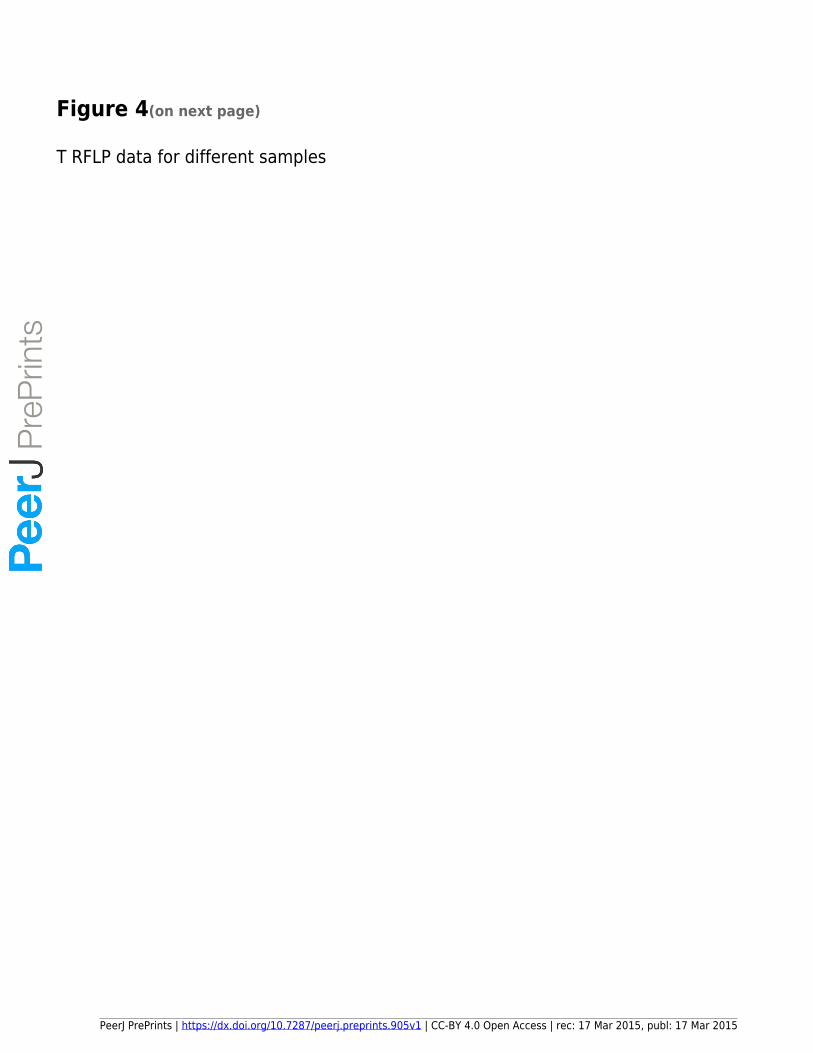
Figure 4(on next page)
T RFLP data for different samples
PeerJ PrePrints | https://dx.doi.org/10.7287/peerj.preprints.905v1 | CC-BY 4.0 Open Access | rec: 17 Mar 2015, publ: 17 Mar 2015
PrePrin
ts
0%
10%
20%
30%
40%
50%
60%
70%
80%
90%
100%
BBW HBW R13 R14 RM SGH
Other
525
516
420
336
198
113
73
32
28
0%
10%
20%
30%
40%
50%
60%
70%
80%
90%
100%
BBW HBW R13 R14 RM SGH
Other
525
410
388
336
292
227
224
192
188
187
181
113
32
A)
B)
PeerJ PrePrints | https://dx.doi.org/10.7287/peerj.preprints.905v1 | CC-BY 4.0 Open Access | rec: 17 Mar 2015, publ: 17 Mar 2015
PrePrin
ts
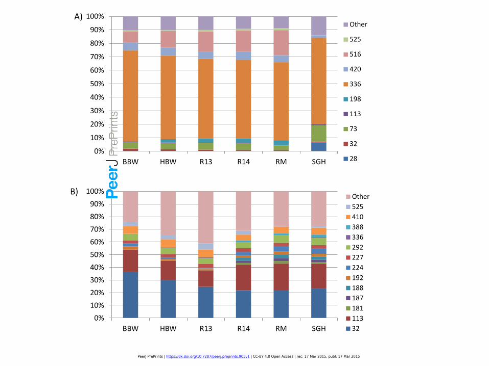
Figure 5(on next page)
Bray-Curtis and NMDS of FISH and TRFLP for all samples
PeerJ PrePrints | https://dx.doi.org/10.7287/peerj.preprints.905v1 | CC-BY 4.0 Open Access | rec: 17 Mar 2015, publ: 17 Mar 2015
PrePrin
ts
1A)
1B)
2A)
2B) 2D Stress = 0.066
2D Stress = 0.028
Cophen r = 0.9425
Cophen r = 0.9896
PeerJ PrePrints | https://dx.doi.org/10.7287/peerj.preprints.905v1 | CC-BY 4.0 Open Access | rec: 17 Mar 2015, publ: 17 Mar 2015
PrePrin
ts

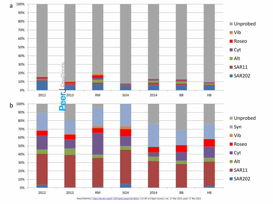
Figure 6(on next page)
FISH adjusted eubacterial percents
PeerJ PrePrints | https://dx.doi.org/10.7287/peerj.preprints.905v1 | CC-BY 4.0 Open Access | rec: 17 Mar 2015, publ: 17 Mar 2015
PrePrin
ts
0%
10%
20%
30%
40%
50%
60%
70%
80%
90%
100%
2012 2013 RM SGH 2014 BB HB
Unprobed
Vib
Roseo
Cyt
Alt
SAR11
SAR202
0%
10%
20%
30%
40%
50%
60%
70%
80%
90%
100%
2012 2013 RM SGH 2014 BB HB
Unprobed
Syn
Vib
Roseo
Cyt
Alt
SAR11
SAR202
a
b
PeerJ PrePrints | https://dx.doi.org/10.7287/peerj.preprints.905v1 | CC-BY 4.0 Open Access | rec: 17 Mar 2015, publ: 17 Mar 2015
PrePrin
ts