molecular landscape of mds and its clinical …...molecular landscape of mds and its clinical...
TRANSCRIPT

Molecular Landscape of MDS and its clinical applications
Papaemmanuil E, PhD
Assistant Attending
Computational Oncology
MSKCC

Disclosure
I have no relevant financial relationships to disclose.
Mye
lod
ysp
last
ic s
ynd
rom
es


The post cancer gene discovery promise
• Biology;
• Molecular oncology;
• Patient tailored medicine;
• Development of rational clinical and therapeutic protocols;

• Well – documented gene mutations, frequencies and prevelance amongst the key MDS subtypes;
• Increasingly understood patterns of co-mutation;
• Recent insights into the biological mechanisms of spliceosome deregulation and downstream
effectors;
Mye
lod
ysp
last
ic s
ynd
rom
es
Papaemmanuil et al, Blood 2013 Hafferlach et al, Leukemia 2014

Mye
lod
ysp
last
ic s
ynd
rom
es
Incorporation into diagnostic and clinical practices is not yet clear;
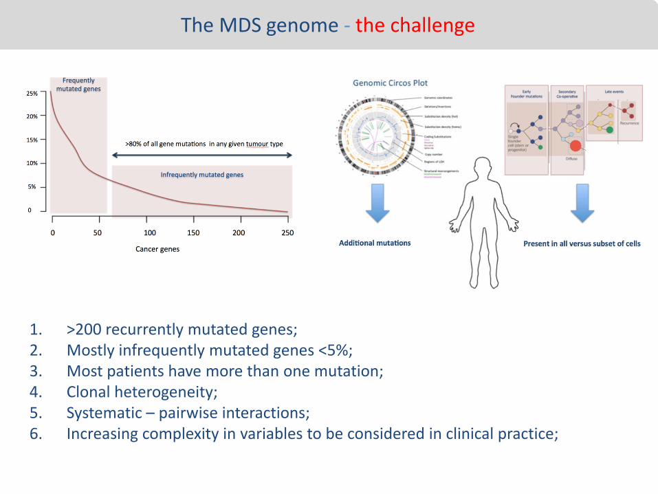
The MDS genome - the challenge
1. >200 recurrently mutated genes;2. Mostly infrequently mutated genes <5%;3. Most patients have more than one mutation;4. Clonal heterogeneity;5. Systematic – pairwise interactions; 6. Increasing complexity in variables to be considered in clinical practice;

Our goal…
1. Insights into the molecular mechanisms of disease biology;
1. Diagnostic biomarkers – genotype, phenotype correlations;
2. Prognostic biomarkers – clinical outcome relationships;
3. Predictive biomarkers – response to therapeutic intervention;
4. Molecular tools to monitor disease progression and response;
5. Therapies tailored to the individual genetic and clonal profiles;
Mye
lod
ysp
last
ic s
ynd
rom
es

Mye
lod
ysp
last
ic s
ynd
rom
es

Splic
ing
fact
or
mu
tati
on
s ar
e d
isea
se d
efin
ing
Splicing factors are disease defining, and much of the focus of biomarker research in MDS

Mye
lod
ysp
last
ic s
ynd
rom
es

SF3
B1
:MD
S Mutations in SF3B1 define a distinct molecular and clinical subgroup in MDS
• Lower blast counts; • Higher presence of ringed sideroblasts; • Lower incidence of multi lineage dysplasia (46.3% vs 82.6%, P < .001); • Lower proportion of dysplastic myeloid cells and megakaryocytes;• Higher absolute neutrophil and platelet counts (P < .001); • Better overall survival (P= 0.003) and lower cumulative incidence of disease progression
Patterns of co-mutation
• SF3B1 mutations define a distinct molecular subgroup that is mutually exclusive tomutations in TP53, and / or cytogenetic abnormalities or complex karyotype.
• Co-mutation with DNMT3A (20%) : - No effect on overall survival or event free survival - Increased involvement of multi lineage dysplasia
• Co-mutation with ASXL1 (7%), RUNX1 (6%) , EZH2 (5%) : - Increased transfusion dependency - Worse prognosis ( P= 0.004), disease progression (P=0.002)
Larger numbers are warranted to study the effects of co-mutation in MDS prognosisMalcovati Blood 2015

SF3
B1
:MD
S 1. Re-evaluation of bone marrow morphology in patients with SF3B1 mutations but
not diagnosed as an MDS with ringed sideroblasts, identifies that SF3B1 predicts for the presence of RS.
2. We show the % of RS is correlated with % VAF of SF3B1 mutations
Malcovati Blood 2012;
Sf3B1 mutation 97% positive predictive value for ringed sideroblastsSF3
B1
:MD
S

SF3
B1
:MD
S 1. Re-evaluation of bone marrow morphology in patients with SF3B1 mutations but
not diagnosed as an MDS with ringed sideroblasts, identifies that SF3B1 predicts for the presence of RS.
2. We show the % of RS is correlated with % VAF of SF3B1 mutations
Sf3B1 mutation 97% positive predictive value for ringed sideroblasts
SF3
B1
:MD
S
Malcovati Blood 2012;

SF3
B1
:MD
S Mutations in SF3B1 define a distinct molecular and clinical subgroup in MDS
• Lower blast counts • Higher presence of ringed sideroblasts • Lower incidence of multi lineage dysplasia (46.3% vs 82.6%, P < .001), • Lower proportion of dysplastic myeloid cells and megakaryocytes• Higher absolute neutrophil and platelet counts (P < .001),
• Better overall survival (P= 0.003) and lower cumulative incidence of disease progression
Patterns of co-mutation
• SF3B1 mutations define a distinct molecular subgroup that is mutually exclusive tomutations in TP53, and / or cytogenetic abnormalities or complex karyotype.
• Co-mutation with DNMT3A (20%) : - No effect on overall survival or event free survival; - Increased involvement of multi lineage dysplasia
• Co-mutation with ASXL1 (7%), RUNX1 (6%) , EZH2 (5%) : - Increased transfusion dependency - Worse prognosis ( P= 0.004), disease progression (P=0.002)
Larger numbers are warranted to study the effects of co-mutation in MDS prognosis
SF3
B1
:MD
S
Malcovati Blood 2015

SF3
B1
:MD
S

SF3
B1
:MD
S Mutations in SF3B1 define a distinct molecular and clinical subgroup in MDS
• Lower blast counts • Higher presence of ringed sideroblasts • Lower incidence of multi lineage dysplasia (46.3% vs 82.6%, P < .001), • Lower proportion of dysplastic myeloid cells and megakaryocytes• Higher absolute neutrophil and platelet counts (P < .001),
• Better overall survival (P= 0.003) and lower cumulative incidence of disease progression
Patterns of co-mutation
• SF3B1 mutations define a distinct molecular subgroup that is mutually exclusive tomutations in TP53, and / or cytogenetic abnormalities or complex karyotype.
• Co-mutation with DNMT3A (20%) : - No effect on overall survival or event free survival - Increased involvement of multi lineage dysplasia
• Co-mutation with ASXL1 (7%), RUNX1 (6%) , EZH2 (5%) : - Increased transfusion dependency - Worse prognosis ( P= 0.004), disease progression (P=0.002)
Larger numbers are warranted to study the effects of co-mutation in MDS prognosis

SF3
B1
:MD
S
Malcovati et al Blood, 2015

SF3
B1
:MD
S
• Defines a distinct clinical and pathologic entity, one characterised by the presence of ringed sideroblasts;
• Molecular testing for SF3B1, more accurately identifies patients in this group, compared to %blast / % ringed sideroblast thresholds;
• SF3B1 is an independent prognostic predictor of clinical outcome associated with favorable prognosis;
• Attention should be given on the pattern of co-mutation: ASXL1 : RUNX1: EZH2 and potentially others..

SRSF
2:M
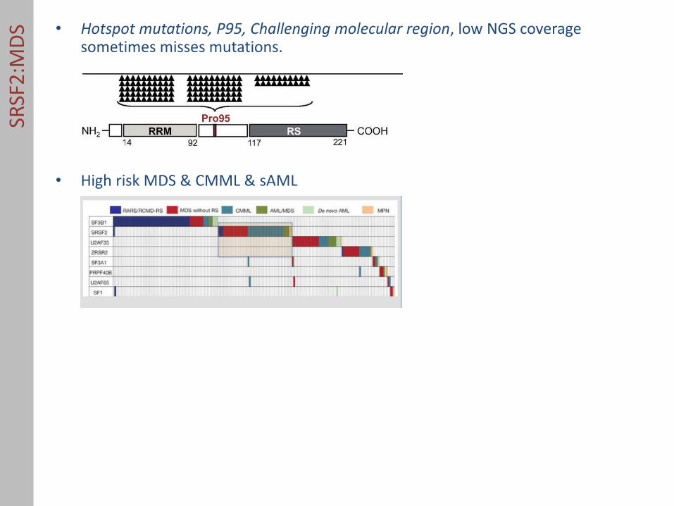
DS • Hotspot mutations, P95, Challenging molecular region, low NGS coverage
sometimes misses mutations.
• High risk MDS & CMML & sAML
• Associated with granulocytic disease, poor outcome and increased risk of leukemic transformation.
• Poor prognosis irrespective of estimates clonal status.
• Is frequently co-mutated with other adverse prognosis genes: ASXL1, STAG2, RUNX1, resulting in further deterioration in overall survival.
SRSF
2:M
DS • Hotspot mutations, P95, Challenging molecular region, low NGS coverage
sometimes misses mutations.
• High risk MDS & CMML & sAML
• Associated with granulocytic disease, poor outcome and increased risk of leukemic transformation.
• Poor prognosis irrespective of estimates clonal status.
• Is frequently co-mutated with other adverse prognosis genes: ASXL1, STAG2, RUNX1, resulting in further deterioration in overall survival.

SRSF
2:M
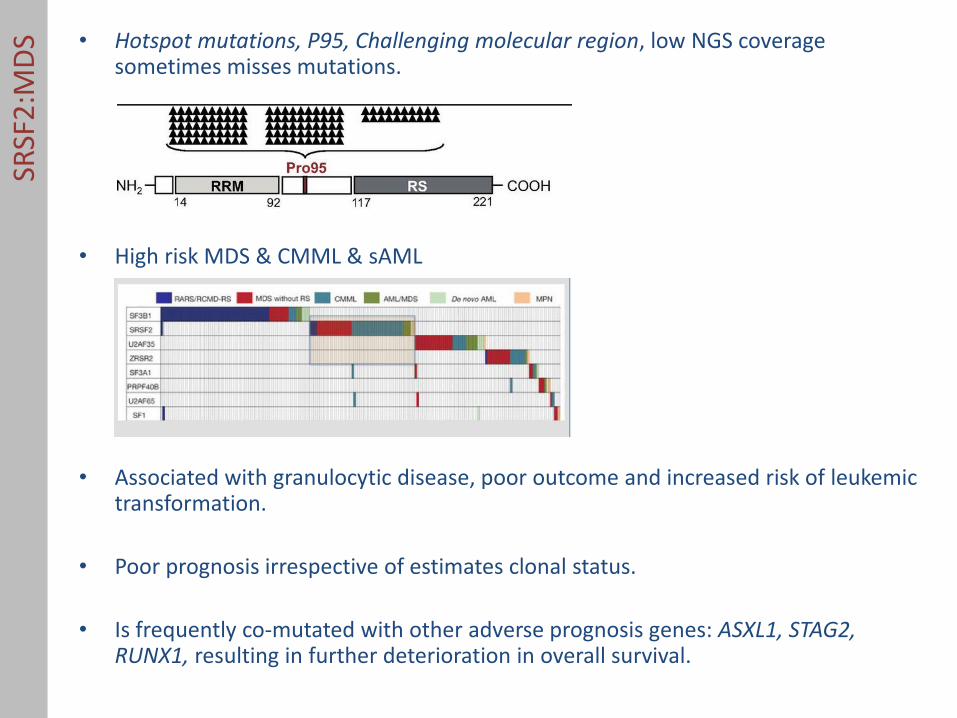
DS • Hotspot mutations, P95, Challenging molecular region, low NGS coverage
sometimes misses mutations.
• High risk MDS & CMML & sAML
• Associated with granulocytic disease, poor outcome and increased risk of leukemic transformation.
• Poor prognosis irrespective of estimates clonal status.
• Is frequently co-mutated with other adverse prognosis genes: ASXL1, STAG2, RUNX1, resulting in further deterioration in overall survival.
SRSF
2:M
DS • Hotspot mutations, P95, Challenging molecular region, low NGS coverage
sometimes misses mutations.
• High risk MDS & CMML & sAML
• Associated with granulocytic disease, poor outcome and increased risk of leukemic transformation.
• Poor prognosis irrespective of estimates clonal status.
• Is frequently co-mutated with other adverse prognosis genes: ASXL1, STAG2, RUNX1, resulting in further deterioration in overall survival.

SRSF
2:M
DS

U2
AF1
:MD
S • Hotspot mutations,
• ~ 7%, mostly high risk MDS & CMML & AML
• Associated with young age, and high-risk of leukemia transformation.
• Adverse prognosis:
Wu et al Am J Hematology 2014; Thol et al Blood 2012

Pro
gno
stic
eff
ect
of
splic
ing
fact
or
mu
tati
on
s
• Whilst SF3B1, associated with favourable prognosis;
• SRSF2; U2AF1, associated with inferior outcomes;
• ZRSR2 – unclear:

MD
S w
ith
sp
licin
g fa
cto
r m
uta
tio
ns
SF3B1
SRSF2 : U2AF1 ZRSR2 SF1 PRPF40B
Clinical presentation and clinical outcomes

Splic
ing
fact
or
mu
tati
on
s in
mye
loid
neo
pla
sms
Positive testing for splicing factor mutations is not unique to MDS

Splic
ing
fact
or
mu
tati
on
s in
mye
loid
neo
pla
sms
SRSF2 has shared genomic landscape between MDS and AML

Splic
ing
fact
or
mu
tati
on
s in
mye
loid
neo
pla
sms
Papaemmanuil under review, Lavallée Blood 2015
SF3B1 mutations occur in distinct genetic background in AML

Dia
gno
stic
cri
teri
a Testing positive for mutations in splicing factor genes..

Busque et al 2012; Jaiswal et al NEJM 2014; Genovese et al NEJM 2014, Xie et al Nat Medicine 2014; McKerrel et al Cell reports
Mu
tati
on
acq
uis
itio
n
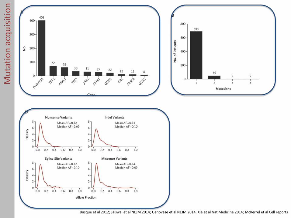
Busque et al 2012; Jaiswal et al NEJM 2014; Genovese et al NEJM 2014, Xie et al Nat Medicine 2014; McKerrel et al Cell reports
Mu
tati
on
acq
uis
itio
n

Clin
ical
pre
sen
tati
on
• Presentation of splicing factor mutations in myeloid neoplasms
% of patients with mutation in panel
21
75
mye
loid
neo
pla
sms
Data presented from panel sequencing

Clin
ical
pre
sen
tati
on
• Presentation of splicing factor mutations in myeloid neoplasms
% of patients with mutation in panel
21
75
mye
loid
neo
pla
sms
Data presented from panel sequencing

Clin
ical
pre
sen
tati
on

Imp
ort
ant
con
sid
erat
ion
s

Formally modeled genomic structure to account for:
Gene mutations + genetic interactions + diagnostic variables + demographic variables.
and build personalised prediction models

26 centers: 2600 cases committed
Meta-analysis ~5000-6000 well annotated MDS cases

2015
Today
Mar Apr May Jun Jul Aug Sep Oct Nov Dec
Invitation to Join Project
Begin receiving clinical data
Finish receiving clinical data
Send out sample queries and MTAs
Begin processing MTA agreements
Begin sending sample tubes
Begin receiving samples at MSKCC
QC and library Construction
Ack
no
wle
dge
me
nts
Peter Campbell Wellcome Trust Sanger Institute• Moritz Gerstung
AML- SG • Hartmut Dohner• Richard Schlenk • Lars Bullinger • Konny Dohner
Papaemmanuil Lab / MSKCC Leukemia Genomics • Gunes Gundem• Matahi Moarii• Franck Rappaport• Juan Medina• Komal Rathi • Noushin Farnhoud • Minal Patel• Kristina Knapp• Irene Phillip• Yesenia Werner• Marc Robert de Massy
MSKCC iGO• Agnes Viale• Kety Huberman