molecular modeling: elucidation of structure/function ...molecular modeling: elucidation of...
TRANSCRIPT

Molecular Modeling: Elucidation of Structure/FunctionRelationships of Proteins and DNA at the Atomic Resolution
Jory Zmuda Ruscio
Dissertation submitted to the Faculty of theVirginia Polytechnic Institute and State University
in partial fulfillment of the requirements for the degree of
Doctor of Philosophyin
Genetics, Bioinformatics & Computational Biology
David R. Bevan, Co-ChairAlexey Onufriev, Co-Chair
Asim EsenT. M. MuraliAdrian Sandu
April 6, 2007Blacksburg, Virginia
Keywords: Molecular Dynamics, Nucleosome Core Particle, β-Glucosidases,Myoglobin
Copyright 2007, Jory Zmuda Ruscio

Molecular Modeling: Elucidation of Structure/Function Relationshipsof Proteins and DNA at the Atomic Resolution
Jory Zmuda Ruscio
(ABSTRACT)
While experiments provide valuable information about biological molecules, currenttechnology cannot yet monitor atomic fluctuations at relevant time scales. Theo-retical computational simulations are able to model the appropriate interactions atatomic resolution. Computational techniques have become widely used for identi-fying interactions in biological systems. Such methods have proven quite accuratein their ability to reproduce experimental data and also in screening and predict-ing pertinent activities. Molecular modeling employs theoretical and computationaltechniques to elucidate biologically relevant information from macromolecular struc-tures. Three biological systems, the nucleosome core particle, myoglobin and gly-cosyl hydrolase family 1 β-glucosidases will be examined with molecular modelingmethods. Results of our analyses provide information about DNA flexibility andpackaging, internal migration of ligands in a small protein, and substrate specificityof an enzyme system.

Attribution
I, Jory Z. Ruscio, performed all of the work reported in this dissertation except forthat which is reported below.
Chapter 2Dr. Alexey Onufriev oversaw the project and contributed to the electrostatic discus-sion
Chapter 3Deept Kumar and Maulik Shukla wrote the PathFinder algorithm used in part ofthe analysis. Drs. Alexey Onufriev and T. M. Murali oversaw the project.
Chapter 4Drs. David Bevan and Asim Esen oversaw the project. Onufriev.
iii

Acknowledgments
This section was the last portion of this document that I wrote, because I find it themost difficult. I have spent almost five of my 25 years studying and living at VirginiaTech in Blacksburg. Numerous people have touched and affected my life during thistime. I can only attempt to acknowledge the influences these individuals have hadon me.
First and foremost, I want to acknowledge the members of my committee, DavidBevan, Alexey Onufriev, Asim Esen, T. M. Murali and Adrian Sandu. I especiallywant to thank Dr. Bevan, who has also my adviser, mentor, teacher and travelingcompanion. He gave me to opportunity to work in his lab, and I appreciate the timeand resources he’s provided to me. I also feel extremely fortunate to have for a co-adviser Dr. Onufriev. Not only has Dr. Onufriev taught me much about science, healways had the time to discuss other topics related to my development as a scientist.The mentorship that Dr. Bevan and Dr. Onufriev have shown me is truly greaterthan the sum of the parts. I am also grateful for the the experience I had during myrotation in Dr. Esen’s lab: it solidified my belief that I did not want to be a wet labscientist; but Dr. Esen’s passion for science was extremely inspiring. The skill setsI learned from Murali and Dr. Sandu, Java and Perl, were extremely instrumentalto my research, and if not for the strong base they taught me, I’m not sure I wouldhave my data analyses finished.
Secondly, I want to thank my friends, with whom I’ve shared the joys and challengesof graduate school. Many nights of stress relief were spent at Rivermill, Bourdreax’s,Bdubs or someones apartment. Without all of you, life in Blacksburg would not havebeen as enjoyable. Thank, you: Ken Hurley, Graham Jack, Craig Tollin, Matt Lieber,Ina and Andrew O’Carroll, Laura and James Freeman, Corban Rivera, Andrew Fen-ley, Ramu Anandakrishnan, Curtis Dahn, John Gordon, Mihaela Babiceanu, DiegoCortes, GSA Board Members, poker companions, and Evergrid friends. I also wantto thank the VT faculty, administrators and staff who have supported me in my
iv

non-research endeavors: Monika Gibson, Dean Karen DePauw, Donna Sanzebach,Dr. Edward Spencer, Dr. Dorris Zallen and Elaine Quenseberry.
I also want to thank my family, who have always supported me in everything that I’vedone . Words cannot express the love and gratitude I have for all of you: my parents,who always believed in my abilities; Ryan, a great brother who has persevered witha smile on his face; Kira, with whom I’ve become much closer through our sharedHokie experience; and Randy, my younger, not little, brother, who has been justdown to the road and contributed much humor to many of the fun gatherings.
Finally, and most importantly, I want to thank my husband Joseph Ruscio. I cannotbegin to describe how important you are to me. I would not be here now if it werenot for you. I cherish the last seven years we have been together and look forwardto the rest of our lives together.
In light of the recent tragic event on Monday, April 16, 2007, I also want to thankthe Virginia Tech community as a whole. The manner in which the community hascome together truly makes me proud to be associated with the Virginia Tech Hokies.
v

Contents
1 Introduction 1
2 A Computational Study of Nucleosomal DNA Flexibility 4
2.1 Abstract . . . . . . . . . . . . . . . . . . . . . . . . . . . . . . . . . . 4
2.2 Introduction . . . . . . . . . . . . . . . . . . . . . . . . . . . . . . . . 5
2.3 Methods . . . . . . . . . . . . . . . . . . . . . . . . . . . . . . . . . . 8
2.4 Results and Discussion . . . . . . . . . . . . . . . . . . . . . . . . . . 13
2.5 Conclusions . . . . . . . . . . . . . . . . . . . . . . . . . . . . . . . . 28
2.6 Acknowledgments . . . . . . . . . . . . . . . . . . . . . . . . . . . . . 31
3 Atomic level identification of ligand migration pathways betweensolvent and heme in myoglobin 32
3.1 Abstract . . . . . . . . . . . . . . . . . . . . . . . . . . . . . . . . . . 32
3.2 Results and Discussion . . . . . . . . . . . . . . . . . . . . . . . . . . 33
3.3 Supplementary Material . . . . . . . . . . . . . . . . . . . . . . . . . 43
4 Computational Investigation of Mechanism and Substrate Speci-ficities of Two Family 1 β-Glucosidases 52
4.1 Abstract . . . . . . . . . . . . . . . . . . . . . . . . . . . . . . . . . . 52
4.2 Introduction . . . . . . . . . . . . . . . . . . . . . . . . . . . . . . . . 53
vi

4.3 Methods . . . . . . . . . . . . . . . . . . . . . . . . . . . . . . . . . . 59
4.4 Results . . . . . . . . . . . . . . . . . . . . . . . . . . . . . . . . . . . 62
4.5 Discussion . . . . . . . . . . . . . . . . . . . . . . . . . . . . . . . . . 74
Bibliography 81
vii

List of Figures
1.1 Explicitly solvated system (a) compared with an implicitly solvatedsystem (b) . . . . . . . . . . . . . . . . . . . . . . . . . . . . . . . . . 2
2.1 Plots of (a) RMSD vs. time of every 10 ps and (b) Atomic PositionalFluctuation by residue. . . . . . . . . . . . . . . . . . . . . . . . . . . 12
2.2 Localization of kinked base-pair steps during the MD simulation ofthe whole nucleosome core particle . . . . . . . . . . . . . . . . . . . 14
2.3 Minor groove width of the bases of the nucleosomal DNA . . . . . . 15
2.4 Axes of curvature . . . . . . . . . . . . . . . . . . . . . . . . . . . . . 17
2.5 End-to-end distance and energy vs. time. . . . . . . . . . . . . . . . . 20
2.6 Conformational energy as a function of the end-to-end distance of thenucleosomal DNA . . . . . . . . . . . . . . . . . . . . . . . . . . . . . 22
2.7 Standard deviation of the base-pair step parameters roll, twist andslide values . . . . . . . . . . . . . . . . . . . . . . . . . . . . . . . . 27
2.8 An example of two plausible structural scenarios of cyclization of shortDNA fragments. . . . . . . . . . . . . . . . . . . . . . . . . . . . . . 28
3.1 A schematic representation of the two channels of ligand migrationand exit/entry in native myoglobin. . . . . . . . . . . . . . . . . . . . 35
3.2 Comparison of ligand trajectories and free volume fluctuation analysis. 36
3.3 Effect of conformation of residue 138 in native myoglobin. . . . . . . 37
3.4 Effects of residue 68 in native myoglobin. . . . . . . . . . . . . . . . . 40
viii

3.5 Effect of residue 68 in V68F myoglobin. . . . . . . . . . . . . . . . . . 41
3.6 Structural origins of ligand migration channels and exit/entry portalsin myoglobin. . . . . . . . . . . . . . . . . . . . . . . . . . . . . . . . 42
3.7 The starting positions of the 8 ligands used in the “CO entry fromsolvent” MD simulations. . . . . . . . . . . . . . . . . . . . . . . . . 45
3.8 Ligand entry and exit times by channel and portal. . . . . . . . . . . 48
3.9 RMSD of backbone atoms during the 90 ns simulations of carbon-monoxy myoglobin (black) and myoglobin with photodissociated CO(red). . . . . . . . . . . . . . . . . . . . . . . . . . . . . . . . . . . . 49
3.10 RMSD per residue during two native myoglobin trajectories. . . . . 50
3.11 The points in native myoglobin that occur with the highest frequency 51
4.1 First step (glycosylation) of double-replacement retaining mechanism. 55
4.2 Structures of DIMBOA-glucoside and dhurrin . . . . . . . . . . . . . 56
4.3 Orientation of the glucosyl moiety of the ligand in the simulations ofGlu1 DIMglc and Dhr1 DIMglc . . . . . . . . . . . . . . . . . . . . . 65
4.4 Number of water molecules within 3.5 A of acid/base catalytic gluta-mate over the course of the simulations. . . . . . . . . . . . . . . . . 68
4.5 Effect of dhurrin on active site of Glu1. . . . . . . . . . . . . . . . . . 69
4.6 The active sites of Glu1 DIMglc, Glu1 dhr and Y473F dhr after min-imization and after 3.5 ns of MD. . . . . . . . . . . . . . . . . . . . . 70
4.7 Different conformations of glucose. . . . . . . . . . . . . . . . . . . . 71
4.8 Residues included in the three different Active Site Profiles. . . . . . 73
4.9 Clustering of active site residues that line the aglycone binding site(Profile20) of the β-glucosidases. . . . . . . . . . . . . . . . . . . . . 75
4.10 Profile20 of the three sequences that did not cluster in the same sub-families based on the full sequences. . . . . . . . . . . . . . . . . . . 76
ix

List of Tables
2.1 Computation time and processor utilization of the simulations. . . . . 11
2.2 Comparison of the nucleosomal DNA bending energies predicted withinthe classical elastic rod theory with those observed in the simulationsreported here. . . . . . . . . . . . . . . . . . . . . . . . . . . . . . . 24
2.3 Destabilization effects of monovalent salt on the “bent” DNA staterelative to the “straight” one as a function of solvent salt concentra-tion. . . . . . . . . . . . . . . . . . . . . . . . . . . . . . . . . . . . . 25
3.1 Surface residues that close to the nine ligand exit/entry portals innative myoglobin shown in Figure 1. . . . . . . . . . . . . . . . . . . 34
3.2 Simulations in which CO escapes/enters native myoglobin and theresidue number of the CO. . . . . . . . . . . . . . . . . . . . . . . . 47
4.1 Distances between the catalytic residues in other Family 1 glycosylhydrolases. . . . . . . . . . . . . . . . . . . . . . . . . . . . . . . . . 54
4.2 Hydrogen bonding between glucose atoms and protein atoms. . . . . 57
4.3 Simulations performed and abbreviations used in the paper. . . . . . 61
4.4 Initial and average distances between two distances that are affectedby substrate specificity. . . . . . . . . . . . . . . . . . . . . . . . . . . 63
4.5 Persistence of hydrogen bonds between glucose moiety and proteincomplexes over the 3.5 ns simulations. . . . . . . . . . . . . . . . . . 66
x

4.6 The change in the dihedral angle that characterizes the conformationof the glucose and the charges of the C1, O5 and O1 atoms when theglycosidic bond (between C1 and O1) is lengthened. . . . . . . . . . 72
xi

Chapter 1
Introduction
The term “molecular modeling” refers to the use of computational and theoreticaltechniques to model the dynamics of molecules. The use of molecular modelingtechniques allows for researchers to interpret or predict interactions at a resolutionthat cannot be detected with current experimental approaches. The availabilityof high-performance computing clusters and the increasing accuracy of theoreticalmethods have allowed molecular modeling and experiment to complement each otherin an attempt to answer biological questions (82).
Molecular dynamics (MD) is the chosen technique for the analysis of the biologicalsystems presented in this work. MD simulations use Newtonian physics to determinetime-dependent behavior of the atomic system. The AMBER (Assisted Model Build-ing with Energy Refinement) suite of programs is used to simulate and analyze thesystems of interest (20). The AMBER MD simulations use the following potentialfunction to calculate the interactions between atoms:
Etotal =∑
bonds
Ki
2(li − li,0)
2 +∑
angles
Ki
2(θi − θi,0)
2 +∑
torsions
Vn
2(1 + cos [nω − γ])
+
N∑
i=1
N∑
j=i+1
(4ǫij
[
(σij
rij
)12 − (σij
rij
)6
]
+qiqj
4πǫ0rij
)
Bond stretching, angle bending, bond rotation and non-bonded interactions con-tribute to the energy of the system. The last term,
qiqj
4πǫ0rij, corresponds to the explicit
solvent treatment. The simulations are typically conducted under NVT conditions
1

2
(a) (b)
Figure 1.1: Explicitly solvated system (a) compared with an implicitly solvated sys-tem (b)
- constant Number of atoms, constant Volume and constant Temperature. Eitherexplicit solvation (Figure 1.1a) or implicit solvation (Figure 1.1b) is used to modelwater molecules. Implicit solvent simulations are described in detail in Sec. 2.3.4.
Atomistic molecular simulations have progressed tremendously over the past coupleof decades. The first simulation of the dynamics of molecules was reported 50 yearsago (3). This hard sphere model, though rather simple, did demonstrate the utility ofusing theoretical calculations to model the dynamic behavior of a system. Since 1957,the use of molecular dynamics simulations has greatly increased, and the applicationof this technique has allowed for the understanding, interpretation and prediction ofproperties and dynamics of many biological systems. The year 1977 saw the first MDsimulation of a biological molecule, bovine pancreatic trypsin inhibitor (85). This500 atom system was simulated for 9.2 ps. Recently in 2006, the complete satellitetobacco mosaic virus, a system of over 1 million atoms, was simulated for over 50ns (51).
This document presents the results of MD simulations of three different biologicalsystems: DNA; a carrier protein; and enzyme-ligand complexes.
Chapter two presents research on the nucleosome core particle (NCP), which is a

3
DNA-protein complex, and the associated DNA. The NCP is the primary unit inDNA packaging in the eukaryotic cell. It is composed of ∼147 bp of DNA wrappedaround a histone octamer protein core. Static crystal structures of the NCP indicatetwo different modes of DNA distortion: local kinking between base-pairs and globalbending of the 147 bp of the DNA. We attempt to understand the mechanism of thesedistortions through the use of implicit solvation MD of the NCP and its associatedDNA free in solution.
The work in chapter three examines ligand migration pathways in myoglobin. Lo-cated in muscle cells, this small globular protein stores oxygen, which binds to aninternal heme iron atom. Although myoglobin was one of the first proteins to have itsthree-dimensional structure solved, uncertainty still exists as to how oxygen migratesbetween the exterior and the interior of the protein; no pathway is evident in anyof the over 250 structures solved to date. We used numerous explicit solvation MDsimulations and a novel algorithm to determine what appears to be a fairly completepicture of ligand migration pathways in myoglobin.
Finally, chapter four describes the investigation into the substrate specificity andmechanism of β-glucosidases. β-glucosidases are proteins involved in the hydrolysisof glucosides. Some of these enzymes exhibit a wide range of substrate specificity,while others are very narrow in specificity. We use two experimentally well charac-terized β-glucosidases, Glu1 and Dhr1, to examine the types of interactions that areimportant in determining substrate specificity and the mechanism of the proteins.Explicit solvation MD simulations are conducted to identify amino acids responsiblefor substrate recognition in Glu1 and Dhr1, which may lead to a better understand-ing of substrate specificity in other β-glucosidases.

Chapter 2
A Computational Study ofNucleosomal DNA Flexibility
Jory Z. Ruscio and Alexey Onufriev
Permission to reprint granted by the Biophysical Journal for this article originallypublished in the Biopysical JournalBiophys. J. 91:4121-4132. 2006
2.1 Abstract
Molecular dynamics simulations of the nucleosome core particle and its isolated DNAfree in solution are reported. The simulations are based on the implicit solventmethodology and provide insights into the nature of large-scale structural fluctua-tions and flexibility of the nucleosomal DNA. In addition to the kinked regions pre-viously identified in the X-ray structure of the nucleosome, the simulations supportthe existence of a biochemically identified distorted region of the DNA. Comparisonof computed relative free energies shows that formation of the kinks is associatedwith little, if any, energy cost relative to a smooth, ideal conformation of the DNAsuperhelix. Isolated nucleosomal DNA is found to be considerably more flexible thanexpected for a 147 bp stretch of DNA based on its canonical persistence length of 500A. Notably, the significant bending of the DNA observed in our simulations occurs
4

5
without breaking of Watson-Crick bonds. The computed relative stability of bentconformations is sensitive to the ionic strength of the solution in the physiologicalrange; the sensitivity suggests possible experiments that might provide further in-sights into the structural origins of the unusual flexibility of the DNA.
Key words: molecular dynamics, nucleosome, DNA flexibility
2.2 Introduction
Evidence is now overwhelming that not only the sequence, but also the details ofDNA packaging inside the cell are an important part of the genetic message. Theprimary level of DNA compaction in eukareotic organisms in vivo is the nucleosome.At this level, a stretch of 147 base-pairs of the DNA is tightly wrapped (∼1.65times) around a set of eight proteins (histones) that carry the charge opposite ofthat of the DNA. Details of the nucleosome dynamics are vital for understandingkey cellular processes such as DNA replication, repair and transcription (55, 70,71, 139, 140). Cell differentiation is also intimately linked with DNA compaction.Despite its importance, the nucleosome system is far from being fully understood.One of the key unanswered questions is the following: how can the whole nucleosomebe highly stable, protective of its genetic material, while at the same time its tightlywrapped DNA be highly accessible, easily revealing its information content?
This dual nature of the DNA packaging in the nucleosome is supported by experimen-tal studies; these reveal the high stability of the whole nucleosome at physiologicalconditions (122), and, at the same time, suggest that small fragments (∼50 bp) ofthe DNA helix can transiently “peel off” (4, 97). The latter observation implies arelatively small free energy barrier associated with such partial unraveling and issuggestive of possible mechanisms behind processes such as transcription, in whichfragments of the nucleosomal DNA become accessible sequentially (97). Since theatomic resolution structures of the nucleosome have become available, there is likelynot much ambiguity left about its static conformation; however, details of nucleoso-mal dynamics are not as clear, especially at atomic resolution. These are importantfor developing molecular mechanisms of the key biological processes involving the nu-cleosome. Theoretical studies using atomistic molecular dynamics simulations mayprovide much needed insights in this area.
A general question that can be addressed by such simulations is exactly how flexible

6
is the DNA wrapped around the nucleosome? Severely restricted mobility of thedouble helix would be suggestive of tight binding, possibly inconsistent with theidea of transient DNA dissociation in processes such as transcription. A relatedquestion is what is the origin of structural distortions that arise when the straightDNA double-helix is forced to adopt the conformation found on the nucleosome. Pastexperimental studies have characterized the persistence length of DNA to be ∼500A, or 150 base pairs (33, 112, 133). Based on this finding, one would expect thatconsiderable force would have to be exerted on the DNA for it to adopt the highlybent superhelical conformation of nucleosomal DNA. Not only would this force needto wrap the DNA around the histone core, but, based on experimental data, itwould also have to locally distort the DNA itself. In particular, the histone corewas shown to have a preference for DNA with an altered helical periodicity (58).Additionally, Hayes et al. showed that DNA sequences with different structuralproperties in solution all adopt a similar, slightly perturbed conformation in thenucleosome (57). The 1.9 A resolution nucleosome core particle crystal structureaffirms that the structure of the DNA on the histone core deviates significantly fromthe best fit ideal superhelical DNA (100). Not only does nucleosomal DNA have twicethe base-pair-step curvature needed for the superhelical conformation, but it is kinkedin several regions. Structurally distorted regions, or “kinks”, have also been identifiedbiochemically. Regions ±1.5 helical turns from the dyad have been shown to be thesites of DNA distortion. Some of these kinked regions have been shown to havepotential biological significance. For example, the DNA located at ±15 bp from thedyad have increased sensitivity to attack by singlet oxygen (61), which preferentiallyattacks denatured or wedge-shaped DNA structures. This region of the DNA is alsorecognized as being distorted by HIV integrase (98) and permanganate (48).
The idea that considerable force is needed to create the large overall bending andlocal distortions of the nucleosomal DNA appears common sense from the classicalpicture of the rather inflexible DNA. However, it may not be all that simple, ac-cording to recent intriguing experimental findings (28, 29). Namely, on short lengthscales (∼100 bp) the DNA double helix was not found to behave as a relatively stiffrod, as might have been expected based on its classical (33, 112, 133) persistencelength value of ∼150 bp. In contrast, short DNA fragments were found to cyclizespontaneously, with an appreciable probability. Exactly how this unusual flexibilityis accommodated structurally is not known (29). Since relatively short, bent DNAfragments participate in many vital biological processes, the issue is important; it hasalready provoked considerable interest and debate in the community. Various, mu-tually exclusive (29, 44, 143), explanations for the phenomenon have been proposed.While the debate does not yet appear to have been settled (44), recent experimental

7
evidence based on techniques such as FRET (113) and AFM(138) – different fromthose used in the original study(28) – provide more evidence in support of enhancedDNA flexibility on short length scales.
In order to investigate the flexibility of nucleosomal DNA and examine the origins ofpossible structural distortions contributing to this flexibility, we have performed sev-eral all-atom molecular dynamics simulations. These simulations explore, at the fullatomic resolution, the dynamics of the entire nucleosome core particle (histone coreand the DNA) as well as its isolated DNA free in solution. In some of the simulations,constraints have been used to model the DNA winding around the nucleosome.
The key to our approach is the use of the “implicit solvent” (93, 126) technique inwhich the effects of aqueous solvation are represented implicitly, via a continuummedium with the properties of bulk water. The electrostatic screening effects of thehigh dielectric medium as well as those of salt ions enter implicitly, via appropriateterms added to the system’s configurational energy. The “hydrophobicity” is also in-cluded as a separate contribution. Within the approach, the system’s configurationalenergy in the presence of solvent becomes an analytical function of the coordinatesof the macromolecule only (and solvent parameters such as ionic strength), whichcontributes to the method’s computational efficiency. The technique effectively elim-inates the need to keep track of the individual water molecules, and focuses the com-putational power on the macromolecule of interest (e.g. the protein and the DNA),often resulting in considerable gains in computational efficiency. Also, compared tothe traditional explicit solvent simulations, the implicit solvent methodology effec-tively eliminates the drag of viscosity, leading to greatly enhanced conformationalsampling. The approach is also particularly well suited for estimations of relativefree energies of various molecular conformations. Over the past decade the method-ology has enjoyed considerable success, especially in the computationally challengingapplications such the protein folding problem(72, 114).
The paper has the following structure. First, we orient the reader by briefly describ-ing the simulations we have performed. Then we present the results of the moleculardynamics simulation of the entire nucleosome, highlighting the structural fluctua-tions and “kinking” of the DNA. Next we describe the simulations and energeticanalyses of two superhelical DNA structures and show that formation of the kinkedstructures on the nucleosomal DNA are unlikely to entail considerable energetic costs,on average. Finally, we discuss the flexibility of isolated nucleosomal DNA observedin our simulations. Analysis of the relative free energies of the conformations withvarious degrees of bending is presented. Computational protocols and comparativevalidation of the methodology are presented Section 2.3.

8
2.3 Methods
2.3.1 Structures
The 1.9 A crystal structure of the nucleosome core particle (PDB code 1KX5) (37)has been used as the initial structure for molecular dynamics of the NCP-DNA andWholeNCP-DNA simulations. The same sequence is used to build DNA in standardB-form and also the Ideal-DNA. The structures are built with NAB (83). The Free-
DNA is built using standard B-form parameters of 35.87o as the twist value and 3.33A as the rise value. The parameters of the WholeNCP-DNA are first analyzed withX3DNA (81), and the corresponding parameter values of 34.65o for twist and 3.38 Afor rise are used to build the Ideal-DNA.
2.3.2 Molecular Dynamics
All MD trajectories have been obtained with AMBER 8 (19), using ff99 force-field.The SHAKE method is used to restrain hydrogen – heavy atom bond distances. Theintegration time-step is 2 fs. The average temperature of the system is maintained at300K by weak coupling (via the Berendsen algorithm ) to a heat bath with couplingconstant of 2 ps.
Simulations in Implicit Solvent
The implicit solvent methodology based on the modified GB model(93) (GBOBC ,igb = 5) is used to describe solvation effects in MD simulations of the WholeNCP-
DNA, Free-DNA, NCP-DNA and Ideal-DNA. The mbondi2 radii are set. The non-
polar contribution is computed via ∆Gsurf = 0.005[kcal/mol]xA[A2], where A is
the solvent accessible surface of the molecule estimated by a fast analytical routinewithin AMBER. A reaction field “cut-off” (rgbmax = 15) is employed to speed-upthe calculation of effective Born radii(20). No cut-off is used for the long-rangeinteractions. The salt concentration is set to 0.2 M during the WholeNCP-DNA andthe Free-DNA simulations. A salt concentration of 10 M is used for the NCP-DNA
and Ideal-DNA simulations in order to dampen the electrostatic repulsions betweenthe “coils” of the nucleosomal DNA and reduce possible artifacts associated with theuse of the restraints. All simulations undergo 100 steps of minimization with a 5kcal/mol all atom constraint relative to the x-ray positions. Two 10 ps equilibration

9
steps are performed, the first using a constraint of 1.0 kcal/mol/A2, the second usinga 0.1 kcal/mol constraint. Following equilibration, the simulations continued withoutconstraints, or with 0.1 or 0.001 kcal/mol/A2 constraints, as in the NCP-DNA andIdeal-DNA simulations. All simulations continue for 1 ns, except for the Free-DNA
for which a total of 5 ns is performed.
Simulations in Explicit Solvent
Explicit simulations of the WholeNCP-DNA and Free-DNA have been performedto provide further validation and a reference point for implicit solvent simulations.Detailed comparisons are presented in the Validation subsection below.
The initial minimization steps are as follows. The systems are immersed in a boxof TIP3 water and are neutralized with Na+ ions. The default PME parametersof AMBER 8 are used (20). The water and ions are first equilibrated to 300K for100 ps while the solute is held frozen. Next the water and ions undergo 300 stepsof minimization. Lastly, the solvent and solute are minimized for 300 steps. Thesystem is then gradually heated to 300 K over 40 ps at constant volume and thenan additional 40 ps of constant volume MD at 300K is performed. The constantpressure explicit solvent MD simulations with Particle Mesh Ewald (PME) of theWholeNCP-DNA and Free-DNA are performed. The following conditions are used:SHAKE on the hydrogen atoms, a 2 fs timestep, temperature of 300 K, a 9 A cutoffapplied to the long-range interactions.
2.3.3 Calculation of structural signatures
The minor groove has been measured with the ptraj module of the AMBER pack-age, using the same procedure as reported by El Hassan and Calladine (46). TheDNA parameter values of roll, twist and slide have been calculated with the packageCurves (77).
The error margins are computed by dividing the 500 snapshots into 5 equal sizedbins in order of the time. The average of each bin is computed, and the error iscomputed from the standard deviations of the 5 averages.

10
2.3.4 Calculation of energies and forces in implicit solvent.
All energies are calculated using the MM-GBSA module in AMBER8, which uses thecontinuum solvent approximation. The same scheme is used to compute configura-tional energy E(xi, yi, zi) as a function of atomic coordinates (xi, yi, zi) for each stepof molecular dynamics. The forces are computed as Fi = ∂E/∂xi, see e.g. Ref. (106).Within the implicit solvent approach, the total energy E of the solvated system iscalculated as the sum of gas-phase energy plus the free energy of solvation that in-cludes the electrostatic and non-polar parts. Namely, E = Eint +Eelec +Evdw +Esurf .Here, Eint represents the additive bond, angular and dihedral degrees of freedomrespectively,
Eint =∑
bonds
Kr(r − req)2 +
∑
angles
Kθ(θ − θeq)2 +
∑
dihedrals
Vn
2[1 + cos(nφ − γ)]
with the constants and their specific values defined in the Cornell et al. forcefield (32); a general discussion of this type of decomposition can be found in (106)
The van der Waals interactions between the atoms are described by Evdw, andEsurf mimics the hydrophobic effect. We use [kcal/mol] Esurf = 0.005A, where A[A2] is the calculated solvent accessible area of the molecule. Eelec is computed asEelec = Evac + Esolv, where Evac is the protein’s Coulomb energy in vacuum, andEsolv is the electrostatic component of the free energy of solvation, computed herewithin the generalized Born approximation. All components of the total energy arecomputed for each snapshot using the AMBER force field parameters. Note thatthe energy computed by the MM-GBSA approach includes the free energy of solventrearrangement implicitly. For the MM-GBSA we use the same force-field, GB modeland non-polar surface parameters as described above in the MD simulation section.The differences are as follows. For the energetic analysis we do not use any reactionfield cut-offs (when calculating effective Born radii we set rgbmax > system size ).Also, we set the salt concentration to 0.2 M in all cases except in the calculation ofthe salt dependence of the relative stability of the “bent” and ”straight” conforma-tions. Naturally, no restraining potentials are included for the “post-MD” analysisof the individual snapshots.
The relative stability is calculated as ∆∆G(salt) = ∆G(salt → ∞) + [∆Eelec(salt)−∆Eelec(salt → ∞)]. Since we are interested only in how the destabilization effectsof salt change as the salt concentration is decreased, we set ∆G(salt → ∞) = 0.The GB model we use here was demonstrated to describe the electrostatic effects ofmonovalent salt adequately(117).

11
2.3.5 Computational resources and times
Table 2.1: Computation time and processor utilization of the simulations.Simulation # Atoms Simulation Computation # CPUs
Time Time (hoursImplicit - WholeNCP-DNA 25,086 1 ns 720 16Explicit - WholeNCP-DNA 223,132 500 ps 70 16Implicit - NCP-DNA 9,346 1 ns 122 32Implicit - Free-DNA 9,346 5 ns 115 128Explicit - Free-DNA 111,629 500 ps 35 16Explicit - DNA decamer 10,277 10 ns 56 16Implicit - DNA decamer 632 10 ns 49 8
All of the MD simulations have been performed on Virginia Tech’s SYSTEM X, the2200 CPU supercomputer (http://www.tcf.vt.edu/systemX.html). The compu-tational times and CPUs used for each simulation are noted in table 2.1. Clearly, forthe large systems such as the entire nucleosome, the implicit simulations reportedhere appear to be more computationally expensive than the explicit simulations.This is due to the fact that we have not used any approximations to speed up thecalculation of the charge-charge interactions to avoid introducing any potential arti-facts into the implicit solvent (GB) approach. As a result, the time complexity of themethod is O(N2), where N is the number of atoms. On the other hand, the tradi-tional explicit solvent simulations typically employ the Particle-Mesh Ewald (PME)approximation, which reduces the time complexity of computing the electrostaticinteractions to O(Nlog(N)) by introducing an artificial periodicity into the system.Still, the seemingly unfavorable computational expense of the implicit simulationsare completely offset by drastically enhanced conformational sampling: for the DNAsystem, the enhancement is believed to be a factor of 20 or even 100, see Refs.(125, 141). Thus, a 5 ns implicit solvent simulation of a DNA fragment effectivelycorresponds to 0.1 - 0.5 µs in explicit solvent.
2.3.6 Additional Validation
The structures from the implicit solvent MD simulations described above have beencompared to the ones obtained by the traditional, explicit solvent approach. Thelatter, in conjunction with modern force-fields, is known to provide a fairly good

12
(a) (b)
Figure 2.1: Plots of (a) RMSD vs. time of every 10 ps and (b) Atomic PositionalFluctuation by residue. Only the last 5 ns of the explicit (black) and implicit (gray)decamer simulations are shown.
representation of nucleic acids as compared to experiment(23, 24). In our implicitsolvent simulation, the all-atom rmsd to the X-ray structure of the WholeNCP-DNA
system stabilizes soon after equilibration to around 4.0 A and exhibits only minorfluctuations around this value for the rest of the simulation. The all-atom rmsd ofthe explicit solvent trajectory is 3.6 A at the end of the 500 ps simulation. Flexiblehistone tails are excluded from the rmsd calculations in all cases.
To provide an additional support for the use of the implicit solvent approach inthe context of the nucleosomal DNA, the number of Watson-Crick base pairs havebeen calculated for each snapshot of the implicit solvent based WholeNCP-DNA andFree-DNA simulations. (The 3DNA package(81) is used). The average number ofpairs for the WholeNCP-DNA is 143. The average number of pairs for the Free-DNA
simulation is 146. When the 10 base-pairs from both ends are excluded from thecalculation, on average, all included base pairs are persistent throughout both theWholeNCP-DNA and Free-DNA simulations.
Relatively short DNA fragments present an opportunity to directly compare theexplicit and the implicit solvent MD on multi-nanosecond time scale. To this endwe have used a 10 bp fragment (decamer GCGCGCGCGC created with the NABprogram using standard B-form DNA parameters). Both the implicit and explicitwater simulations are run for 10 ns using the protocols described above. All Watson-

13
Crick base-pairs have persisted throughout both the implicit and explicit solventtrajectories. In the explicit solvent simulation the backbone rmsd to the t = 0conformation is 1.8 ± 0.4, averaged over the last 5 ns; for the same time frame, thermsd of the implicit simulation is only slightly larger, 2.2 ± 0.4. As is evident formFig.2.1, the decamer in the implicit simulation is as stable as in the in the explicitsolvent simulation. We attribute the fluctuations to the shortness of the fragment,and note that similar results and rmsd values were reported earlier by Tsui and Casefor a DNA fragment of similar size (125). The agreement between the explicit andthe implicit solvent results provides an additional support for the use of GB-basedimplicit solvent model for the DNA simulations, at least on the time scale of 10 ns.
2.4 Results and Discussion
The molecular dynamics (MD) simulations we have performed can be divided intothree groups: 1) MD of the whole nucleosome core particle; 2) MD of just the nucle-osomal DNA constrained to the wound-up conformation found on the nucleosome;and 3) MD of the “free” nucleosomal DNA in solution, initially set in a straight,classical B-form. All structures used have the same sequence as the DNA in the 1.9A NCP crystal structure of Richmond and Davey (100). In this section we describeresults and analysis of the simulations in each group identified above. In all simu-lations, an implicit solvent model based on the generalized Born approximation hasbeen used to represent solvent effects, including the screening effects of salt.
2.4.1 The Whole Nucleosome
The 1 ns long simulation of the whole nucleosome core particle (WholeNCP-DNA:histones + the DNA) is based on the X-ray structure of Richmond and Davey asthe starting conformation. The main motivation is to estimate the size of the DNAfluctuations and explore the nature of the “kinked” regions originally identified in theX-ray structure of the NCP (100). In particular, Richmond and Davey identified sixkinked base-pair steps (16/17, 26/27, 38/39, 109/110, 121/122, and 131/132), threeon either side of the dyad. Minor groove bending causes kinking of the DNA in the1.9 A crystal structure of the NCP and is defined by, relative to the classical B-form,increased slide values, increased twist values, and decreased roll values. The resultsof our simulation are summarized in Fig. 2.2. Our definition of a kink is slightly lessstringent than Richmond’s ( roll < -11.8o; twist > 40o; and slide > 1.25A) in order
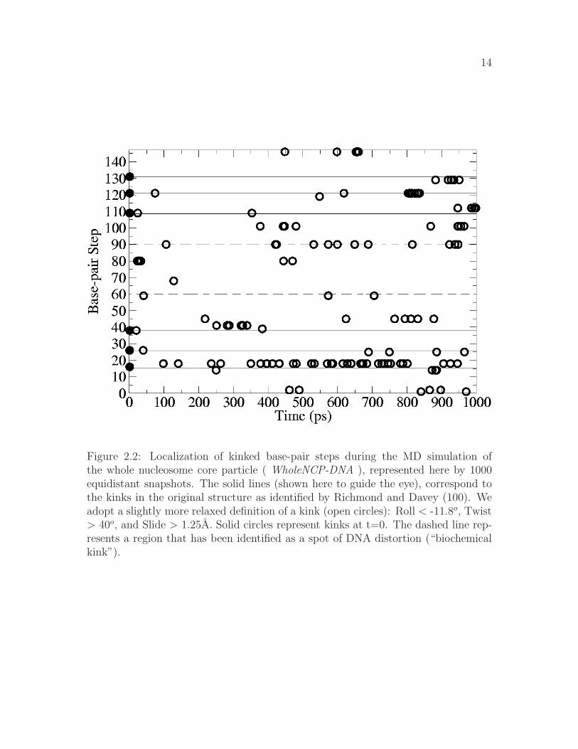
14
Figure 2.2: Localization of kinked base-pair steps during the MD simulation ofthe whole nucleosome core particle ( WholeNCP-DNA ), represented here by 1000equidistant snapshots. The solid lines (shown here to guide the eye), correspond tothe kinks in the original structure as identified by Richmond and Davey (100). Weadopt a slightly more relaxed definition of a kink (open circles): Roll < -11.8o, Twist> 40o, and Slide > 1.25A. Solid circles represent kinks at t=0. The dashed line rep-resents a region that has been identified as a spot of DNA distortion (“biochemicalkink”).

15
Figure 2.3: Minor groove width of the bases of the nucleosomal DNA ( WholeNCP-
DNA ) during the 1 ns MD simulation. Thick blue solid line: values averaged overlast 500 ns of the MD, the corresponding standard deviations are shown as red dottedlines. For reference, the initial values of minor grove width are also shown (blackdashed line). An extreme narrowing of the minor groove occurs near a region locatedat ±15 base pairs from the dyad.

16
to ensure the inclusion of all of the six kinked base-pairs from the X-ray structure.Overall, during the course of the 1 ns long MD simulation, the kinks do appearto persist predominantly at the locations found in the X-ray structure, providingsupport for the methodology we use. The simulation also reveals the presence of akinked region (dashed line at base-pair step 90 in Fig. 2.2) previously not identifiedin the X-ray structure. This region corresponds with the biochemically identifieddistorted structure (48, 61, 98). Analysis of the minor groove width during the MDsimultion supports the existence of a distorted structure located at ±15 bp from thedyad (Fig 2.3). Near residues 60 and 90, the minor groove width decreases from∼9 A the initial value in the crystal structure, to an average of ∼6.5 A over thelast 500 ps of the simulation. During DNA bending, the minor groove will becomecompressed when it faces the histone octamer. Sharp bending is evident ±15 bpfrom the dyad, as the width of the minor groove at this location is much less thanthe other minor grooves that also face the histone octamer.
2.4.2 The Superhelical DNA: energetics of the kink forma-
tion
Experimental determination of the amount of free energy stored in the kinks ofthe nucleosomal DNA is difficult, if not impossible; certainly, this information is notdirectly available from the X-ray structure of the nucleosome. At the same time, MDsimulations based on implicit solvent offer an opportunity to estimate this additionalconformational energy, relative to a hypothetical “smooth” superhelical DNA, ina fairly straightforward manner (125, 126). To this end, we have performed MDsimulations of two 147 bp fragments of DNA starting from the structures describedbelow and illustrated in Fig. 2.4 (a):
1. NCP-DNA - nucleosome core particle DNA in the same conformation as in thecore particle, but stripped of histones.
2. Ideal-DNA - an “ideal” (“smooth”, no kinks) superhelical DNA with structuralparameters corresponding to the average values of NCP-DNA (see “Methods”for details). No histones.
The Ideal-DNA and NCP-DNA have each undergone 1 ns of MD in implicit solvent.
Cartesian harmonic restraints ( 0.1 kcal/mol/A2
) have been used to hold all theatoms close to their original positions. The results are illustrated in Fig. 2.4 (b),

17
(a) (b)
Figure 2.4: Axes of curvature of the initial (a), and final (b) snapshots of the NCP-
DNA (dark) and the Ideal-DNA (light) simulations. Both structures represent thewinding of the DNA in the nucleosome, but the “kinks” are initially present only inthe NCP-DNA ; the Ideal-DNA is in an ideal, smooth superhelical conformation atthe start of the simulation. No histone core is present in either case, but all-atom
harmonic restraints (0.1 kcal/mol/A2
) are used to keep the structures close to theirrespective initial conformations.

18
which shows the qualitative difference between the two structures at the beginningand at the end of the simulation. Clearly, the overall superhelical shape of the DNAfound on the nucleosome is preserved by the constraints throughout the simulation. Adetailed analysis reveals the persistence of most of the kinks in the NCP-DNA andalmost a complete absence of those in the Ideal-DNA throughout the simulation.Namely, an average of 379 ± 27 kinked base-pair steps occur in a 100 ps intervalin the NCP-DNA while only an average of 2.6 ± 0.4 kinks occur in the Ideal-DNA
during the same time interval. To put these numbers in perspective: if all six kinksidentified by Richmond and Davey persisted in every snapshot throughout the entiresimulation, the number of kinked base-pair steps would be 600 per a 100 ps interval.
To determine whether an ideal, smooth superhelical conformation of the nucleosomalDNA is energetically preferred over the conformation (with kinks) found in the actualnucleosome, the difference in (free) energy of the two systems has been estimatedon the ensemble of snapshots from the last 500 ps of the MD trajectories describedabove. The result is that the energy of the NCP-DNA is 172.42± 7.09 kcal/mol less
than that of the Ideal-DNA. Therefore, it appears that, if anything, kinks do notcost additional energy relative to an ideal, smooth DNA constrained to the overallconformation found in the nucleosome. One has to be careful though as to the exactmeaning of this finding: it does not guarantee that no extra energy (conformationalstrain) is stored locally in the kinked regions, but if the strain does build up, it iscompensated for by conformational adjustments elsewhere on the structure so thatthe overall energy of the 147 bp fragment is still lowered. Of course, force-fieldartifacts are always a possibility, but we are reasonably confident that this is notthe case: in an earlier work by Tsui and Case (125), a similar approach based onAMBER force-field was reported to be adequate to correctly describe the subtle freeenergy differences between A and B forms of the DNA.
To assess the effects of the strength of theharmonic constraints, we have performedanother set of simulations, this time with the restraining force holding the DNA
reduced by two orders of magnitude, from 0.1 to 0.001 kcal/mol/A2. The same
structures have again undergone 1 ns of MD with all other conditions being thesame as described above. With these much softer restraints the overall global shapeof the DNA still remains superhelical and similar to what is shown in Fig. 2.4,however the kinks practically disappear in the NCP-DNA during the course of thesimulations. The structures become similar to each other; namely, an average of 2.4±1.2 kinked base-pair steps occur over a 100 ps interval of the NCP-DNA while anaverage of 4.8 ±4.6 kinked base-pair steps occur in a 100 ps interval of the Ideal-
DNA. The structure that has evolved from the Ideal-DNA appears to be slightly

19
more favorable energetically, by 21.96 ± 9.16 kcal/mol, than the one evolved fromthe NCP-DNA. Again, formation of the small local distortions (kinks) appear to beenergetically favorable for the entire structure.
2.4.3 The Free B-form nucleosomal DNA
Accepted theories of DNA flexibility (111, 145) backed by classic experiments (112,133) predict the persistence length of DNA to be about 150 bp (∼500 A) at phys-iological conditions. Therefore, DNA fragments shorter than this perceived length,were, until very recently, believed to be generally inflexible. Nucleosome core particleDNA contains approximately 147 bp of DNA that is wound ∼1.65 times around thehistone core, and therefore one might expect that considerable force would be neededto bend the DNA to assume its orientation on the nucleosome. Recent experiments,however, suggested that DNA as short as 94 bp can spontaneously cyclize (28), indi-cating that at the length scales on an order of or smaller than 500 A, the DNA maybe much more flexible than previously thought.
To investigate the flexibility of nucleosomal DNA free in solution, we have performeda 5 ns MD simulation of the DNA segment 147 bp long (Free-DNA) with the samesequence as the DNA in the 1.9 A NCP structure of Richmond and Davey The mainresults are summarized in Fig. 2.5. A natural question arises: is it realistic to ob-serve the large fluctuations seen in Fig. 2.5 on the seemingly short time scale of theMD reported here, only 5 ns? Of course, in an experiment, the relevant time scalefor the structural fluctuations of this size would be much larger, but note that in oursimulations the drag of viscosity is eliminated through the use of the implicit solventmethodology, and so the Free-DNA can sample conformations on a much faster timescale than it would in an explicit solvent simulation or experiment. Therefore, whilethe simulations reported here are only several nanosecond long, the conformationalsampling for the DNA systems is expected to be enhanced 20-100 fold (125, 141),which would make these simulations effectively correspond to hundreds of nanosec-onds or more of “real” time. The implicit solvent approach can be considered asa sampling enhancement technique, which preserves the thermodynamic propertiesof the system but changes its dynamic time-scales. The presence of quasi-periodicmotions, Fig. 2.5, on time-scales atypical for traditional simulations in viscous sol-vents is therefore not surprising. In what follows, we will not discuss any dynamicalproperties of the system.
Several qualitative conclusions can be made by inspecting Fig. 2.5. First, the nucleo-

20
Figure 2.5: End-to-end distance and energy vs. time. End-to-end distance (thicksolid line) of the the free B-DNA during the course of the 5 ns MD simulation atroom temperature. Horizontal line represents the average potential energy of thelast 4 ns. Dashed line is a running average of the potential energy over a windowof 25 data points; this energy includes the molecular mechanical energy and the freeenergy of solvent rearrangement (see “Methods”). Some of the structures and theircorresponding energies relative to the 4 ns average are also shown. The DNA helix isshown as VDW representation. The complete MD trajectory is available as a movieat http://people.cs.vt.edu/%7Eonufriev/RESEARCH/dna.html.

21
somal DNA appears quite flexible on the length scale of 150 bp. Its shape undergoessubstantial fluctuations, indicating that DNA that forms a nucleosome does not be-have like a fairly rigid rod. Assuming contour length of the nucleosomal DNA tobe roughly equal to its canonical persistence length of Lp ∼500 A (∼150 bp), thestandard polymer theory (43, 49) predicts the average end-to-end distance to be500×
√
2/e ∼ 425A. However, according to Fig. 2.5, the end-to-end distance barelyreaches this value (apart from the starting point). These results are difficult to rec-oncile with the classic models of the DNA flexibility; they are in better qualitativeagreement with experiments of Cloutier and Widom, who estimated the persistencelength for a nucleosome positioning sequence to be ∼350A (105 bp) (29). We avoidmaking quantitative comparisons here because, on one hand, the measured persis-tence length appears to be sequence-dependent (28, 29), but also because its reliableestimation through computed end-to-end distance would require longer MD simula-tions than reported here. Still, we are confident that our qualitative conclusion –that the persistence length of the nucleosomal DNA is appreciably shorter than theclassical value of 500 A is valid. Given the careful equilibration protocol, a relativelytight binding to the thermostat (see “Methods”) and the analysis of the energy fluc-tuations presented below, we are reasonably sure that during the simulated dynamicsthe system has not traveled far from the state of thermal equilibrium at 300K – thesimulation temperature.
A more detailed analysis of the relative free energies of the DNA snapshots will nowbe presented to confirm the qualitative observations made above. Again, the timescales are irrelevant here: the only requirement is that enough of the conformationalspace has been sampled to make a statistically meaningful analysis. The implicitsolvent methodology (MM-GBSA) (69, 116) allows straightforward calculation ofthe energy of each snapshot as a sum of the full molecular mechanics energy of themacromolecule plus the free energy of solvent rearrangement (see “Methods”).
As with the size of the conformational fluctuations, one may wonder if the consider-able energy fluctuations seen in Fig. 2.5 are reasonable to expect? In fact, they arequite reasonable for the system of this size. That is if the DNA fragment used here isapproximated by a classical solid with 3N degrees of freedom, general thermodynam-ics argument predict, in the Dulong-Petit limit, the size of the energy fluctuations√
〈(E− < E >)2〉 = kT√
3N ∼ 100 kcal/mol at 300 K and for the number of atomsin the nucleosomal DNA N ∼ 9000. This is in qualitative agreement with the valueof 52 kcal/mol calculated directly from the MD trajectory.
Looking at the conformational snapshots presented in Fig. 2.5 and their associated(free) energies, there appears to be no large penalty for significant (semi- circle or

22
Figure 2.6: Conformational energy as a function of the end-to-end distance of thenucleosomal DNA free in solution ( Free-DNA ). Each point represents an averageover a bin of 100 structures: the average energy and distance for each bin is shown.The lowest energy “bent” and “straight” conformations are denoted by an “x” and“*”, respectively. All the energies are offset by a constant (the highest energy).

23
even more) bending of nucleosomal DNA at room temperature. This conclusion isconsistent with a recent theoretical model proposed by Wiggins et al. (137) and withexperimental observations on the contours of DNA adsorbed to a surface (138). Thepoint is investigated further in Fig. 2.6. Clearly, the expected trend is present:conformations with smaller end-to-end distances are generally less energetically fa-vorable. The scatter is also not surprising, as more than one spacial conformationcorresponds to a given end-to-end distance, with the exception of the completelystraight polymer. At the same time, some obviously bent conformations (end-to-end distance as low as 250 A) have virtually the same energy as relatively straightones (those with end-to-end distances around 400 A). In fact, among the 5 lowestenergy conformations (all within the error margin from each other) in Fig. 2.6, onecorresponds to snapshots with an end-to-end distance of ∼250 A.
It is worth mentioning that there appears to be no obvious sampling bias in oursimulation due to possible dynamic effects. As evident from Fig. 2.6, the numberof bent conformations (end-to-end distance less than 318 A) is roughly equal to thenumber of stretched ones (end-to-end distance >318 A ). It does not appear thathighly bent conformations of the nucleosomal DNA are very rare, as one might expectfrom the classical picture of the “stiff” DNA on the length scale of 150 bp. A moreaccurate quantitative analysis of the relative populations would require considerablylonger simulations to generate an adequate ensemble of states; and the numbers maystill be somewhat dependent on the molecular mechanical force-fields used.
Still, it is instructive to compare the relative energies of the snapshots in Fig. 2.6with the predictions of the “classical” picture of DNA flexibility based on the semi-flexible polymer model(43). The latter assumes that the DNA can be representedas an elastic rod with quadratic dependence of the elastic energy upon the bendingdegree of freedom. Within Landau’s model of thin elastic rod, the bending energyis(75):
Ebend ∼ kTLp
∫ Lp
0
ds
(
∂~t
∂s
)2
≥ kT (φ)2 (2.1)
where the rod’s axis is given by parametric curve ~t(s) ( ~t is the tangent unit vectorat position s). The inequality reflects the fact that the quadratic function stronglypenalizes sharp bends, and so the most energetically favorable conformation for agiven end-to-end distance is that of a perfect circular arc of arclength φ radians. Theinequality becomes an equality only for an unbent rod. In the case of the nucleosomalDNA, contour length ∼ Lp ∼ 500 A which gives about ∼ kT of elastic energy forthe classical DNA bent into a circular arc of exactly 1 radian.

24
Table 2.2: Comparison of the nucleosomal DNA bending energies predicted withinthe classical elastic rod theory with those observed in the simulations reported here.The classical values are computed assuming bending through perfectly circular arcs ofspecified end-to-end distance; Eq. 2.1 is used with the persistence length of Lp = 500A. Conformational energies are computed as averages over the following representa-tive points in Fig. 2.6, as one moves along the horizontal axis: five rightmost points,five in the middle (interval [300, 325] A ), and 5 leftmost points. Their average ener-gies 〈E〉 and end-to-end distances are reported. Note that each interval contains 500of the original molecular dynamics conformations, 100 per each point. Since onlythe snapshots from the last 4 ns of the MD are included into the energy analysis, theenergy of the initially fully stretched DNA conformation (end-to-end distance ∼ 500A, φ = 0) is not computed, and can be considered as an unknown constant offset.
arc length, φ, rad 2.0 π 3.7end-to-end distance, A 411 318 257”classical” Ebend, kcal/mol 2.3 5.8 8.0〈E〉 of snapshots in Fig. 2.6, kcal/mol -19.1 -12.8 -12.5
In Table 2.2 we compare predictions of the classical theory, Eq 2.1, with the averagesof points in Fig. 2.6 taken over three intervals: the two extremes of the observed rangeof end-to-end distances and the middle of the range. Due to a constant (unknown)offset of the energy of the snapshots in Fig. 2.6 relative to that of the completelystraight conformation of the nucleosomal DNA, it is only meaningful to comparethe differences in energies as the end-to-end distance changes. We start from therelatively lightly bent conformations. The extra cost in the DNA bending energy ingoing from the end-to-end distance of 411 A to 318 A, assuming bending throughperfect circular arcs, is 5.8 − 2.3 = 3.5 kcal/mol according to the classical theory,Table 2.2. This is to be compared with −12.8−(−19.1) = 6.3 kcal/mol correspondingto the same change in end-to-end distance of the conformations observed in oursimulation. In view of the inequality in Eq.2.1, the latter value is reasonable, anddoes not contradict the classical picture. However, the classical picture predicts analmost equal increase in bending energy of 8.0 − 5.8 = 2.2 kcal/mol in going fromthe half-circle (318 A or φ = π) to the most bent conformations in Fig. 2.6, withthe average end-to-end distance of 257 A. This predicted lower bound of the energyincrement is more difficult to reconcile with the much smaller value observed in oursimulations −12.5 − (−12.8) = 0.3 kcal/mol, especially since the strength of the

25
inequality in Eq.2.1 is only expected to increase as the contour shape of the polymerdeviates more and more from the straight line. We would like to stress, however,that generalizing this observation made for the specific nucleosomal sequence usedin this work to arbitrary DNA may not be appropriate, as the the flexibility of theDNA is known to be sequence dependent.
Quantitative predictions can be made for the ionic strength dependence of therelative free energies of conformers (Table 2.3). Success of the the atomic-detailcontinuum electrostatics approach in describing the salt dependent conformationalchanges was recently reported on various molecular systems such as proteins (94),the nucleosome(8), and chromatin fibers (120). The analysis can potentially provideclues into the nature of the enhanced DNA flexibility and how it is consistent withthe strong repulsion between parts of this highly charged polymer.
Table 2.3: Destabilization effects of monovalent salt on the “bent” DNA state rel-ative to the “straight” one as a function of solvent salt concentration. The bentstate corresponds to an average of the 100 conformations represented by (x) and thestraight state corresponds to the average of the 100 conformations represented by (*)in Fig.2.6. Progressively higher values of ∆∆G indicate decreasing relative stabilityof the bent conformations as ionic strength of the solution decreases. Computationaldetails are in the ”Methods” Section.
Salt concentration, M 10.0 1.0 0.4 0.2 0.1 0.05 0.01Debye screening length, A 1.0 3.1 5.0 7.0 10.0 14.1 31.6
∆∆G, kcal/mol 0.0 0.0 1.85 1.48 3.22 5.19 19.17
Note that the presence of salt introduces an important length scale into the system– the Debye screening length, D[A ] ≈ 3.161/(
√
[salt][mol/L]); electrostatic interac-tions over distance L scale as ∼ exp(−L/D). From Table 2.3 there is no apparent saltdependence above 1M salt concentration, or D < 3 A, which makes sense given thatthe distance between consecutive negative charges along the backbone is about 6.6A. In the physiological range of salt concentrations, there is a small, but noticeabledifference in relative free energies of straight and bent conformations. For example,changing the salt concentration from 0.2 to 0.1 M will result in destabilization ofthe “bent” state by 1.74 kcal/mol, corresponding to an ∼18 fold decrease in therelative population. The observation suggests that particular care of ionic strengthconditions must be taken when comparing quantitative results from experiments thatmeasure end-to-end distance of DNA fragments comparable in length to the nucleo-

26
somal DNA. When the salt concentration is reduced to 0.01 M, the low end-to-enddistance conformations become virtually non-existing, separated by at least a 10 to15 kcal/mol barrier from the lowest energy “straight” conformations. Interestingly,the 0.01 M salt corresponds to Debye length of only ∼ 30A ≪ lowest end-to-enddistance observed in our simulations ( 250 A ). This suggests that electrostatic re-pulsion on length scales of ∼ 30 A become important. The observation also suggeststhat salt dependence of end-to-end distance fluctuations, or related experimentallyaccessible quantities such as salt-dependence of cyclization probability, may be usedto gain insights into the mechanism of the unusual DNA flexibility discussed above.
The idea is that these salt-dependenct quantities should be strongly affected by thedetails of the molecular mechanism behind the observed phenomenon of unusuallyhigh bending probability of short DNA fragments. In particular, if formation of sharp“kinks” (conformation (1) in Fig. 2.8 on left) is key, one should expect a noticeablystronger dependence on the screening effects of salt compared to the case of a moreuniform bending, as in conformation (2) in the same figure. This is because, for apolyion, the electrostatic repulsion disfavors sharply bent conformations much morestrongly compared to the uniformly bent.
The analysis presented above and Fig. 2.6 also suggest that the elastic energy com-ponent associated with the curvature of the nucleosomal DNA may be small. This,by itself, does not necessarily mean that the over-all energy cost of creating the DNAin the conformation found in the nucleosome is small: the cost of bringing the twostrands right “on top” of each other to create the 1.65 turns found in the nucleo-some is unlikely to be small, due to strong electrostatic repulsion between the twostrands at physiological conditions. This cost is expected to be compensated by theattractive interactions with the oppositely charged histone core.
Atomic-level MD simulations provide an opportunity to complement experiment innarrowing down the list of competing theories that claim to explain the unusualpersistence length of the DNA. For example, in Ref. (29) several different theo-retical models were found consistent with the available experimental data on DNAcyclization, in particular the “melted-bubble” model of Yan and Marko (143). Notethat the melted-bubble mechanism, as proposed, postulates the breaking of at least 2consecutive WC bonds to create a sharp bend in the DNA. Analysis of our Free-DNA
MD trajectory shows that two adjacent base-pair unpairings never occurs (and evensingle breaks occur rarely, and only at the ends of the fragment) in the simulationthat nevertheless exhibits sharp bends. Therefore, the melted-bubble may not bean appropriate model to describe the unusually high flexibility of the DNA, at leaston length scales of ∼150 bp. This conclusion is likely to be model independent: the

27
Figure 2.7: Standard deviation of the base-pair step parameters roll, twist and slidevalues for the WholeNCP-DNA (thick dashed line) and the Free-DNA(thin solid line)simulations. Values used to compute the standard deviation are from the last 500 psof the WholeNCP-DNA simulation and that last 4.5 ns of the Free-DNA simulations.
drastic conformational changes such as formation of “bubbles” of broken WC bonds,should not be sensitive to details of modern force-fields used here. Similarly, whilethe fine-grain conformational details may be force-field dependent, the salt depen-dence computed within the implicit solvent approach appears to be insensitive tothem (94). This is because non-electrostatic components cancel out, and what re-mains are “∆∆” estimates, which are typically more accurate than just “∆” involvedin computing of the electrostatic part of solvation energy. Our previous experiencewith a similar size system—unfolding apomyoglobin—shows that the computed saltdependence of free energy can be within kT of the experimental numbers (94).

28
Figure 2.8: An example of two plausible structural scenarios of cyclization of shortDNA fragments. Scenario (1) implies stronger electrostatic repulsion between thestrands and is therefore expected to be more sensitive to the screening effects of salt.
2.4.4 Fluctuations of the DNA on the nucleosome
The many interactions between the histone core and the nucleosomal DNA arethought to severely constrain the motion of the DNA. Our MD simulations pro-vide evidence that these interactions may not be so restrictive after all. To supportour point, we compare structural fluctuations between the DNA on the nucleosomeand fluctuations of the same fragment free in solution. Comparison of the standarddeviation of the base-pair step parameters roll, twist and slide of the Free-DNA withthose of the WholeNCP-DNA trajectory shows that these are similar, (Fig. 2.7). Thatis, the degrees of freedom of the nucleosomal DNA determined by these parametersdo not appear to be noticeably restricted by interactions with the core histones. Thesimilarity of the structural fluctuations between the DNA bound in the nucleosomeand the corresponding free DNA was also reported in a recent molecular dynamicsstudy that used explicit solvent (121).
2.5 Conclusions
This work is motivated by the biological importance of structural fluctuations in thenucleosome; these are related to the inherent flexibility of its DNA. Several multi-

29
nanosecond molecular dynamics simulations of the nucleosome core particle and itscorresponding DNA in isolated form in solution have been performed. Implicit sol-vent methodology has been used to represent effects of aqueous solvation at phys-iological conditions, including the electrostatic screening effects of monovalent salt.Despite the relatively large molecular size of the system, the use of the continuumsolvent methodology has allowed us to observe considerable structural fluctuations,such as significant bending of the 147 bp stretch of isolated nucleosomal DNA. Theimplicit solvent approach (MM-GBSA) has also been used to estimate relative freeenergies of conformations observed in the simulation. The analysis of the computedstructural signatures and energies has led to a number conclusions.
A biochemically identified region of DNA distortion localized at ±15 base-pairs awayfrom the dyad becomes apparent during the simulation of the entire nucleosome coreparticle. This region was not identified as distorted in the 1.9 A crystal structure ofthe nucleosome, but, during the simulation the distortion of the DNA in this regionbecomes clear. This region may have importance in the binding of the DNA to thehistones; short DNA sequences that can be easily distorted may indicate where DNAbinds to the histones. At the same time, the six kinked base-pair steps identified inthe 1.9 A crystal structure by Richmond and Davey, three on either side of the dyad,are not always present throughout the entire simulation. Rather, the six regions arethe predominant regions of kink persistence, dynamically adapting to the fluctuationsof the superhelical DNA. The result is consistent with our finding that the freeenergy cost of forming the kinks cannot be large: in fact, the formation of kinks islikely to be energetically favorable over-all. This result follows from comparing freeenergy of kinked superhelical nucleosomal DNA conformation to the corresponding“smooth” conformation making the same number of turns around the histone core.Our finding does not exclude the possibility that additional conformational strainbuilds up locally in the kinked regions, but if it the case, this extra energy appears tobe compensated by structural adjustments elsewhere in the nucleosomal DNA thatlower the overall energy of the fragment, relative to the corresponding ”smooth”conformation.
A related observation is that the nucleosomal DNA bound to the histones experi-ences substantial structural fluctuations. In particular, the fluctuations of the keystructural parameters such as roll, twist and tilt are essentially the same as thoseobserved in the simulations of the isolated nucleosomal DNA. This observation sup-ports the idea that the histone core does not impose strong structural constraints onthe nucleosomal DNA. The histones most likely stabilize the superhelical structureof the DNA through electrostatic interactions, which are long-range and therefore

30
more conducive of substantial structural rearrangements in the core particle com-pared to strong short-range van der Waals forces. Thus, the structure of the coreparticle appears to be more “fluid” than one might expect from its “static” X-rayrepresentation. From a biological standpoint, an ability of the nucleosomal DNAto easily change conformation locally while preserving the overall global structureof the nucleosome may be important. In particular, it could hint at how the entirenucleosome can be highly stable, protective of its genetic material, while at the sametime its tightly wrapped DNA be highly accessible, easily revealing its informationcontent.
The simulations of the isolated nucleosomal DNA free in solution have provided inter-esting insights into the nature of its flexibility. First, we have found the nucleosomalDNA to be considerably more bendable than might be expected from the the classicalpicture of a DNA polymer with a persistence length of about 150 bp. The conclusionis further supported by the analysis of relative free energies of the conformers seenalong the simulated trajectory: the energetic cost associated with substantial degreesof bending is smaller than expected from the classical picture. Our results are in qual-itative agreement with the recent cyclization experiments of Cloutier and Widom,which suggest that small DNA fragments are capable of sharp bending unaccountedfor by the classical models. Notably, no breaking of Watson-Crick bonds is observedin our simulations, suggesting that the unusual flexibility of nucleosomal DNA doesnot imply formation of local “bubbles” of single stranded DNA. This conclusion re-lies on the observed absence of drastic conformational changes during the simulation,and is robust to subtle details of the methodology and the force-fields used. Com-puted dependence of relative free energies of the simulated conformations upon ionicstrength of the solution suggests that the relative population of the highly bent andrelatively straight conformations is sensitive to the salt conditions in the physiologi-cal range. This relative population is also predicted to be sensitive to the structuraldetails responsible for the enhanced DNA flexibility. This observation suggests thatby measuring this dependence experimentally one might gain further insights into themolecular mechanism underlying this newly discovered phenomenon. For example, ascenario that involved formation of sharp bends would produce a much stronger saltdependence of some experimentally accessible quantities (e.g. cyclization probabil-ity ) than would a mechanism based on smooth, uniform bending. The methodologythat we have used here to compute the salt effects was shown before to give quan-titatively reliable predictions for molecular systems of similar size. Insight into themolecular mechanism underlying DNA flexibility can better our understanding ofprocesses such as gene regulation and nucleosome positioning. Flexible DNA canform loops with much lower energetic consequence, thus providing some explanation

31
for the many looped regulatory complexes, and regions of DNA that are of higherflexibility may be indicators for nucleosome positioning.
Perhaps one of the main methodological conclusion of this work is that the useof implicit solvent methodology allows one to observe and analyze conformationalchanges on a scale that would have been difficult or impossible with the traditionalexplicit solvent approach. This is mainly due to the fact that the implicit solventapproach eliminates the viscous forces, and so a considerably larger part of theavailable conformational space is explored. Also, since solvent degrees of freedomare taken into account implicitly, estimating free energies of solvated structures ismuch more straightforward than with explicit water models: for the systems as largeas the nucleosome, an approach based on implicit solvation maybe the only practicalway to estimate relative stabilities of various conformations. Potential artifacts ofperiodic boundary conditions (often used to speed up explicit solvent simulations)are also avoided. This may be important for “free moving”, highly charged systemssuch as the DNA in our simulation. While obtaining atomic-resolution pictures ofconformational fluctuations in such large systems experimentally is difficult, theycome within reach through the methodology described here.
2.6 Acknowledgments
The authors thank Jonathan Widom and Philip Nelson for many helpful suggestions.The authors also thank Alexander Birger and Intelligent Software Solutions, Inc. forthe help with the animation and artistic rendering issues. This work was supportedby NIH grant GM076121 and ASPIRES seed grant from Virginia Tech.

Chapter 3
Atomic level identification ofligand migration pathways betweensolvent and heme in myoglobin
Jory Z. Ruscio, Deept Kumar, Maulik Shukla, T. M. Muraliand Alexey Onufriev
To be submitted to Science
3.1 Abstract
Myoglobin, the “hydrogen atom” of structural biology, is a small globular proteininvolved in oxygen storage in muscle cells. The oxygen binds to an internal hemeiron. For several decades there has been significant interest in identifying the path-way of ligand (e.g. O2 or CO) diffusion in myoglobin. Does the ligand travel throughmyoglobin along a single dominant pathway, or is it able to reach the heme iron bymany different routes? Our study provides strong support for the existence of onlytwo distinct ligand migration channels going through the protein bulk with a smallnumber of distinct exit/entry points (portals). We identify one dominant, primarychannel and one secondary channel. This conclusion is based upon the analysis ofnumerous room temperature explicit solvent molecular dynamics simulations, total-ing over four microseconds, of two native forms of myoglobin and its V68F mutant.
32

33
We identify the two pathways by studying a) the diffusion of ligand (CO, in ourMD simulations) both in and out of the protein; and b) the connectivity pattern ofcomputed transient cavities that connect the iron atom to the surface of the protein.
3.2 Results and Discussion
The route through which small molecular ligands move to reach the heme iron inmyoglobin, a process vital for its function in oxygen transport, has remained un-resolved for many years, despite extensive study (50). None of the over 250 staticcrystallographic structures in the Protein Data Bank show an obvious path betweenthe external solvent to the heme iron. Precisely identifying how ligands exit/enterthe protein is important, as this small protein is integral for oxygen transport. Isthere a single dominant pathway or do multiple pathways exist? Atomistic simula-tions may have the potential to fill the gap of ligand migration, but have so far failedto directly simulate ligand escape, or entry, at experimentally relevant conditions.
Forty years ago, Perutz and Matthews proposed that the distal histidine may rotatein order for the heme iron to become accessible in hemoglobin (96). The histidine actslike a gate to the sole entrance, controlling the accessibility of the heme. In Case andKarplus’ simulations, their high-energy modeled ligands escaped primarily betweenthe distal histidine and residues 67 and 68 (18). Crystal structures of myoglobinwith large bound ligands have shown displacement of the distal histidine (68, 101).The size of the distal histidine has been shown to affect ligand entry and exit (9, 92,109, 115).
Many X-ray crystallography studies and computational studies provide alternativesto the histidine-gate hypothesis for ligand entry/escape. Tilton et al. identified fourxenon binding cavities, Xe1, Xe2, Xe3 and Xe4, in the interior of myoglobin (123).These four cavities are the basis of many studies contending that they are the in-termediates in one of several exit pathways for a ligand. Elber and Karplus (47)observed through LES MD simulations at least five different pathways through whichthe high temperature ligand escaped from the protein. Experimental results havealso correlated with MD results: random mutagenesis identified clusters of residues,not located near the distal histidine, that affect ligand binding kinetics (63). Basedon observed CO escape dependence on Xe pressure, Nishihara et al. proposed abranched escape model, with 3 intermediate states (91).
The existence of internal cavities in myoglobin and hemoglobin have been analyzed

34
in snapshots from MD simulations to investigate ligand migration (12, 13, 64, 87).While the correlation of these cavities identified in single frames of MD simulationsto experimentally identified cavities is beneficial for understanding internal ligandmigration, the question of how the ligands enter/escape from myoglobin is still notcompletely understood. Very recently, Cohen et al. used three-dimensional mapsof potential of mean forces of ligands over MD simulations to identify regions offavorable ligand placement, which supported multiple escape points (30).
This work tries reconciles the two seemingly mutually exclusive views by examiningligand migration into myoglobin at the atomic resolution. Our molecular dynamicssimulations attempts to provide what other techniques, experimental or theoretical,have yet to resolve: the path of ligand migration between external solvent and theinternal heme iron (Figure 1). Our conclusion is that ligands migrate along twomajor channels, from which they can escape or into which they can enter the moleculethrough several portals located near the surface (Figure 1). This conclusion is basedon: (a) direct observation of ligand (CO) migration in and out in MD simulations ofnative myoglobin on relevant time scales; (b) direct observation of ligand migrationand protein dynamics of mutant V68F myoglobin; and (c) a computational analysisof free volume fluctuations in native myoglobin using our PathFinder algorithm.
Table 3.1: Surface residues that close to the nine ligand exit/entry portals in nativemyoglobin shown in Figure 1. Highlighted residues are identified by mutagenesis (63)as greatly affecting ligand binding kinetics.
Channel Portal Residues near portal Escapes Entries Total
1
1 64 67 154 7 5
312 101 104 139 143 146 4 13 71 74 75 82 85 89 3 54 43 45 1 35 34 51 55 2 0
2
6 7 134 137 2 2
77 16 20 24 118 119 1 08 18 21 70 1 09 8 11 79 1 0
We analyzed a total of 68 CO trajectories in 38 explicit solvent all-atom MD simu-lations of ligand entry and escape from native myoglobin. Each simulation is 90 ns
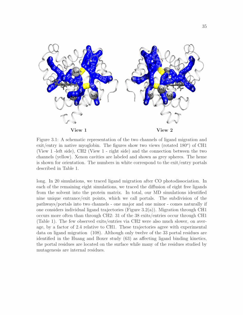
35
View 1 View 2
Figure 3.1: A schematic representation of the two channels of ligand migration andexit/entry in native myoglobin. The figures show two views (rotated 180o) of CH1(View 1 -left side), CH2 (View 1 - right side) and the connection between the twochannels (yellow). Xenon cavities are labeled and shown as grey spheres. The hemeis shown for orientation. The numbers in white correspond to the exit/entry portalsdescribed in Table 1.
long. In 20 simulations, we traced ligand migration after CO photodissociation. Ineach of the remaining eight simulations, we traced the diffusion of eight free ligandsfrom the solvent into the protein matrix. In total, our MD simulations identifiednine unique entrance/exit points, which we call portals. The subdivision of thepathways/portals into two channels - one major and one minor - comes naturally ifone considers individual ligand trajectories (Figure 3.2(a)). Migration through CH1occurs more often than through CH2: 31 of the 38 exits/entries occur through CH1(Table 1). The few observed exits/entries via CH2 were also much slower, on aver-age, by a factor of 2.4 relative to CH1. These trajectories agree with experimentaldata on ligand migration (108). Although only twelve of the 33 portal residues areidentified in the Huang and Boxer study (63) as affecting ligand binding kinetics,the portal residues are located on the surface while many of the residues studied bymutagenesis are internal residues.

36
In 55% of our native myoglobin simulations, the CO escaped. In 33% of the nativesimulations in which CO was originally in the solvent, CO successfully migrated todistal heme pocket; 69% of these COs later escaped. The migration pathways tracedby the COs that never escaped do in fact coincide with the ones from trajectories inwhich CO escaped/reached the heme.
Even though we have traced the trajectories of 38 COs that exited/entered myo-globin, MD simulation may not completely explore the universe available for ligandmigration. To address this possibility, we developed the PathFinder algorithm, whichsystematically analyzes the dynamic free volume fluctuations of the protein. Thisanalysis, PathFinder, computes cavities present in each static frame and integratescavities across multiple frames to reveal connected migratory channels. Portionsof the channel transiently occur in internal cavities, influenced by protein motion.By carefully connecting these cavities across time, PathFinder reveals the ligandmigration pathways.
(a) (b)
Figure 3.2: Comparison of ligand trajectories and free volume fluctuation analysis.(a) The positions occupied by CO during the trajectories in which CO escapes/entersmyoglobin. Light blue points are positions of CO that exited/entered via portals inCH1. Darker blue points are positions of CO that exited/entered via portals in CH2.(b) The results of PathFinder analysis in the native protein. Each point representsa cavity large enough to hold a ligand molecule.
The results of PathFinder analysis in native myoglobin shown in Figure 3.2(b) arein qualitative agreement with the migration points of the MD simulations (Fig-ure 3.2(a)). Most free volume fluctuations occur in regions previously identified as

37
(a) (b)
(c) (d)
Figure 3.3: Effect of conformation of residue 138 in native myoglobin. (a) Morefrequent, and crystal structure, orientation of F138, which is parallel to the heme.(b) Less frequent orientation of F138, which is perpendicular to the heme. (c) Theresults of PathFinder analysis of all snapshots of the more frequent orientation ofF138, which allows access between CH1 and CH2. (d) The results of PathFinderanalysis of all snapshots of the less frequent orientation of F138, which blocks accessbetween CH1 and CH2.
Xe1, Xe3, Xe4 and a fourth region that corresponds to a xenon cavity observed un-der alkaline conditions (107) (Supplementary Material). It is interesting to note thatthe protein fluctuations allow numerous points to accommodate a ligand moleculeduring the course of the 15 ns simulation.
In our MD simulations, the pattern of paths along which CO entered/exited through

38
CH1 is different from the pattern of paths along which CO entered/exited throughCH2 (Figure 3.2(a)). The COs that exit/enter in CH1 traverse Xe1, Xe3 and Xe4whereas the COs that exit/enter in CH2 traverse Xe2, Xe3 and Xe4. Since COsthat exit/enter via CH1 portals do not enter Xe2 and COs that enter/exit via CH2portals do not enter Xe1, we reasoned that a gating mechanism may control the accessbetween CH1 and CH2. Visualization of the MD trajectories implicates residue F138as the gate between CH1 and CH2. We divided the snapshots from the trajectoriesinto two groups based on the orientation of F138, and then used PathFinder toidentify the effect of the position of F138 on the connectivity between CH1 and CH2.When the phenyl ring of F138 is parallel to the heme (Figure 3.3(a)), CO is able totravel between CH1 and CH2 (Figure 3.3(c)). In about 5% of the simulation, though,the phenyl ring is oriented perpendicular to the heme (Figure 3.3(b)), effectivelyblocking access between CH1 and CH2 (Figure 3.3(d)). The result is consistent withthe observation by Huang and Boxer that F138 affects ligand binding kinetics (63).
In native myoglobin, the analysis usingPathFinder identified numerous cavities thatconnect the distal pocket to the rest of the protein. The visualization of the MD sim-ulations indicated that the orientation of V68 may influence the connection betweenthe distal pocket and the rest of the protein. V68 adopts two different configurations:“open” in which the side chain methyl groups are oriented parallel to the heme (Fig-ure 3.4(a)); and “closed” in which the side chain methyl groups are not parallel tothe heme (Figure 3.4(b)). The PathFinder analysis on the snapshots of V68 in the“open” position show that migration between the distal pocket and the rest of theprotein funnels through Xe4 but ligands are able to migrate from the distal pocketto the external solvent near the distal histidine (Figure 3.4(c)). The PathFinderanalysis on the snapshots of V68 in the “closed” position show a more direct routebecomes accessible, but the access to the external solvent near the distal histidinedecreases (Figure 3.4(d)).
Additional support for our picture of ligand migration in the native myoglobin comesfrom the analysis of the V68F mutant. While the kinetics of tens of myoglobinmutants have been studied, only a select few can provide a clear-cut test case to studyligand migration. We chose to simulate the V68F mutant because V68 residue ishighly conserved in myoglobin and the V68F mutant only affects the ligand pathways,not binding affinities (45, 99, 135). In the analysis of free volume fluctuations in theV68F mutant, the bulky phenylalanine side chain (Figure 3.5(a)) blocks the routebetween the distal pocket and the rest of the interior of the protein and also theaccess between the distal pocket and the external solvent near the distal histidineis decreased (Figure 3.5(b)). The orientation of the CB, CB1 and CG2 atoms

39
of F68 in the V68F mutant are similar to the orientation of these atoms in the“closed” conformation of the V68 of the native structure. The V68F MD simulationsconfirm this result: all of the ligands that do not escape within 90 ns remain in thedistal pocket; of the ones that do escape the distal pocket, the F68 must rotateapproximately 90o from the position in the V68F crystal structure.
One possible explanation for the ligand migration channels is to look at the highlyalpha-helical structure of the myoglobin protein. These helices are relatively rigidand therefore the volume fluctuations must occur in-between the helices (Figure 3.6).The access to the interior may only be accessible near the helix joints.
This study integrates (i) an analysis of free volume fluctuations in native myoglobinusing the PathFinder algorithm, (ii) extensive MD simulations of both photodissoci-ated and free CO in native myoglobin, and (iii) MD simulations of the V68F mutantmyoglobin to provide a new characterization of migration pathways by which ligandsreach the internal heme iron of myoglobin. This analysis reveals that ligand migra-tion is characterized by two distinct channels with multiple portals. Migration viaCH1 dominates migration via CH2. In CH1, exit/entry via portals near the distalhistidine and near the Xe1 cavity dominate other portals. This atomic level char-acterization of ligand exit, entry and migration leads to the conclusion that thereexists one major channel, with two dominant portals, and one minor channel forligand migration in myoglobin.

40
(a) (b)
(c) (d)
Figure 3.4: Effects of residue 68 in native myoglobin. (a) Sidechain conformationof V68 (yellow) in native myoglobin in crystal structure and in 18% of the MDsimulation. (b) Sidechain conformation of V68 (yellow) in native myoglobin witha rotation of C-CA-CB-CG2 compared with crystal structure conformation (82%of the MD simulation). (c) PathFinder identifies a narrow connection, which goesthrough Xe4, between the distal pocket and the rest of the protein (right circle),and an opening exists between the distal pocket and the solvent (left circle). (d)PathFinder identifies more free volume between the distal pocket and the rest ofthe protein interior (right circle), but the opening between the distal pocket and thesolvent is less open (left circle).

41
(a) (b)
Figure 3.5: Effect of residue 68 in V68F myoglobin. (a) Sidechain conformation ofF68 (yellow) in V68F myoglobin. (b) PathFinder confirms that bulky phenyl sidechain of F68 blocks access between the distal pocket and the rest of CH1 and CH2(right circle) and cuts off all access between the distal pocket and the solvent (leftcircle).

42
Figure 3.6: Structural origins of ligand migration channels and exit/entry portalsin myoglobin. Migration occurs between the helices and the portals occur near thejoints of the helices. Alpha-helices A, E and F and the heme molecular surface aretransparent. Alpha-helices are rendered with a radius of 4.0 A.

43
3.3 Supplementary Material
Methods
StructuresTwo different structures were used for the native myoglobin: PDB ID 1A6G - car-bonmonoxy myoglobin solved at 1.15A resolution by X-ray diffraction (132); andPDB ID 2MB5 - carbonmonoxy myoglobin solved by neutron diffraction (25). TheV68F mutant was represented by PDB ID 1MLJ - carbonmonoxy myoglobin solvedat 2.0A resolution by X-ray diffraction.All water molecules found in the PDB 2MB5 structure were retained. In the othertwo structures the original water molecules found in the PDB files were strippedoff. The initial placement of protons for 1A6G and 1MLJ X-ray structures wasperformed by MOLPROBITY (41) server; no dihedral flips were suggested at thevalidation step. As the 2MB5 structure already had most of the protons, these wereretained. In agreement with earlier computational prediction and experiment(7), theHIS residues were set in the following states of protonation: HIS 12, 36, 81, 116 inthe doubly protonated (charged) form, HIS 64 and 93 as singly protonated in the δposition, and HIS 24, 48, 82, 97, 113, 119 as singly protonated in the ǫ position. Allother titratable groups were set in their standard protonation states, in agreementwith the predictions based on the continuum solvent methodology(7). Missing heavyatoms and protons were added, and pKs of titratable groups computed with the helpof H++(54) – web-based tool for structure completion and pK prediction.
3.3.1 Molecular Dynamics Simulations
AMBER (version 9) suite of programs(19) was used to further prepare the structuresand perform all of the MD simulations described here. Each structure was solvatedin a box of pre-equilibrated TIP3P water, with a 7 A solvent buffer. A bond be-tween the “C” atom of the CO ligand and the “Fe” atom of the heme was set duringthe preparation stage (tleap module of AMBER). All of the MD simulations wereperformed using pmemd module. Periodic boundary conditions were used, with thedefault parameters for the PME. The ff99 force-field was employed. (Parameters ofthe heme group and CO ligand are not part of the standard ff99 and are discussedbelow.) The SHAKE method was used to restrain hydrogen – heavy atom bonddistances. The systems first underwent 100 steps of minimization, during which

44
all non-solvent atoms were restrained to their original (PDB) positions with a 5.0kcal/mol/A2 harmonic potential. This was followed by two steps of equilibration,200 ps each, under NPT conditions. All non-solvent atoms were constrained to theiroriginal positions with 1.0 kcal/mol/A2 and 0.1 kcal/mol/A2 harmonic potentialsrespectively. The temperature was gradually raised to 300 K during the first 200ps step of the equilibration, and then kept constant during the second step. Theintegration time-step was 2 fs. The average temperature of the system was main-tained by weak coupling (via the Berendsen algorithm ) to a heat bath with couplingconstant of 2 ps. The pressure coupling constant was 2 ps. All of the productionMD runs were performed at 300 K and constant pressure. All of them share thecommon preparation, minimization and equilibration protocol. The production MDsimulations can be separated into three groups. Features of the MD protocols com-mon to all of the simulations are given above, and the differences are presented below.
Generation of snapshots for the PathFinder analysis
The ensemble of structures used in the PathFinder analysis was generated by 3MD simulations, each corresponding to one of the structures described above. Eachsimulation is 15 ns in length, with the protein and solvent coordinates saved everypicosecond. The initial structures were prepared as described above. To model thebound ligand we used the parameters corresponding to the 6-coordinated Fe, seebelow. The first 200 ps of the production run for each simulation was not includedin the PathFinder analysis.
Simulation of Photodissociated CO
We use the “instantaneous” model for photodissociation(47, 86), in which the Fe-C bond is cut and the heme force field is switched instantly from that of the 6-coordinated Fe to that of the 5-coordinated Fe, see below. The LJ interactions withthe ligand also change instantly at t = 0, corresponding to the absence of the bondbetween the ”C” and ”Fe” atoms. Each complete trajectory is 90 ns. All of thetrajectories were different, which was achieved by using randomized initial velocities(drawn from a thermal distribution at 300K) at t = 0. Solvent and solute coordinateswere saved every 10 ps.

45
Figure 3.7: The starting positions of the 8 ligands used in the “CO entry fromsolvent” MD simulations. A planar view. Each ligand is initially placed at least 15A from the protein surface.
Simulation of ligand entry from solvent
The starting structures for these simulations were snapshots from the photodissoci-ated CO trajectories of each of the two native myoglobin structures described above.The snapshots in which the ligand had escaped and moved at least 15 A away fromthe protein were chosen. Seven additional CO molecules were added to each solutebox by replacing the water molecules at the edges of the box, see Fig. 3.7. Includingthe originally escaped CO, the eight COs are initially located outside of the protein,at least 15A from the protein surface. All water molecules were then deleted and theprotein along with the 8 added ligands were re-solvated in a box of pre-equilibratedTIP3P water as described above. The minimization, equilibration, and productionMD protocols are described above, and correspond to the 5-coordinated heme ironsince the heme is now in ”deoxy” state. Each MD simulation is 90 ns long, withthe solvent and solute coordinates saved every 10 ps. All of the trajectories were

46
different, which was achieved by using randomized initial velocities (drawn from athermal distribution at 300K) at t = 0. A total of 6 MD simulations of this typewere performed, 3 per each native structure. This gives a total of (3+3)×8 = 48individual ligand diffusion trajectories.
heme and ligand force field parameters
Force field parameters for the ligated (6-coordinated Fe) are available with the AM-BER distribution, these include the LJ parameters of the CO ligand. To model theunligated (5-coordinated Fe) heme, we modified the above parameters as follows:Fe-NB and FE-NO bond lengths were increased by 0.1A, and Fe-Np bond lengthswere increased by 0.1431A. These changes correspond to the difference between Fe-Nbonds for 5- and 6- coordinated Fe published in Table 1 of Ref. (79). We employthe two-site carbon monoxide model that was used earlier in a similar context(86).It consists of two atoms bearing small charges (0.021 au) of opposite sign, located atthe ligand atomic positions. This model approximately reproduces the experimentaldipole moment of carbon monoxide as well as several other experimental trends. Ina systematic comparative analysis(86), Meller at al. did not find a clear evidencethat a more sophisticated ”three site” model should be preferred to the two-site oneused here, at least as long as one is not interested in short time scale dynamics. Thegeneral conclusion of insensitivity of the ligand migration pathways to this level ofdetail of the ligand charge model is consistent with recent recent work of of Cohenet al.(30).
3.3.2 CO escapes/entries
The simulations in which CO escaped/entered and reached the iron atom are notedin Table 3.2. The amount of time in which CO traveled between the surface andthe iron is noted for each entry/escape. These times are plotted in Figure 3.8. Thesimulations are divided into groups by the portal in which they enter/escape.

47
Table 3.2: Simulations in which CO escapes/enters native myoglobin and the residuenumber of the CO. The times (in ns) at which the CO reached the surface and atwhich the CO reached the iron atom, along with the total time of travel between thetwo, and the portal through which it escapes/enters are shown.Simulation Res. Entry Exit
Surf Fe Time Portal Fe Surf Time Portal1A6G.IG10 153 - - - - 0 36.71 36.71 11A6G.IG20 153 - - - - 0 0.20 0.20 51A6G.IG30 153 - - - - 0 36.70 36.70 21A6G.IG40 153 - - - - 0 12.44 12.44 21A6G.IG60 153 - - - - 0 1.4 1.4 51A6G.IG0.eq2 153 - - - - 0 42.7 42.7 31A6G.IG50.eq2 153 - - - - 0 38.58 38.58 61A6G.8CO 158 11.05 11.26 0.21 4 11.26 29.33 18.07 41A6G.8CO.prop 153 14.39 15.21 0.82 1 15.21 33.51 18.31 11A6G.8CO.prop 160 87.78 89.99 2.22 1 - - - -1A6G.8CO.prop.IG10 153 15.72 16.57 0.86 1 16.57 22.33 5.77 11A6G.8CO.prop.IG10 158 3.86 4.10 0.26 1 4.10 23.82 19.73 11A6G.8CO.prop.IG10 160 1.31 1.93 0.03 1 1.92 4.59 2.67 12MB5.IG0 155 - - - - 0 76.15 76.15 72MB5.IG20 155 - - - - 0 17.10 17.10 12MB5.IG30 155 - - - - 0 11.62 11.62 12MB5.IG50.eq2 155 - - - - 0 45.91 45.91 32MB5.8CO 156 12.74 37.35 24.62 3 37.35 53.58 16.24 92MB5.8CO 157 24.30 48.24 23.99 6 - - - -2MB5.8CO.prop 156 37.70 38.40 0.70 6 38.40 48.00 9.55 22MB5.8CO.prop 159 69.60 72.70 3.31 3 - - - -2MB5.8CO.prop 160 45.80 53.50 7.90 3 53.50 65.40 11.87 32MB5.8CO.prop 161 33.74 33.88 0.14 2 33.88 50.84 16.96 62MB5.8CO.prop 161 64.10 66.60 2.62 3 66.60 78.05 11.34 22MB5.8CO.prop 162 33.80 34.10 0.50 4 34.10 63.20 28.81 82MB5.8CO.prop.IG10 158 8.61 12.00 3.40 3 - - - -2MB5.8CO.prop.IG10 162 44.80 52.38 7.59 4 - - - -
3.3.3 Validation
RMSD
The RMSD of backbone atoms during a 90 ns simulation in which CO was bound tothe iron atom and the RMSD of backbone atoms during a 90 ns simulation in which8 COs were initially outside the protein are shown in Figure 3.9. The overall RMSDsshown the the simulation is relatively stable and does not deviate much from the

48
Figure 3.8: Ligand entry and exit times by channel and portal. Circles - time ittook for the ligand to enter the protein and reach the distal pocket. Triangles - timeit took for the ligand to escape from the distal pocket to the protein surface. Thestring ’CHiPj’ denotes channel i and portal j.
initial crystal structures.
The RMSD per residue during the two native myoglobin trajectories were measured(Figure 3.10). The residues from both trajectories that exhibited the largest fluctua-tions (RMSD gt 1) were: 1, 2, 4, 6, 8, 11, 12, 21, 24, 31, 40, 41, 47, 49, 50, 59, 63, 64,68, 69, 75, 76, 83, 85, 89, 91, 97, 98, 101, 102, 104, 109, 118, 119, 122, 126, 136, 138,140, 141, 146, 151, and 152. The residues located in the interior of the protein are:24, 69, 76, 89, 104, 138. Residues 76 and 138 are located near the connection betweenCH1 and CH2. The conformation of residue 138 is shown to affect the accessibilitybetween CH1 and CH2 at the lower connection point; the conformation of residue 76does slightly affect the accessibility between CH1 and CH2 at the upper connection,but not as clearly as residue 138 affects the lower connection point.
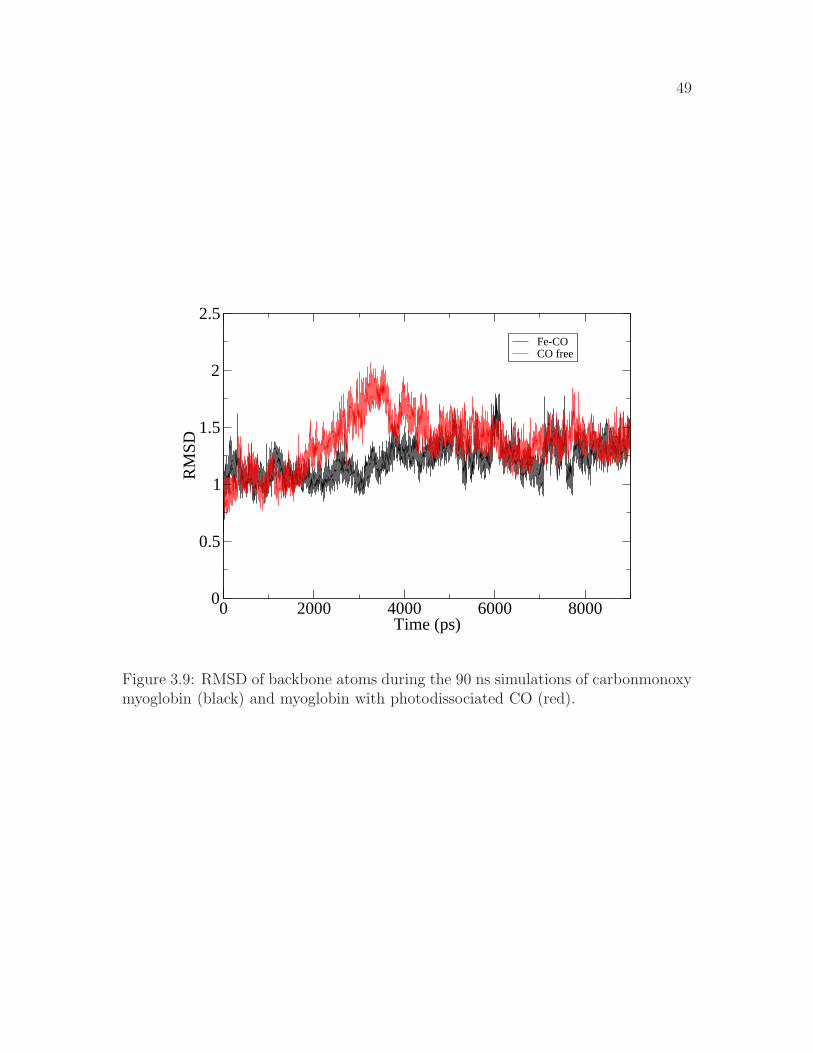
49
0 2000 4000 6000 8000Time (ps)
0
0.5
1
1.5
2
2.5
RM
SD
Fe-COCO free
Figure 3.9: RMSD of backbone atoms during the 90 ns simulations of carbonmonoxymyoglobin (black) and myoglobin with photodissociated CO (red).

50
0 25 50 75 100 125 150Residue
0
0.25
0.5
0.75
1
1.25
1.5
RM
SD
2MB5RMSD per Residue
0 25 50 75 100 125 150Residue
0
0.25
0.5
0.75
1
1.25
1.5
RM
SD
1A6GRMSD per Residue
A B
Figure 3.10: RMSD per residue during two native myoglobin trajectories.

51
Figure 3.11: The points in native myoglobin that occur with the highest frequency(over 2500 times in 11,200 snapshots) are shown in red. Three of the four regionscorrespond to Xe1, Xe3 and Xe4, while the fourth region is near a xenon bindingsite observed by Schoenborn (107).

Chapter 4
Computational Investigation ofMechanism and SubstrateSpecificities of Two Family 1β-Glucosidases
Jory Z. Ruscio, Asim Esen and David R. Bevan
4.1 Abstract
Computational methods have been used to characterize substrate specificity and in-vestigate the catalytic mechanism in two family 1 β-glucosidases, Glu1 and Dhr1.Glu1, from Zea mays, hydrolyzes DIMBOA-glc, its natural substrate, and is inhib-ited by dhurrin. Dhr1, from Sorghum, hydrolyzes dhurrin, its natural substrate,and is inhibited by DIMBOA-glc. Experimental data do not provide a completeunderstanding as to how these two proteins interact with their respective substrateand inhibitor. Molecular dynamics (MD) simulations have been performed on Glu1with DIMBOA-glc, Glu1 with dhurrin, Dhr1 with DIMBOA-glc, Dhr1 with dhurrin,and a Glu1-Y473F mutant with dhurrin. Our MD simulations also have implica-tions on the current view of the mechanism of retaining glucosidases. A distance of∼5.5A between the catalytic glutamates has generally been accepted. The results
52

53
of the MD simulations indicate that the distance between the catalytic glutamatesis closer to 3.0A which supports an in-plane protonation hypothesis. Additionally,the observed interactions between ligands and proteins provide information on thesubstrate specificities of each protein.
4.2 Introduction
β-glucosidases (β-D-glucoside glucohydrolase, EC 3.2.1.21) occur in all organismsand play essential roles in numerous biological processes. Their function in plantsis of basic importance, as they are involved in functions such as defense, lignifica-tion (42), growth, signaling and cell wall metabolism. β-glucosidases are membersof families 1 and 3 of glycosyl hydrolases (60) and hydrolyze substrates of type G-O/S-X, where G is a glycosyl residue and X is either another glycosyl residue ora non-glycosyl aglycone moiety. G and X are connected in the β-configuration byeither an oxygen(O) or sulfur(S) atom. Two glutamate residues, which are highlyconserved in the FNEP and ITENG motifs, act as a general acid/base and a nu-cleophile, respectively. The substrate is cleaved by a two-step retaining mechanism.The first step involves nucleophilic attack at the anomeric carbon (C1) of the gly-cosyl moiety; the concerted protonation of the glycosidic oxygen by the general acidyields a covalent glycosyl-enzyme intermediate and release of the leaving group (131).The deprotonated glutamate then acts as a general base, extracting a hydrogen froma water molecule. The activated water molecule attacks at the anomeric carbon,causing release of the glycosyl moiety from the enzyme, and the catalytic glutamateresidues return to their original protonation states. The anomeric configuration ofthe glycosyl residue is retained from substrate to product (144).
In order for enzymatic catalysis to occur, a substrate must be oriented such that theanomeric carbon of the glycosyl moiety and the catalytic glutamates are positionedappropriately. These distances have been approximated based on crystal structures ofretaining enzymes. The distance between the catalytic glutamates was determinedto be ∼5.5A (40). To date, eleven Family 1 glycosyl hydrolase unligated nativestructures have been solved (Table 4.1). While the distance between the δ carbonsof the glutamate residues is about 5A, the ǫ oxygens are much closer (around 3.5A).
Crystal structures of glucosidases complexed with transition state inhibitors de-pict the protonation occurring above the plane of the glucose moiety, rather thanabove (127, 130) (Figure 4.1). For in-plane protonation, the catalytic glutamateswould need to be positioned much closer than the conventionally accepted 5.5A (59).

54
Table 4.1: Distances between the catalytic residues in other Family 1 glycosyl hy-drolases.
Protein Organism PDB ID Catalytic Cδ-Cδ Oǫ-Oǫ YearResidues
cyanogenic β-glucosidase
Trifolium
repens
1CBG 183/397 5.21 3.75 1995 (6)
6-phospho-β-galactosidase
Lactococcus
lactis
1PBG 160/375 4.82 3.35 1995 (136)
β-glucosidase Sulfolobus
solfataricus
1GOW 206/387 4.93 4.06 1997 (1)
β-glucosidaseA
Bacillus
Polymyxa
1BGA 166/352 5.1 3.22 1998 (104)
myrosinase Sinapis alba 1MYR 187/409 5.02 3.61 1997 (16)β-glucosidase Zea mays 1E1E 191/406 4.97 3.49 2000 (35)β-glucosidase Bacillus cir-
culans
1QOX 166/355 4.24 3.23 2000 (56)
β-glycosidase Thermus
nonprote-
olyticus
1NP2 164/338 5.2 3.65 2003 (134)
β-glucosidase Sorghum bi-
color
1V02 189/404 4.92 3.47 2004 (129)
β-glucosidase Pyrococcus
horikoshii
1VFF 155/324 5.23 4.37 2004 (2)
myrosinase Brevicoryne
brassicae
1WCG 167/374 5.18 3.89 2005 (66)

55
Figure 4.1: First step (glycosylation) of double-replacement retaining mechanism.The catalytic residues may be oriented for (A) above plane protonation or (B) inplane protonation (39, 59).

56
A shorter distance indicates that the catalytic glutamates would be on the sameside of the glycosidic bond, and not on opposite sides, as sometimes depicted. Thedistance between the ǫ oxygens of the catalytic glutamic acids in the proteins withsolved structures provides support for in-plane protonation. These distances are sim-ilar to those described by Heightman et al (59). Additionally, the optimal distancebetween the anomeric carbon and nucleophile for nucleophilic attack should be <3.25A (14, 80, 105, 124).
The discrepancies of the catalytic glutamate distance between each other in modelswith inhibitor and those with substrate indicate that the mechanism of glycosylhydrolases, and more specifically, family 1 β-glucosidases, needs to be examined inmore depth. If the interaction distance of the catalytic glutamates can be definedwhen substrate is present, it will be much easier to define whether or not a smallmolecule is a potential substrate.
Figure 4.2: Structures of DIMBOA-glucoside and dhurrin
Some β-glucosidases exhibit a wide range of substrate specificities (5, 6, 10, 134) whileothers have a quite narrow substrate specificity (27, 62). Kinetic studies with nativeand chimeric enzymes provide much useful information about the substrate specifici-ties of Glu1 and Dhr1. Glu1 from Zea mays and Dhr1 from Sorghum bicolor have∼70% sequence identity (27), yet their substrate specificities are extremely different.In addition to its natural substrate, 2-O-β-D-glucopyranosyl-4-hydroxy-7-methoxy-1,4-benzoxanzin-3-one (DIMBOA-glc), Glu1 can hydrolyze a wide variety of artificialsubstrates (5, 34), but is inhibited by dhurrin (Ki of 0.076mM (26))(Figure 4.2) .Dhr1 is highly specific for its natural substrate dhurrin (β-D-glucopyranosyl-oxy-(S)-p-hydroxymandelonitrile) and is inhibited by DIMBOA-glc (Ki of 0.009mM (26)).
The crystal structures of Glu1 and Dhr1 provide information that can help identifyresidues important in determining substrate specificity. Czjzek et al. (35) showedthat the aglycone binding pocket of Glu1 is defined by Phe198, Phe205, Phe466and W378, which form a hydrophobic slot. The aglycone moiety is believed to be

57
Table 4.2: Hydrogen bonding between glucose atoms and protein atoms in theglycosyl-enzyme myrosinase structure (16), glucotetrazole in myrosinase (15), dhur-rin in Dhr1-E189D (129), and DIMBOA-glc in Glu1-E191D (35) (hydrogen bondsin glucotetrazole-myrosinase complex, dhurrin-Dhr1-E189D complex and DIMBOA-glc-Glu1-E191D complex from Verdoucq et. al (129)).
Glucose atom Glycosylated Glucotetrazole in Dhurrin in DIMBOA-Glc inMYR MYR Dhr1-E189D Glu1-E191D
O2 Gln187-Oǫ1 Asp189-Oδ2 Asp191-Oδ2Asn186-Nδ2 Asn186-Nδ2 Asn188-Nδ2
His143-Nǫ2O3 His151-Nǫ2 His141-Nǫ2 His143-Nǫ2 His152-Nǫ2
Gln39-Oǫ1 Gln39-Oǫ1 Gln39-Oǫ1Gln39-Nǫ2 Gln39-Nǫ2
Glu406-Oǫ1Glu406-Oǫ2
Trp461-Nǫ2O4 Gln39-Nǫ2 Gln39-Nǫ2 Gln39-Nǫ2 Gln38-Oǫ1
Glu464-Oǫ1 Glu464-Oǫ1 Glu464-Oǫ1Trp457-Nǫ1 Trp453-Nǫ1 Trp465-Nǫ1
His142-Nǫ2O6 Glu464-Oǫ2 Glu464-Oǫ2 Glu460-Oǫ2 Glu464-Oǫ2
Glu464-Oǫ1Gln38-Nǫ2Trp457-Nǫ1

58
positioned by Trp378 through aromatic stacking interactions (35, 36, 146). Thispositioning is thought to determine the orientation of the glucosyl moiety in relationto the catalytic glutamates. Incorrect positioning of the glucosyl moiety in relationto the catalytic glutamates will inhibit catalysis, as is evident with Glu1 inhibitorsdhurrin and p-nitrophenyl β-D-thioglucoside (35, 36).
The residues involved in aglycone recognition are different in Dhr1 than in Glu1. Thethree phenylalanines that compose one side of the active site opening are replacedwith valine, leucine and serine. The opening of the active site is small and circularin Dhr1, compared to the slot-like opening in Glu1. Although native Glu1 cannothydrolyze dhurrin, some chimeric and altered Glu1 proteins have gained the abilityto hydrolyze dhurrin (26). Analysis of chimeric protein with the fewest residuesdifferent between Glu1 and Dhr1 eventually helped identify residue Tyr473 (Glu1)and Phe469 (Dhr1) as important for conferring dhurrin hydrolysis ability to Glu1.The opposite substitution in Dhr1 does not confer DIMBOA-glc hydrolysis ability,however. Glu1 appears uses aromatic stacking to interact with the aglycone whileDhr1 utilizes polar interactions (129), and therefore the difference may be due to thefact the two proteins use different modes of aglycone recognition.
With the availability of protein-ligand complexes, the conformation of the ligandin the active site can now be examined. To some extent, the interactions betweenthe protein and ligand are similar to the interactions in the glycosylated protein(Table 4.2). But, for instance, the O6 atom in DIMBOA-glc in Glu1-E191D makesseveral interactions with the protein that are not seen in glycosylated MYR, glucote-trazole in MYR or dhurrin in Dhr1-E189D. Do atoms of DIMBOA-glc in Glu1 reallyinteract with several protein atoms, or do the definition of these interactions need tobe further studied, taking into account the dynamics of the protein-ligand complex?
Additionally, the conformation of the glucose moiety appears to have implications onthe mechanism and the protein residues with which it interacts. Several of the crystalstructures of protein-ligand complexes show distortion of the glycosyl moiety of theligand. A proposed pathway, based on these distortions, describes a 1S3 → 4H3 → 4C1
glucose conformation (Fig 4.1) in which the 4H3 is a transition state structure (38) .The electron density of the glycone binding pocket of Glu1 with DIMBOA-glc was notclear enough to accurately define the glucose conformation (35). The conformationcould be modeled as either the 4C1 chair, which is the ground state conformation,or the 1S3 skew-boat, which is a proposed Michaelis conformation (40). The crystalstructure of Glu1 with glucotetrazole, which is a transition state-mimicking inhibitor,depicts the glucose in the 4H3 conformation. Additionally, the crystal structure ofDhr1 with dhurrin depicts the glucose in the 1S3 skew-boat conformation (129). It

59
has been proposed that Glu1 can bind substrate with the glucose as 4C1 or as theskew-boat, and the glycone binding pocket can stabilize either one, while Dhr1 canonly bind substrate with the glucose in the 1S3 conformation. It has been proposedthat for retaining glycosyl hydrolases that bind with a distorted 1S3 glycosyl moietyconformation, the glycosyl moiety in the transition state would adopt a 4H3 confor-mation (38). A computational study involving a retaining family 11 xylanase showedthat the enzyme binds the substrate more tightly when the sugar is the in skewedconformation than in the 4C1 chain conformation (74).
While the kinetic data show whether or not an enzyme can hydrolyze a ligand, it doesnot provide any information as to how or why one ligand is a substrate and one isan inhibitor. Likewise, while the crystal structures have increased our understandingof the enzyme-ligand complexes, these are still only static snapshots of dynamicsystems. Additionally, all of the enzyme-ligand complexes were crystallized withan altered form of the enzyme (the catalytic acid/base glutamate changed to anaspartate), rendering the enzyme inactive, and thus not a mechanistically relevantrepresentation.
In order to gain a better understanding of what is occurring at the atomic level, wehave applied computational techniques to the Glu1 and Dhr1 systems. The use ofmolecular dynamics simulations has enabled us to model the protein-ligand interac-tions and help answer the question of how ligands affect protein dynamics and inturn, determine catalytic activity. We first quantify the importance of the distancebetween the catalytic glutamate residues, as well as the distance between the nucle-ophilic glutamate and the anomeric carbon. Then, we determine how interactionsbetween the ligand and the protein and the ligand and the solvent may affect binding.The atomic fluctuations of the active site are then examined so that we may have abetter understanding of the changes that are caused by the inhibition of dhurrin inGlu1. We then investigate the affects of catalysis on the conformation of the glycosylmoiety. Finally, we extend some of the substrate specificity based information gainedfrom Glu1 and Dhr1 to 41 putative β-glucosidases in Arabidopsis thaliana.
4.3 Methods
In this investigation, molecular dynamics was the primary tool used to characterizesubstrate specificity in Glu1 and Dhr1. The crystal structures of Glu1 and Dhr1with and without ligands are available. All MD setup and MD simulations wereperformed using the AMBER 7 suite of programs (21). To fully examine the differ-
60
ences in the active site architecture, many perturbations of the two proteins wereused for simulations. Catalytically inactive forms, such as when an aspartic acid issubstituted for one of the glutamic acids and when the acid/base catalytic glutamicacid is unprotonated, as well as protein-inhibitor complexes were simulated in orderto compare interactions with catalytically active forms. The simulations performedas well as the abbreviations used in the rest of the paper are noted in Table 4.3.
Preparation of Structures for MD: The structures of Glu1 with ligand (1E56)and Dhr1 with ligand (1V03) obtained from the Protein Data Bank were of alteredforms (acid/base glutamate residue changed to an aspartate residue) or of the pro-teins with no ligand (1E1E and 1V02). Y473F was created by deleting the hydroxylgroup of tyrosine 473 and renaming the residue PHE. The DIMBOA-glc and dhurrinstructures were geometry optimized and partial charges calculated using Hartree-Fock 3-21G in Gaussian98 (52). To prepare for MD, the ligand was docked into thenative enzyme by superimposing the structures of an altered enzyme (e.g., 1E56)with ligand and a native enzyme (e.g., 1E1E) and then removing the altered en-zyme. To alter the protonation state of the acid/base catalyst, in order to accuratelydescribe the catalytic state, residue 191 was changed from GLU to GLH, to adhereto AMBER notation. The cysteine residues involved in a disulfide bond (103) werechanged from CYS to CYX, and connected using the bond command in Leap. Allthe systems were processed with Leap to add hydrogens, add counterions and solvatein a box of TIP3P water.
Running MD: The following conditions were used: default particle mesh Ewald(PME) parameters; SHAKE on the hydrogen atoms; a 2fs timestep; and a 9A cutoffapplied to long-range interactions. Before running MD, the systems were equilibratedand minimized. The first step involved 100 ps equilibration of all the water moleculesand ions at 300K while holding the protein and ligand atoms frozen. Next the watermolecules and ions were subjected to 300 steps of steepest descent minimizationwhile again holding the protein and ligand atoms frozen. Finally, the entire systemunderwent 300 steps of steepest descent minimization. To begin MD, the entiresystem was slowly heated from 0 to 300K in 30ps at constant volume. Constantvolume MD was continued for an additional 50ps, after which the system was rununder constant pressure conditions. Twelve simulations were carried out to 3.5 ns.Eight simulations involving catalytically inactive states were only of 1.5 ns. Thesesimulations were not carried out to 3.5 ns because the results at 1.5 ns were alreadysufficient to show why they are not catalytically active and had provided enoughdata to compare with catalytically active complexes.
Analysis of MD: All MD analysis was performed using the ptraj module of the

61
Table 4.3: Simulations performed and abbreviations used in the paper. †Catalyticallyactive
Simulation Protein Description Ligand
3.5
ns
Glu1 Glu1 N/A†Glu1 DIMglc Glu1 DIMBOA-glcGlu1 dhr Glu1 dhurrinDhr1 Dhr1 N/A†Dhr1 dhr Dhr1 dhurrinDhr1 dhr tors Dhr1 w/torsion angle restraint on C2 C1
O5 C5 of glucosyl moietydhurrin
Dhr1 DIMglc Dhr1 DIMBOA-glcDhr1 DIMglc tors Dhr1 w/torsion angle restraint on C2 C1
O5 C5 of glucosyl moietyDIMBOA-glc
Y473F Glu1 w/Tyr473 changed to Phe N/A†Y473F DIMglc Glu1 w/Tyr473 changed to Phe DIMBOA-glc†Y473F dhr Glu1 w/Tyr473 changed to Phe dhurrin†Chim21 dhr Glu1 w/466FAGFTERY473 w/Dhr1
462SSGYTERF469
dhurrin
F198V Glu1 w/residue Phe198 changed to Val N/AF198V DIMglc Glu1 w/residue Phe198 changed to Val DIMBOA-glcF198V dhr Glu1 w/residue Phe198 changed to Val dhurrin
1.5
ns
Glu1 a/b un Glu1 w/acid/base catalyst unprotonated N/AGlu1 a/b un DIMglc Glu1 w/acid/base catalyst unprotonated DIMBOA-glcGlu1 nuc prot DIMglc Glu1 w/nucleophile protonated
(acid/base unprotonated)DIMBOA-glc
Glu1 E/D DIMglc Glu1 w/Glu191 changed to Asp DIMBOA-glcGlu1 E/D un Glu1 w/Glu191 changed to Asp (unpro-
tonated)N/A
Glu1 E/D un DIMglc Glu1 w/Glu191 changed to Asp (unpro-tonated)
DIMBOA-glc
Glu1 E/D un dhr Glu1 w/Glu191 changed to Asp (unpro-tonated)
dhurrin
Dhr1 E/D dhr Dhr1 w/Glu189 changed to Asp dhurrin

62
AMBER package. Calculation of hydrogen bonds used the default values of a distancecutoff of 3.0A and angle cutoff of 120◦.
Glucosyl moiety conformation analysis: The charges and conformations of theglucosyl moiety were calculated using Jaguar (67). The DIMBOA-glucoside aftersander minimization of the Glu1 DIMglc complex was used as the initial structure.A geometry optimization was performed while the glycosidic bond (between theanomeric carbon and the glycosidic oxygen) was held constant. After each opti-mization, the glycosidic bond length was increased by 0.2A, and the structure wassubjected to another geometry optimization. This cycle was continued until the en-ergy stabilized (final bond length = 3.5A). The geometry optimizations were donewith B3LYP and basis set 6-31G**.
Active Site Profiling: Three Active Site Profiles of the Glu1 and Dhr1 activesites were created using the two catalytic glutamate residues in Glu1 and Dhr1(191/406 and 189/404, respectively) as the reference residues. The Glu1 and Dhr1sequences were aligned with 39 putative β-glucosidases from Arabidopsis thaliana.The residues of the A. thaliana sequences that aligned with the residues definingthe Glu1/Dhr1 Active Site Profiles were included in the Active Site Profile for β-glucosidases. Clustering of these alignments was done using the ClustalW server(http://www.ebi.ac.uk/clustalw/).
The first, and largest, Profile59, based on 59 amino acids from Glu1 and Dhr1, in-cluded the glycone and aglycone binding pockets. The second, Profile39, includedthe glycone binding pocket (red residues in sequences and Figure 4.8). The thirdActive Site Profile, Profile20, contained only the aglycone binding pocket (yellowresidues in sequences and Figure 4.8). The clustering of the partial sequences iscompared with the clustering of the full sequences (142). The seven putative pseu-dogenes (At5g16580,At1g60260, At1g60270, At2g44480, Ag1g51490, Ag5g48375 andAt3g18070) were not included in the clustering.
Glu1AATQRFHWFNEPQTFTFLAFDVNFGLNYYNWIITENGIGDVDRYFAWSLDNEATERYGI
Dhr1AATQRFHWFNEPETFCVLALNVCFGINYYNWIITENGMGDIDRYFAWSLDNESTERFGI
4.4 Results
Orientation of the catalytic glutamates: Several structural features within theactive site of the glucosidases examined in these simulations were analyzed. In each of

63
Simulation 191Oǫ2-406Oǫ2 C1-406Oǫ2Initial Final Initial Final
3.5
ns
Glu1 4.94 3.32 ±0.54 n/a n/a†Glu1 DIMglc 4.94 2.60±0.13 5.92 3.74±0.25Glu1 dhr 4.94 2.76±0.32 3.95 3.60±0.25Dhr1 3.24 5.31 ±0.51 n/a n/a†Dhr1 dhr 3.24 3.80±0.34 3.56 3.38±0.20Dhr1 DIMglc 3.24 4.34±0.40 4.19 4.61±0.14Y473F 4.94 3.65±0.61 n/a n/a†Y473F DIMglc 3.49 3.51±0.29 3.53 5.00±0.39†Y473F dhr 4.64 3.13±0.29 3.95 3.25±0.14†Chim21 dhr 4.08 3.62±0.43 5.03 4.84±0.21F198V 3.64 3.41±0.60 n/a n/aF198V DIMglc 3.64 3.73 ±0.55 5.21 6.57 ±1.05F198V dhr 3.64 4.17 ±0.21 4.65 5.35 ±0.27
1.5
ns
Glu1 a/b unprot 4.94 3.51±0.25 n/a n/aGlu1 a/b unprot DIMglc 4.94 4.81±0.42 5.92 4.66±0.15Glu1 nuc prot DIMglc 4.94 3.12±0.45 5.92 4.77±0.36Glu1 E/D DIMglc 6.24 4.40±0.47 4.46 5.21±0.25Glu1 E/D unprot 6.24 4.12±0.47 n/a n/aGlu1 E/D unprot DIMglc 6.24 7.92±0.94 4.46 5.82±0.81Glu1 E/D unprot dhr 3.46 7.58±1.29 4.59 6.52±1.25Dhr1 E/D dhr 4.16 4.95±0.61 3.63 5.19±0.39
Table 4.4: Initial and average distances between two distances that are affected bysubstrate specificity: the Oǫ2 oxygen atoms of catalytic glutamate residues; and thenucleophilic Oǫ2 and C1 of the glucosyl moiety over the course of the simulations.Bold rows indicate simulations in which the distance between the catalytic gluta-mates and the distance between the nucleophile and C1 are close to the expectedvalues for catalysis to occur. †Indicates catalytically active complex.
the 20 different simulations, two key distances were measured: the distance betweenthe catalytic glutamates (Oǫ2 to Oǫ2) and, if relevant, the distance between thenucleophilic glutamate and C1 of the glucosyl moiety (Oǫ2 to C1) (Table 4.4). Thesedistances provide information as to how the residues are oriented in catalyticallyactive complexes versus catalytically inactive complexes and may distinguish betweenthe above-plane and in-plane protonation hypotheses. Also, since the nucleophile

64
must be close enough to attack at the anomeric carbon, the C1-O2 distance maydistinguish between catalytically active and catalytically inactive complexes. Fora complex to be considered catalytically active, the distance between the catalyticglutamates must be less than 5A and the distance between the nucleophilic glutamateand the anomeric carbon should be around 3.25A.
The distances measured in the systems that only underwent MD for 1.5 ns weresufficient to determine that these complexes did not represent active enzymes. InGlu1 a/b unprot DIMglc, Glu1 E/D DIMglc, Glu1 E/D unprot,Glu1 E/D unprot DIMglc, Glu1 E/D unprot dhr and Dhr1 E/D dhr, the distancesbetween the catalytic glutamates are too great for catalysis to occur. InGlu1 nuc prot DIMglc, although the distance between the catalytic glutamates maybe appropriate, the distance between the nucleophilic glutamate and the anomericcarbon is too great.
Of the 12 simulations (eight of which contain a ligand) carried out to 3.5 ns, onlyfour of the protein-ligand complexes show the distance between the catalytic glu-tamates and the distance between the nucleophile and C1 of the ligand to be closeenough for catalysis to occur: Glu1 DIMglc, Glu1 dhr, Dhr1 dhr and Y473F dhr.Three of these four complexes are known to be catalytically active. Glu1 dhr is notcatalytically active, but the effective binding is consistent with the ability of dhurrinto inhibit Glu1. Although the initial distances between the anomeric carbon and thenucleophilic glutamate in these four complexes prior to MD vary between ∼3.5A to5.9A, only the Dhr1 DIMglc distance is significantly different than the others afterMD (4.61A vs. ∼3.4A). After MD, the glucosyl moieties in Glu1 DIMglc, Glu1 dhr,Dhr1 dhr and Y473F dhr are positioned at a similar distance from the nucleophilicglutamate, most likely the position optimal for catalysis; conversely, the glucosylmoiety in Dhr1 DIMglc is rotated, such that the anomeric carbon aligns with thering oxygen of the other simulations (Figure 4.3).
One thing to note is that the distances between the catalytic glutamates inGlu1 DIMglc and Glu1 dhr are slightly closer than the distances in Dhr1 DIMglc andDhr1 dhr, regardless of ligand. The distances in Chim21 dhr, Y473F DIMglc andY473F dhr are intermediate. This suggests that enzyme specific interactions, ratherthan protein-ligand interactions, have greater influences on the slight variations inthis distance.
In the five catalytically active complexes (Glu1 DIMglc, Dhr1 dhr, Y473 DIMglc,Y473F dhr and Chim21 dhr), the distances between the catalytic glutamates supportin-plane protonation of the glycosidic oxygen, as this distance is much closer than

65
Initial Final
Figure 4.3: Orientation of the glucosyl moiety of the ligand of Glu1 DIMglc (licorice)and Dhr1 DIMglc (cpk) after minimization (left) and after 3.5 ns of MD (right). Theanomeric carbon in Glu1 DIMglc is better oriented for catalysis to occur, while theanomeric carbon in Dhr1 DIMglc is too far from the nucleophilic glutamic acid.While not shown, the anomeric carbons in the Glu1 dhr, Dhr1 dhr and Y473F dhrsimulations align well with Glu1 DIMglc.
the conventionally accepted distance of ∼5A for retaining glycosyl hydrolases. Thedistance between the catalytic glutamates in these five simulations was significantlyless than 5A (2.60, 3.80, 3.51, 3.13, 3.62).
In the simulations of the protein in absence of ligand (Glu1, Dhr1, Y473F, F198V,Glu1 a/b unprot and Glu1 E/D unprot), the distances between the catalytic gluta-mates is slightly larger than when a ligand is present (e.g. Glu1 - 3.32 vs Glu1 DIMglc- 2.63 and Glu1 dhr - 2.76), but still much less than 5A. Additionally, the standarddeviation of the distance between the catalytic glutamates is slightly higher (Ta-ble 4.4 ) than when is ligand is present, indicating that they are able to fluctuatemore freely.
The F198V complex is known to have drastically decreased catalytic ability (128). Inthe F198V simulations (with and without ligand), the distances between the catalyticglutamates are over 1A greater than of the native enzyme in complex with a ligand.Additionally, the nucleophilic glutamate is located too far away from the anomericcarbon for catalysis. Taken together, these distances would indicate the the F198Vcomplexes are catalytically inactive, correlating with the experimental data.

66
Table 4.5: Persistence of hydrogen bonds between glucose moiety and protein com-plexes over the 3.5 ns simulations (percentage). Only interactions that occur in atleast 20% of the simulation are reported.
Glucose Protein Glu1 Dhr1 Y473F F198Vatom atom DIMglc dhr DIMglc dhr DIMglc dhr DIMglc dhr
O-2Glu191-Hǫ2 25.5Glu406-Oǫ2 97.1 95.4 99.1 97.9 99.9 96.5
O-3Gln38-Oǫ1 50.8 20.6 35.9 81.5Glu406-Oǫ1 55.8Trp465-Nǫ1 45.8 59.1 59.9 22.2 78.2
O-4
Gln38-Oǫ1 47.8Gln38-Nǫ2 69.7 48.3 22.5 29.1 68.4Trp457-Nǫ1 58.2 24.4 65.5 33.1Glu464-Oǫ1 48.3 81.1 90.1 72.1 70.7Glu464-Oǫ2 25.8 21.4 33.0
O-6Asn462-Nδ2 28.7 21.0Glu464-Oǫ1 97.6 63.4 35.8 77.2Glu464-Oǫ2 91.0 85.7 90.7 97.7
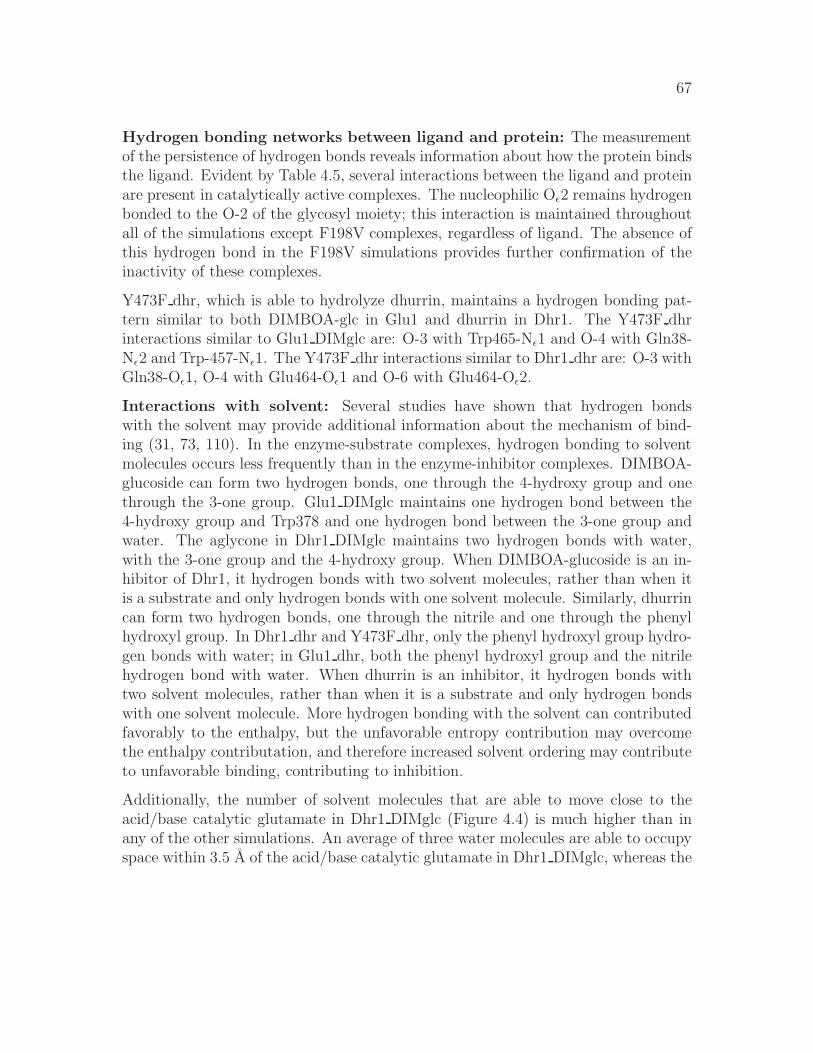
67
Hydrogen bonding networks between ligand and protein: The measurementof the persistence of hydrogen bonds reveals information about how the protein bindsthe ligand. Evident by Table 4.5, several interactions between the ligand and proteinare present in catalytically active complexes. The nucleophilic Oǫ2 remains hydrogenbonded to the O-2 of the glycosyl moiety; this interaction is maintained throughoutall of the simulations except F198V complexes, regardless of ligand. The absence ofthis hydrogen bond in the F198V simulations provides further confirmation of theinactivity of these complexes.
Y473F dhr, which is able to hydrolyze dhurrin, maintains a hydrogen bonding pat-tern similar to both DIMBOA-glc in Glu1 and dhurrin in Dhr1. The Y473F dhrinteractions similar to Glu1 DIMglc are: O-3 with Trp465-Nǫ1 and O-4 with Gln38-Nǫ2 and Trp-457-Nǫ1. The Y473F dhr interactions similar to Dhr1 dhr are: O-3 withGln38-Oǫ1, O-4 with Glu464-Oǫ1 and O-6 with Glu464-Oǫ2.
Interactions with solvent: Several studies have shown that hydrogen bondswith the solvent may provide additional information about the mechanism of bind-ing (31, 73, 110). In the enzyme-substrate complexes, hydrogen bonding to solventmolecules occurs less frequently than in the enzyme-inhibitor complexes. DIMBOA-glucoside can form two hydrogen bonds, one through the 4-hydroxy group and onethrough the 3-one group. Glu1 DIMglc maintains one hydrogen bond between the4-hydroxy group and Trp378 and one hydrogen bond between the 3-one group andwater. The aglycone in Dhr1 DIMglc maintains two hydrogen bonds with water,with the 3-one group and the 4-hydroxy group. When DIMBOA-glucoside is an in-hibitor of Dhr1, it hydrogen bonds with two solvent molecules, rather than when itis a substrate and only hydrogen bonds with one solvent molecule. Similarly, dhurrincan form two hydrogen bonds, one through the nitrile and one through the phenylhydroxyl group. In Dhr1 dhr and Y473F dhr, only the phenyl hydroxyl group hydro-gen bonds with water; in Glu1 dhr, both the phenyl hydroxyl group and the nitrilehydrogen bond with water. When dhurrin is an inhibitor, it hydrogen bonds withtwo solvent molecules, rather than when it is a substrate and only hydrogen bondswith one solvent molecule. More hydrogen bonding with the solvent can contributedfavorably to the enthalpy, but the unfavorable entropy contribution may overcomethe enthalpy contributation, and therefore increased solvent ordering may contributeto unfavorable binding, contributing to inhibition.
Additionally, the number of solvent molecules that are able to move close to theacid/base catalytic glutamate in Dhr1 DIMglc (Figure 4.4) is much higher than inany of the other simulations. An average of three water molecules are able to occupyspace within 3.5 A of the acid/base catalytic glutamate in Dhr1 DIMglc, whereas the

68
0
2
4
6
Wat
er m
olec
ules
DIMBOA-glucosideDhurrin
Glu1 Dhr1
0 1000 2000 3000Time (ps)
0
2
4
6
Wat
er m
olec
ules
Y473F
0 1000 2000 3000Time (ps)
F198V
Figure 4.4: Number of water molecules within 3.5 A of acid/base catalytic glutamateover the course of the simulations.
69
A B
Figure 4.5: Effect of dhurrin on active site of Glu1. A - In Glu1 DIMglc simulation,Tyr473 directly hydrogen bonds with Trp378 and can either form direct or indirecthydrogen bonds with Asn376 and Glu471. B - In Glu1 dhr simulation, due to lackof interaction between aglycone and Trp378, Tyr473 cannot directly hydrogen bondwith Trp378. Tyr473 can then shift slightly and form direct hydrogen bonds withAsn376 and Glu471.
average number of water molecules within that distance in Glu1 DIMglc, Glu1 dhr,Dhr1 dhr, Y473F DIMglc, Y473F dhr, F198V DIMglc and F198V dhr is about one,which is consistent with the need for a water molecule to be near the acid/baseglutamic acid during the second step of the mechanism. .
Insight into dhurrin inhibition in Glu1: The ligand-protein interactions greatlyaffect catalysis ability. Glu1 residue Y473 has been identified in several studies ascontributing to the conformational flexibility of the active site (119, 128, 146). InGlu1, this tyrosine residue’s hydroxyl group has the ability to form a hydrogen bondwith other close residues or water; in Dhr1, this residue is a phenylalanine and doesnot have the ability to form a hydrogen bond due to the lack of the hydroxyl group.
During the Glu1 DIMglc simulation, Trp378 is stabilized through its interaction withthe 4-hydroxy group of the DIMBOA moiety, and a hydrogen bond between Trp378and Tyr473 stabilizes the active site opening. Tyr473, situated near the C-6 of

70
Glu1 DIMglc Glu1 dhr Y473F dhr
Figure 4.6: The active sites of Glu1 DIMglc, Glu1 dhr and Y473F dhr after min-imization (top) and after 3.5 ns of MD (bottom). The molecular surface of theproteins is shown in grey, residues 198 and 378 are colored yellow and the substratesare shown as van Der Waals in atom coloring In Glu1 dhr, the active site appears tobecome closed by the interaction of Trp378 and Phe198. Pictures were generated inVMD (65).

71
A B C
Figure 4.7: Different conformations of glucose. Initial conformation of glucose moietyof A) DIMBOA-glucoside in chair 4C1, B) dhurrin in skew boat 1S3 C) and geometryoptimized DIMBOA-glucoside with the C1-O1 bond length extended to 2.7A in half-chair 4H3. The aglycone moieties are omitted for clarity.
glucose, also forms direct or indirect hydrogen bonds, through water molecules, withAsn376 and Glu471 (Figure 4.5A). These four interactions stabilize the active siteopening (Figure 4.6), so the entrance to the active site remains accessible.
In the Glu1 dhr simulation, the aglycone does not interact with Trp378, and thereforeTrp378 is not positioned to hydrogen bond with Tyr473. Tyr473 then has enoughfreedom to shift slightly from beneath the C-6 of glucose and form a direct hydrogenbond with Asn376 and Glu471 (Figure 4.5B). Without the Trp378-Tyr473 hydrogenbond, Trp378 is free to fluctuate, and Asn376 shifts slightly upwards. These motionscause the active site opening to become distorted, as Trp378 and Phe198 becomecloser and block the entrance to the active site (Figure 4.6).
In the Y473F simulation, the lack of a side chain hydroxyl group on residue Phe473has an impact on the dynamics of the protein with affect the ability of the protein tohydrolyze dhurrin. Phe473 is unable to form hydrogen bonds and therefore Asn378,Trp378 and Glu471 are not constrained by Phe473. The entrance to the active siteremains open, and the fluctuations of Trp378 do not distort the ligand or accessibilityof the active site(Figure 4.6).
Glucosyl moiety conformations: The conformation of the glucosyl moiety in theinitial DIMBOA-glucoside structure from the crystal structure was 4C1 and the con-formation of the glucosyl moiety in the initial dhurrin structure was 1S3 (Figure 4.7).It was proposed the Glu1 can bind the ligand in the 4C1 or a skewed conformationbut Dhr1 may only bind the ligand in the skewed conformation, as it is closer to the

72
Table 4.6: The change in the dihedral angle that characterizes the conformation ofthe glucose and the charges of the C1, O5 and O1 atoms when the glycosidic bond(between C1 and O1) is lengthened.
dist C2-C1-O5-C5 C1 O5 O11.42 -53.9 0.50468 -0.44677 -0.368691.70 -55.9 0.43459 -0.41143 -0.382141.90 -53.8 0.38239 -0.37148 -0.395852.10 -46.2 0.39260 -0.35481 -0.430912.30 -23.7 0.37428 -0.33299 -0.479952.50 -11.2 0.31414 -0.30074 -0.516482.70 -8.6 0.31962 -0.27899 -0.526752.90 -6.3 0.30315 -0.24979 -0.542093.10 -4.0 0.31222 -0.25770 -0.557733.30 -4.0 0.28763 -0.25580 -0.560283.50 -4.2 0.27480 -0.25430 -0.57216
transition state conformation (35, 129).
To investigate the conformation of the glucose in the transition state, quantum me-chanical calculations were performed on DIMBOA-glucoside, with the glucosyl moi-ety in the 4C1 conformation. The glycosidic bond was lengthened to mimic thereaction, and the dihedral (C2-C1-O5-C5) that characterizes the conformation ofthe glucose was measured, as well as the change in charges on the C1, O5 and O1atoms (Table 4.6).
As the glycosidic bond lengthens, the glucose conformation changes from 4C1 to 4H3.In the 4H3 conformation, the C2, C1, O5, C5 atoms lie in a plane, which must occurat the transition state. The 4H3 glucose conformation and the lower negative chargeon the O5 is consistent with the oxocarbenium ion transition state (38, 78, 118).
Active Site Profiling: Conventionally, entire sequences of proteins are used forclustering the proteins to determine functionality. Several groups have recognized,though, that clustering using the entire sequences, where identities are often as lowas 20%, may not be the most accurate (17, 22, 102). Clustering of active site residuesmay be more effective in providing informative clustering of sequences. This cluster-ing analysis requires many sequences, so we have used the putative β-glucosidases

73
Figure 4.8: Residues included in the three different Active Site Profiles. Profile59 -Red & Blue, Profile39 - Red, and Profile20 - Blue. DIMBOA-glucoside shown as vander Waals in atom coloring.
from A. thaliana and the Glu1, Dhr1 and Myr proteins to possibly gain insight intothe substrate specificities of the A. thaliana proteins.
The clustering of the alignments based on the full sequences (142), residues in theglycone binding site (Profile39), aglycone binding site (Profile20) and both the gly-cone and aglycone binding site (Profile59) were compared to determine if residues inthe active site will yield information about substrate specificities. Surprisingly, onlyincluding active site residues did not change the overall clustering of the sequenceswhen compared with the full sequences. Possibly this is because 40 of the 59 residuesare in the glycone binding pocket, which one would expect to be fairly conserved inβ-glucosidases.
Profile20, Profile39 and Profile59 identified four putative β-glucosidases that clusterdifferently than when the full sequences are used. At2g44480, in the full sequencesubclassification 2, clusters near sequences in either full sequence subclassification 1

74
(Profile20) or 7 (Profile39 and Profile59) . While the sequences in subclassification 2and At2g44480 have three insertions compared to the sequences in subclassification1, the residues in At2g44480 aglycone binding pocket appear more similar to theaglycone binding pockets in subclassification 1 (Figure 4.10). The second deviationfrom the full sequence clustering occurs with subclassifications 5 and 7. The puta-tive myrosinases compose subclassification 7, so it is interesting that At5g25980 andAt5g26000 cluster separately from At1g47600 and At1g51470. Thirdly, At5g36890was not clustered with the other two proteins from subclassification 8. Instead,At3g18080 (original subclassification 9) clustered with the two subclassification 8proteins. At5g36890 has several charged residues (E,D,Q and K) aligning with un-charged residues. Finally, Profile39, which contained the residues in the glyconebinding site, showed that At3g03640 clustered differently from the full sequence sub-classification. At3g03640 is extremely different from all of the other sequences, asinstead of a glutamic acid in the nucleophilic position, it contains a glycine. Exam-ining the effect of this alternation on the functionality, if any, of this protein wouldbe interesting.
4.5 Discussion
This study confirms the ability of MD simulations to model the dynamics of protein-ligand interactions. The crystal structures of Glu1 and Dhr1 with and without ligandhave provided much information about some of these interactions. A major caveatabout the experimentally determined crystal structures is that the structures of thecomplexes were solved with the acid/base catalytic glutamic acid substituted withan aspartic acid. This exchange eliminates the catalytic functioning of these en-zymes, and therefore the interactions in these crystal structure complexes may beslightly different from interactions that occur with the native protein. Additionally,the experimental structures do not provide information about the dynamic behaviorof these systems, only a static picture. We are now able to better understand thedynamics and interactions of the native structures because of the application of ex-tensive MD of several different complexes of the Glu1 and Dhr1 systems. The MDsimulations of Glu1 DIMglc, Glu1 dhr, Dhr1 dhr, Dhr1 DIMglc and Y473F dhr pro-vide valuable information about the protein-ligand interactions that affect substratespecificity in these closely related proteins.
The protonation states of the catalytic glutamic acids are integral to the mech-anism of these proteins. The MD simulation results confirm that only when the

75
Figure 4.9: Clustering of active site residues that line the aglycone binding site (Pro-file20) of the β-glucosidases. The putative subfamilies based on the entire sequencesin Xu et. al (142) are appended to each A. thaliana gene ID. Clusters that aredifferent from the full sequence subfamilies are boxed in red. The alignments of theresidues of Profile20 are shown in Figure 4.10.

76
Figure 4.10: Profile20 of the three sequences that did not cluster in the same sub-families based on the full sequences. The gene identifiers are appended with the fullsequences subfamily number At2g44480 clustered with Profile20 subcluster 1, notwith subfamily 1. Full sequences subfamilies 5 and 7 were intermixed in the Pro-file20 clustering. At3g18080, from full sequences subfamily 9, clustered with Profile20cluster 8. The ClustalW physio-chemical coloring (red - small + hydrophobic (AVF-PMILW); blue - acidic (DE); magenta - basic (RHK); and green - hydroxyl + amine+ basic (STYHCNGQ)) and conservation symbols are shown in each clustering (’*’- identical; ’:’ - conserved substitutions; ’.’ - semi-conserved substitutions)

77
acid/base catalyst is protonated and the nucleophile is unprotonated are the cat-alytic residues oriented properly. When the acid/base catalyst is unprotonated, thecatalytic residues repel each other, probably due to unfavorable electrostatic inter-actions, and are oriented too far away from each other for catalysis to occur. Severalother computational studies have demonstrated the importance of accounting for theproper protonation states when observing dynamics (76, 95).
The distances between the catalytic residues in this study support the in-plane pro-tonation mechanism for glycosyl hydrolases. Our results show that the carboxyloxygens are ∼3 A apart in the protein-ligand complexes and not the current pro-posed distance of 5.5 A. This closer distance would require a different orientationbetween the catalytic glutamic acids and the substrate, which leads to the in-planeprotonation hypothesis. Under the in-plane protonation hypothesis, members of fam-ily 1 glycosyl hydrolases have been shown to be anti protonators, which is consistentwith the orientation of the acid/base residue with respect to the glycosyl moietyplane (59). In Glu1, Dhr1 and in all of the putative A. thaliana β-glucosidases,a tyrosine near the ring oxygen of the glucose moiety is highly conserved and be-lieved to be involved in the anti in-plane protonation mechanism (90). This tyrosineis also thought to be a part of a hydrophobic platform, with a tryptophan and aphenylalanine, that stabilizes the transition state (89). The A. thaliana putativeβ-glucosidases all show conservation of these residues.
Only one of the interactions between the glucosyl moiety and the protein is observedin all of the simulations of catalytically active complexes. The O-2 oxygen of glucoseand the nucleophilic glutamic acid form a hydrogen bond persistent throughout thesimulations, and this hydrogen bond is observed in the crystal structure complexesof Glu1-191D with DIMBOA-glucoside, Dhr1-189D with dhurrin and MYR withglucotetrazole (129). This interaction is known to stabilize the transition state (53,88).
The interaction between DIMBOA-glucoside and Glu464 in Glu1 and Glu460 in Dhr1is slightly different than the interactions between dhurrin and Glu464 in Glu1 andGlu460 in Dhr1. This glutamic acid has been shown to have a role in discriminat-ing between glucosides and galactosides (84). The O-6 of the glucosyl moiety ofDIMBOA-glucoside interacts with Glu464-Oǫ1 in Glu1 and Dhr1, but it is the O-4of the glucosyl moiety of dhurrin that interacts with Glu464-Oǫ1 atom of Glu1 andDhr1. The O-6 of the glucosyl moiety of dhurrin interacts with Glu464-Oǫ2 atom ofGlu1 and Dhr1. The Glu1-E191D with DIMBOA-glucoside crystal structure showsthe O-6 of the glucosyl moiety of the DIMBOA-glucoside forming a hydrogen bondwith both Glu464-Oǫ1 and Glu464-Oǫ2 atoms. The Dhr1-E189D with dhurrin crys-

78
tal structure shows the same hydrogen bonding pattern of Glu464 carboxyl oxygenatoms as the MD simulation of Dhr1 dhr.
Also, the interactions between Glu1 and DIMBOA-glucoside substrate are differ-ent from the interactions between Dhr1 and dhurrin. Besides the O-2 atom of theglucosyl moiety interacting with nucleophilic glutamic acid, none of the Glu1 withDIMBOA-glucoside interactions are observed in the Dhr1 with dhurrin interactions.These differences in the glycone binding suggest possibly slightly different modes ofbinding between these two ligands. Additional support for different binding modesbetween Glu1 and Dhr1 comes from the differences in the distance between the cat-alytic glutamic acids, which, in the catalytically active complexes, is much closer inGlu1 than in Dhr1. Also, while substituting a Dhr1 residue for a Glu1 residue (e.g.Y473/F471) allows Glu1 to hydrolyze dhurrin, the opposite substitution in Dhr1does not confer DIMBOA-glucoside hydrolysis ability to Dhr1 (26, 128).
The conformation of the glucosyl moiety may also contribute to the binding of thesubstrate. Based on the lack of electron density of several atoms of the glucosylmoiety, it was proposed that Glu1 can bind the glucose moiety in both the chairand the skew boat conformation (35); electron density of Dhr1 shows binding ofthe glucosyl moiety in the skew boat conformation (129). While dhurrin is initiallyin the skew boat position, the computationally derived parameters used during theMD simulations are for glucose in the chair position, as that was the conformationof the glucosyl moiety in the dhurrin structure available at the time of the study.During the MD simulations, the glucosyl moiety of dhurrin switched from the skewboat to the chair conformation; the distances between the anomeric carbon andthe nucleophilic glutamate may change if the glucosyl moiety was in the skew boatconformation. A study of the affect of the glucose moiety was done by rerunning theMD simulations of Dhr1 DIMglc and Dhr1 dhr with the dihedral restraint on theC2-C2-O5-C5 atoms to keep the sugar in the skew boat conformation. While thedistance between the anomeric carbon and the nucleophilic glutamic acid were closerin the the Dhr1 DIMglc simulation, this distance is slightly larger in the Dhr1 dhrsimulation. More relevant results may be obtained by rerunning the simulations withthe ligand reparameterized for the skew boat conformation.
A recent Car-Parrinello MD (CPMD) and QM/MM CPMD study of 4C1 versus the1S3 glucose conformation presented an interesting proposal about the effect of atomiccharge of the anomeric carbon on the glucose conformation (11). They noted thatthe glucose remained in the 1S3 only if the charge on the anomeric carbon was in-creased by 0.6 electrons, though no experimental or theoretical reason was providedfor choosing this number. Additionally, they saw the charge on the anomeric carbon

79
increase and the charge on the ring oxygen decrease from the 4C1 to 1S3 confor-mations. This change in the charges of the anomeric carbon and ring oxygen isdifficult to reconcile with our charge calculations during the 4C1 to 4H3 conforma-tional change. We see the charge on the anomeric carbon decrease and the chargeon the ring oxygen increase. An increase on the charge of the ring oxygen, though,is consistent with the oxocarbenium ion intermediate of the proposed mechanism.
The interaction with solvent may play an important role in differentiating substratesfrom inhibitors. When DIMBOA-glucoside was in Dhr1 and dhurrin was in Glu1,the aglycone portion of each ligand hydrogen bonded with two water molecules. Butwhen DIMBOA-glucoside was in Glu1 and dhurrin was in Dhr1, the aglycone portionof each ligand hydrogen bonded with only one water molecule. The number of watermolecules within 3.5 A of the acid/base catalytic glutamate in Dhr1 DIMglc is threetimes greater than the number of water molecules within 3.5 A of the acid/basecatalytic in all of the other simulations. The increased solvent ordering caused bythe inhibitors may contribute to unfavorable binding.
Moreover, water appears to play an important role in stabilizing the active site archi-tecture (73). A water molecule bridges interactions between Asn376, Trp378, Glu471and Tyr473 in Glu1 DIMglc. The absence of this water molecule in Glu1 dhr causesshifts in the above mentioned residues, and distortion of the active site. The abilityof the Tyr473 phenolic hydroxyl to form hydrogen bonds with water also appears toaffect the efficiency of Glu1. Phe473 cannot form a hydrogen bond with water, as isseen in the Glu1 DIMglc simulations. Several studies have shown that, while the lossof a hydrogen bond contributes to unfavorable binding enthalpy, the increased en-tropy may not compensate for the unfavorable binding enthalpy associated with theloss of that hydrogen bond (31, 110). A Glu1/Dhr1 chimera in which the Tyr473 ischanged to a Phe473 shows only 65% of the relative efficiency of the native Glu1 (26).
One major finding of this work is that, even assuming no prior knowledge about thesubstrate specificities of Glu1 and Dhr1, by evaluating all of the interactions betweenthe substrate and active site residues, we could computationally differentiate betweencatalytically active and inactive complexes. Fifteen MD simulations of protein-ligandcomplexes were conducted. Based on the distance between the catalytic glutamatesand the distance between the nucleophilic glutamic acid and the anomeric carbon,we would propose that eight of the fifteen simulations did not model a catalyticallyrelevant system, as these distances were too large. The skewed position of the glucosylmoiety of DIMglc and the high number of water molecules that were able to be within3.5 A of the acid/base catalytic glutamic acid would indicate that the Dhr1 DIMglcmay not be catalytically active. The distortion of the opening to the active site in the

80
Glu1 dhr simulations would also indicate that this complex may not be catalyticallyactive. In the simulations of Glu1 DIMglc, Dhr1 dhr, Y473F DIMglc, Y473F dhrand Chim21 dhr, we do not observe any unusual interactions or structural distortions.The five proteins in these simulations have demonstrated activity towards the ligandwith which they are complexed.
While it was known that dhurrin inhibits Glu1 and DIMBOA-glucoside inhibits Dhr1,how this inhibition occurred was not known. DIMBOA-glucoside is a much morepotent inhibitor of Dhr1 than dhurrin is of Glu1 (Ki 9µM vs 76µM). While thedhurrin inhibition of Glu1 is associated with conformational change of the activesite, no obvious protein conformational changes occur during the act of DIMBOA-glucoside inhibition of Dhr1. The lack of major conformational changes in Dhr1with DIMBOA-glucoside may explain why DIMBOA-glucoside is more potent thendhurrin in their respective proteins, as the residues in the active site of Dhr1 are notgreatly perturbed.
The MD simulations of Glu1 and Dhr1 allow for atomic level insight into the pro-tein fluctuations of protein-ligand complexes. These simulations help to bridge ourunderstanding as to what interactions occur in these different complexes. Hopefullythe application of this atomic level information of known systems can be applied toadditional proteins, especially the putative β-glucosidases in A. thaliana.

Bibliography
[1] C F Aguilar, I Sanderson, M Moracci, M Ciaramella, R Nucci, M Rossi, andL H Pearl. Crystal structure of the beta-glycosidase from the hyperthermophilicarcheon sulfolobus solfataricus: resilience as a key factor in thermostability. J
Mol Biol, 271(5):789–802, Sep 1997.
[2] T Akiba, M Nishio, I Matsui, and K Harata. X-ray structure of amembrane-bound beta-glycosidase from the hyperthermophilic archaeon py-rococcus horikoshii. Proteins, 57(2):422–431, Nov 2004.
[3] B.J. Alder and T.E. Wainwright. Phase transition for a hard sphere system.J. Chem. Phys., 27(3):1208–1209, 1957.
[4] J.D. Anderson, A. Thastrom, and J. Widom. Spontaneous access of proteinsto buried nucleosomal dna target sites occurs via a mechanism that is distinctfrom nucleosome translocation. Mol. Cell. Biol., 22(20):7417–7157, 2002.
[5] G.D. Babcock and A. Esen. Substrate specificity of maize β-glucosidase. Plant
Science, 101(1):31–39, 1994.
[6] T. Barrett, C.G. Suresh, S.P. Tolley, E.J. Dodson, and M.A. Hughes. Thecrystal structure of a cyanogenic beta-glucosidase from white clover, a family1 glycosyl hydrolase. Structure, 3:951–60, Sep 1995.
[7] D Bashford, D A Case, C Dalvit, L Tennant, and P E Wright. Electrostaticcalculations of side-chain pk(a) values in myoglobin and comparison with nmrdata for histidines. Biochemistry, 32(31):8045–8056, Aug 1993.
[8] D. Beard and T. Schlick. Modeling salt-mediated electrostatics of macro-molecules: The discrete surface charge optimization algorithm and its applica-tion to the nucleosome. Biopolymers, 58:106–115, 2001.
81

82
[9] A. Bellelli, R.S. Blackmore, and Q.H. Gibson. Ligand binding to a hemoproteinlacking the distal histidine. J. Biol. Chem., 265:13595–13600, 1990.
[10] J G Berrin, M Czjzek, P A Kroon, W R McLauchlan, A Puigserver,G Williamson, and N Juge. Substrate (aglycone) specificity of human cytosolicbeta-glucosidase. Biochem J, 373(Pt 1):41–48, Jul 2003.
[11] X Biarnes, J Nieto, A Planas, and C Rovira. Substrate distortion in themichaelis complex of bacillus 1,3-1,4-beta-glucanase. insight from first prin-ciples molecular dynamics simulations. J Biol Chem, 281(3):1432–1441, Jan2006.
[12] C. Bossa, A. Amadie, I. Daidone, M. Anselmi, B. Vallone, M. Brunori, andA. Di Nola. Molecular dynamics simulation of sperm whale myoglobin: effectsof mutations and trapped co on the structure and dynamics of cavities. Biophys.
J., 89:465–474, 2005.
[13] C. Bossa, M. Anselmi, D. Roccatano, A. Amadei, B. Vallone, M. Brunori, andA. Di Nola. Extended molecular dynamics simulation of the carbon monoxidemigration in sperm whale myoglobin. Biophys. J., 86:3855–3862, 2004.
[14] H. B. Burgi, J. D. Dunitz, and E. Shefter. Chemical reaction paths. IV. As-pects of O· · ·C = O interactions in crystals. Acta Crystallographica Section B,30(6):1517–1527, Jun 1974.
[15] W P Burmeister, S Cottaz, P Rollin, A Vasella, and B Henrissat. High resolu-tion x-ray crystallography shows that ascorbate is a cofactor for myrosinase andsubstitutes for the function of the catalytic base. J Biol Chem, 275(50):39385–39393, Dec 2000.
[16] W.P. Burmeister, S. Cottaz, H. Druquez, R. Iori, Palmieri S., and B. Henrissat.The crystal structures of sinapis alba myrosinase and a covalnd glycosyl-enzymeintermediate provide insights into th substrate recognition and active-site ma-chinery of an s-glycosidase. Structure, 5:663–75, 1997.
[17] S.A. Cammer, B.T. Hoffman, J.A. Speir, M.A. Canady, M.R. Nelson, S. Knut-son, M. Gallian, S.M. Baxter, and J.S. Fetrow. Structure-based active site pro-files for genome analysis and functional family subclassification. J.Mol.Biol.,334:387–401, 2003.
[18] D A Case and M Karplus. Dynamics of ligand binding to heme proteins. J
Mol Biol, 132(3):343–368, Aug 1979.
83
[19] D.A. Case, T. Darden, T. Cheatham, H. Gohlke, K. Merz, A. Onufriev, R. Luo,and R. Woods. The amber biomolecular simulation programs. J. Comp. Chem.,26:1668–1688, 2005.
[20] D.A Case, T.A. Darden, T.E. Cheatham III, C.L. Simmerling, J. Wang, R.E.Duke, R. Luo, K.M. Merz, B. Wang, D.A. Pearlman, M. Crowley, S. Brozell,V. Tsui, H. Gohlke, J. Mongan, V. Hornak, G. Cui, P. Beroza, C. Schafmeis-ter, J.W. Caldwell, W.S. Ross, and P.A. Kollman. Amber 8. University of
California, San Francisco, 2004.
[21] D.A. Case, D.A. Pearlman, J.W. Caldwell, T.E. III Cheatham, J. Wang, W.S.Ross, C.L. Simmerling, T.A. Darden, K.M. Merz, R.V. Stanton, A.L. Cheng,J.J. Vincent, M. Crowley, V. Tsui, H. Gohlke, R.J. Radmer, Y. Duan, J. Pitera,I. Massova, G.L. Seible, U.C. Singh, P.K. Weiner, and P.A. Kollman. AMBER7,2002.
[22] R Chakrabarti, A M Klibanov, and R A Friesner. Computational predictionof native protein ligand-binding and enzyme active site sequences. Proc Natl
Acad Sci U S A, 102(29):10153–10158, Jul 2005.
[23] TE Cheatham. Simulation and modeling of nucleic acid structure, dynamicsand interactions. Curr. Opin. Struct. Biol., 14(3):360–367, Jun 2004.
[24] TE Cheatham and MA Young. Molecular dynamics simulation of nucleic acids:successes, limitations, and promise. Biopolymers, 56(4):232–256, Dec 2001.
[25] X D Cheng and B P Schoenborn. Neutron diffraction study of carbon-monoxymyoglobin. J Mol Biol, 220(2):381–399, Jul 1991.
[26] M. Cicek, D. Blanchard, D. R. Bevan, and A. Esen. The aglyconespecificity-determining sites are different in 2,4-dihydroxy-7-methoxy-1,4-benzoxazin-3-one (DIMBOA)-glucosidase (Maize beta-Glucosidase) and Dhur-rinase (Sorghum beta-Glucosidase). J. Biol. Chem., 275(26):20002–20011,2000.
[27] M. Cicek and A. Esen. Structure and expression of a dhurrinase (β-glucosidase)from sorghum. Plant Physiol., 116:1469–1478, 1998.
[28] T.E. Cloutier and J. Widom. Spontaneous sharp beinding of double-strandeddna. Molecular Cell, 14:355–362, 2004.
84
[29] TE. Cloutier and J. Widom. DNA twisting flexibility and the formation ofsharply looped protein-DNA complexes. PNAS, 102(10):3645–3650, Mar 2005.
[30] Jordi Cohen, Anton Arkhipov, Rosemary Braun, and Klaus Schulten. Imagingthe migration pathways for o2, co, no, and xe inside myoglobin. Biophys. J.,91(5):1844–1857, 2006.
[31] PR Connelly, RA Aldape, FJ Bruzzese, SP Chambers, MJ Fitzgibbon,MA Fleming, S Itoh, DJ Livingston, mA Navia, JA Thomson, and KP Wilson.Enthalpy of Hydrogen Bond Formation in a Protein-Ligand Binding Reaction.PNAS, 91(5):1964–1968, 1994.
[32] W.D. Cornell, P. Cieplak, C.I. Bayly, I.R. Gould, K.M. Merz, D.M. Fergu-son, D.C. Spellmeyer, T. Fox, J.W. Caldwell, and P.A. Kollman. A secondgeneration force field for the simulation of proteins, nucleic acids, and organicmolecules. J. Am. Chem. Soc., 117:5179–5197, 1995.
[33] DM Crothers, J Drak, JD Kahn, and SD Levene. DNA bending, flexibility,and helical repeat by cyclization kinetics. Methods Enzymol., 212:3–29, 1992.
[34] L. Cuevas, H.M. Niemeyer, and L.M.V. Jonsson. Partial purification and char-acterization of a hydroxamic acid glucoside β-glucosidase from maize. Phy-
tochem., 31:2609–2612, 1992.
[35] M. Czjzek, M. Cicek, V. Zamboni, D. R. Bevan, B. Henrissat, and A. Esen.The mechanism of substrate (aglycone) specificity in beta -glucosidases isrevealed by crystal structures of mutant maize beta-glucosidase-DIMBOA, -DIMBOAGlc, and -dhurrin complexes. PNAS, 97(25):13555–13560, 2000.
[36] M. Czjzek, M. Cicek, V. Zamboni, W.P. Burmeister, D.R. Bevan, B. Henrissat,and A. Esen. Crystal structure of a monocotyledon (maize zmglu1) beta-glucosidase and a model of its complex with p-nitrophenyl beta-d-thioglucoside.Biochem. J., 354:37–46, 2001.
[37] C.A. Davey, D.F. Sargent, K. Luger, A.W. Maeder, and T.J. Richmond. Sol-vent mediated interactions in the structure of the nucleosome core particle at1.9a resolution. J.Mol.Biol., 319:1097–1113, 2002.
[38] G J Davies, V M Ducros, A Varrot, and D L Zechel. Mapping the confor-mational itinerary of beta-glycosidases by x-ray crystallography. Biochem Soc
Trans, 31(Pt 3):523–527, Jun 2003.

85
[39] G.J. Davies and B. Henrissat. Structures and mechanisms of glycosyl hydro-lases. Structure, 3:853–859, September 1995.
[40] G.J. Davies, L. Mackenzie, A. Varrot, M. Dauter, A. M. Brzozowski,M. Schulein, and S.G. Withers. Snapshots along an enzymatic reactioncoordinate: analysis of a retaining β-glycoside hydrolase. Biochemistry,37(34):11707–11713, 1998.
[41] IW. Davis, LW. Murray, JS. Richardson, and DC. Richardson. MOLPRO-BITY: structure validation and all-atom contact analysis for nucleic acids andtheir complexes. Nucleic Acids Res, 32(Web Server issue):615–619, Jul 2004.
[42] D P Dharmawardhana, B E Ellis, and J E Carlson. A beta-glucosidase fromlodgepole pine xylem specific for the lignin precursor coniferin. Plant Physiol,107(2):331–339, Feb 1995.
[43] M. Doi and S.F. Edwards. Theory of Polymer Dynamics. Oxford Press, NewYork, 1985.
[44] Q. Du, C. Smith, N. Shiffeldrim, M. Vologodskaia, and A. Vologodskii. Cy-clization of short DNA fragments and bending fluctuations of the double helix.Proc Natl Acad Sci U S A, 102(15):5397–5402, Apr 2005.
[45] K.D. Egeberg, B.A. Springer, S.G. Sligar, T.E. Carver, R.J. Rohlfs, and J.S.Olson. The role of val68(e11) in ligand binding to sperm whale myoglobin. J.
Biol. Chem., 265:11788–11795, 1990.
[46] M.A. El Hassan and C.R. Calladine. Two distinct modes of protein-inducedbending in dna. J. Mol. Biol., 282:331–343, 1998.
[47] R. Elber and M. Karplus. Enhanced sampling in molecular dynamics: use ofthe time-dependent hartree approximation for a simulation of carbon monoxidediffusion through myoglobin. J. Am. Chem. Soc., 112:9161–9175, 1990.
[48] D.J. Fitzgerald and J.N. Anderson. Dna distortion as a factor in nucleosomepositioning. J. Mol. Biol., 293:477–491, 1999.
[49] P.J. Flory. Statistical Mechanics of Chain Molecules. Interscience Publishers,New York, 1969.
[50] H. Frauenfelder, B. H. McMahon, and P. W. Fenimore. Myoglobin: The hydro-gen atom of biology and a paradigm of complexity. PNAS, 100(15):8615–8617,2003.

86
[51] Peter L. Freddolino, Anton S. Arkhipov, Steven B. Larson, Alexander McPher-son, and Klaus Schulten. Molecular dynamics simulations of the completesatellite tobacco mosaic virus. Structure, 14(3):437–449, 2006.
[52] M.J. Frisch. et. al, Gaussian98, 1998.
[53] T M Gloster, S Roberts, V M Ducros, G Perugino, M Rossi, R Hoos,M Moracci, A Vasella, and G J Davies. Structural studies of the beta-glycosidase from sulfolobus solfataricus in complex with covalently and nonco-valently bound inhibitors. Biochemistry, 43(20):6101–6109, May 2004.
[54] J.C. Gordon, J.B. Myers, T. Folta, V. Shoja, L.S. Heath, and A. Onufriev.H++: a server for estimating pkas and adding missing hydrogens to macro-molecules. Nucleic Acids Research, 33:368–371, 2005.
[55] J.M. Gottesfeld, J.M. Belitsky, C. Melander, P.B. Dervan, and K. Luger. Block-ing transcription through a nucleosome with synthetic dna ligands. J. Mol.
Biol., 321:263, 2002.
[56] N Hakulinen, S. Paavilainen, T. Korpela, and J. Rouvinen. The crystal struc-ture of β-glucosidase from Bacillus circulans sp. alkalophilus : ability to formlong polymeric assemblies. J Struct Biol, 129:69–79, 2000.
[57] JJ. Hayes, J. Bashkin, TD. Tullius, and AP. Wolffe. The histone core exerts adominant constraint on the structure of DNA in a nucleosome. Biochemistry,30(34):8434–8440, Aug 1991.
[58] JJ. Hayes, TD. Tullius, and AP. Wolffe. The structure of DNA in a nucleosome.Proc Natl Acad Sci U S A, 87(19):7405–7409, Oct 1990.
[59] T.D. Heightman and A.T. Vasella. Recent insights into inhibition, structure,and mechanism of configuration retaining glycosidases. Angew. Chem. Int. Ed.,38:750–770, 1999.
[60] B. Hernrissat. A classification of glycosyl hydrolases based on amino acidsequence similarities. Biochem. J., 280:309–316, 1991.
[61] M.E. Hogan, T.F. Rooney, and R.H. Austin. Evidence for kinks in dna foldingin the nucleosome. Nature, 328:554–557, 1987.

87
[62] W Hosel, I Tober, S H Eklund, and E E Conn. Characterization of beta-glucosidases with high specificity for the cyanogenic glucoside dhurrin insorghum bicolor (l.) moench seedlings. Arch Biochem Biophys, 252(1):152–162, Jan 1987.
[63] X. Huang and S.G. Boxer. Discover of new ligand binding pathways in myo-globin by random mutagenesis. Nat. Struct. Biol., 1(4):226–229, 1994.
[64] G. Hummer, F. Schotte, and P.A. Anfinrud. Unveiling functional protein mo-tions with picosecond x-ray crystallography and molecular dynamics simula-tions. Proc. Natl. Acad. Sci. USA, 101:15330–15334, 2004.
[65] W. Humphrey, A. Dalke, and K. Schulten. VMD – Visual Molecular Dynamics.Journal of Molecular Graphics, 14:33–38, 1996.
[66] H. Husebye, S. Arzt, W.P. Burmeister, F.V. Hartel, A. Brandt, J.T. Rossiter,and A.M. Bones. Crystal structure at 1.1 aresolution of an insect myrosinasefrom Brevicoryne brassicae shows its cloe relationship to β-glucosidases. Insect
Biochem Mol Biol, 35:1311–1320, 2005.
[67] Jaguar. version 6.0 Schrodinger, LLC, New York, NY, 2005.
[68] K A Johnson, J S Olson, and G N Phillips. Structure of myoglobin-ethyl iso-cyanide. histidine as a swinging door for ligand entry. J Mol Biol, 207(2):459–463, May 1989.
[69] P.A. Kollman, I.R. Massova, C.A. Reyes, B. Kuhn, S.A. Huo, L. Chong, M. Lee,T. Lee, Y. Duan, W. Wang, O. Donini, P. Cieplak, J. Srinivasan, D.A. Case,and T.E. III. Cheatham. Calculating structures and free energies of com-plex molecules: Combining molecular mechanics and continuum models. Acc.
Chem. Res., 33:889–897, 2000.
[70] R.D. Kornberg and Y. Lorch. Interplay between chromatin structure and tran-scription. Curr. Opin. Cell Biol., 7:371–375, 1995.
[71] R.D. Kornberg and Y. Lorch. Twenty-five years of the nucleosome, fundamentalparticle of the eukaryote chromosome. Cell, 98:285–294, 1999.
[72] B Kuhlman, G Dantas, G C Ireton, G Varani, B L Stoddard, and D Baker.Design of a novel globular protein fold with atomic-level accuracy. Science,302(5649):1364–1368, Nov 2003.

88
[73] J E Ladbury. Just add water! the effect of water on the specificity of protein-ligand binding sites and its potential application to drug design. Chem Biol,3(12):973–980, Dec 1996.
[74] T Laitinen, J Rouvinen, and M Perakyla. Mm-pbsa free energy analysis ofendo-1,4-xylanase ii (xynii)-substrate complexes: binding of the reactive sugarin a skew boat and chair conformation. Org Biomol Chem, 1(20):3535–3540,Oct 2003.
[75] L. D. Landau and E. M. Lifshitz. Theory of Elasticity. Pergamon, New York,1986.
[76] E Langella, R Improta, O Crescenzi, and V Barone. Assessing the acid-baseand conformational properties of histidine residues in human prion protein(125-228) by means of pk(a) calculations and molecular dynamics simulations.Proteins, 64(1):167–177, Jul 2006.
[77] R. Lavery and H. Sklenar. The definition of generalized helicoidal parametersand of axis curvature for irregular nucleic acids. J. Biomol. Struct. Dyn.,6(1):63–91, 1988.
[78] J K Lee, A D Bain, and P J Berti. Probing the transition states of four glucosidehydrolyses with 13c kinetic isotope effects measured at natural abundance bynmr spectroscopy. J Am Chem Soc, 126(12):3769–3776, Mar 2004.
[79] H Li, R Elber, and JE Straub. Molecular dynamics simulation of NO recom-bination to myoglobin mutants. J. Biol. Chem., 268(24):17908–17916, 1993.
[80] F.C. Lightstone and T.C. Bruice. Ground state conformations and entropic andenthalpic factors in the efficiency of intramolecular and enzymatic reactions.1. cyclic anhydride formation by substituted glutarates, succinate, and 3,6-endoxo-4-tetrahydrophthalate monophenyl esters. Journal of the American
Chemical Society, 118(11):2595–2605, 1996.
[81] Xiang-Jun Lu and Wilma K. Olson. 3dna: a software package for the analysis,rebuilding and visualization of three-dimensional nucleic acid structures. Nuc.
Acids Res., 31:5108–5121, 2003.
[82] Buyong Ma and Ruth Nussinov. From computational quantum chemistryto computational biology: experiments and computations are (full) partners.Physical Biology, 1(4):P23–P26, 2004.
89
[83] T. Macke and D.A Case. Modeling unusual nucleic acid structures. MolecularModeling of Nucleic Acids, pages 379–393. American Chemical Society, 1998.
[84] S.R. Marana, W.R. Terra, and C. Ferreira. The role of amino-acid residuesQ39 and E451 in the determination of substrate specificity of the Spodoptera
frugiperda β-glycosidase. Eur. J. Biochem., 269:3705–3714, 2002.
[85] J. Andrew McCammon, Bruce R. Gelin, and Martin Karplus. Dynamics offolded proteins. Nature, 267(5612):585–590, 1977.
[86] J Meller and R Elber. Computer simulations of carbon monoxide photodisso-ciation in myoglobin: structural interpretation of the b states. Biophys J, 74(2Pt 1):789–802, Feb 1998.
[87] L. Mouawad, J-D. Marechal, and D. Perahia. Internal cavities and ligandpassageways in human homoglobin characterized by molecular dynamics sim-ulations. Biochim. Biophys. Acta, 1724:385–393, 2005.
[88] M N Namchuk and S G Withers. Mechanism of agrobacterium beta-glucosidase: kinetic analysis of the role of noncovalent enzyme/substrate inter-actions. Biochemistry, 34(49):16194–16202, Dec 1995.
[89] W Nerinckx, T Desmet, and M Claeyssens. A hydrophobic platform as amechanistically relevant transition state stabilising factor appears to be presentin the active centre of all glycoside hydrolases. FEBS Letters, 538(1):1–7, 2003.
[90] W Nerinckx, T Desmet, K Piens, and M Claeyssens. An elaboration on the syn–anti proton donor concept of glycoside hydrolases: electrostatic stabilisation ofthe transition state as a general strategy. FEBS Letters, 579(2):302–312, 2005.
[91] Y. Nishihara, M. Sakakura, Y. Kimura, and M. Terazima. The escape processof carbon monoxide to solution at physiological temperature. J. Am. Chem.
Soc., 126:11877–11888, 2004.
[92] J.S. Olson, A.J. Mathews, R.J. Rohlfs, B.A. Springer, K.D. Egeberg, S.G.Sligar, J. Tame, J.P. Renaud, and K. Nagai. The role of the distal histidine inmyoglobin and haemoglobin. Nature, 336:265–266, 1988.
[93] A. Onufriev, D. Bashford, and D.A Case. Exploring protein native states andlarge-scale conformational changes with a modified generalized born model.Proteins, 55:383–394, 2004.
90
[94] A Onufriev, DA Case, and D Bashford. Structural details, pathways, andenergetics of unfolding apomyoglobin. J. Mol. Biol., 325:555–67, 2003.
[95] H Park and S Lee. Determination of the active site protonation state of beta-secretase from molecular dynamics simulation and docking experiment: impli-cations for structure-based inhibitor design. J Am Chem Soc, 125(52):16416–16422, Dec 2003.
[96] M.F. Perutz and F.S. Mathews. An x-ray study of azide methaemoblogin. J.
Mol. Biol., 21:199–202, 1966.
[97] K.J. Polach and J. Widom. Mechanism of protein access to specific dna se-quences in chromati: a dynamic equilibrium model for gene regulation. J. Mol.
Biol., pages 130–149, 1995.
[98] D. Pruss, FD. Bushman, and AP. Wolffe. Human immunodeficiency virusintegrase directs integration to sites of severe DNA distortion within the nu-cleosome core. Proc Natl Acad Sci U S A, 91(13):5913–5917, Jun 1994.
[99] M.L. Quillin, T. Li, J.S. Olson, G.N. Phillips Jr, Yi Dou, M. Ikeda-Saito,R. Regan, M. Carlson, Q.H. Gibson, H. Li, and R. Elber. Structural andfunctional effecs of apolar mutations of the distalvaline in myoglobin. J. Mol.
Biol., 245:416–436, 1995.
[100] T.J. Richmond and C.A. Davey. The structure of dna in the nucleosome core.Nature, 423:145–150, 2003.
[101] Dagmar Ringe, Gregory A. Petsko, David E. Kerr, and Paul R. Ortiz de Mon-tellano. Reaction of myoglobin with phenylhydrazine: a molecular doorstop.Biochemistry, 23(1):2–4, 1984.
[102] B Rost. Enzyme function less conserved than anticipated. J Mol Biol,318(2):595–608, Apr 2002.
[103] Vladimir Rotrekl, Eliska Nejedla, Igor Kucera, Fuad Abdallah, Klaus Palme,and Bretislav Brzobohaty. The role of cysteine residues in structure and enzymeactivity of a maize beta-glucosidase. Eur J Biochem, 266(3):1056–1065, 1999.
[104] J. Sanz-Aparicio, J.A. Hermoso, J.L. Martınez-Ripoll, Lequerica, and J. Po-laina. Crystal structure of β-glucosidase a from Bacillus polymyxa: insightsinto the catalytic activity in family 1 glucosyl hydrolases. J Mol Biol, 275:491–502, 1998.

91
[105] Steve Scheiner, William N. Lipscomb, and Daniel A. Kleier. Molecular or-bital studies of enzyme activity. 2. nucleophilic attack on carbonyl systemswith comments on orbital steering. Journal of the American Chemical Society,98(16):4770–4777, 1976.
[106] T. Schlick. Molecular Modeling and Simulation. Springer, 2002.
[107] B P Schoenborn. Structure of alkaline metmyoglobin-xenon complex. J Mol
Biol, 45(2):297–303, Oct 1969.
[108] F Schotte, M Lim, T A Jackson, A V Smirnov, J Soman, J S Olson, G NPhillips, M Wulff, and P A Anfinrud. Watching a protein as it functions with150-ps time-resolved x-ray crystallography. Science, 300(5627):1944–1947, Jun2003.
[109] E.E. Scott, Q.H. Gibson, and J.S. Olson. Mapping the pathways for o2 entryinto and exit for myoglobin. J. Biol. Chem., 276(7):5177–5188, 2001.
[110] Scott D. Sharrow, Katherine A. Edmonds, Michael A. Goodman, Milos V.Novotny, and Martin J Stone. Thermodynamic consequences of disruptinga water-mediated hydrogen bond network in a protein:pheromone complex.Protein Sci, 14(1):249–256, 2005.
[111] J. Shimado and H. Yamakawa. Ring-closure probabilities for twisted wormlikechains. application to dna. Macromolecules, 17:689–698, 1984.
[112] D. Shore, J. Langowski, and RL. Baldwin. DNA flexibility studied by covalentclosure of short fragments into circles. Proc Natl Acad Sci U S A, 78(8):4833–4837, Aug 1981.
[113] H Shroff, B M Reinhard, M Siu, H Agarwal, A Spakowitz, and J Liphardt.Biocompatible force sensor with optical readout and dimensions of 6 nm3.Nano Lett, 5(7):1509–1514, Jul 2005.
[114] C Simmerling, B Strockbine, and A E Roitberg. All-atom structure predictionand folding simulations of a stable protein. J. Am. Chem. Soc., 124(38):11258–11259, Sep 2002.
[115] B.A. Springer, K.D. Egeberg, S.G. Sligar, R.J. Rohlfs, A.J. Mathews, and J.S.Olson. Discrimination between oxygen and carbon monoxide and inhibition ofautooxidation by myoglobin. J. Biol. Chem., 264:3057–3060, 1989.
92
[116] J Srinivasan, T.E Cheatham, P Cieplak, PA Kollman, and DA Case. Con-tinuum solvent studies of the stabiity of dna, rna and phosphoramidate-dnahelices. J. Am. Chem. Soc., 120(37):9401–9409, 1998.
[117] J Srinivasan, MW Trevathan, P Beroza, and DA Case. Application of a pairwisegeneralized Born model to proteins and nucleic acids: Inclusion of salt effects.Theor. Chem. Acc., 101:426–434, 1999.
[118] J M Stubbs and D Marx. Aspects of glycosidic bond formation in aqueoussolution: chemical bonding and the role of water. Chemistry, 11(9):2651–2659,Apr 2005.
[119] Masayuki Sue, Kana Yamazaki, Shunsuke Yajima, Taiji Nomura, Tetsuya Mat-sukawa, Hajime Iwamura, and Toru Miyamoto. Molecular and Structural Char-acterization of Hexameric beta-D-Glucosidases in Wheat and Rye. Plant Phys-
iol., 141(4):1237–1247, 2006.
[120] J. Sun, Q. Zhang, and T. Schlick. Electrostatic mechanism of nucleosomalarray folding revealed by computer simulation. PNAS, 102:8180–8185, 2005.
[121] Bishop T. Molecular dynamics simulations of a nucleosome and free dna. J
Biomol Struct Dyn, 22(6):673–685, 2005.
[122] A. Thastrom, J.M. Gottesfeld, K. Luger, and J. Widom. Histone-dna bindingfree energy cannot be measured in dilution-driven dissociation experiments.Biochemistry, 43:736–741, 2004.
[123] R.F. Tilton, I.D. Kuntz, and G.A. Petsko. Cavities in proteins: Structure ofa metmyoglobin-xenon complex solved to 1.9a. Biochemistry, 23:2849–2857,1984.
[124] Rhonda A. Torres and Thomas C. Bruice. Molecular dynamics study displaysnear in-line attack conformations in the hammerhead ribozyme self-cleavagereaction. PNAS, 95(19):11077–11082, 1998.
[125] V. Tsui and D.A. Case. Molecular dynamics simulations of nucleic acids witha generalized born solvation model. J. Am. Chem. Soc., 122(11):2489–2498,2000.
[126] V. Tsui and D.A. Case. Theory and applications of the generalized born sol-vation model in macromolecular simulations. Biopolymers, 56:275–291, 2001.

93
[127] A. Varrot, M. Schulein, M. Pipelier, A. Vasella, and G.J. Davies. Lateral proto-nation of a glycosidase inhibitor. structure of the bacillus agaradhaerens cel5ain complex with a cellobiose-derived imidazole at 0.97 a resolution. Journal of
the American Chemical Society, 121(11):2621–2622, 1999.
[128] L. Verdoucq, M. Czjzek, J. Moriniere, D. R. Bevan, and A. Esen. Mutationaland structural analysis of aglycone specificity in maize and sorghum beta-glucosidases. J. Biol. Chem., 278(27):25055–25062, 2003.
[129] L. Verdoucq, J. Moriniere, D.R. Bevan, A. Esen, A. Vasella, B. Henrissat, andM. Czjek. Structural determinants of substrate specificity in family 1 beta-glucosidases: novel insights from the crystal structure of sorghum dhurrinase-1, a plant beta-glucosidase with strict specificity, in complex with its naturalsubstrate. J. Biol. Chem, 279(30):31796–31803, 2004.
[130] F Vincent, T M Gloster, J Macdonald, C Morland, R V Stick, F M Dias,J A Prates, C M Fontes, H J Gilbert, and G J Davies. Common inhibition ofboth beta-glucosidases and beta-mannosidases by isofagomine lactam reflectsdifferent conformational itineraries for pyranoside hydrolysis. Chembiochem,5(11):1596–1599, Nov 2004.
[131] D.J. Vocadlo, G.J. Davies, R. Laine, and S.G. Withers. Catalysis by hen egg-white lysozyme proceeds via a covalent intermediate. Nature, 412:835–838,2001.
[132] Jaroslav Vojtchovsky, Kelvin Chu, Joel Berendzen, Robert M. Sweet, andIlme Schlichting. Crystal Structures of Myoglobin-Ligand Complexes at Near-Atomic Resolution. Biophys. J., 77(4):2153–2174, 1999.
[133] M. Vologodskaia and A. Vologodskii. Contribution of the intrinsic curvatureto measured DNA persistence length. J Mol Biol, 317(2):205–213, Mar 2002.
[134] Xinquan Wang, Xiangyuan He, Shoujun Yang, Xiaomin An, Wenrui Chang,and Dongcai Liang. Structural Basis for Thermostability of beta-Glycosidasefrom the Thermophilic Eubacterium Thermus nonproteolyticus HG102. J.
Bacteriol., 185(14):4248–4255, 2003.
[135] Yuhong Wang, J. Spencer Baskin, Tianbing Xia, and Ahmed H. Zewail. Humanmyoglobin recognition of oxygen: Dynamics of the energy landscape. PNAS,101(52):18000–18005, 2004.

94
[136] C. Wiesmann, G. Beste, W. Hengestenberg, and G. E. Schulz. The three-dimensional structure of 6-phospho-β-galactosidase from Lactococcus lactis.Structure, 3:961–968, Sep 1995.
[137] P. A. Wiggins and P. C.Nelson. Generalized theory of semiflexible polymers.Phys. Rev. E, 73:031906, 2006.
[138] Paul A. Wiggins, Thijn van der Heijden, Fernando Moreno-Herrero, AndrewSpakowitz, Rob Phillips, Jonathan Widom, Cees Dekker, and Philip C. Nelson.High flexibility of dna on short length scales probed by atomic force microscopy.Nat Nano, 1(2):137–141, 2006.
[139] A.P. Wolffe and D. Guschin. Review: chromatin structural features and targetsthat regulate transcription. J. Struct. Biol., 129:102–122, 2000.
[140] J.L. Workman and R.E. Kingston. Alteration of nucleosome structure as amechanism of transcriptional regulation. Annu. Rev. Biochem., 67:545–79,1998.
[141] B. Xia, V. Tsui, D.A. Case, H.J. Dyson, and P.E. Wright. Comparison ofprotein solution structures refined by molecular dynamics simultion in vacuum,with a generalized born model, and with explicit water. J. Biomol. NMR, pages317–331, 2002.
[142] Zhiwi Xu, Luis L. EscamillaTrevino, Lihui Zeng, Mallikarjun Lalgonar, D. R.Bevan, Brenda S. J. Winkel, Ali Mohamed, Chi-Lien Cheng, Ming-Che Shih,Jonathan E. Poulton, and Asim Esen. Functional genomic analysis of Ara-
bidopsis thaliana glycoside hydrolase family 1. Plant Mol. Biol., 55:343–367,2004.
[143] J. Yan and JF. Marko. Localized single-stranded bubble mechanism for cy-clization of short double helix DNA. Phys. Rev. Lett., 93(10):108108–108108,Sep 2004.
[144] D.L. Zechel and S. Withers. Glycosidase mechanisms: anatomy of a finelytuned catalyst. Acc. Chem. Res., 33(1):11–18, 2000.
[145] Y Zhang and DM Crothers. Statistical mechanics of sequence-dependent cir-cular DNA and its application for DNA cyclization. Biophys J, 84(1):136–153,Jan 2003.

95
[146] J. Zouhar, J. Vevodova, J. Marek, J. Damborsky, X.D. Su, and B. Brzobohaty.Insights into the functional architecture of the catalytic center of a maize β-glucosidase zm-p60.1. Plant Physiology, 127:973–985, 2001.