mona phd presentation
TRANSCRIPT


بسم ا الرحمن الرحيم ّل ِإ َمْل اَم اَل اَن ا ِع َمْب اَح ا اَن اَك اَل بُس َمْا بُلاو اَق ابُم ِلي َمْل اَع ّن اَك اَأن اَت ا ِإ َمْم اَت اَن ا ّل اَم ا اَع
بُم ِكي َمْل اَح اصدق ا العظيم
( 32ساورة البقرة آيه: ).

Dr. Mohamed Saied Mahdy NadaEmeritus Professor of Parasitology,
Department of ParasitologyFaculty of Vet. MedicineZagazig Univers ity

Dr. Ahmed Ibrahem I. BadawyProfessor & head of Parasitology
Department Faculty of Vet. MedicineZagazig Univers ity

Dr. Ahmed Anwar Alsayed Abdelaal Professor of Parasitology, Faculty of Veterinary Medicine,
Suez Canal University.

Dr. Bassiouny Abd El-Hafez Ahmed
Emeritus Professor of Parasitology, Faculty of Veterinary Medicine,
Zagazig University.
Immunomolecular Studies On Buffaloe 's Sarcocystis Species
At Sharkia Province

By
Mona Mohammed Ibrahim Abd-ElRahman
(B.V.Sc., Zagazig University, 2007) (M.V.Sc., Zagazig
University, 2010(

INTRODUCTION
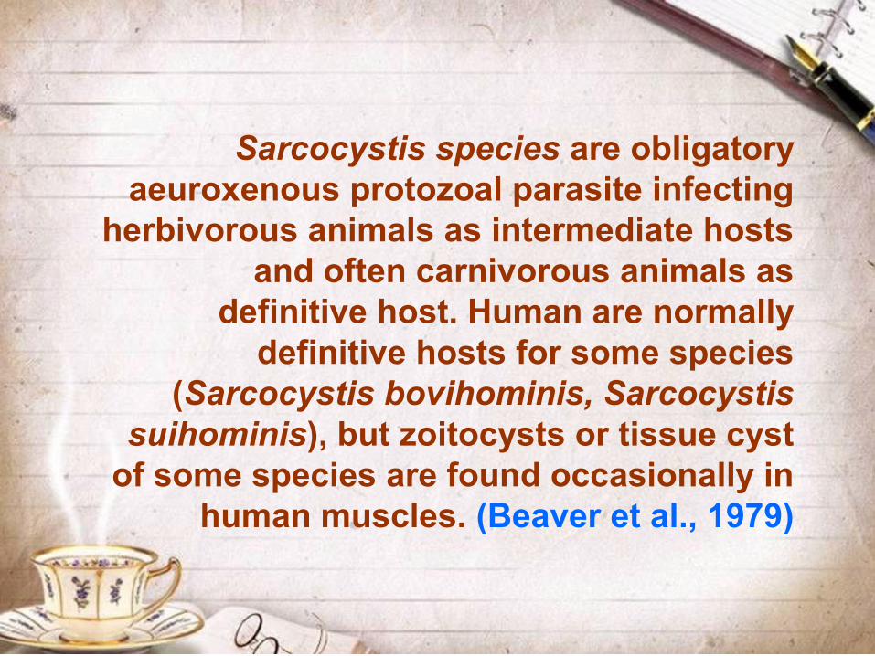
Sarcocystis species are obligatory aeuroxenous protozoal parasite infecting
herbivorous animals as intermediate hosts and often carnivorous animals as
definitive host. Human are normally definitive hosts for some species
(Sarcocystis bovihominis, Sarcocystis suihominis), but zoitocysts or tissue cyst
of some species are found occasionally in human muscles. (Beaver et al., 1979)
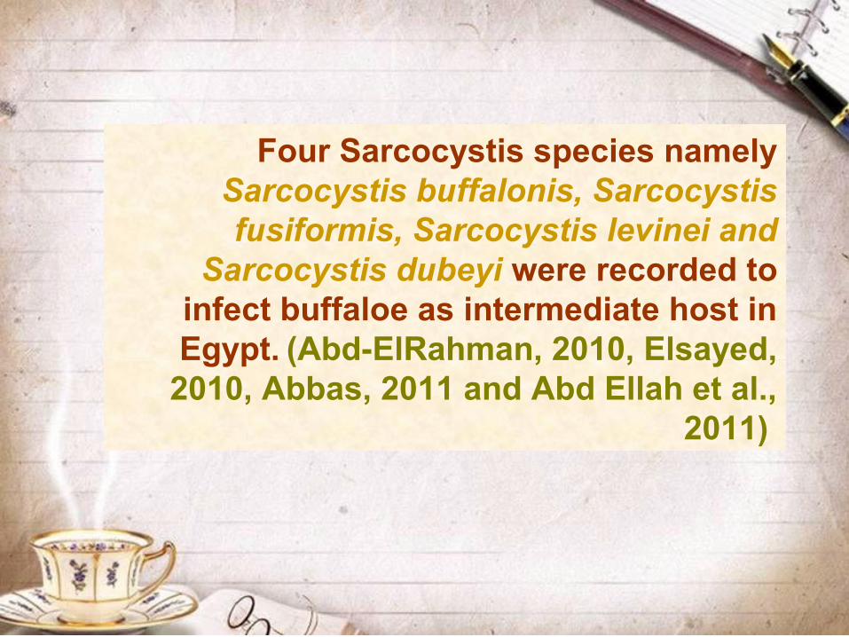
Four Sarcocystis species namely Sarcocystis buffalonis, Sarcocystis fusiformis, Sarcocystis levinei and
Sarcocystis dubeyi were recorded to infect buffaloe as intermediate host in Egypt. (Abd-ElRahman, 2010, Elsayed,
2010, Abbas, 2011 and Abd Ellah et al., 2011)
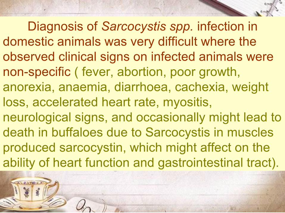
Diagnosis of Sarcocystis spp. infection in domestic animals was very difficult where the observed clinical signs on infected animals were non-specific ( fever, abortion, poor growth, anorexia, anaemia, diarrhoea, cachexia, weight loss, accelerated heart rate, myositis, neurological signs, and occasionally might lead to death in buffaloes due to Sarcocystis in muscles produced sarcocystin, which might affect on the ability of heart function and gastrointestinal tract).
Also histological or digestion techniques was not a valid procedure for diagnosing species of Sarcocystis in livestock animals. Several studies had been performed to diagnose Sarcocystis spp. by (ELISA).
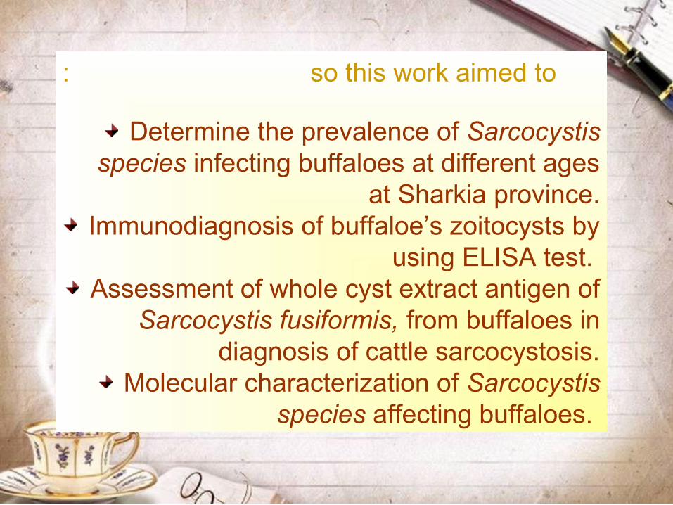
so this work aimed to:
Determine the prevalence of Sarcocystis species infecting buffaloes at different ages
at Sharkia province.Immunodiagnosis of buffaloe’s zoitocysts by
using ELISA test. Assessment of whole cyst extract antigen of
Sarcocystis fusiformis, from buffaloes in diagnosis of cattle sarcocystosis.
Molecular characterization of Sarcocystis species affecting buffaloes.

MATERIAL AND METHODS

1. Collection of materials
1.1. Meat samples:
A total of 110 oesophageal meat samples (22 from young males aged less than 45 days, 82 from adult males aged 2-3 years and 6 from old females aged over 5 years) were collected from slaughtered buffaloes (Bubalus bubalis) at abattoir of Zagazig city. Also another 65 oesophageal meat samples were collected from slaughtered adult males of cattle (Bos Taurus) aged 2-3 years during the period from May 2012 to November 2013 at the main abattoir of Zagazig City.
1.2. Serum samples:
65 and 110 venous blood samples were taken from the examined cattle and buffaloes, respectively. Also at the same time oesophageal muscle samples were taken from each animal at the time of slaughtering. The collected blood samples were left to clot at room temperature for one hour and centrifuged at 3000 r.p.m for 10 minutes. Sera were aspirated by Pasteur pipette in clean dry crocked bottle which labeled in a serial after being kept in the refrigerator for overnight and stored at -20°C until used for detection of anti Sarcocystis species antibodies.

2. Examination of the collected meat
samples.
2.1.Gross inspection of macroscopic cyst:
Cysts were found in skeletal muscle and were distinguished by their size, wall thickness and shape. The cysts were teased out with a needle and forceps.
2.2.Digestion method:
2.2.1. Peptic Digestion.
2.2.2.Trypsin Digestion.
2.3. Muscle squash:
About 1 gm of tissue was cut into small pieces, approximately 3-5 mm thick, and crushed firmly between two glass slides and examined under the microscope (Jackson et al., 1981).
2.4. Squeezing:
About 20 gm of meat was placed in the pocket of a metal instrument for garlic crushing and squeezing. One drop of the fluid from holes of instrument was placed on glass slide covered with cover slip and examined microscopically
(Latif et al., 1999).
2.5. Histopathology.(Do et al., 2008)
3. Detection of circulating Sarcocystis species antibodies by
ELISA test
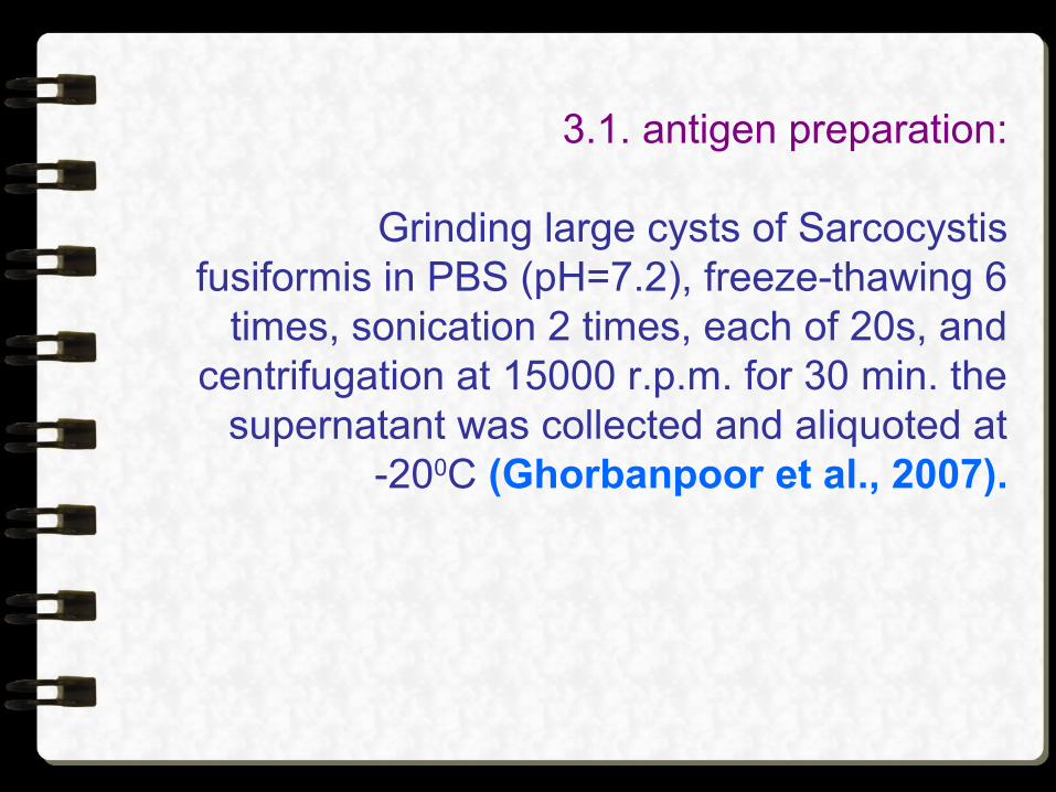
3.1. antigen preparation:
Grinding large cysts of Sarcocystis fusiformis in PBS (pH=7.2), freeze-thawing 6
times, sonication 2 times, each of 20s, and centrifugation at 15000 r.p.m. for 30 min. the
supernatant was collected and aliquoted at -200C (Ghorbanpoor et al., 2007).
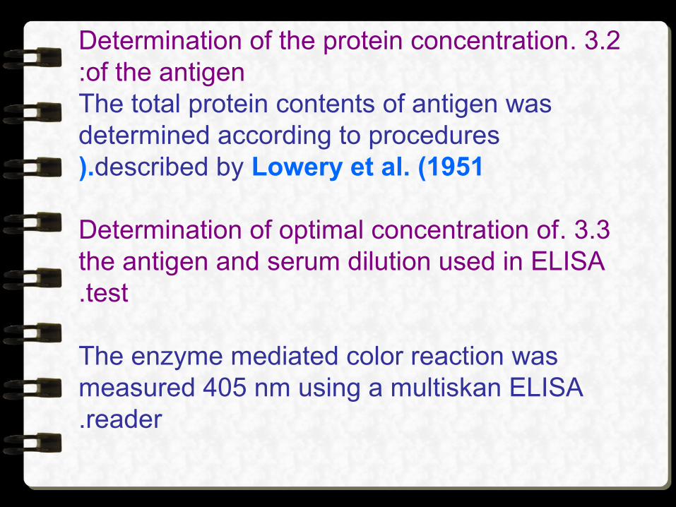
3.2. Determination of the protein concentration of the antigen:
The total protein contents of antigen was determined according to procedures
described by Lowery et al. (1951).
3.3. Determination of optimal concentration of the antigen and serum dilution used in ELISA test.
The enzyme mediated color reaction was measured 405 nm using a multiskan ELISA reader.

4. Polymerase chain reaction (PCR)
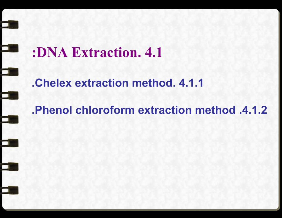
4.1. DNA Extraction:
4.1.1. Chelex extraction method.
4.1.2. Phenol chloroform extraction method.
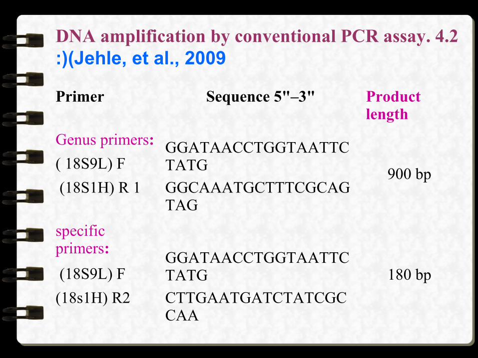
4.2. DNA amplification by conventional PCR assay (Jehle, et al., 2009(:
Primer Sequence 5"–3" Product length
Genus primers: GGATAACCTGGTAATTCTATG
900 bp( 18S9L) F
(18S1H) R 1 GGCAAATGCTTTCGCAGTAG
specific primers:
GGATAACCTGGTAATTCTATG 180 bp (18S9L) F
(18s1H) R2 CTTGAATGATCTATCGCCAA

After gently mixing and brief centrifugation, all tubes were placed in the thermal cycler, and amplification program
was as follow:Initial denaturation at 94o C for 3 minutes.
Fourty cycles:Denaturation step 94o C for 40 seconds. Annealing step 53o C for 60 seconds.Extension step 72o C for 80 seconds.
The final extension 72o C for 5 minutes.
Amplification products as well as a negative control (nuclease-free water) were separated on a 1.5% agarose gel stained with ethidium bromide (TBE-buffer, 55V, 45 min).
4.3. Restriction Fragment Length Polymorphism Polymerase Chain Reaction (RFLP-PCR(:
Amplified PCR products were digested separately with restriction enzyme (Bsl1). A total of 30 ml reaction mixture was used containing 10 ml PCR product, 1 unit restriction
enzyme, 2 ml appropriate buffer and 17 ml nuclease free H2o.The restriction mixture was incubated for 1 h at 37 0C.
Enzyme was inactivated for 5 min at 70 0C. The obtained restriction fragments were
separated on a 2% agarose gel stained with ethidium bromide (TBE-buffer, 55V, 45 min(.

RESULTS

1. Prevalence of Sarcocystis in the
examined buffaloes
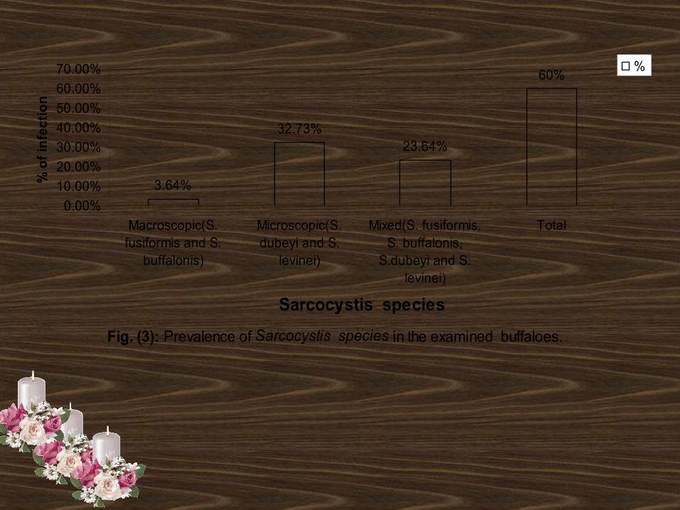
Fig. (3(: Prevalence of Sarcocystis species in the examined buffaloes.
3.64%
32.73%23.64%
60%
0.00%
10.00%
20.00%
30.00%
40.00%
50.00%
60.00%
70.00%
Macroscopic(S.fusiformis and S.
buffalonis)
Microscopic(S.dubeyi and S.
levinei)
Mixed(S. fusiformis,S. buffalonis,
S.dubeyi and S.levinei)
Total
Sarcocystis species
% o
f in
fect
ion
%
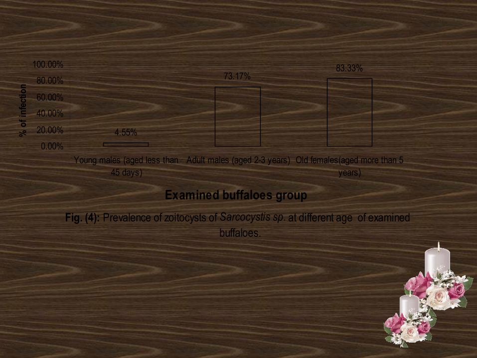
Fig. (4(: Prevalence of zoitocysts of Sarcocystis sp. at different age of examined buffaloes.
4.55%
73.17%83.33%
0.00%
20.00%
40.00%
60.00%
80.00%
100.00%
Young males (aged less than45 days)
Adult males (aged 2-3 years) Old females(aged more than 5years)
Examined buffaloes group
% o
f inf
ectio
n
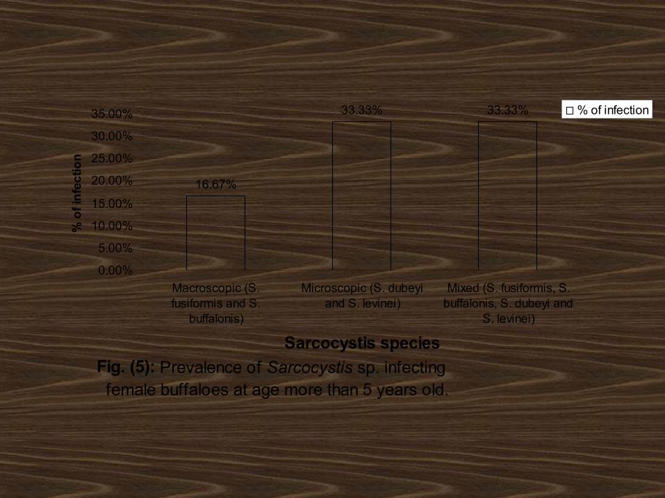
Fig. (5(: Prevalence of Sarcocystis sp. infecting female buffaloes at age more than 5 years old.
16.67%
33.33% 33.33%
0.00%
5.00%
10.00%
15.00%
20.00%
25.00%
30.00%
35.00%
Macroscopic (S.fusiformis and S.
buffalonis)
Microscopic (S. dubeyiand S. levinei)
Mixed (S. fusiformis, S.buffalonis, S. dubeyi and
S. levinei)
Sarcocystis species
% o
f in
fec
tion
% of infection
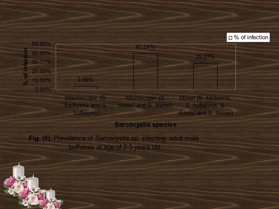
Fig. (6(: Prevalence of Sarcocystis sp. infecting adult male buffaloes at age of 2-3 years old.
3.66%
40.24%
29.27%
0.00%
10.00%
20.00%
30.00%
40.00%
50.00%
Macroscopic (S.fusiformis and S.
buffalonis)
Microscopic (S.dubeyi and S. levinei)
Mixed (S. fusiformis,S. buffalonis, S.
dubeyi and S. levinei)
Sarcocystis species
% o
f in
fect
ion
% of infection
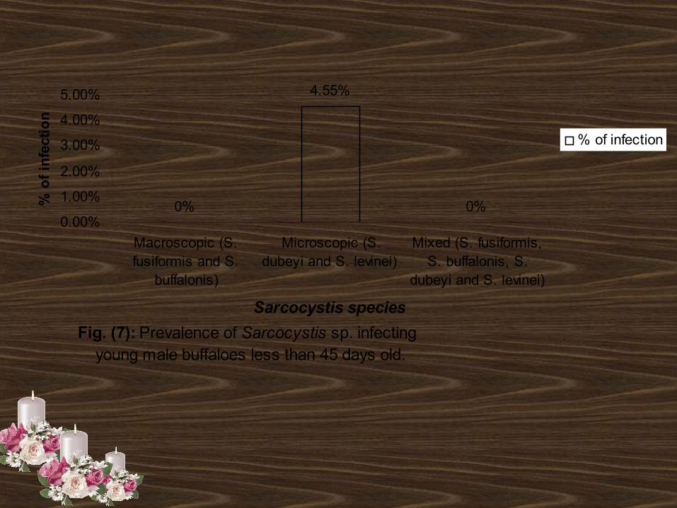
Fig. (7(: Prevalence of Sarcocystis sp. infecting young male buffaloes less than 45 days old.
0%
4.55%
0%0.00%
1.00%
2.00%
3.00%
4.00%
5.00%
Macroscopic (S.fusiformis and S.
buffalonis)
Microscopic (S.dubeyi and S. levinei)
Mixed (S. fusiformis,S. buffalonis, S.
dubeyi and S. levinei)
Sarcocystis species
% o
f in
fect
ion
% of infection
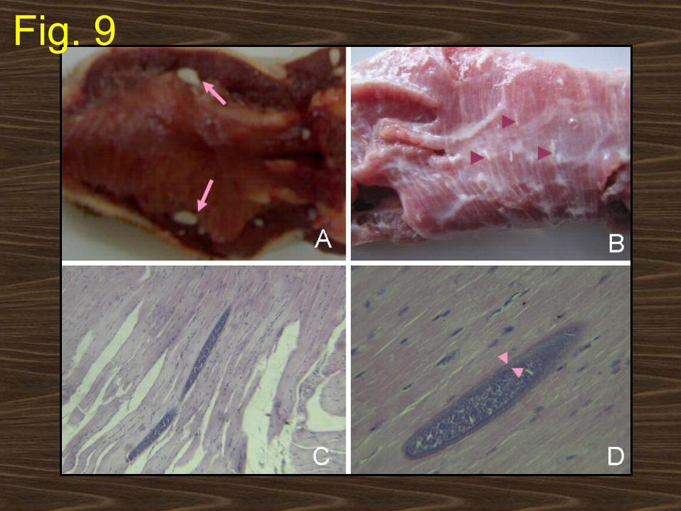
Fig. 9
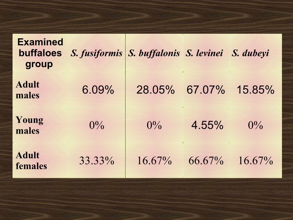
Examined buffaloes
group S. fusiformis S. buffalonis S. levinei S. dubeyi
Adult males 6.09% 28.05% 67.07% 15.85%
Young males 0% 0% 4.55% 0%
Adult females 33.33% 16.67% 66.67% 16.67%
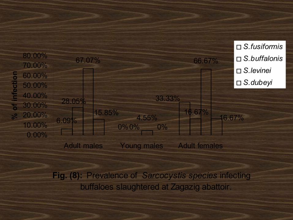
Fig. (8): Prevalence of Sarcocystis species infecting buffaloes slaughtered at Zagazig abattoir.
6.09%
67.07% 66.67%
33.33%
0%
16.67%
0%
28.05%
4.55% 16.67%0%
15.85%
0.00%10.00%20.00%30.00%40.00%50.00%60.00%70.00%80.00%
Adult males Young males Adult females
% o
f in
fect
ion
S.fusiformis
S.buffalonis
S.levinei
S.dubeyi
2- ELISA test for diagnosis of
sarcocystosis of buffaloes and
cattle
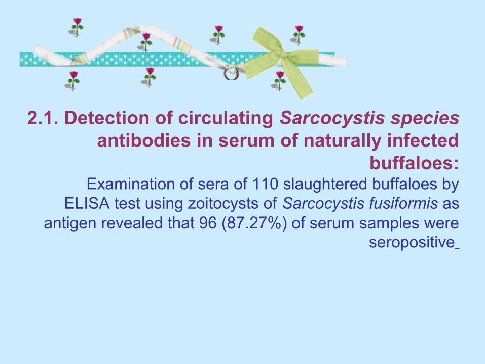
2.1. Detection of circulating Sarcocystis species antibodies in serum of naturally infected
buffaloes: Examination of sera of 110 slaughtered buffaloes by
ELISA test using zoitocysts of Sarcocystis fusiformis as antigen revealed that 96 (87.27%) of serum samples were
seropositive
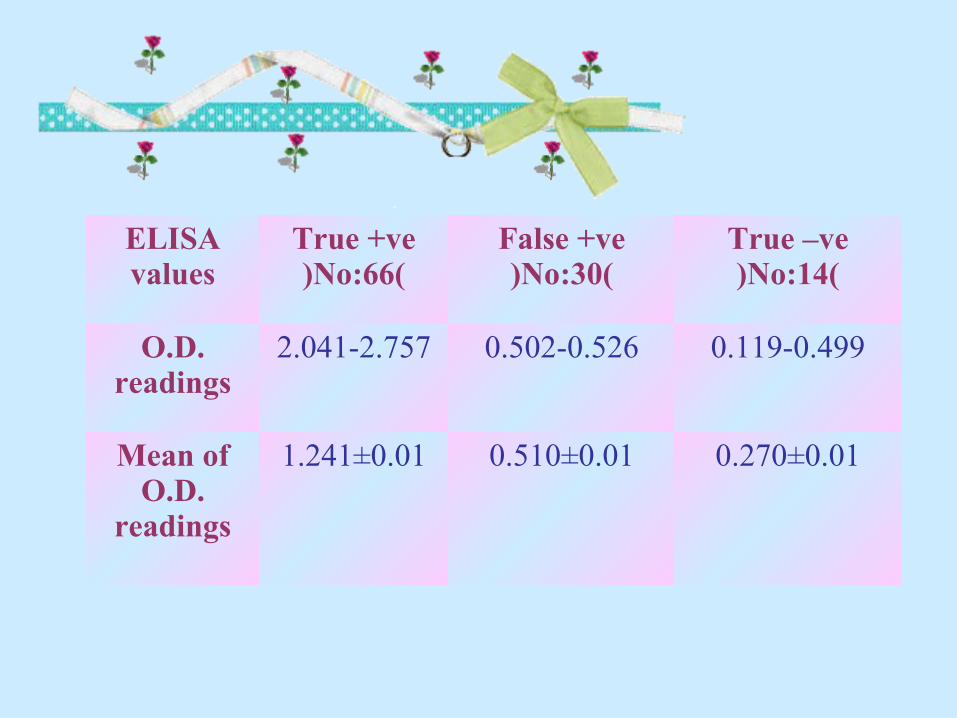
ELISA values
True +ve)No:66(
False +ve)No:30(
True –ve)No:14(
O.D. readings
2.041-2.757 0.502-0.526 0.119-0.499
Mean of O.D.
readings
1.241±0.01 0.510±0.01 0.270±0.01
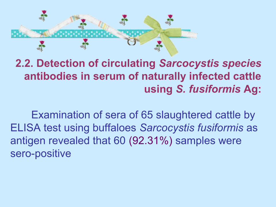
2.2. Detection of circulating Sarcocystis species
antibodies in serum of naturally infected cattle using S. fusiformis Ag:
Examination of sera of 65 slaughtered cattle by ELISA test using buffaloes Sarcocystis fusiformis as antigen revealed that 60 (92.31%) samples were sero-positive
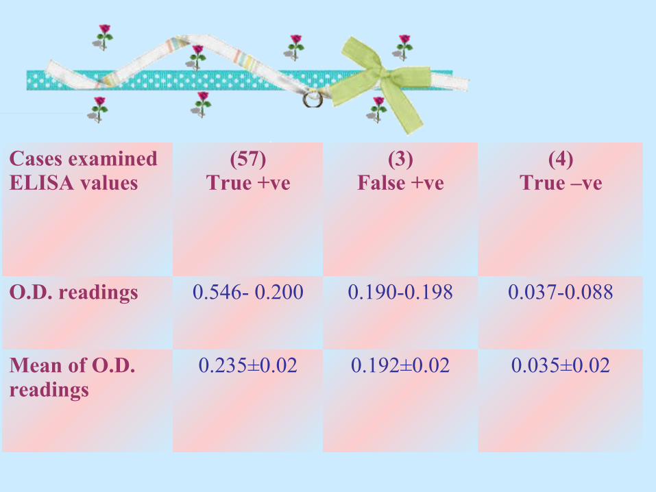
Cases examinedELISA values
(57)True +ve
(3)False +ve
(4)True –ve
O.D. readings 0.200- 0.546 0.190-0.198 0.037-0.088
Mean of O.D. readings
0.235±0.02 0.192±0.02 0.035±0.02
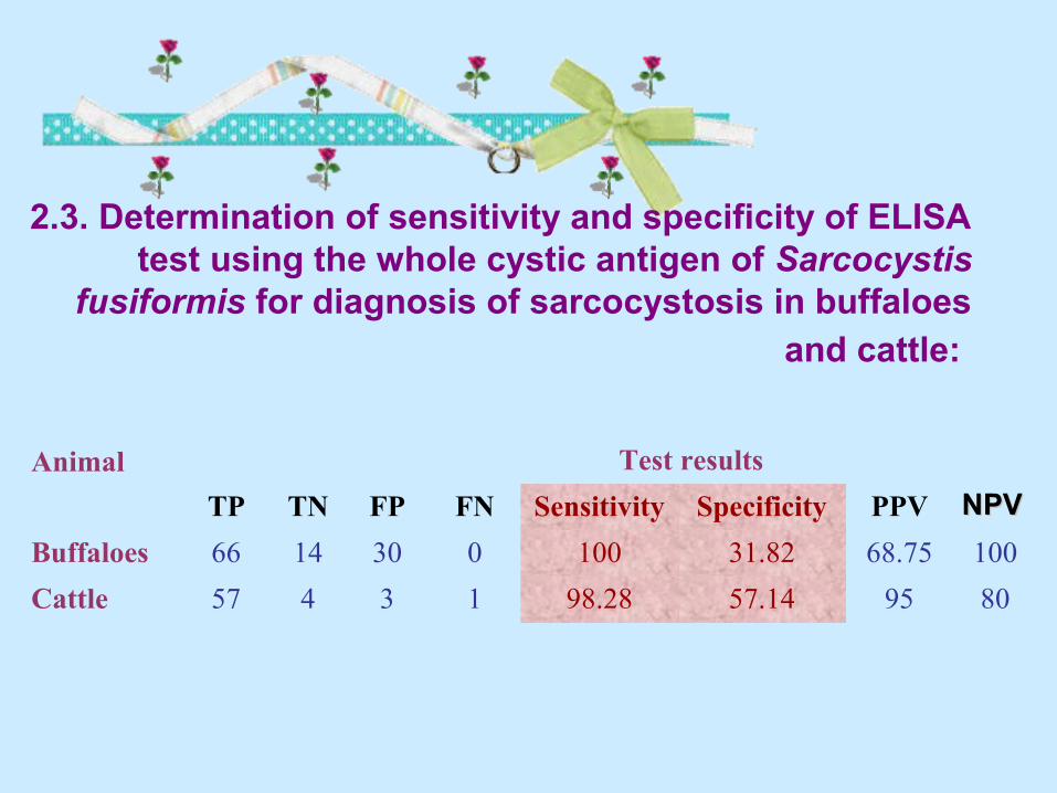
2.3. Determination of sensitivity and specificity of ELISA test using the whole cystic antigen of Sarcocystis
fusiformis for diagnosis of sarcocystosis in buffaloes and cattle:
Animal Test results
TP TN FP FN Sensitivity Specificity PPV
Buffaloes 66 14 30 0 100 31.82 68.75 100
Cattle 57 4 3 1 98.28 57.14 95 80
NPVNPV
3. Molecular characterization of
Sarcocystis species affecting buffaloes using
PCR-based approach
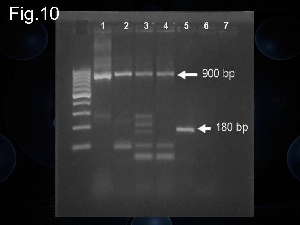
Fig.10
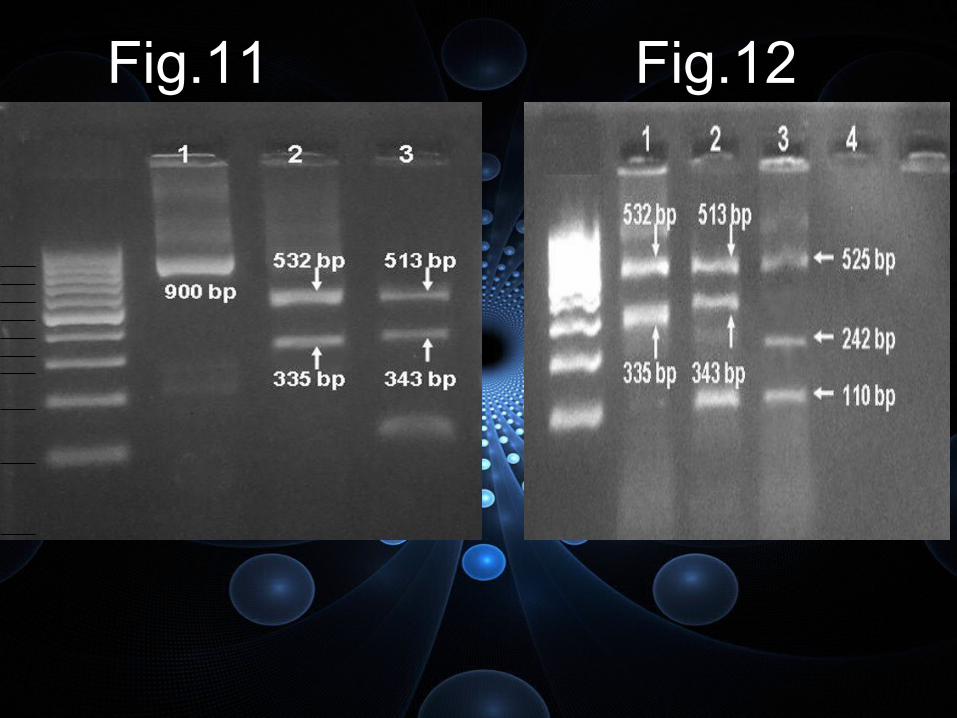
Fig.11 Fig.12