monoclonal antibodies against all known variants of espa ... · ... centre for biotechnology; cmc,...
TRANSCRIPT

Downloaded from www.microbiologyresearch.org by
IP: 143.210.121.140
On: Tue, 16 Feb 2016 14:20:45
Monoclonal antibodies against all known variants ofEspA: development of a simple diagnostic test forenteropathogenic Escherichia coli based on a keyvirulence factor
Uta Praekelt,1 Rolf Reissbrodt,2 Andreas Kresse,2
Asalapuram Pavankumar,33 Krishnan Sankaran,3 Roger James,4
Mary Jesudason,5 Shalini Anandan,5 Agila Prakasam,5
Veeraraghavan Balaji,5 Shanta Dutta,6 Sanjucta Dutta,6
Thandavarayan Ramamurthy,6 Renate Fischer,7 Peter Sander,7
Reiner Schaumann,8 Armando Navarro9 and Peter Williams1
Correspondence
Uta Praekelt
Received 25 March 2014
Accepted 16 September 2014
1Department of Genetics, University of Leicester, Leicester, UK
2Abteilung fur Infektionskrankheiten, Robert Koch Institut, Wernigerode, Germany
3Centre for Biotechnology, Anna University, Chennai, India
4Department of Infection, Immunity and Inflammation, University of Leicester, Leicester, UK
5Christian Medical College, Vellore, India
6National Institute of Cholera and Enteric Diseases, Kolkata, India
7R-Biopharm AG, Darmstadt, Germany
8Institut fur Medizinische Mikrobiologie, Universitat Leipzig, Leipzig, Germany
9Department of Public Health, Faculty of Medicine, National Autonomous University of Mexico,Mexico City, Mexico
Enteropathogenic Escherichia coli (EPEC) are a major cause of infant diarrhoea in developing
countries and a significant public health issue in industrialized countries. Currently there are no
simple tests available for the diagnosis of EPEC. Serology of O-antigens is widely used routinely
in many laboratories throughout the world, even though it has been known for many years to be an
unreliable indicator of EPEC virulence. We have developed a simple, low-cost immunodiagnostic
test based on the EspA filament, an essential virulence factor of EPEC and the related
enterohaemorrhagic E. coli (EHEC). Using recombinant proteins of the five major variants of EspA
as immunogens, we raised a panel of three monoclonal antibodies in mice that detects all variants
of the native target in bacterial cultures. The antibodies proved suitable for application in
sandwich-type assays, including ELISA and lateral flow immunoassays (LFI). Prototypes for both
assays were specific for EPEC and EHEC strains when tested against a panel of control micro-
organisms. We have also developed a simple, affordable culture medium, A/E medium, which
optimizes expression of EspA allowing improved sensitivity of detection compared with standard
Dulbecco’s modified Eagle’s medium. Together these reagents form the basis of robust,
informative tests for EPEC for use especially in developing countries but also for routine screening
in any clinical laboratory.
3Present address: Science for Life Laboratory, Stockholm University, Stockholm, Sweden.
Abbreviations: A/E, attaching and effacing; CBT, Centre for Biotechnology; CMC, Christian Medical College; EHEC, enterohaemorrhagic E. coli; EPEC,enteropathogenic E. coli; LFI, lateral flow immunoassay; NICED, National Institute of Cholera and Enteric Diseases; RKI, Robert Koch Institut; TMB,tetramethyl benzidine.
The GenBank/EMBL/DDBJ accession numbers for the 22 sequences of espA determined in this study are given in Table 2.
Journal of Medical Microbiology (2014), 63, 1595–1607 DOI 10.1099/jmm.0.076323-0
076323 G 2014 The Authors Printed in Great BritainThis is an Open Access article distributed under the terms of the Creative Commons Attribution License (http://creativecommons.org/licenses/by/3.0/). 1595

Downloaded from www.microbiologyresearch.org by
IP: 143.210.121.140
On: Tue, 16 Feb 2016 14:20:45
INTRODUCTION
Enteropathogenic Escherichia coli (EPEC) are a major causeof infant diarrhoea in developing countries, accounting foran estimated 10 % of the approximately 1.4 billion paediatricdiarrhoeal episodes annually in children under the age of5 (O’Ryan et al., 2005). In the absence of treatment, particu-larly among very young children also affected by malnutri-tion, EPEC diarrhoea can be fatal or lead to irreversibledamage to the intestine (Chen & Frankel, 2005). Industrializedcountries have experienced an overall decline in childhooddiarrhoea during the past 50 years, but EPEC still accountsfor a similar proportion of diarrhoeal incidences (Afset et al.,2003; Robins-Browne et al., 2004). In addition in industria-lized countries, the closely related pathotype enterohaemorr-hagic E. coli (EHEC) is responsible for occasional, mainlyfood-borne outbreaks of diarrhoea in adults and children,frequently accompanied by severe complications such ashaemorrhagic colitis and haemolytic uraemic syndrome dueto the action of shigatoxins not present in EPEC (Frankelet al., 1998; Smith et al., 2004).
EPEC and EHEC are a heterogeneous group of E. coli strains.For many years the diagnosis of EPEC has been basedprimarily on the identification of O : H serotypes accordingto WHO guidelines dating from 1987, which recognized the12 so-called classical EPEC serogroups associated withchildhood diarrhoea: O26, O55, O86, O111, O114, O119,O125, O126, O127, O128, O142 and O158 (Campos et al.,2004). EHEC strains are commonly associated withserogroups O103, O145 and O157, while some serogroups,in particular O55, O26 and O111, include both EPEC andEHEC strains. However, the serotyping scheme wasdeveloped before EPEC and EHEC virulence mechanismswere elucidated and many subsequent studies have shownthat there is only partial correlation between serology andpathotype (Afset et al., 2003; Ajjampur et al., 2008; Camposet al., 2004; Yang et al., 2007). Nevertheless, despite the factthat O-serology is non-informative of virulence, it continuesto be used in many clinical laboratories throughout theworld as one of the routine tests to establish the cause ofdiarrhoea (Kozub-Witkowski et al., 2008). While EHEC canbe identified by immunological tests for shigatoxins, there isa real need for simple diagnostic tests for EPEC, based onknown virulence factors, especially in developing countrieswhere EPEC diarrhoea is endemic, but also in industrializedcountries where studies indicate that EPEC may be moreprevalent than was previously thought.
EPEC and EHEC colonize the intestinal epithelium causingattaching and effacing (A/E) lesions by a mechanism thatinvolves the intimate attachment of bacteria to the hostcell (Kenny et al., 1997). Various virulence factors essentialfor this process are encoded on a pathogenicity island, thelocus of enterocyte effacement, including intimin, a bacterialmembrane adhesion protein encoded by the eae gene andthe EspA (E. coli secreted protein A) filament, a hollow tubethat acts like a molecular syringe for delivery of the Tir(translocated intimin receptor) protein and other effector
molecules into the host cell (Crepin et al., 2005; Frankel et al.,1998; Knutton et al., 1998). Methods that target the presenceof virulence genes, such as PCR and DNA microarray testsfor the eae gene, are ideal as the basis for reliable diagnos-tic tests, but such methods are generally not applicable toroutine diagnostic testing in peripheral health centres indeveloping countries where resources and skills may belimited. In these circumstances, simple antibody-based testsare much more suitable.
There have been several reports of antibodies raised againstvarious secreted or surface-located EPEC virulence factors(Batchelor et al., 1999; Giron et al., 1995; Kuhne et al.,2004; Lu et al., 2002; Menezes et al., 2010); however, theseeither detected only a limited subset of EPEC strains orrequired denaturation of the target for detection and to ourknowledge have not been developed further. We chose theEspA filament, a 5-stranded helical polymer of identical21 kDa monomers (Daniell et al., 2003; Delahay et al.,1999), as the immunological target for the development ofa low cost EPEC diagnostic test initially intended for useprimarily in the Indian subcontinent. We characterizedespA gene sequences from a set of clinical isolates collected insouth India and identified five major variants, all of whichwere represented, sometimes with minor variations, in theDNA and protein databases. Using recombinant proteins ofthese five variants as immunogens, we raised monoclonalantibodies capable of detecting all the EspA variantspublished to date. We also designed a low cost mediumfor optimal expression of EspA in culture. Together thesereagents comprise a simple and reliable replacement for O-serogrouping for the identification of EPEC diarrhoea.
METHODS
Bacterial strains and growth conditions. Clinical isolates wereobtained from the following laboratories: 16 eae+ strains fromChristian Medical College (CMC), Vellore, India and four eae+ strainsfrom the Centre for Biotechnology (CBT), Anna University, Chennai,India of known O : H serotype; 61 strains from the National Instituteof Cholera and Enteric Diseases (NICED), Kolkata, India, isolatedon the basis of a positive PCR for intimin (eae+) but of unknownserotype; 242 strains from the Institut fur Medizinische Mikrobiologie,Universitat Leipzig, Germany, isolated on the basis of O-serogroupstypical for EPEC and EHEC, and of these only 104 were eae+; 34 eae+
strains from the Department of Public Health, Faculty of Medicine,National Autonomous University of Mexico with known O : H serotypes;14 eae+ strains from the Robert Koch Institut (RKI), Wernigerode,Germany, which had been O : H-serotyped and also tested for virulencefactors associated with EHEC to distinguish EPEC (8 strains) fromEHEC (3 stx1 and 3 stx2 strains). Non-EPEC reference strains (as listedin Fig. 6) were also from RKI.
Strains were maintained on Luria agar. To induce production ofvirulence factors, strains were inoculated into Dulbecco’s modifiedEagle’s medium (DMEM; Gibco) containing 1 % glucose and incubatedovernight at 37 uC in 5 ml volumes without agitation.
Improved medium for virulence induction. To improve the expre-ssion of EspA in some strains, we developed an alternative medium,A/E medium, based on soy peptone and yeast extract, using somepoorly expressing strains as indicators of improvement in a dot blot
U. Praekelt and others
1596 Journal of Medical Microbiology 63

Downloaded from www.microbiologyresearch.org by
IP: 143.210.121.140
On: Tue, 16 Feb 2016 14:20:45
(see below). Taking into account the reported importance of calciumand sodium bicarbonate (Abe et al., 2002), and considering thecomponents of DMEM that might be important, such as vitamins,we arrived at the following formulation: 4.5 g Difco Select Soytonel21 (BD); 6 g HEPES l21 (free acid; Melford); 2 g yeast extract l21
(Oxoid); 10 g lactose l21 (Fisher Scientific); 0.2 g CaCl2 . H2O l21
(Sigma); 0.2 g ferric ammonium chloride l21 (Sigma); and 0.4 g KCll21 (Sigma). The medium was sterilized either by autoclaving or byfilter-sterilization. NaHCO3 powder (Sigma) was added to a finalconcentration of 7.5 g l21 immediately prior to use. Cultures (5–10 ml) were inoculated and incubated overnight at 37 uC withoutagitation.
Cloning and mutagenesis. Variant espA genes were amplified byPCR from genomic DNA using flanking primers UP1 F/UP1 R(727 bp) or UP2 F/UP2 R (1010 bp; Table 1). Products were purifiedfrom agarose gels and the DNA sequences determined using the sameprimers. For cloning and expression of recombinant proteins, espAcoding regions were amplified from five strains using primers EspAF1 and EspA R1 (isolate III-3, EspA a; 591 bp); EspA F2 and EspA R2(isolate A5, EspA b and isolate A7, EspA d; 592 bp); EspA F5 andEspA R2 (isolate III5, EspA c; 592 bp) and EspA F6 and EspA R4(isolate C2; EspA e; 582 bp). PCR products were digested with the
appropriate restriction enzymes (as indicated in Table 1) and cloned
into similarly cut vector pET28a (Novagen) in E. coli strain XL10 Gold
(Stratagene). After confirmation by DNA sequencing using primer T7
(homologous to vector sequence) the recombinant plasmids were
transformed into strain BL21(DE3) (Stratagene).
For epitope mapping, overlapping fragments of the espA genes
from the three variants representing the three mAbs used in this work
were cloned into pET28a. Templates for amplification were recom-
binant plasmids containing EspA b for mapping mAb 14, EspA d for
mapping mAb 209 and EspA e for mapping mAb 2. EspA b fragments
were amplified using primer combinations Epi F21/Epi R12 (299 bp;
amino acids 110–192) and Epi F20/Epi R1 (371 bp; amino acids 1–
118). EspA d fragments were amplified with primers Epi F12/Epi R10
(428 bp; amino acids 1–137) and Epi F21/Epi R12 (299 bp; amino
acids 100–192). EspA e fragments were amplified with primers Epi F6/
Epi R9 (422 bp; amino acids 1–135), Epi F7/Epi R5 (356 bp; amino
acids 78–190) and Epi F6/Epi R20 (485 bp; amino acids 1–156).
Fragments were digested with restriction enzymes as indicated in
Table 1 and cloned into similarly digested pET28a.
EspA mutant proteins containing single amino acid changes were
produced by first amplifying two separate PCR fragments primed
O103:H2
O26:H– *A7
O49:H12δ
O125:H– *H2
O111:H2– CA12348
O126:H– *A5
O26:H11– YP003232165
O111:NM ZP03059068(B171)β
O128:H2
O103:H2
O119:H2
O111:H– YP003236076
O55:H7– CAA12350
O127:H6
O127:H6– *III3
Ont *G6
O157:H7
α
O157:H7
O55:H7
O55:H–
C. freundii AAL06381
O119:H9
γ
O119:H6
O157:H18
O111:H9
O86:H34
ZP18692421
ε
O157:H39
B155ND
YP003223463
AA066617
*H1
ZP03043278
CAA12345
YP002331398 (E2348/69)
ZP11668346
*III5
YP003501862
* III6 (EPEC)
CAG17641
*C2
ZP18671299
ZP03051294
*D2
Fig. 1. Phylogenetic tree of deduced EspAamino acid sequences using CLUSTAL W.*Representative clinical isolates from southIndia for which espA gene sequences weredetermined in this study (Table 2). B155represents a strain of unknown serotype fromNICED. All others are sequences from theDNA (GenBank) and protein (NCBI) data-bases. NM, non-motile; Ont, non-typable forO-antigen; ND, serotype not determined.
EPEC diagnosis with monoclonal antibodies against EspA
http://jmm.sgmjournals.org 1597

Downloaded from www.microbiologyresearch.org by
IP: 143.210.121.140
On: Tue, 16 Feb 2016 14:20:45
with overlapping internal primers containing the appropriate base
changes and external primers, and then mixing the resulting frag-
ments together and amplifying with external primers only. The
resulting products were digested with restriction enzymes, cloned and
expressed in pET28a using the following combinations: EspA b
(N112K) Mut 1/EspA R2 (268 bp) and Mut 2/EspA F2 (354 bp);
EspA b (D117G) Mut 15/EspA R2 (261 bp) and Mut 16/EspA F2
(378 bp); EspA d (N96E) Mut 5/EspA R2 (317 bp) and Mut 6/EspA
F2 (313 bp); EspAd (K112Q) Mut 7/EspA R2 (269 bp) and Mut 8/
EspA F2 (361 bp); EspA e (E94D) Mut 17/EspA R4 (319 bp) and Mut
18/EspA F6 308 bp).
Recombinant protein expression and purification. Recombinant
plasmids were transformed into BL21(D3) (Stratagene) by the calcium
chloride method. Transformants were grown at 37 uC to mid-exponential
phase in 400 ml Luria broth, induced by addition of IPTG to 1 mM
and grown for a further 4 h. Recombinant proteins were isolated in
the form of inclusion bodies. Cells in 50 ml culture volumes were
pelleted by centrifugation and resuspended in 2.5 ml 0.1 M sodium
phosphate (pH 8.5), 0.3 M sodium chloride. After incubation for
30 min at 37 uC in the presence of lysozyme (10 mg ml21) the cells
were disrupted by sonication on ice. Suspensions were centrifuged at
6000 r.p.m. for 10 min in an Eppendorf Minispin centrifuge and
pellets were resuspended in wash buffer (50 mM Tris, pH 8, 0.1 M
sodium chloride, 2 M urea, 0.5 % Nonidet) by syringing to break up
clumps. Washing was repeated and suspensions were stored in wash
buffer at 220 uC overnight, resulting in almost complete clearing of
the suspension. Suspensions were centrifuged at 13 500 r.p.m. for
5 min and the clear supernatants were removed and dialysed against
several changes of PBS. Essentially pure EspA protein, which was
Table 1. Primers for cloning and mutagenesis
Primer Sequence (5§ to 3§) Tm (6C)
UP1 F TAATACATTATTAATGATTGGTAAAG 54.8
UP1 R TATCGYTATTTACRTTAAGCATAG 56.6
UP2 F CTCGGGTATCGATTGTCGAAGAT 67.4
UP2 R CAGAGGGCGTCACTAATGAGTG 66.2
EspA F1 (a; NdeI) GCCATATGGATACATCAACTACAGCAC 67.2
EspA R1 (a; SacI) GCGAGTCTTATTTACCAAGGGATATTCCTG 70.0
EspA F2 (b,d; NdeI) GCCATATGGATACATCAACTGCAACATC 70.4
EspA R2 (b,d,c; SacI) GCGAGCTCTTATTTACCAAGGGATATTGCTG 72.7
EspA F6 (e; NcoI) CACCATGGATAATTCAGTTACATCATC 65.2
EspA R4 (e; XhoI) CGCTCGAGTTTACCAAAACTTATTGCC 70.4
EspA F5 (c; NdeI) GCCATATGGATACATCAAATGCAACATC 70.1
Epi F21 (b,d; BamHI) GCGGATCCATGAAAGCCAAACTTCCTCAA 77.6
Epi R12 (b,d; HindIII) GCAAGCTTATTTACCAAGGGATATTG 65.4
Epi F20 (b; BamHI) GCGGATCCATGGATACATCAACTGCAACA 77.2
Epi R1 (b; HindIII) GCAAGCTTAAATGTCATTGCGAGGATCATT 73.5
Epi F12 (d; BamHI) GCGGATCCATGGATACATCAACTGCAACA 77.2
Epi R10 (d; HindIII) GCAAGCTTACGTTTGCAAATCACCAGCGCC 80.1
Epi F6 (e; BamHI) GCGGATCCATGGATAATTCAGTTACATCA 72.1
Epi R9 (e; HindIII) GCAAGCTTAGGTTTGCAGGTCACCAGCACC 78.7
Epi F7 (e; BamHI) GCGGATCCATGGCCAATTTGGTGGATGCC 83.3
Epi R5 (e; HindIII) GCAAGCTTATTTACCAAAACTTATTGC 64.7
Epi R20 (e; HindIII) GCAAGCTTAAAGCTGGTTATTATTCACCGT 70.3
Mut 1 (b N112K) GTGATTGACTATATAAAAGATCCTCGCAATG 67.7
Mut 2 (b N112K) CATTGCGAGGATCTTTTATATAGTCAATCAC 67.7
Mut 15 (b D117G) CTATATAAATGATCCTCGCAATGGCATTACA... 76.8
...GTAAGTGGTATTAGCG
Mut 16 (b D117G) CGCTAATACCACTTACTGTAATGCCATTGCG... 76.8
...AGGATCATTTATATAG
Mut 5 (d N96E) GTTCAGAGCAGTTCTGAGAAGGATGCGAAAGCCAAACTT 80.2
Mut 6 (d N96E) AAGTTTGGCTTTCGCATCCTTCTCAGAACTGCTCTGAAC 80.2
Mut 7 (d K112Q) GTGATCGACTATATACAAGATCCTCGTAATGGCATTAC 72.2
Mut 8 (d K112Q) GTAATGCCATTACGAGGATCTTGTATATAGTCGATCAC 72.2
Mut 17 (e E94D) CTGAAGTTCAGTCTAGTTCGGACAAGGATAAA... 77.0
...AAGGTCAAACTTC
Mut 18 (e E94D) GAAGTTTGACCTTTTTATCCTTGTCCGAACTA... 77.0
...GACTGAACTTCAG
T7 AATACGACTCACTATAGGG 51.2
Where appropriate, primer names indicate EspA type and restriction sites (in italics) used for cloning into vector DNA. Primers used for
mutagenesis (Mut) also indicate the amino acid substitution achieved (underlined). Tm values are based on the Tm Calculator from Thermo
Scientific.
U. Praekelt and others
1598 Journal of Medical Microbiology 63

Downloaded from www.microbiologyresearch.org by
IP: 143.210.121.140
On: Tue, 16 Feb 2016 14:20:45
recovered as a fine translucent precipitate, was quantified by SDS-PAGE against dilutions of a BSA standard.
mAb production. The five major espA variants (as shown in Fig. 2)were used as targets for raising monoclonal antibodies. BALB/c micewere immunized with recombinant EspA proteins from differentEPEC strains, presented in the form of inclusion bodies, eitherindividually or as mixtures, in the presence of Freund’s adjuvant orTitermax and boosted on two or three subsequent occasions at two-weekly intervals. Serum titres were determined by whole cell ELISA(see below). Splenocytes from mice with a high antibody titre werefused with NS0 cells by standard procedures using PEG and dispensedinto 96-well plates. Hybridomas were again screened by whole cellELISA and positive wells were cloned twice and expanded forproduction of antibody and storage of cell lines. Large-scale productionand purification of antibody were carried out by Sifin Diagnostics andsupplied at 1–2 mg ml21. Antibodies were isotyped using a mousemonoclonal antibody isotyping kit (Zymed Laboratories). Microtitreplates (NUNC Maxisorp) were coated with each antibody at 10 mgml21 in coating buffer (3.03 g Na2CO3 l21, 6.0 g NaHCO3 l21 indistilled water, pH 9.6). ELISA was performed according to themanufacturer’s instructions.
Western blots and dot blots. Proteins were resolved on 12 %polyacrylamide gels and blotted onto nitrocellulose membranes.These were blocked in PBS containing 4 % skimmed milk and probedwith hybridoma supernatants at dilutions of 1 : 100 to 1 : 500 for 1 h.Bound antibody was detected with goat antimouse horseradishperoxidase (Sigma) for 30 min, followed by tetramethyl benzidine(TMB) for up to 3 min. Membranes were washed with distilled waterand dried, and the results were recorded by scanning. For dot blots,2 ml volumes of culture were mixed with an equal volume of ethanol,
spotted onto polyvinylidene fluoride membrane and dried before
processing as for Western blots.
Direct whole cell ELISA. EPEC strains expressing EspA variants were
grown overnight in DMEM containing 1 % glucose. Cultures were
either mixed together in equal proportions (for initial screening of
hybridomas) or used separately (for subsequent testing) and diluted
with an equal volume of coating buffer. For testing, 100 ml of mixture
was dispensed into each well of 96-well microtitre plates (NUNC
Maxisorp), dried overnight at 37 uC, washed twice with PBS and then
blocked for 30 min with PBS containing 4 % skimmed milk. Doubling
dilutions of serum from test bleeds or undiluted hybridoma super-
natants were added to wells and incubated for 1 h, washed three times
with PBS, incubated with goat antimouse alkaline phosphatase (Sigma)
for 30 min and then washed five times with PBS. Detection was by p-
nitrophenyl phosphate (Sigma); reactions were stopped by the addition
of 1/4 volume of 3 M sodium hydroxide and absorbance was read at
405 nm.
Sandwich ELISA. Purified mAbs were conjugated to horseradish
peroxidase using the Lightning-Link kit (Innova Biosciences)
according to the manufacturer’s instructions. Microtitre plates were
coated with purified mAbs, either individually or as a mixture, at
10 mg ml21 in coating buffer and stored at 4 uC overnight. After
rinsing twice with PBS the plates were blocked with 4 % skimmed
milk in PBS for 30 min. Overnight cultures were added to wells either
directly or after dilution with PBS and incubated for 1 h. After five
washes detection was by horseradish peroxidase-conjugated mAbs,
again either separately or as a mixture, followed by substrate TMB
(liquid substrate system for ELISA, Sigma). After 15 min results were
recorded by scanning.
MDTSTATSVASANASTSTSTVYDLGSMSKDEVVQLFNKVGVFQAALLMFAYMYQAQSDLSMDTSTATSVASANASTSTSTVYDLGSMSKDEVVQLFKEVGVFQAALLMFAYMYQAQSDLSMDTSTTASVASANASTSTSMAYDLGSMSKDDVIDLFNKLGVFQAAILMFAYMYQAQSDLSMDTSNATSVVNVSASSSTSTIYDLGNMSKDEVVKLFEELGVFQAAILMFSYMYQAQSNLSMDNSVTSSIS—-SASTSTSMTYDLGNMSKDEVVELFKKVGVFQAALIMFAYMYQAQSELS
IAKFADMNEASKESTTAQKMANLVDAKIADVQSSSDKNKKAKLPQEVIDYINDPRNDITVIAKFADMNEASKESTTAQKMANLVDAKIAEVQSSSNKDAKAKLPQEVIDYIKDPRNGITVIAKFADMNEASKESTTAQKMANLVDAKIADVQSSSDKNAKAQLPDEVISYINDPRNDITIIAKFADMNEASKASTTAQKMANLVDAKIADVQSSTDKNAKAKLPQDVIDYINDPRNDISVIATYADMNESSKESTEAQKMANLVDAKIAEVQSSSEKDKKVKLPDEVISYIQDSRNGISV
SGISDLNAELGAGDLQTVKAAISAKSNILTTVVNNSQLEIQQMSNTLNLLTSARSDIQSL SDVKDITVELGAGDLQTVKAAISAKSNNLTTVVNNSQLEIQQMSNTLNLLTSARADIQSLSGIDNINAQLGAGDLQTVKAAISAKANNLTTTVNNSQLEIQQMSNTLNLLTSARSDMQSLTGISDLSGDLSAGDLQTVKAAISAKANNLTTVVNNSQLEIQQMSNTLNLLTSARSDVQSLSSDIDITKELGAGDLQTVKAAISAKANNLTTTVNNNQLTLQQMSNTLNLLTNARSDMQSL
QYRTISAISLGK QYRTISTISVGKQYRTISGISLGKQYRTISAISLGKQYRTIQAISFGK
A5
A7
III3
III5
C2
A5
A7
III3
III5
C2
A5
A7
III3
III5
C2
A5
A7
III3
III5
C2
60
60
60
60
58
120
120
120
120
180
180
180
180
180
178
192
192
192
192
190
Fig. 2. Alignment of five representative EspA amino acid sequences used for raising mAbs, using CLUSTAL 2. Source strains areA5, EspA a; A7, EspA d; III3, EspA b; III5, EspA c; C2, EspA e. Shading indicates residues that differ from the consensus.
EPEC diagnosis with monoclonal antibodies against EspA
http://jmm.sgmjournals.org 1599

Downloaded from www.microbiologyresearch.org by
IP: 143.210.121.140
On: Tue, 16 Feb 2016 14:20:45
R-Biopharm prototype sandwich ELISA. The sandwich ELISA was
carried out in 96-well plates with 12 separate strips of eight wells each
in a strip holder, which were supplied ready coated with a mixture of
mAbs, stabilized and blocked. All solutions except wash buffer and
sample diluent were provided in dropper bottles. To carry out the
test, samples were diluted with an equal volume of diluent (protein-
buffered NaCl solution) and 100 ml was applied to each well, followed
immediately by two drops of conjugate 1 (biotin-conjugated mAbs
in stabilized protein solution). After incubation for 1 h at room
temperature, wells were rinsed five times with wash buffer (PBS
containing 0.1 % thimerosal) and incubated for 30 min with two
drops of conjugate 2 (streptavidin-enhanced horseradish peroxidase
conjugate in stabilized protein solution). After five washes two drops
of substrate (hydrogen/TMB ready to use) were added and left to
incubate for 15 min. Results were recorded either by scanning for a
visual record, or by stopping the reaction with one drop of stop
solution (2 M H2SO4) followed by measurement of absorbance at
450 nm.
Lateral flow immunoassay (LFI). A prototype LFI was developed in
collaboration with Forsite Diagnostics Ltd, York, UK. Purified mAbs
were adsorbed to blue latex beads and incorporated into the release pad.
The capture antibody was line sprayed onto Prima 40 nitrocellulose
membranes (GE Health Care), initially using a single antibody for
each device. A control line to indicate a functional assay contained
antimouse antibody for capture of the mAb-conjugate. Devices were
assembled into cassettes and supplied in sealed foil pouches. For the
final prototype device all three antibodies were applied as mixtures to
both release pad and capture line. For the assay, bacterial cultures were
grown overnight in A/E medium, mixed with 1/10 volume of 5 M
sodium chloride and applied either to the devices directly or following
brief centrifugation to remove bacterial cells; 70 ml of suspensions or
supernatants were applied to the test devices and results recorded
photographically after 5 min.
RESULTS
EspA sequences of strains from south India
The EspA proteins encoded by the 21 confirmed EPEC orEHEC strains from south India (Table 2) were found to beeither identical or closely related to published EspA proteinsequences. A phylogenetic tree illustrating the relationship ofthese EspA proteins to those in protein and DNA databases isshown in Fig. 1, which also includes the EspA sequences ofB155, an A/E lesion-forming strain of unknown serotype fromNICED (see below) and Citrobacter freundii, an organism thatis commonly found in soil and water but may be present inthe intestinal tract of humans and animals (Tschape et al.,1995).
To rationalize the nomenclature of EspA we have assignedthe sequences to five groups based on similarity and desig-nated them using letters of the Greek alphabet by analogywith the nomenclature for intimin. Three of these groups,EspA a, EspA b and EspA c, exemplified by the well-characterized serotypes O127 : H6, O111 : H2 and O157 : H7,respectively, had already been designated as such inaccordance with the intimin variants carried by these strains.We suggest the designation EspA d for the group of variantsincluding O49 : H12, which was previously named EspA‘beta variant’ (Bertin et al., 2004), and EspA e for the group
including O119 : H6 that showed the greatest sequencedivergence of all variants and which had been variouslycalled EspA ‘beta variant’ or EspA ‘b2’ (Afset et al., 2006;Garrido et al., 2006).
Production and characterization of mAbs
We chose representatives of the five major EspA proteins(Fig. 2) as targets for the production of mAbs. The initialscreen of hybridoma supernatants by whole cell ELISA wasdesigned to select only those reacting with the native target.The resulting antibodies gave various patterns of cross-reactions with the different EspA types. Three hybridomaswhich produced strong signals in various applications andtogether detected all EspA variants were selected for furtherwork.
Western blots against recombinant EspA confirmed thatthe positive ELISA signals were due to specific interactionswith EspA protein (Fig. 3a, b); mAb 14 reacted stronglywith EspA types a, b and c and weakly with EspA d, whilemAb 2 and mAb 209 detected one EspA type each, e and d,
Table 2. Clinical isolates from India
Leic. no. Collection Serotype EspA type GenBank
III 3* RKI 05-02944 O127 : H6 a KJ549668
B5 RKI 04-02142 O127 : H6 a KJ549669
E4 RKI 04-02177 O127 : H6 a KJ549670
F6 RKI 04-02191 O127 : H6 a KJ549671
G6* RKI 04-02203 Ont : H2 a KJ549672
B3 RKI 04-02140 Ont : H2 b KJ549673
H1* RKI 04-02210 O128 : H2 b KJ549674
G1 RKI 04-02198 O128 : H2 b KJ549675
A5* RKI 04-02130 O126 : H2 b KJ549676
H3 RKI 04-02212 O126 : H2 b KJ549677
III 5* RKI 05-02946 O157 : H7 c KJ549678
III 14 RKI 05-02948 O157 : H7 c KJ549679
III 6* RKI 05-02947 O55 : H2 c KJ549680
H2* RKI 04-02211 O125 : H2 d KJ549681
A6 RKI 04-02131 O125ab : H2 d KJ549682
A7* RKI 04-02132 O26 : H2 d KJ549683
C2* RKI 04-02151 O119 : H6 e KJ549684
G2 RKI 04-02199 O119 : H6 e KJ549685
D2* RKI 04-02163 O86 : H34 e KJ549686
F5 RKI 04-02190 O55 : H2 e KJ549687
H6 RKI 04-02215 O55 : H2 e KJ549688
B155* NICED B155 ND e KJ549689
Strains were collected at CBT and CMC; B155 was from NICED. All
except B155 were logged and serotyped at RKI and espA DNA
sequences were determined at the University of Leicester. Deduced
EspA protein sequences were classified into five subtypes according to
similarity and denoted by a, b, c, d or e.
*Strains included in the phylogenetic tree shown in Fig. 1; Ont non-
typable for O-antigens of classical EPEC/EHEC strains (O26, O55,
O86, O103, O111, O114, O119, O125, O127, O128, O126, O142
O145, O157); ND, not determined.
U. Praekelt and others
1600 Journal of Medical Microbiology 63

Downloaded from www.microbiologyresearch.org by
IP: 143.210.121.140
On: Tue, 16 Feb 2016 14:20:45
respectively. Recombinant EspA e produced multiple bandsvisible in both the Coomassie stained gel and a Westernblot with mAb 2; unlike the other recombinant EspAvariants, EspA e contains a C-terminal histidine tag whichmay have resulted in translation initiation at downstreammethionine codons within the espA gene. Western blots ofcell lysates from corresponding overnight cultures of EPECstrains confirmed detection of a single band migrating atthe expected position for a 21 kDa protein (Fig. 3c). Thepattern of reactivity was similar to that with recombinantproteins, although no cross-reaction of mAb 14 was seenwith a lysate of strain H2 carrying EspA d.
Similar patterns of reactions were obtained with two versionsof a direct whole cell ELISA of immobilized cultures, usingeither horseradish peroxidase-conjugated secondary antibodyfor rapid visual inspection of results, or alkaline phospha-tase-conjugated secondary antibody for absorbance meas-urement using a plate reader (Fig. 3d, e). Strain B155, an A/Elesion-forming strain of unknown serotype from NICED,was included here as it was unexpectedly detected by bothmAb 2 and mAb 209.
For testing large numbers of samples simultaneously duringthe development phase of the work, we used a dot blot.This gave results essentially identical to those obtained with
ELISA (Fig. 3f), although mAb 209 also showed a slightcross-reaction in this test with strain C2 carrying EspA e.
Improved medium for EspA induction
DMEM has traditionally been used to study the virulencefactors of EPEC, but we found it to be suboptimal for EspAexpression in some strains in our collections. DMEM isalso likely to be too expensive for routine use in developingcountries. We therefore developed an alternative cheapermedium, which we called A/E (for attaching and effacing)medium, that enhanced the detection of EspA after overnightgrowth compared with DMEM for a representative sample ofstrains (Fig. 3g). In a time-course of EspA induction, most ofthese strains were detectable by dot blot after 4 h and allstrains after 5 h in A/E medium, starting with a 1/100dilution from an overnight culture in Luria broth (data notshown).
Epitope mapping and IgG isotyping
Overlapping fragments of espA from isolates A5 (b), H2 (d)and C2 (e) were tested by dot blots with mAbs 14, 209 and2, respectively (Fig. 4a). Corresponding Western blots (notshown) confirmed that the signal was specific for EspA
III3KDa
97/66
45
31
2114
EspA variant
III5 B155A5 C2
Strain
Strain
(d)
H2 mAb
14
2
209
Mix
14
2
209
mAb
14
2
Mix
14
209
mAb
2
209
Mix
14
2
209
(g) C2H1A5III3 G6 III14III5A7D2 H2 Strain
EspA variant
A/E
DMEM
III3 III5 B155A5 C2 H2
III3 III5 B155A5 C2 H2III3 III5A5 C2 H2
(e)
(a)
(b)
(f)(c)
α β γ δε
α α β β γ γδ δε ε
EspA variant
α β γ δε
Fig. 3. Detection of EspA variants with mAbs14, 2 and 209. SDS-PAGE of partially purifiedrecombinant EspA proteins stained withCoomassie blue (a). Western blots showingcross-reactivity of mAbs with recombinant EspAproteins (b) and SDS-PAGE-separated totalcell protein of EPEC strains III3 (EspA b), A5 (a),C2 (e), H2 (d) and III5 (c) (c). Direct whole cellELISA of the same five strains plus strain B155,developed with horseradish peroxidase-anti-mouse antibody and TMB (d) and thendeveloped with alkaline phosphatase-anti-mouse antibody and p-nitrophenyl phosphate;absorbance was read at 405 nm (e). Dot blotsusing the same strains (f). Dot blots with amixture of three mAbs using ten strains repre-senting the range of EspA types showingimproved detection after overnight growth inA/E medium compared with DMEM (g).
EPEC diagnosis with monoclonal antibodies against EspA
http://jmm.sgmjournals.org 1601

Downloaded from www.microbiologyresearch.org by
IP: 143.210.121.140
On: Tue, 16 Feb 2016 14:20:45
protein. In the case of mAb 14 the complete antigenicregion was present on a short peptide between amino acids100 and 118; the other two required larger fragments,including the central variable region of the EspA protein,for full recognition by the cognate antibody.
Individual amino acid residues were chosen for mutagenesisof the three EspA targets on the basis of sequence homologyin the central region and antibody cross-reactions. The full-length mutated EspA proteins were tested in dot blots (Fig. 4a)and Western blots (not shown) as above. In all three casesmutation of a single charged amino acid within the predictedsurface exposed loop of the EspA filament abolished antibodybinding. Interestingly, an amino acid change in EspA d ofasparagine (N) to glutamic acid (E) at position 96, equivalentto the essential E at position 94 in EspA e, resulted inrecognition by mAb 2 in addition to mAb 209. Fig. 4bsummarizes the results, and includes the EspA of strain B155in the alignment, which also cross-reacted with both mAbs 2and 209 and carries the residues essential for binding of bothantibodies. Inspection of additional EspA sequences depos-ited in the database showed that all contained at least one ofthe essential amino acids required for recognition by ourthree mAbs. The C. freundii EspA sequence, on the otherhand, contains none of the amino acids residues importantfor binding of our three mAbs. Antibody isotyping indicatedIgG1 for mAb 14 and IgG2b for both mAb 2 and mAb 209.
Sandwich ELISA and LFI platforms
Fig. 5a shows the results of a sandwich ELISA using bothindividual and mixed antibodies for capture and detection.The pattern of antibody cross-reactions with the variousEPEC strains was the same as that obtained with Westernand dot blots. At high antibody concentration mAb 14 (a,b, c) showed a slight cross-reaction also with EspA d andmAb 209 (d) also significantly cross-reacted with EspA e.The most specific antibody was mAb 2 (e). The prototypeELISA developed by R-Biopharm AG uses a mixture of allthree antibodies for both capture and detection.
In initial tests with the LFI devices we occasionally observedfalse positive bands at the test line. This is illustrated in Fig.5b, where strain III20, an Orough : H6 strain from the CBTcollection (RKI no. 05-02949) that carries an espA deletion,gave no signal by dot blot but a positive test line by LFI,however removal of bacterial cells by brief centrifugationyielded a supernatant that was negative in both tests. Bycontrast, strains A5, C2 and H2, which express EspAproteins, gave positive signals in both tests with or withoutcentrifugation. Subsequent testing of many other EPEC andEHEC strains confirmed that most, if not all, of the antigenicactivity was present in the culture supernatant afterovernight growth, suggesting that prior centrifugation couldbe used to prevent false positive results.
QSSTDKNAKAKLPQDVIDYINDPRNDISVTQSSSDKNAKAQLPDEVISYINDPRNDITISQSSSDKNKKAKLPQEVIDYINDPRNDITVSQSSSNKDAKAKLPQEVIDYIKDPRNGITVSQSSSEKDKKVKLPDEVISYIQDSRNGISVSQSSSEKDKKVKLSDEVINYIKDSRNGISISQSSSDKNAKAKLPQEVIDYISDSRNSITVS
(a)
β 100-192
β 1-118
δ 1-137
δ 100-192
δ 1-192
ε 1-135
ε 78-190
mAb 14 (αβγ ) mAb 209 (δ ) mAb 2 (ε )
β 1-192
β N112K
β D117G
δ N96E
δ K112Q
ε 1-156
ε 1-190
ε E94D
δ N96E
(b) MutationmAb
EspA
EspA
EspA
121WT sequence92
abolishing
III5 γ binding:
14III3 α14
14
209
2
2;209
None
A5 β β D117GH2 δ δ K112QC2 ε ε E94DB155 ε
C. freundii
Fig. 4. Epitope mapping. (a) Overlapping fragments of EspA b, d and e were expressed in pET28a and confirmed recombinantcultures were tested in dot blots with mAbs as indicated. Fragments not detected by antibodies are indicated by dotted lines.Mutations are marked with stars. (b) Alignment of antibody binding regions of the five major variants of EspA with the mAb thatbinds each variant indicated. Residues essential for binding are shown in bold type and mutations that abolished binding areshown. The EspA e of strain B155 has the essential residues for binding both mAb 2 and mAb 209 and is recognized by bothantibodies. The closely related EspA of C. freundii has none of the essential residues and is not detected by any of the threemAbs.
U. Praekelt and others
1602 Journal of Medical Microbiology 63

Downloaded from www.microbiologyresearch.org by
IP: 143.210.121.140
On: Tue, 16 Feb 2016 14:20:45
Specificity and sensitivity
To determine the specificity of our mAbs we tested a panelof reference strains held in the collection at RKI by dot blotand sandwich ELISA following growth in A/E medium(Fig. 6a, b). While all known EPEC (n54) and EHEC(n53) strains gave positive results in both tests, all strainsof the following species tested were negative: Aeromonashydrophila; Bacillus cereus; C. freundii; enteroinvasive E.coli; enterotoxigenic E. coli; Morganella morganii; Proteusmirabilis; Proteus vulgaris; Providencia rettgeri; Providenciastuartii; Pseudomonas aeruginosa; Salmonella Enteritidis;Salmonella Typhimurium and Shigella boydii.
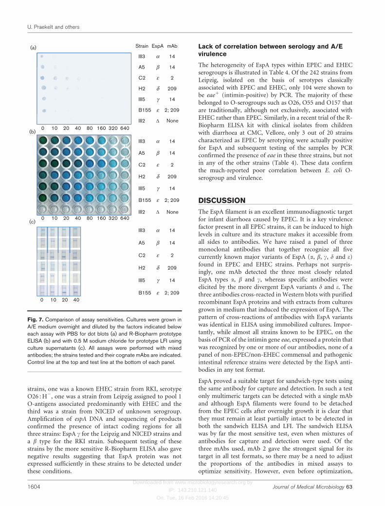
To determine the relative sensitivity of the various plat-forms, cultures were grown overnight in A/E medium andserial dilutions in PBS were tested by dot blots and by theprototype ELISA and LFI kits (Fig. 7a–c). ELISA showedmuch greater sensitivity than the other two tests, withdilution of cultures to at least 160-fold giving positivesignals with all strains. Strains C2 and B155, recognized bymAb 2, showed even greater detectability to a dilution of640-fold.
Coverage of mAbs
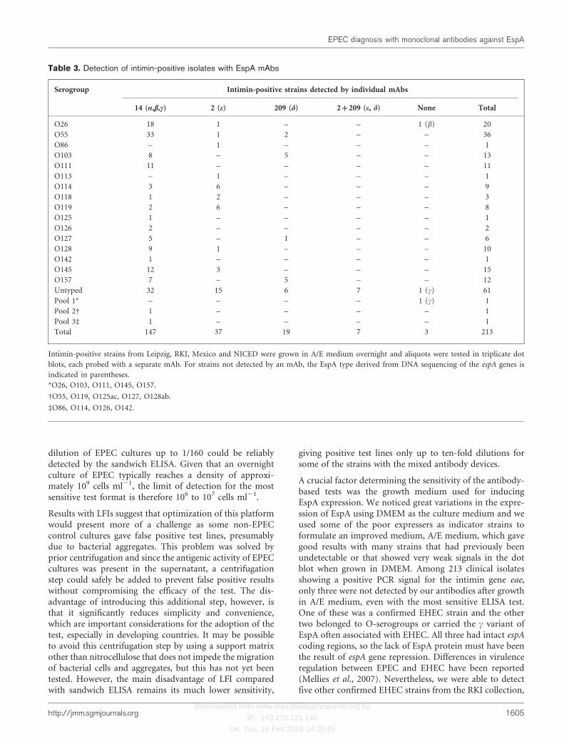
We tested all eae+ strains in our collection for EspA detec-tion after overnight growth in A/E medium using the threeantibodies separately in triplicate dot blots. The results aresummarized in Table 3. Among the 213 eae+ isolates, 210were detected with at least one antibody. Seven strainsfrom NICED (including B155) were detected by both mAb2 and mAb 209; DNA sequencing confirmed that their espAgenes were identical to that of B155, encoding the variantof EspA e containing both essential amino acids for recog-nition by the two mAbs. Of the three dot blot negative
(a) (b) (c)
1 7 13 19
2 8 14 20
3 9 15 21
4 10 16 22
5 11 17 23
6 12 18 24
Fig. 6. Specificity of antibodies. Cultures grown overnight in A/E medium were tested by dot blots (a) and by the prototype sandwichELISA from R-Biopharm (b), using mixed mAbs 2, 14 and 209. The layout of micro-organisms in the tests is shown in (c): 1, EHECO91 : H14 eaeA”; 2, EHEC O5 : H”; 3, EHEC O157:H7; 4, EIEC; 5, ETEC; 6, Proteus vulgaris; 7, Salmonella Typhimurium; 8,Aeromonas hydrophila; 9, Morganella morganii; 10, Providencia stuartii; 11, Bacillus cereus; 12, A. hydrophila; 13, Providencia rettgeri;14, Citrobacter freundii; 15, B. cereus; 16, Proteus mirabilis; 17, Shigella boydii; 18 Salmonella Enteritidis; 19, Pseudomonas
aeruginosa; 20, EPEC O127 : H6 (strain III3, EspA a); 21, EPEC O126 : H” (strain A5, EspA b); 22, EPEC O119 : H6 (strain C2, EspAe); 23, EPEC O125 : H” (H2, EspA d); 24, EPEC O157 : H7 (strain III5, EspA c). Strains positive in both tests are indicated in bold type.
T CT C
HRP-mAb dilution
1:5000
(a) Capture and detection antibody14 2 209 Mix
1:10 000
1:15 000
1:20 000
1:30 000
1:50 000
A5 C2H2 A5 C2H2 A5 C2H2 A5 C2H2
EPEC strain
LFI Dot blot(b)
Tot Sup Tot Sup Strain EspA mAb
A5 β 14
C2 ε 2
H2 δ 209
III20 D None
Fig. 5. Development of sandwich ELISA and LFI for EPEC detection.(a) ELISA plates were coated with capture antibody, either individuallyor mixed. Representative cultures expressing EspA b (A5), EspA e
(C2) and d (H2) detected by the three mAbs were grown in A/Emedium overnight and added undiluted to wells in columns as shown.Horseradish peroxide-conjugated antibodies (HRP-mAb) were addedat the dilutions indicated. Detection was with tetramethyl benzidine for15 min. (b) Induced cell cultures were subjected to dot blot bothbefore (Tot) and after (Sup) centrifugation to remove bacterial cells.Aliquots were supplemented with 0.5 M sodium chloride and loadedonto mixed antibody LFIs before (Tot) and after (Sup) centrifugation.Test (T) lines on LFIs are indicated, as are strains, EspA type and theantibody recognizing the strain. Results were recorded after 5 min.
EPEC diagnosis with monoclonal antibodies against EspA
http://jmm.sgmjournals.org 1603
Downloaded from www.microbiologyresearch.org by
IP: 143.210.121.140
On: Tue, 16 Feb 2016 14:20:45
strains, one was a known EHEC strain from RKI, serotypeO26 : H2, one was a strain from Leipzig assigned to pool 1O-antigens associated predominantly with EHEC and thethird was a strain from NICED of unknown serogroup.Amplification of espA DNA and sequencing of productsconfirmed the presence of intact coding regions for allthree strains: EspA c for the Leipzig and NICED strains anda b type for the RKI strain. Subsequent testing of thesestrains by the more sensitive R-Biopharm ELISA also gavenegative results suggesting that EspA protein was notexpressed sufficiently in these strains to be detected underthese conditions.
Lack of correlation between serology and A/Evirulence
The heterogeneity of EspA types within EPEC and EHECserogroups is illustrated in Table 4. Of the 242 strains fromLeipzig, isolated on the basis of serotypes classicallyassociated with EPEC and EHEC, only 104 were shown tobe eae+ (intimin-positive) by PCR. The majority of thesebelonged to O-serogroups such as O26, O55 and O157 thatare traditionally, although not exclusively, associated withEHEC rather than EPEC. Similarly, in a recent trial of the R-Biopharm ELISA kit with clinical isolates from childrenwith diarrhoea at CMC, Vellore, only 3 out of 20 strainscharacterized as EPEC by serotyping were actually positivefor EspA and subsequent testing of the samples by PCRconfirmed the presence of eae in these three strains, but notin any of the other strains (Table 4). These data confirmthe much-reported poor correlation between E. coli O-serogroup and virulence.
DISCUSSION
The EspA filament is an excellent immunodiagnostic targetfor infant diarrhoea caused by EPEC. It is a key virulencefactor present in all EPEC strains, it can be induced to highlevels in culture and its structure makes it accessible fromall sides to antibodies. We have raised a panel of threemonoclonal antibodies that together recognize all fivecurrently known major variants of EspA (a, b, c, d and e)found in EPEC and EHEC strains. Perhaps not surpris-ingly, one mAb detected the three most closely relatedEspA types a, b and c, whereas specific antibodies wereelicited by the more divergent EspA variants d and e. Thethree antibodies cross-reacted in Western blots with purifiedrecombinant EspA proteins and with extracts from culturesgrown in medium that induced the expression of EspA. Thepattern of cross-reactions of antibodies with EspA variantswas identical in ELISA using immobilized cultures. Impor-tantly, while almost all strains known to be EPEC, on thebasis of PCR of the intimin gene eae, expressed a protein thatwas recognized by one or more of our antibodies, none of apanel of non-EPEC/non-EHEC commensal and pathogenicintestinal reference strains were detected by the EspA anti-bodies in any test format.
EspA proved a suitable target for sandwich-type tests usingthe same antibody for capture and detection. In such a testonly multimeric targets can be detected with a single mAband although EspA filaments were found to be detachedfrom the EPEC cells after overnight growth it is clear thatthey must remain at least partially intact to be detected inboth the sandwich ELISA and LFI. The sandwich ELISAwas by far the most sensitive test, even when mixtures ofantibodies for capture and detection were used. Of thethree mAbs used, mAb 2 gave the strongest signal for itstarget in all test formats, so there may be a need to adjustthe proportions of the antibodies in mixed assays tooptimize sensitivity. However, even before optimization,
III3
A5
C2
H2
III5
B155
III2
Strain EspA mAb
14
14
(a)
β
α
ε 2
δ 209
γ 14
ε 2; 209
D None
A5
C2
H2
III5
B155
III3 14
14β
α
ε 2
δ 209
γ 14
ε 2; 209
2; 209
A5
C2
H2
III5
B155
III3 14
14β
α
ε 2
δ 209
γ 14
ε
III2 D None
(b)
(c)
0 10 20 40
0 10 20 40 80 160 320 640
0 10 20 40 80 160 320 640
Fig. 7. Comparison of assay sensitivities. Cultures were grown inA/E medium overnight and diluted by the factors indicated beloweach assay with PBS for dot blots (a) and R-Biopharm prototypeELISA (b) and with 0.5 M sodium chloride for prototype LFI usingculture supernatants (c). All assays were performed with mixedantibodies; the strains tested and their cognate mAbs are indicated.Control line at the top and test line at the bottom of each panel.
U. Praekelt and others
1604 Journal of Medical Microbiology 63
Downloaded from www.microbiologyresearch.org by
IP: 143.210.121.140
On: Tue, 16 Feb 2016 14:20:45
dilution of EPEC cultures up to 1/160 could be reliablydetected by the sandwich ELISA. Given that an overnightculture of EPEC typically reaches a density of approxi-mately 109 cells ml21, the limit of detection for the mostsensitive test format is therefore 106 to 107 cells ml21.
Results with LFIs suggest that optimization of this platformwould present more of a challenge as some non-EPECcontrol cultures gave false positive test lines, presumablydue to bacterial aggregates. This problem was solved byprior centrifugation and since the antigenic activity of EPECcultures was present in the supernatant, a centrifugationstep could safely be added to prevent false positive resultswithout compromising the efficacy of the test. The dis-advantage of introducing this additional step, however, isthat it significantly reduces simplicity and convenience,which are important considerations for the adoption of thetest, especially in developing countries. It may be possibleto avoid this centrifugation step by using a support matrixother than nitrocellulose that does not impede the migrationof bacterial cells and aggregates, but this has not yet beentested. However, the main disadvantage of LFI comparedwith sandwich ELISA remains its much lower sensitivity,
giving positive test lines only up to ten-fold dilutions forsome of the strains with the mixed antibody devices.
A crucial factor determining the sensitivity of the antibody-based tests was the growth medium used for inducingEspA expression. We noticed great variations in the expre-ssion of EspA using DMEM as the culture medium and weused some of the poor expressers as indicator strains toformulate an improved medium, A/E medium, which gavegood results with many strains that had previously beenundetectable or that showed very weak signals in the dotblot when grown in DMEM. Among 213 clinical isolatesshowing a positive PCR signal for the intimin gene eae,only three were not detected by our antibodies after growthin A/E medium, even with the most sensitive ELISA test.One of these was a confirmed EHEC strain and the othertwo belonged to O-serogroups or carried the c variant ofEspA often associated with EHEC. All three had intact espAcoding regions, so the lack of EspA protein must have beenthe result of espA gene repression. Differences in virulenceregulation between EPEC and EHEC have been reported(Mellies et al., 2007). Nevertheless, we were able to detectfive other confirmed EHEC strains from the RKI collection,
Table 3. Detection of intimin-positive isolates with EspA mAbs
Serogroup Intimin-positive strains detected by individual mAbs
14 (a,b,c) 2 (e) 209 (d) 2+209 (e, d) None Total
O26 18 1 – – 1 (b) 20
O55 33 1 2 – – 36
O86 – 1 – – – 1
O103 8 – 5 – – 13
O111 11 – – – – 11
O113 – 1 – – – 1
O114 3 6 – – – 9
O118 1 2 – – – 3
O119 2 6 – – – 8
O125 1 – – – – 1
O126 2 – – – – 2
O127 5 – 1 – – 6
O128 9 1 – – – 10
O142 1 – – – – 1
O145 12 3 – – – 15
O157 7 – 5 – – 12
Untyped 32 15 6 7 1 (c) 61
Pool 1* – – – – 1 (c) 1
Pool 2D 1 – – – – 1
Pool 3d 1 – – – – 1
Total 147 37 19 7 3 213
Intimin-positive strains from Leipzig, RKI, Mexico and NICED were grown in A/E medium overnight and aliquots were tested in triplicate dot
blots, each probed with a separate mAb. For strains not detected by an mAb, the EspA type derived from DNA sequencing of the espA genes is
indicated in parentheses.
*O26, O103, O111, O145, O157.
DO55, O119, O125ac, O127, O128ab.
dO86, O114, O126, O142.
EPEC diagnosis with monoclonal antibodies against EspA
http://jmm.sgmjournals.org 1605

Downloaded from www.microbiologyresearch.org by
IP: 143.210.121.140
On: Tue, 16 Feb 2016 14:20:45
as well as a large number of likely EHEC isolates in theLeipzig collection. Indeed, 75 of 104 intimin-positive strainsfrom Leipzig belonged to serogroups usually associated withEHEC or shared between EPEC and EHEC.
Our screening results with clinical isolates provide furtherevidence for the lack of reliability of serological classifica-tion as a method of determining pathotypes of E. coli strains.It is clear that on the one hand a large proportion of samplesare currently misdiagnosed as EPEC by serogrouping, whileon the other hand an unknown number are not detected.This highlights the superiority of our immunological test forroutine diagnostic purposes. Our panel of three antibodiesdetects all currently known variants of EspA, includingminor local variants such as the unusual e variant fromNICED that reacted with two of our antibodies.
In summary, when used together with the improved mediumfor EspA induction the prototype sandwich ELISA basedon a panel of three monoclonal antibodies was shown to bespecific and sensitive and thus has potential as a routinediagnostic tool for the identification of EPEC. Where EHECrather than EPEC is suspected this can be confirmed by aparallel test for shigatoxins. The level of sensitivity is suffi-cient for testing mixed cultures grown from stool samples ofchildren with diarrhoea using simple visual interpretation ofresults without the need for a plate reader. Further work isunder way in our laboratories to develop a routine protocolfor the detection of EPEC in stool samples. The prototypeLFI was less sensitive than the ELISA platform and so is not
as promising for the identification of EPEC in mixedcultures. However, for reference and diagnostic laboratoriesworking with pure cultures our LFI represents a valuable,informative and reliable tool in the repertoire of diagnostictests, particularly as a replacement for conventional serology.
ACKNOWLEDGEMENTS
This work was funded by EU grant ‘EACh.Child’ ICA4-CT-2002-10032, and by the Wellcome Trust Translational award ‘Developmentof a simple rapid test kit for the early diagnosis of enteropathogenicEscherichia coli (EPEC) in children with diarrhoea’. We would like tothank Gad Frankel for the gift of polyclonal anti-EspA antiserum usedin the exploratory stages of this work; Jennifer Bailey for excellenttechnical assistance; Chris Danks for expert advice on lateral flowtechnology; and Julie Pratt for critical reading of the manuscript.Conflict of interest: the materials described have been licensed to R-Biopharm, and in case of commercialization, any royalties due aresplit between U. P., R. R., P. W., the Wellcome Trust and theUniversity of Leicester.
REFERENCES
Abe, H., Tatsuno, I., Tobe, T., Okutani, A. & Sasakawa, C. (2002).Bicarbonate ion stimulates the expression of locus of enterocyteeffacement-encoded genes in enterohemorrhagic Escherichia coli O157:H7. Infect Immun 70, 3500–3509.
Afset, J. E., Bergh, K. & Bevanger, L. (2003). High prevalence ofatypical enteropathogenic Escherichia coli (EPEC) in Norwegianchildren with diarrhoea. J Med Microbiol 52, 1015–1019.
Table 4. Frequency of intimin-positive strains among O-serogroups classically associated with EPEC and EHEC
Serogroup IMM, Leipzig CMC, Vellore
Intimin-positive Intimin-negative Intimin-positive Intimin-negative
O55 25 6 1 3
O128 9 22 – –
O26 16 3 2 7
O125 1 18 – –
O126 0 18 0 3
O145 13 4 – –
O103 12 4 – –
O86 1 15 – –
O114 7 8 – –
O127 3 10 0 1
O157 8 0 – –
O111 0 8 0 3
O119 3 3 – –
O118 3 0 – –
O142 0 3 – –
O158 0 2 – –
Pools 1,2,3* 3 14 – –
Total 104 138 3 17
Heterogeneity of EspA types within EPEC and EHEC serogroups in strains from IMM, Institut fur Medizinische Mikrobiologie, Leipzig, Germany
and CMC, Christian Medical College, Vellore, India.
*Pool 1, O26, O103, O111, O145, O157; pool 2, O55, O119, O125ac, O127, O128ab; pool 3, O86, O114, O126, O142. Intimin genes were amplified
from bacterial cell extracts using universal intimin primers as described by Batchelor et al. (1999).
U. Praekelt and others
1606 Journal of Medical Microbiology 63

Downloaded from www.microbiologyresearch.org by
IP: 143.210.121.140
On: Tue, 16 Feb 2016 14:20:45
Afset, J. E., Bruant, G., Brousseau, R., Harel, J., Anderssen, E.,Bevanger, L. & Bergh, K. (2006). Identification of virulence geneslinked with diarrhea due to atypical enteropathogenic Escherichia coliby DNA microarray analysis and PCR. J Clin Microbiol 44, 3703–3711.
Ajjampur, S. S. R., Rajendran, P., Ramani, S., Banerjee, I., Monica, B.,Sankaran, P., Rosario, V., Arumugam, R., Sarkar, R. & other authors(2008). Closing the diarrhoea diagnostic gap in Indian children by theapplication of molecular techniques. J Med Microbiol 57, 1364–1368.
Batchelor, M., Knutton, S., Caprioli, A., Huter, V., Zanial, M., Dougan,G. & Frankel, G. (1999). Development of a universal intiminantiserum and PCR primers. J Clin Microbiol 37, 3822–3827.
Bertin, Y., Boukhors, K., Livrelli, V. & Martin, C. (2004). Localizationof the insertion site and pathotype determination of the locus ofenterocyte effacement of shiga toxin-producing Escherichia colistrains. Appl Environ Microbiol 70, 61–68.
Campos, L. C., Franzolin, M. R. & Trabulsi, L. R. (2004). DiarrheagenicEscherichia coli categories among the traditional enteropathogenicE. coli O serogroups–a review. Mem Inst Oswaldo Cruz 99, 545–552.
Chen, H. D. & Frankel, G. (2005). Enteropathogenic Escherichia coli:unravelling pathogenesis. FEMS Microbiol Rev 29, 83–98.
Crepin, V. F., Shaw, R., Knutton, S. & Frankel, G. (2005). Molecularbasis of antigenic polymorphism of EspA filaments: development of apeptide display technology. J Mol Biol 350, 42–52.
Daniell, S. J., Kocsis, E., Morris, E., Knutton, S., Booy, F. P. & Frankel,G. (2003). 3D structure of EspA filaments from enteropathogenicEscherichia coli. Mol Microbiol 49, 301–308.
Delahay, R. M., Knutton, S., Shaw, R. K., Hartland, E. L., Pallen, M. J. &Frankel, G. (1999). The coiled-coil domain of EspA is essential for theassembly of the type III secretion translocon on the surface ofenteropathogenic Escherichia coli. J Biol Chem 274, 35969–35974.
Frankel, G., Phillips, A. D., Rosenshine, I., Dougan, G., Kaper, J. B. &Knutton, S. (1998). Enteropathogenic and enterohaemorrhagicEscherichia coli: more subversive elements. Mol Microbiol 30, 911–921.
Garrido, P., Blanco, M., Moreno-Paz, M., Briones, C., Dahbi, G.,Blanco, J., Blanco, J. & Parro, V. (2006). STEC-EPEC oligonucleotidemicroarray: a new tool for typing genetic variants of the LEEpathogenicity island of human and animal Shiga toxin-producingEscherichia coli (STEC) and enteropathogenic E. coli (EPEC) strains.Clin Chem 52, 192–201.
Giron, J. A., Qadri, F., Azim, T., Jarvis, K. J., Kaper, J. B. & Albert, M. J.(1995). Monoclonal antibodies specific for the bundle-forming pilusof enteropathogenic Escherichia coli. Infect Immun 63, 4949–4952.
Kenny, B., DeVinney, R., Stein, M., Reinscheid, D. J., Frey, E. A. &Finlay, B. B. (1997). Enteropathogenic E. coli (EPEC) transfers itsreceptor for intimate adherence into mammalian cells. Cell 91, 511–520.
Knutton, S., Rosenshine, I., Pallen, M. J., Nisan, I., Neves, B. C., Bain,C., Wolff, C., Dougan, G. & Frankel, G. (1998). A novel EspA-associated surface organelle of enteropathogenic Escherichia coli
involved in protein translocation into epithelial cells. EMBO J 17,2166–2176.
Kozub-Witkowski, E., Krause, G., Frankel, G., Kramer, D., Appel, B. &Beutin, L. (2008). Serotypes and virutypes of enteropathogenic andenterohaemorrhagic Escherichia coli strains from stool samples of
children with diarrhoea in Germany. J Appl Microbiol 104, 403–410.
Kuhne, S. A., Hawes, W. S., La Ragione, R. M., Woodward, M. J.,Whitelam, G. C. & Gough, K. C. (2004). Isolation of recombinant
antibodies against EspA and intimin of Escherichia coli O157:H7.J Clin Microbiol 42, 2966–2976.
Lu, Y., Toma, C., Honma, Y. & Iwanaga, M. (2002). Detection of EspBusing reversed passive latex agglutination: application to determina-
tion of enteropathogenic Escherichia coli. Diagn Microbiol Infect Dis
43, 7–12.
Mellies, J. L., Barron, A. M. S. & Carmona, A. M. (2007). Enteropatho-
genic and enterohemorrhagic Escherichia coli virulence gene regulation.Infect Immun 75, 4199–4210.
Menezes, M. A., Rocha, L. B., Koga, P. C., Fernandes, I., Nara, J. M.,Magalhaes, C. A., Abe, C. M., Ayala, C. O., Burgos, Y. K. & otherauthors (2010). Identification of enteropathogenic and enterohae-
morrhagic Escherichia coli strains by immunoserological detection ofintimin. J Appl Microbiol 108, 878–887.
O’Ryan, M., Prado, V. & Pickering, L. K. (2005). A millennium updateon pediatric diarrheal illness in the developing world. Semin Pediatr
Infect Dis 16, 125–136.
Robins-Browne, R. M., Bordun, A.-M., Tauschek, M., Bennett-Wood,V. R., Russell, J., Oppedisano, F., Lister, N. A., Bettelheim, K. A.,Fairley, C. K. & other authors (2004). Escherichia coli and community-acquired gastroenteritis, Melbourne, Australia. Emerg Infect Dis 10,
1797–1805.
Smith, H., Willshaw, G. & Cheasty, T. (2004). E. coli as a cause ofoutbreaks of diarrhoeal disease in the UK. Microbiol Today 31, 117–
118.
Tschape, H., Prager, R., Streckel, W., Fruth, A., Tietze, E. & Bohme,G. (1995). Verotoxinogenic Citrobacter freundii associated with severe
gastroenteritis and cases of haemolytic uraemic syndrome in a nurseryschool: green butter as the infection source. Epidemiol Infect 114, 441–
450.
Yang, J.-R., Wu, F.-T., Tsai, J.-L., Mu, J.-J., Lin, L.-F., Chen, K.-L., Kuo,S. H.-S., Chiang, C.-S. & Wu, H.-S. (2007). Comparison between
O serotyping method and multiplex real-time PCR to identifydiarrheagenic Escherichia coli in Taiwan. J Clin Microbiol 45, 3620–
3625.
EPEC diagnosis with monoclonal antibodies against EspA
http://jmm.sgmjournals.org 1607