morphology of the kk - univerzita karlovaanat.lf1.cuni.cz/souhrny/aofa7.pdf · derivates of the 1....
TRANSCRIPT

Morphology of the periodontium, tongue, soft and hard palati. Muscles attaching
mandible; TMJ joint
Ivo Klepáček KK

Cavitas oris dutina ústní
Dental arches divide it into vestibulum oris and cavitas oris propria
boundaries:
ventrally: lips – labia štěrbina-rima oris laterally: buccal parts – buccae
cranially: roof – palatum
causdally: m. mylohyoideus + m. geniohyoideus - tongue - lingua
dorsally: isthmus faucium KK

Specialized mucosa: free endings (pain, temperature) , Paccini (vibration), Ruffini (traction), taste buds Lining (buccal) mucosa: free endings (pain, temperature) , Paccini (vibration), Meissner (pressure), Merkel (pressure)
Mucosa Masticatory mucosa: free endings (pain, temperature) , Paccini (vibration), Ruffini (traction), Meissner (pressure) KK

Parodontal functions Tooth fixation, elasticity, (hydroelastic cushion -pillow) nutrition, asistency during eruption
Parodontium Clinical unit involving gum,
periodontal ligaments, cementum and lamina dura
(cortical layer) of the alveolus KK

Arrangement of the periodontal ligaments. a - ligamenta marginalia, b - ligamenta dentalia superiora, c - ligamenta dentalia media, d - ligamenta dentalia inferiora (apicalia)
Interdental circumdental,
dentoalveolar and intraalveolar
ligaments
0.3-0.5mm
0.1-0.2mm
about 0.2mm
Periodontium Free endings - pain Ruffini bodies - traction Paccini bodies - pressure , vibration KK

formation of the suspensory systém before and after eruption (after Levy and Bernick 1968, modified). A – tooth primordium inside developing alveolus, B – eruption starts, C - alveolus is formed, stadium, D – fully erupted tooth
KK

Periodontium (Parodontium)
Cells Fibers Matrix Ligaments Plasma Vessels Nerves Interstitial plasma and blood in vessels -
“hydroelastic pillow“ KK

Ion exchange between inner (pulp) and outer (oral cavity) environment KK

Gingival sulcus (pocket) Sulcus
gingivae
Free gingival groove Paramarginal sulcus
Gingiva = tightly surrounds tooth neck – “cuff (collar-like) connection“
free: Interdental; circumdental adjacent: closely attached, fixing
KK

Alveolar mucosa (“loose gingiva“) = shiny red, nonkeratizing
Gingiva proper (attached) = pink, stippled, keratizing KK

Interdental, circumdental, dentoalveolar, interalveolar ligaments
Ligamentous slings and circles help to tight attachment between gingiva and tooth KK

Free gingival groove changes
into periodontal
True pocket (deeper 3.5 mm) appears, if the tooth attachment is lost
True pocket
KK

nipple compression
nipple compression
nipple sucking
bite combined
boxed-like
folding-like KK

Lingua, glossa
Mucous and muscularorgan;
Inside cavum oris and pharynx apex
corpus (body) dorsum facies inferior
radix (root) margo (margin) sulcus (groove)
terminalis medianus (midline)
foramen caecum tonsilla lingualis
KK

Week 6 - 7
Papillae filiformes fungiformes vallatae foliatae
KK

hořko
slano
kyselo
sladko KK

Inervace
motorická: n. XII
(x m.palatoglossus)
senzitivní
V/3
IX
X
senzorická
VII
vegetativní:
parasympatická
ggl. submandibulare
sympatická
plexus lingualis
KK

f – plica fimbriata e – frenulum linguae g – vena lingualis profunda b – caruncula sublingualis a – opening of the ductus gl.sublingualis on the plica sublingualis KK

Ankyloglossia – influence on oral cavity bottom formation
Tongue-tie – Ankyloglossia – fusion between
tongue and floor of the mouth,
Tongue frenulum
extends to the tongue top
Combined with (Pierre-Robin, Treacher Collinssyndroms)
KK

Styloglossus Palatoglossus
Hyoglossus
Genioglossus
Zevní svaly jazyka mění
polohu jazyka KK

Vnitřní svaly jazyka mění tvar jazyka
mm. Longitudinales superiores et inferiores, transversalis, verticalis KK

Mm. Genioglossal muscles are separated from each other with
septum linguae (linguale) Gap between m.
hyoglossus and m. genioglossus is for vessels and nerves
Septum linguae is made from sparse connective tissue; it can be socked by pus - abscessus KK

KK

Abscessus v septu KK
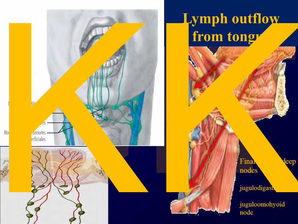
Lymph outflow from tongue KK

vestibulum
vestibule
KK

c - frenulum linguae sup.
b,a - plicae bucco-alveolares (buccales) KK

KK

Palatum durum Palatum molle
Hard palate Soft palate
Premaxilla Maxilla Os palatinum
Papilla, rugae (folds) pits (foveolae) Lines (crests)
KK

Palatum – relief
Lacey
Papilla incisiva
Rugae palatinae
Raphe mediana
Top of the interalveolar septum; At level of papilla incisiva
The most ventrally found rugae palatinae are at level of caninus alveoli
KK

Relief patra; rugae, foveolae a papilla incisiva
KK

KK

podle Petrovického2002
KK

Palatum molle (soft)
– posterior view
– anterior view KK

Palate – relief
Lacey
Palatum durum
Palatum molle
Raphe mediana
Fat zone
Foveolae palatinae
A
H
Hauptmayer line – between soft and hard palatum
KK

Chrápání Snoring
KK

Masticatory muscles
Musculi masticatorii Muscles of mastication
V3 – MANDIBULARIS Derivates of the 1. branchial arch KK

Masticatory muscles n. mandibularis - 3rd branch of the n. V.
M. masseter From the outer surface of the zygomatic arch; deep part
of the muscle run from the internal bone surface, too Superficial part runs mandibular angle; deep to the
„fovea zygomaticomandibularis“
M. temporalis From inferior temporal line (+adjacent bone) Proc. coronoideus mandibulae (coronoid process)
Fascia temporalis and fascia parotideomasseterica KK

M. temporalis et fascia temporalis
Spatium interfasciale; Interfascial
space KK

M. masseter KK

Fascia parotideomasseterica, Parotideomasseteric fascia
KK

Jost , G, Levet, V.: Parotid fascia and Face lifting: A critical Evaluation of the SMAS concept. Plastic and Reconstructive Surg, 74:42-51, 1983
KK

Mm. pterygoidei Medialis
From the pterygoid fossa and from the tuber maxillae Tuberositas pterygoidea
Lateralis From the processus pterygoideus (lamina lateralis) and
from the infratemporal face of the greater wing of the sphenoid bone
below mandibular head, pterygoid fossa and onto the joint capsule KK

pterygomandibularis
pterygoideus proprius
pterygospinosus
KK

a – lig. pterygospinosum
b – n. alveolaris inferior
c, d – n. lingualis e – lig. pterygomandibulare
(raphe buccopharyngea)
f – sulcus mylohyoideus
g – angulus mandibulae et lig. stylomandibulare
h – lig. sphenomandibulare
KK

Main and ´assisting´ masticatory muscles
(orthodontic and prosthetic point of view)
anterior belly of digastric m., mylohyoid m.
Innervation: CN V3
Geniohyoid m. KK

JAW MUSCLES There is differentiation of the jaw-closing musculature.
In basal synapsids, the major jaw-closing muscle is the adductor mandibulae (externus). It originates from the back of the skull and inserts on the posterior end of the lower jaw.
In derived synapsids, the adductor mandibulae divides into two major sets of jaw-closing muscles, the temporalis and masseter. The temporalis originates from the skull roof near the sagittal crest and inserts on the coronoid process. The masseter in turn divides into two parts. The deep masseter originates on the zygomatic arch and inserts on the lower jaw; the superficial masseter part arises beneath the eye, passes across the deep masseter, to insert on the angle of the dentary.
Evolution of Synapsid jaw adductor muscles
KK

Paleontological evidence for mammalian middle ear evolution. (A) Diagrams of lateral views of jaw skeletal elements showing modifications leading to the mammalian condition (after Allin, ’75). The geological record and occurrence of each animal are indicated on the left. For clarity of comparison, no teeth are shown. Note that a set of postdentary elements (articular, surangular, and angular) and the upper jaw elements (quadrate and quadratojugal), indicated by gray, became separated from the dentary and reduced in size during the transition from pelycosaurs to mammals. The sequence of changes in the fossil record does not represent a true ancestor–descendent relationship, but only structural grades. (B) Changes in jaw articulation during mammalian evolution. In a pelycosaur, Dimetrodon (top), the quadrate and articular formed a functional jaw joint (black arrow). In an ‘‘advanced’’ cynodont, Diarthrognathus (middle), an additional jaw joint was observed between the squamosal and dentary (white arrow). In an extant marsupial, Didelphis (bottom), the functional jaw joint has been taken over only by the squamosal and dentary.
Takechi M, Kuratani S. 2010. History of studies on mammalian middle ear evolution: a comparative morphological and developmental biology perspective. J. Exp. Zool. (Mol. Dev. Evol.) 314B:[page range].
KK

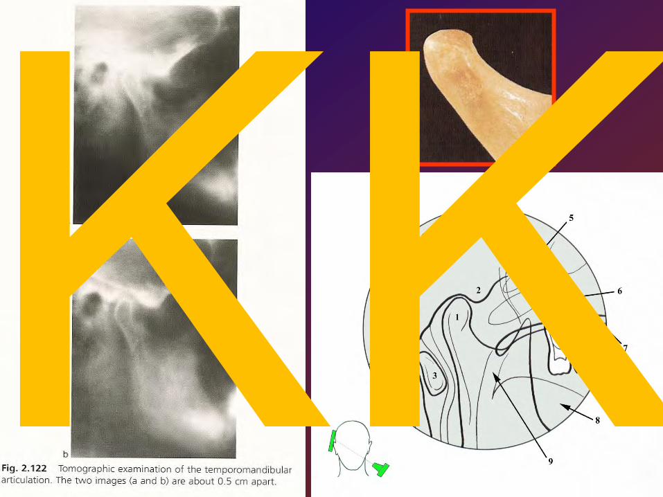
Compound joint Similar to hinge joint type
Temporomandibular (craniomandibular) joint Articulatio
temporo- mandibularis
ATM lat.
TM , TMJ engl.
Morphological
findings:
• The great variability of all the
articular structures
• The absence of hyaline cartilage
•The two separate compartments,
allowing a wider range
of mandibular movements
• The mared weakness of the
articular ligaments, allowing
hypertranslation and dislocation
without tearing the capsule
KK

KK

KK
KK

Late triassic early mammal Mesozoic early mammal 125 millions year ago
Water mole (duckbill)
KK

rovina okluze occlusal plane protetická rovina Camper plane
x vodorovná rovina horizontal plane
M.Doskočil: Chrupavka ve vývoji mandibuly. (cartilage in the development of the mandible) Cs.Stomatologie, 1:10-18, 1988
Meckelova chrupavka a chrupavčité deriváty v krčku dolní čelisti KK

END
bundle of cartilaginous cels – Meckel derivate ?
KK

KK

Os tympanicum zvýrazníte dalším kliknutím Tuberc. pharyngicum Foramen lacerum Spina sphenoidalis Foramen spinosum Foramen ovale
Fossa mandibularis Tuberculum articulare
Arcus zygomaticus
Condylus occipitalis
Processus styloideus Foramen jugulare For. stylomastoideum Processus mastoideus Fissura tympanomastoidea Fis.tympanopetrosa Fis.petrosquamosa Fis.tympanosquamosa Porus acusticus ext.
Basis cranii externa – semiview on the tympanic bone
KK

Processus postglenoidalis Post-glenoid process
Trigonum suprameatum Suprameatal triangle
KK

Articular Capsule is a sac that encloses TMJ. Borders: Superior: Capsule is positioned underneath inferior side of Articular Eminence. Inferior: Capsule wraps around condyle's neck (Collum Mandibulae)
It is a fibro-cartilageus disc. It divides synovial cavity of TMJ into: 1. Superior synovial cavity 2. Inferior synovial cavity Both cavities are filled with synovial fluid, secreted by inner side of articular capsule (clear, viscous fluid). Attachments of articular disc:
1.Anterior: a. Anterio-Superior: indirectly to articular eminence through capsule
b.Anterio-inferior: to condyl's neck
2.Posterior: a.Posterio-superior: to post-glenoid process spina supra meatum ?
b. Posterio-inferior: to condyl's neck
Gray´s anatomy, The classic collector´s edition KK

Salentijn, L. Biology of Mineralized Tissues: Prenatal Skull Development, Columbia University College of Dental Medicine post-graduate dental lecture series, 2007 Moss, ML. The non-existent hinge axis, Am. Inst, Oral Biol. 1972, 59-66 Rodríguez-Vázquez JF, et al., JF; Mérida-Velasco, JR; Mérida-Velasco, JA; Jiménez-Collado, J (1998). "Anatomical considerations on the discomalleolar ligament". J Anat.. 192 (Pt 4): 617–621. PMC 1467815. PMID 9723988. //www.ncbi.nlm.nih.gov/pmc/articles/PMC1467815/. Rodríguez-Vázquez JF, et al. (1993). "Relationships between the temporomandibular joint and the middle ear in human fetuses.". J Dent Res.. 72 (1): 62–66. T Rowicki, J Zakrzewska. (2006). "A study of the discomalleolar ligament in the adult human.". Folia Morphol. (Warsz).. 65 (2): 121–125. S Zhang, N Gersdorff, J Frahm (2011) Real-Time Magnetic Resonance Imaging of Temporomandibular Joint Dynamics. The Open Medical Imaging Journal, 2011, 5, 1-7, [1] Zadik, Yehuda; Aktaş Alper; Drucker Scott; Nitzan W Dorrit (2012). "Aneurysmal bone cyst of mandibular condyle: A case report and review of the literature". J Craniomaxillofac Surg 40.
KK

Joint surface is composed form the fourth layers: • Superficial layer: superficial articular layer = connective tissue character • Very cellular layer: • Proliferating layer: • Hypertrophic layer:
discus articularis is rich by vessels in early age
Loose of vascularization supports degenerative changes of the disc
KK

Upper space - cavitas discosquamosa – 581 mm2
Lower space - cavitas discocondylaris – 396 mm2 KK

Condylar atrophy follows age
KK

A) Fissura tympanosquamosa
B) Stratum superius C) genu vasculosum D) Stratum inferius E) Capsule F) Glandula parotis G) Discus articularis ( Dreger 1994 )
Discus napojen na fascia parotis, a podkožní struktury; pouzdro je slabé
Discus napojen přes pouzdro
Discus je napojen samostatně
Discus a pouzdro jsou pevně spojeny
Dreger H (1994) Untersuchungen zur posterioren Anheftung Des Diskus artikularis im menschlichen Kiefergelenk. Med Diss Kiel
Vasili Naroushvili: Wechselwirkungen zwischen Okklusionsarten und Anheftungsarten des Musculus pterygoideus lateralis bei der Entstehung von Diskus Dislokation des Kiefergelenkes Hamburg 2006 KK

Minarelli, AM, DelSanto, M, Liberti, EA: The structure of the human temporomandibular joint disc: A scanning electron microscopy study. J Orof Pain 11:95-98, 1997
Examined discs: 16-39 weeks of intrauterine life Up to 4 months of age 30-39 years 60-69 years KK

Superior head, inferior head, and ´third´head of the lateral pterygoid are shown (dissected) 1-Discus articularis, 2-M.pteryg.lat (superior head), 3-M.pteryg.lat (inferior head), 4- third head of the m. pteryg. lat (attached inferior head)
1
2
3 4
Discus articularis (dissected) 1-Discus articularis, 2-insertion of the mm. masseter and temporalis, 3-insertion of the m. pteryg.lat (superior head), 4-insertion of the m. pteryg.lat (inferior head)
V. Naroushvili 2006
KK

KK

KK

•J. Chen, U. Akyuz, L. Xu, R.M.V. Pidaparti : Stress analysis of the human temporomandibular joint •Medical Engineering & Physics 20/8/: 565-572, October 1998
KK

Thin elastic lamina
fibroelastic lamina
Retroarticular hydroelastic cushion Zenker (contains vessels) KK

KK

Kondylová dráha Condyle path a Transverse b Longitudinal
Rest position
Central position
Ventral position
Habitual (high) End of the masticatory cycle
Ventral (low)
muscle tone is the smallest
central (zenit)
through swalloving
KK

N. facialis: • lateral surface of the joint capsule KK

N. auriculotemporalis nerve is branched into four nerves: • lateral branch •Medial branch • branch from the middle nerve segment • branch from the area where nerve crosses n. temporalis superficialis KK

n. massetericus send four branches:
• branch below oval foramen • branch from the first nervous segment closely below skull base • two branches from the first segment below zygomatic bone
m. temporalis profundus:
• supplies rostromedial part of the disc and capsule
Ganglion oticum (otic ganglion):
• supplies dorsal part (pars discosquamalis) of the joint capsule
KK

KK

Literature R. Čihák: Anatomie 1, 2, 3 Grada Publishing 2003
M. Dykes : Anatomy 2th edition, Mosby 2002
S.Snell: Clinical anatomy for Medical Students 6th edition, Lippincott, Williams & Wilkins
I.Klepáček, J.Mazánek et al.: Klinická anatomie ve stomatologii Grada Publishing 2001
G.J.Tortora : Principles of Human Anatomy 4th edition, Williams & Wilkins
K.L.Moore, A.F.Dalley: Clinically Oriented Anatomy 4th edition, Williams & Wilkins
F.H.Netter: anatomický atlas člověka Vlastní archív
KK