mri imaging for patients with cardiac implantable...
TRANSCRIPT

MRI imaging for patients with cardiac
implantable electronic devices (CIEDs)
13th annual International Winter Arrhythmia School
Collingwood, Ontario, Canada
February 6, 2016
Andrew C.T. Ha, MD, MSc, FRCPC
Cardiac Electrophysiology
University Health Network

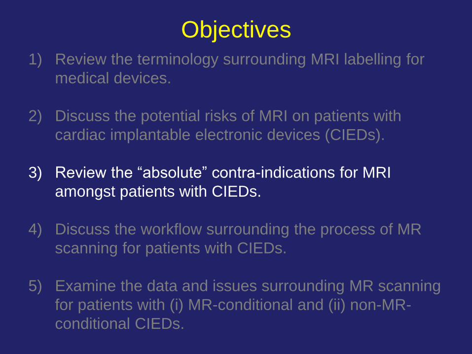
1) Review the terminology surrounding MRI labelling for
medical devices.
2) Discuss the potential risks of MRI on patients with
cardiac implantable electronic devices (CIEDs).
3) Review the “absolute” contra-indications for MRI
amongst patients with CIEDs.
4) Discuss the workflow surrounding the process of MR
scanning for patients with CIEDs.
5) Examine the data and issues surrounding MR scanning
for patients with (i) MR-conditional and (ii) non-MR-
conditional CIEDs.
Objectives

MRI labelling for medical devices:
terminology
Adapted from: Woods T.O. Standards for medical devices in MRI: present and future.
J Magn Reson Imaging 2007;26:1186-9.
1) Review the terminology surrounding MRI labelling for
medical devices.
2) Discuss the potential risks of MRI on patients with
cardiac implantable electronic devices (CIEDs).
3) Review the “absolute” contra-indications for MRI
amongst patients with CIEDs.
4) Discuss the workflow surrounding the process of MR
scanning for patients with CIEDs.
5) Examine the data and issues surrounding MR scanning
for patients with (i) MR-conditional and (ii) non-MR-
conditional CIEDs.
Objectives

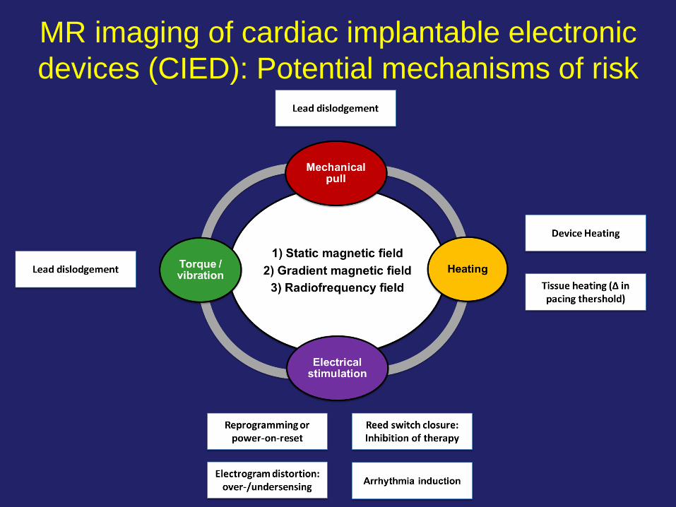
Risks associated with MR imaging arise
from 3 distinct mechanisms:
1) Static magnetic field.
2) Gradient magnetic field.
3) Radiofrequency (RF) energy.
How can MRI affect CIEDs?

Static magnetic field
• In general, the magnetic field strength of 1.5 to 3.0
Tesla MRI scanners is about 30,000 to 60,000 times
that of the Earth’s magnetic field strength.
• Ferromagnetic objects may move, rotate, or dislodge
in the presence of such powerful fields.
How can MRI affect CIEDs?
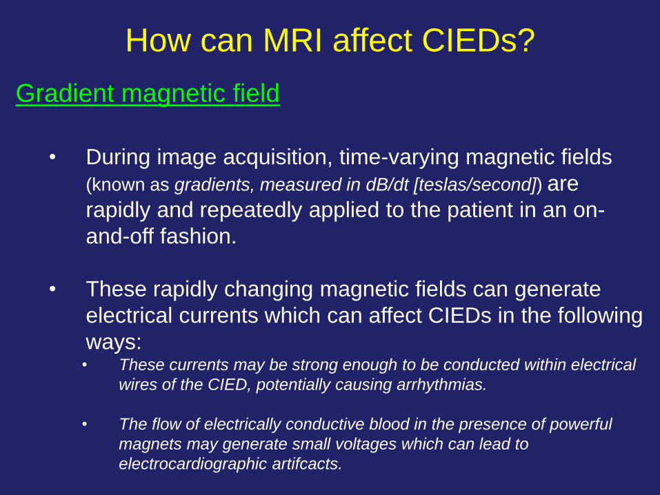
Gradient magnetic field
• During image acquisition, time-varying magnetic fields
(known as gradients, measured in dB/dt [teslas/second]) are
rapidly and repeatedly applied to the patient in an on-
and-off fashion.
• These rapidly changing magnetic fields can generate
electrical currents which can affect CIEDs in the following
ways: • These currents may be strong enough to be conducted within electrical
wires of the CIED, potentially causing arrhythmias.
• The flow of electrically conductive blood in the presence of powerful
magnets may generate small voltages which can lead to
electrocardiographic artifcacts.
How can MRI affect CIEDs?
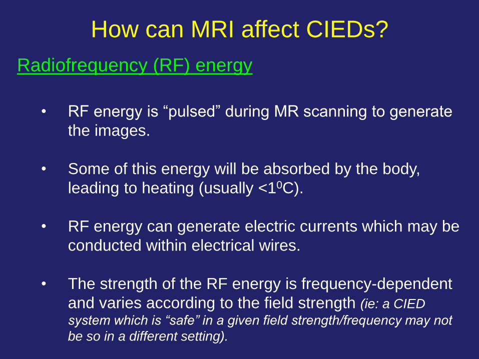
Radiofrequency (RF) energy
• RF energy is “pulsed” during MR scanning to generate
the images.
• Some of this energy will be absorbed by the body,
leading to heating (usually <10C).
• RF energy can generate electric currents which may be
conducted within electrical wires.
• The strength of the RF energy is frequency-dependent
and varies according to the field strength (ie: a CIED
system which is “safe” in a given field strength/frequency may not
be so in a different setting).
How can MRI affect CIEDs?
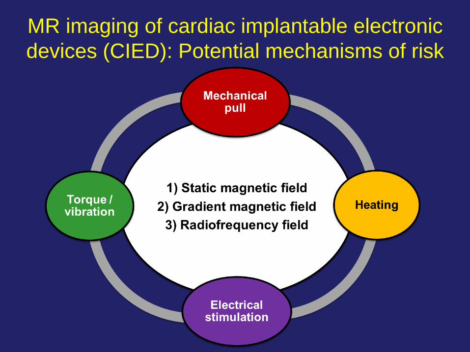
MR imaging of cardiac implantable electronic
devices (CIED): Potential mechanisms of risk
MR imaging of cardiac implantable electronic
devices (CIED): Potential mechanisms of risk
1) Review the terminology surrounding MRI labelling for
medical devices.
2) Discuss the potential risks of MRI on patients with
cardiac implantable electronic devices (CIEDs).
3) Review the “absolute” contra-indications for MRI
amongst patients with CIEDs.
4) Discuss the workflow surrounding the process of MR
scanning for patients with CIEDs.
5) Examine the data and issues surrounding MR scanning
for patients with (i) MR-conditional and (ii) non-MR-
conditional CIEDs.
Objectives

CIED components which are considered to
be contra-indicated for MR imaging • Broken or fractured lead(s) – known or suspected.
• Abandoned (capped) or extraneous lead(s), lead
extender(s), or lead adaptor(s).
• Remnants of a lead which persist in the patient’s body (e.g.
pacemaker pocket, vascular space, or cardiac chamber).
• A pacemaker-dependent patient with a non-MR-conditional
ICD system.
• Permanent epicardial pacing or ICD lead(s). (Note: the
presence of temporary epicardial wire(s) inserted at the time of cardiac
surgery is not considered to be an absolute contra-indication for MR
scanning.*) Verma A. et al. Can J Cardiol. 2014 Oct;30(10):1131-41.
*Hartnell GG et al. Am J Roentgenol. 1997 May;168(5):1157-9

1) Review the terminology surrounding MRI labelling for
medical devices.
2) Discuss the potential risks of MRI on patients with
cardiac implantable electronic devices (CIEDs).
3) Review the “absolute” contra-indications for MRI
amongst patients with CIEDs.
4) Discuss the workflow surrounding the process of MR
scanning for patients with CIEDs.
5) Examine the data and issues surrounding MR scanning
for patients with (i) MR-conditional and (ii) non-MR-
conditional CIEDs.
Objectives
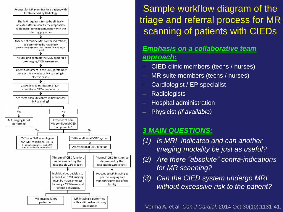
Sample workflow diagram of the
triage and referral process for MR
scanning of patients with CIEDs
Verma A. et al. Can J Cardiol. 2014 Oct;30(10):1131-41.
Emphasis on a collaborative team
approach:
– CIED clinic members (techs / nurses)
– MR suite members (techs / nurses)
– Cardiologist / EP specialist
– Radiologists
– Hospital administration
– Physicist (if available)
3 MAIN QUESTIONS:
(1) Is MRI indicated and can another
imaging modality be just as useful?
(2) Are there “absolute” contra-indications
for MR scanning?
(3) Can the CIED system undergo MRI
without excessive risk to the patient?
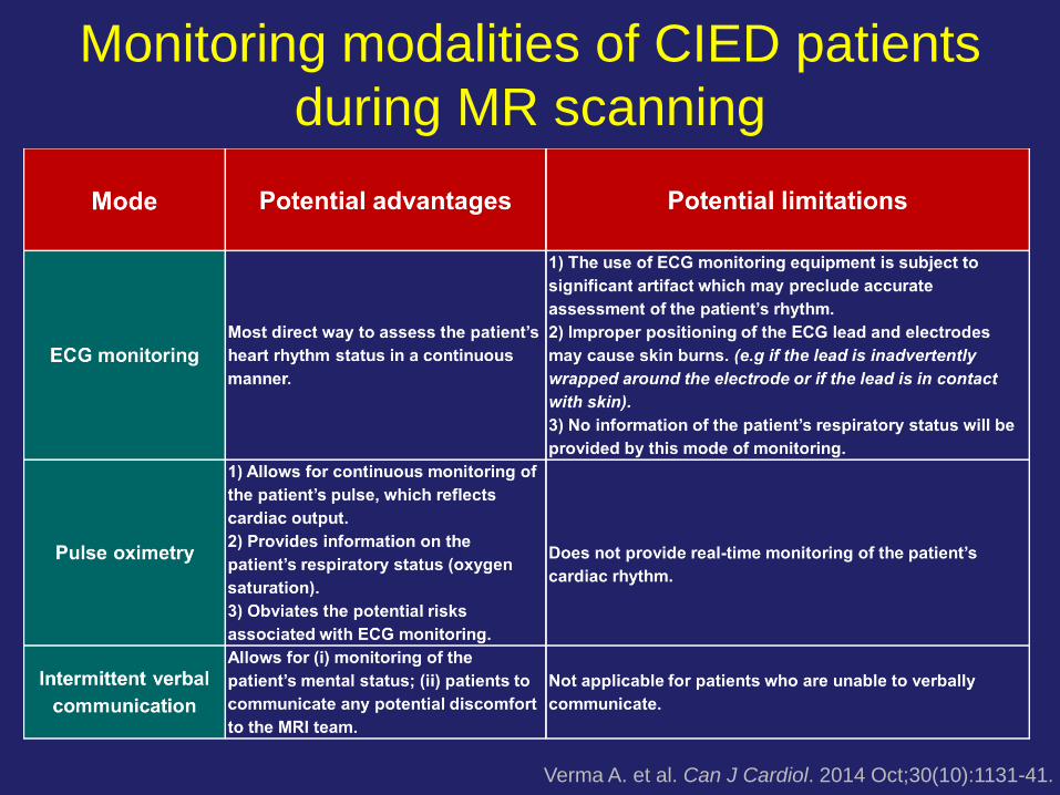
Monitoring modalities of CIED patients
during MR scanning
Verma A. et al. Can J Cardiol. 2014 Oct;30(10):1131-41.

1) Review the terminology surrounding MRI labelling for
medical devices.
2) Discuss the potential risks of MRI on patients with
cardiac implantable electronic devices (CIEDs).
3) Review the “absolute” contra-indications for MRI
amongst patients with CIEDs.
4) Discuss the workflow surrounding the process of MR
scanning for patients with CIEDs.
5) Examine the data and issues surrounding MR scanning
for patients with (i) MR-conditional and (ii) non-MR-
conditional CIEDs.
Objectives

• No CIED system is “MR-safe”.
• Some CIED systems are “MR-conditional”; this means that
patients may undergo MR scanning without additional
known risks as long as manufacturer-specified scanning
parameters are followed.
• Recommended scanning parameters vary amongst CIED
manufacturers, meaning that the MR scanning protocol will
vary in accordance to the patient’s CIED system.
What is a “MR-conditional” CIED system? What is the “fine print”?
Verma A. et al. Can J Cardiol. 2014 Oct;30(10):1131-41.

MRI for patients with MR-conditional pacemakers
Bailey WM et al. Hearth Rhythm 2015;10:1-8 and 2015; 12:1183–1191
• Prospective, single-arm, non-randomized study evaluating the safety of MR scanning
for patients with single or dual-chamber Biotronik Entovis pacemakers (Setrox S 53-cm
and/or 60-cm leads).
• Patients undergo thoracic spine, cardiac, head, and lower lumbar MR scanning at ≥5
weeks post pacemaker implant.
• 245 patients were enrolled from 31 centres between Dec 13 2013 to July 11 2014.

MRI for patients with MR-conditional pacemakers
Bailey WM et al. Hearth Rhythm 2015;10:1-8 and
2015; 12:1183–1191
Comments:
(1) Patients were excluded from
MRI scan if they had pre-MRI
threshold(s) >2.0 V @ 0.4ms;
lead impedance <200 or >1500
Ω; phrenic nerve stimulation at
4.8Va @ 1.0 ms; or threshold
variation exceeding 0.5 V from
baseline.
(2) Otherwise, no appreciable
change in atrial or ventricular
pacing thresholds or sensing
was noted in this study.
MRI for patients with MR-conditional
pacemakers: adverse events
• The freedom rate of serious adverse event was 99.6% (220/221
subjects).
• One subject experienced an adverse event which was felt to be
possibly related to MR scanning.
• This subject developed chest pain with pericarditis and a pericardial
effusion 4 days post-MRI. There was no imaging evidence of
perforation. This was eventually treated with repositioning of the
ventricular lead.
Bailey WM et al. Hearth Rhythm 2015;10:1-8 and 2015; 12:1183–1191

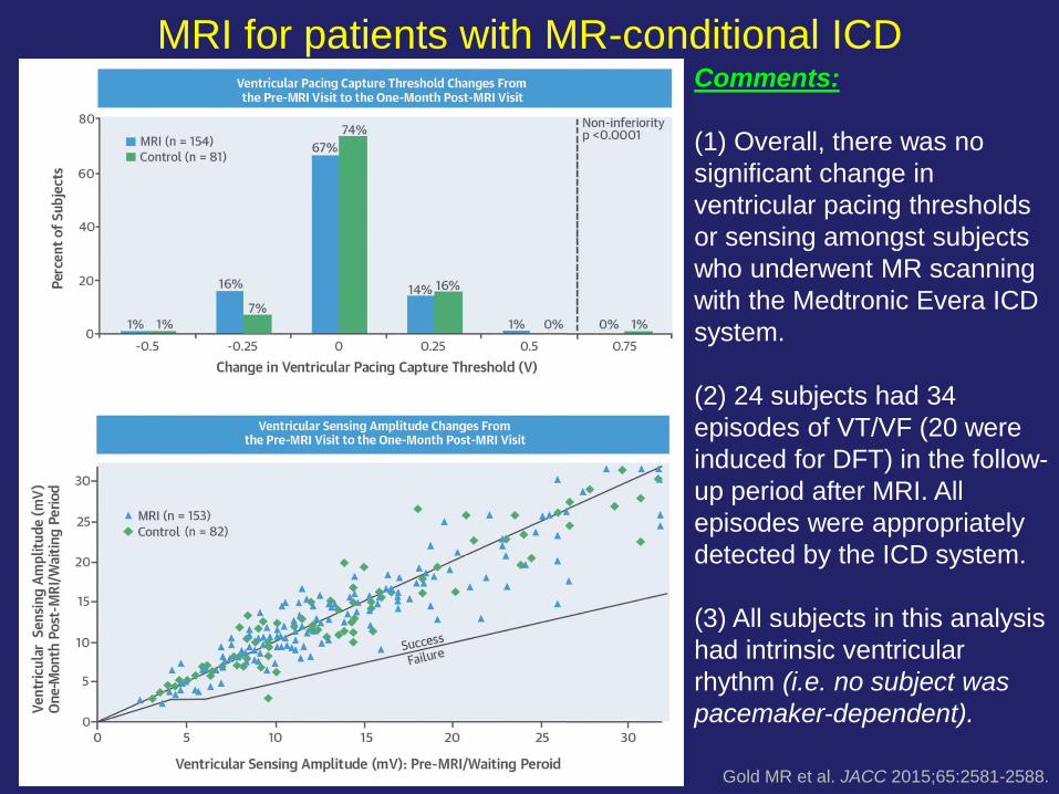
MRI for patients with MR-conditional ICD
Gold MR et al. JACC 2015;65:2581-2588.
• Prospective, 2-arm, randomized study evaluating the safety of MR scanning for patients with
single or dual-chamber Medtronic DF-4 Evera ICDs (6935M or 6947M 55-/62-cm ICD leads ±
Medtronic 5076 atrial leads).
• If randomized to the MR scanning group (n=175), patients underwent “full-body” MRI (cardiac,
thoracic spine, lumbar spine, head) at 9-12 weeks post ICD implant.
• Patients in the control group (n=88) did not undergo protocol-mandated MR scanning.
• 275 patients were enrolled from 42 centres.
MRI for patients with MR-conditional ICD
Gold MR et al. JACC 2015;65:2581-2588.
Comments:
(1) Overall, there was no
significant change in
ventricular pacing thresholds
or sensing amongst subjects
who underwent MR scanning
with the Medtronic Evera ICD
system.
(2) 24 subjects had 34
episodes of VT/VF (20 were
induced for DFT) in the follow-
up period after MRI. All
episodes were appropriately
detected by the ICD system.
(3) All subjects in this analysis
had intrinsic ventricular
rhythm (i.e. no subject was
pacemaker-dependent).

MRI for patients with MR-conditional ICD:
adverse events
Gold MR et al. JACC 2015;65:2581-2588.
• There were 5 MR-imaging-related events occurring in 5 subjects.
• 2 subjects reported site warmth and 1 reported back pain during
scanning (no additional action was required).
• 1 subject reported a burning sensation in the forehead – no specific
pathology was noted (X-ray ruled out presence of metallic foreign
body).
• 1 subject experienced atrial tachycardia which was terminated by
atrial ATP. The MR scan was suspended and was eventually
completed. This patient had multiple episodes of atrial tachycardia
during follow-up.
• There were 12 deaths (4 control, 8 MRI group). After adjudication by
the clinical events committee, none of the deaths was felt to be
related to the MR-ICD system or the MRI procedure.
• There are a number of published reports which
examined this topic.
• In general, these studies are retrospective and
included relatively small numbers of patients.
MR scanning of patients with non-MR-
conditional CIEDs
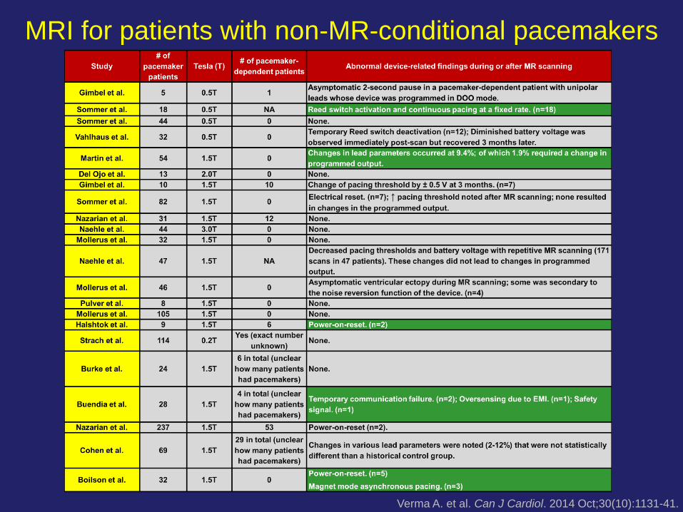
MRI for patients with non-MR-conditional pacemakers
Verma A. et al. Can J Cardiol. 2014 Oct;30(10):1131-41.

MR scanning of patients with non-MR-
conditional ICD systems
Verma A. et al. Can J Cardiol. 2014 Oct;30(10):1131-41.

• Considered to be “off-label” and not “standard of care” in
many institutions.
• Potentially serious complications may occur as a
consequence of MR scanning in this patient population
(e.g. death, system malfunction or damage, arrhythmia
induction).
• If this is to be done, we suggest: (i) development of an
institutional-specific standardized protocol; (ii) clear
discussion of risks and benefits amongst patient and
physicians; (iii) written informed consent.
MR scanning of patients with non-MR-
conditional CIEDs: Comments
Verma A. et al. Can J Cardiol. 2014 Oct;30(10):1131-41.

(1) No CIED system is “MRI-safe”.
(2) There are a number of CIED systems which are MR-conditional. This
means that patients with these systems may undergo MR scanning
without additional/excessive risk, provided that: (i) manufacturer-specified MR scanning parameters are followed.
(ii) the CIED is checked pre-MRI and its function is deemed satisfactory.
(iii) the CIED is reprogrammed to the “MR scanning” mode during MRI.
(3) Published studies of MR scanning of patients with MR-conditional
CIEDs included very few, if any, patients who are pacemaker-
dependent.
(4) MR scanning of non-MR-conditional CIEDs is considered “off-label”
and not part of “standard of care” in most institutions. They may be
performed but a rigorous system of checks and balances is needed.
MR scanning of patients with CIEDs: Conclusions