mrs. marr sports medicine ii. bones of the lower limb pelvis (hip): 3 bones that have grown together...
TRANSCRIPT

Mrs. Marr
Sports Medicine IISports Medicine II
Bones of the Bones of the Lower LimbLower Limb
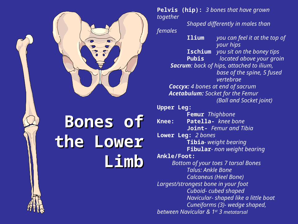
Pelvis (hip): 3 bones that have grown togetherShaped differently in males than
femalesIlium you can feel it at the top of
your hipsIschium you sit on the boney tips Pubis located above your groin
Sacrum: back of hips, attached to ilium, base of the spine, 5 fused vertebrae
Coccyx: 4 bones at end of sacrum Acetabulum: Socket for the Femur
(Ball and Socket joint)Upper Leg:
Femur ThighboneKnee: Patella- knee bone
Joint- Femur and TibiaLower Leg: 2 bones
Tibia- weight bearing Fibular- non weight bearing
Ankle/Foot: Bottom of your toes 7 tarsal Bones
Talus: Ankle Bone Calcaneus (Heel Bone)
Largest/strongest bone in your footCuboid- cubed shapedNavicular- shaped like a little boatCuneiforms (3)- wedge shaped,
between Navicular & 1st 3 metatarsal
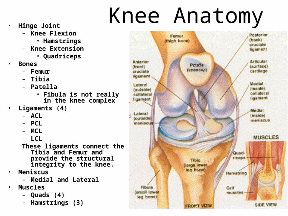
Knee Anatomy• Hinge Joint
– Knee Flexion• Hamstrings
– Knee Extension• Quadriceps
• Bones– Femur– Tibia– Patella
• Fibula is not really in the knee complex
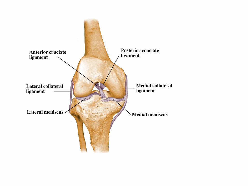
• Ligaments (4)– ACL– PCL– MCL– LCLThese ligaments connect
the Tibia and Femur and provide the structural integrity to the knee.
• Meniscus– Medial and Lateral
• Muscles– Quads (4)– Hamstrings (3)

Cadaver KneePCL
ACL
LCL MCL
MM
LM
Patella Tendon
Patella

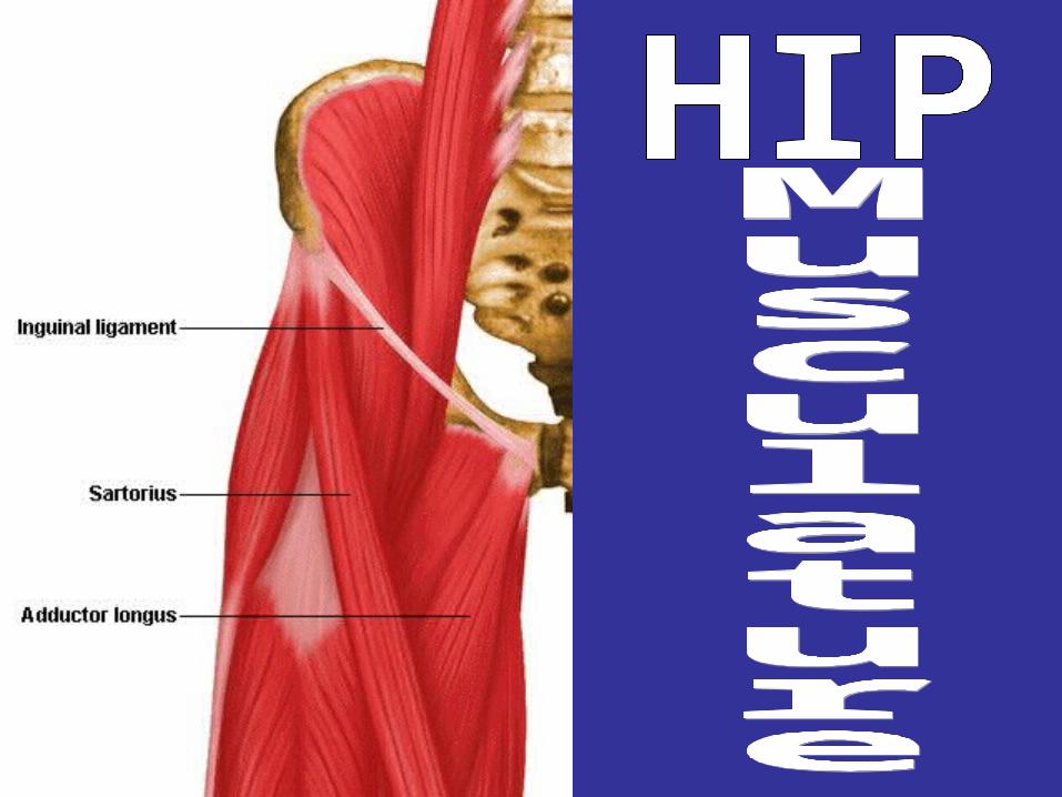

ADDUCTORS
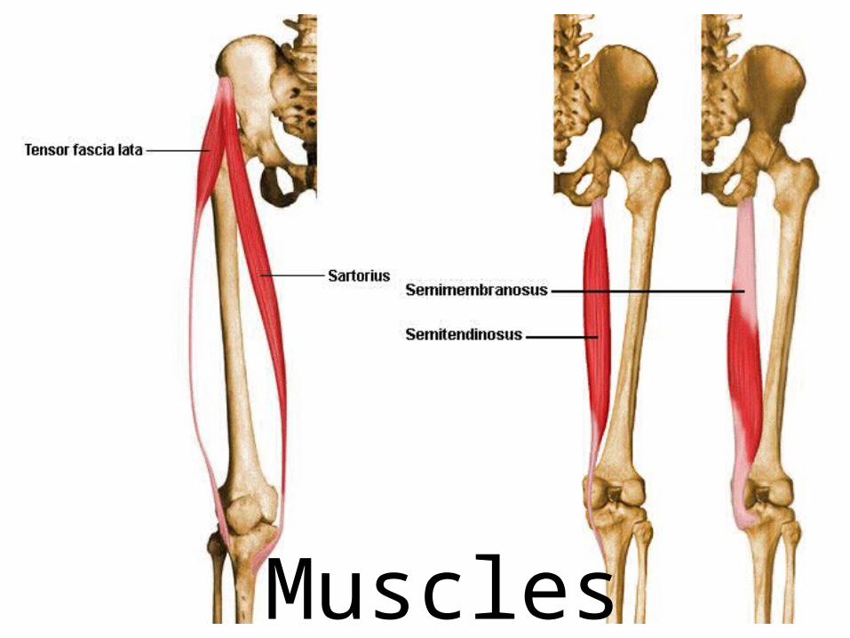
Muscles
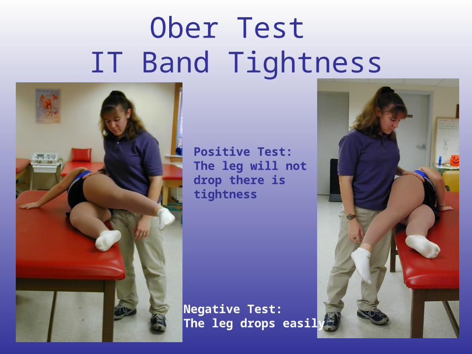
Ober Test IT Band Tightness
Positive Test: The leg will not drop there is tightness
Negative Test:The leg drops easily

Lachman’s Test (ACL)
• Alternative ACL Test– Anterior Drawer – Modified Lachman’s
– Forward translation of the Femur on the Tibia


Posterior Sag (PCL)• Can also be done with
heel resting on chair• Look for sag of the Tibia/
Backward translation of the Tibia

Stress Test Valgus Varus
MCL LCL

What would you do…
• You are a new athletic trainer at a high school and the head football coach tells you that last year his team had too many injuries in the quad, hip, and groin region. What are you going to do to help eliminate this problem for the upcoming season?
Preventing Hip Injuries• Because the hip is a very stable joint, we
will tend to see very few sprains, but many hip strains and contusions.
• Therefore, proper flexibility training and stretching prior to vigorous exercise or activity is warranted.
• Because the thigh is exposed to contact in many athletic activities it is important to have the proper equipment.
• Also the iliac crest (the point of the hip), must be protected because it has very little natural protection.
• Proper strength training is also very important for these muscles to maintain normal balance and stability.

Quadriceps Strain Treatment
• Quad strains should initially be treated with PRICE and wrapped with a supportive elastic bandage.
• After 48-72 hours gradually begin using moist heat and gentle stretching.
• Rehabilitation will focus on regaining strength and range of motion and enhancing flexibility.
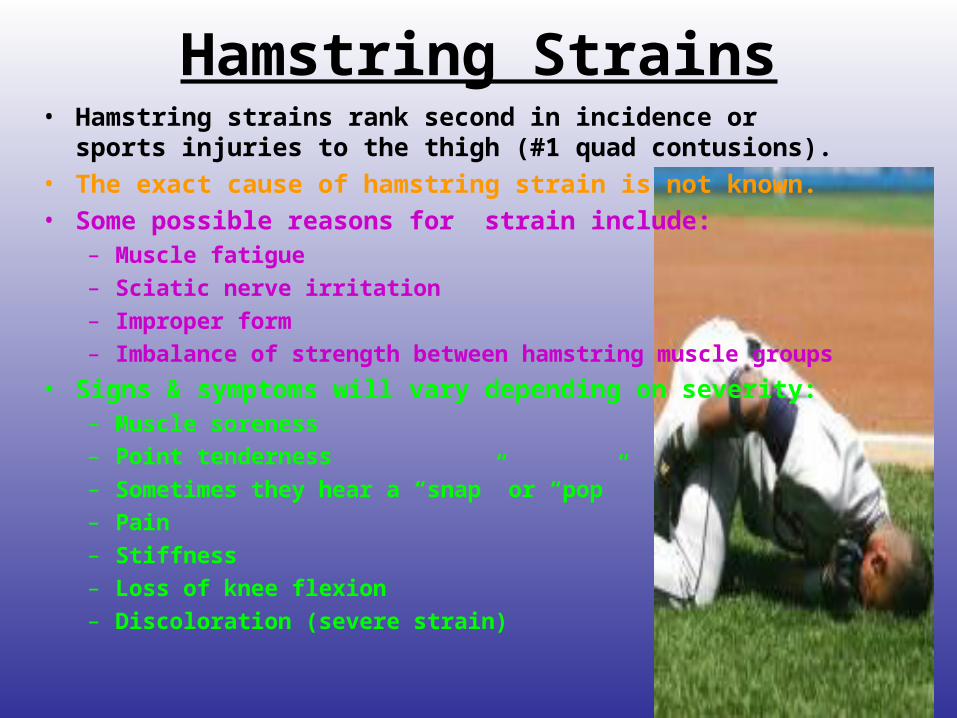
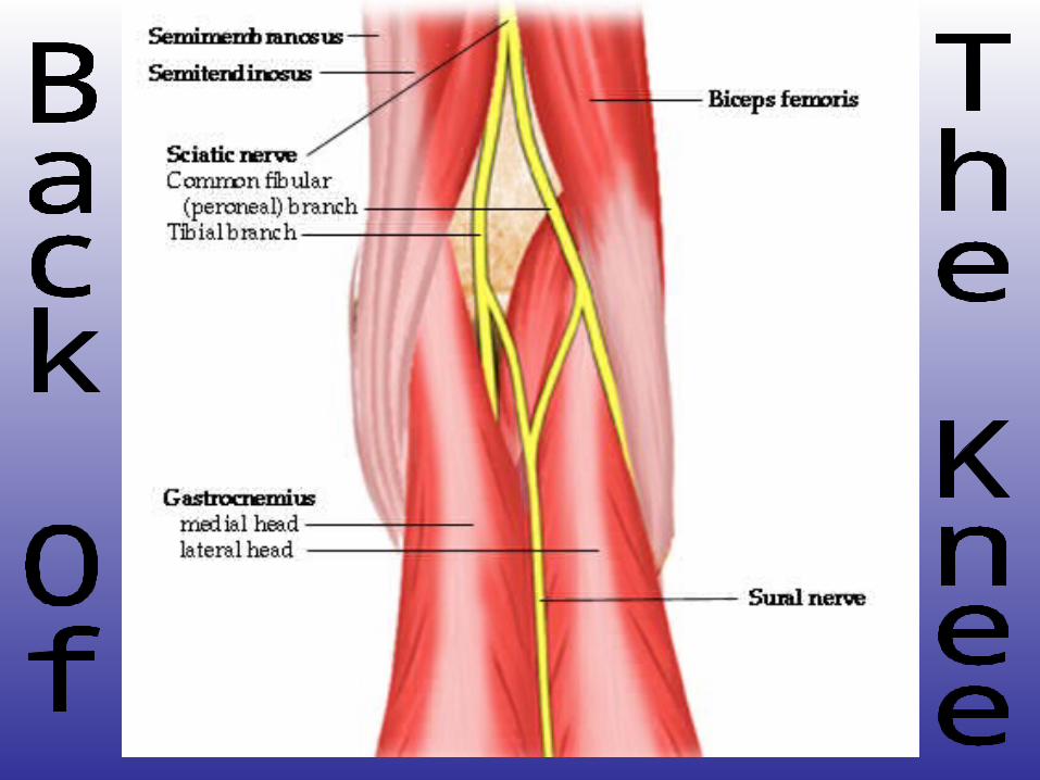
Hamstring Strains• Hamstring strains rank second in incidence or
sports injuries to the thigh (#1 quad contusions).• The exact cause of hamstring strain is not known.• Some possible reasons for strain include:
– Muscle fatigue– Sciatic nerve irritation– Improper form– Imbalance of strength between hamstring muscle
groups
• Signs & symptoms will vary depending on severity:– Muscle soreness– Point tenderness– Sometimes they hear a “snap” or “pop”– Pain– Stiffness– Loss of knee flexion– Discoloration (severe strain)
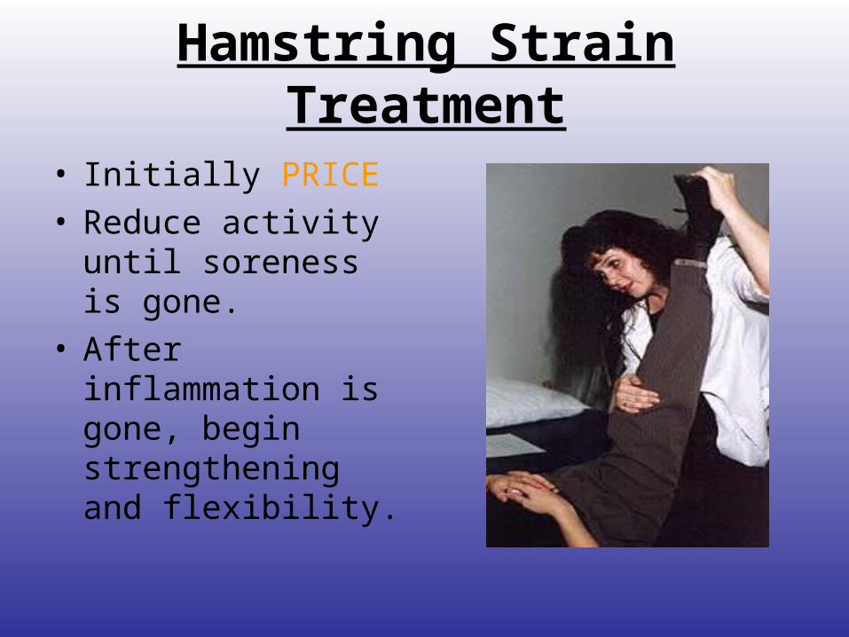
Hamstring Strain Treatment
• Initially PRICE• Reduce activity
until soreness is gone.
• After inflammation is gone, begin strengthening and flexibility.
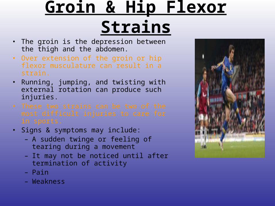
Groin & Hip Flexor Strains• The groin is the depression between the
thigh and the abdomen.• Over extension of the groin or hip flexor
musculature can result in a strain.• Running, jumping, and twisting with
external rotation can produce such injuries.
• These two strains can be two of the most difficult injuries to care for in sports.
• Signs & symptoms may include:– A sudden twinge or feeling of tearing
during a movement– It may not be noticed until after
termination of activity– Pain– Weakness

Groin & Hip Flexor Treatment
• The strain should be treated with intermittent ice, pressure, and rest for 48 to 72 hours.
• Rest has been found to be the best treatment.
• Exercise after pain free.• Gradual stretching and
restoring of normal ROM• A protective spica
should be used until full strength and flexibility are restored.
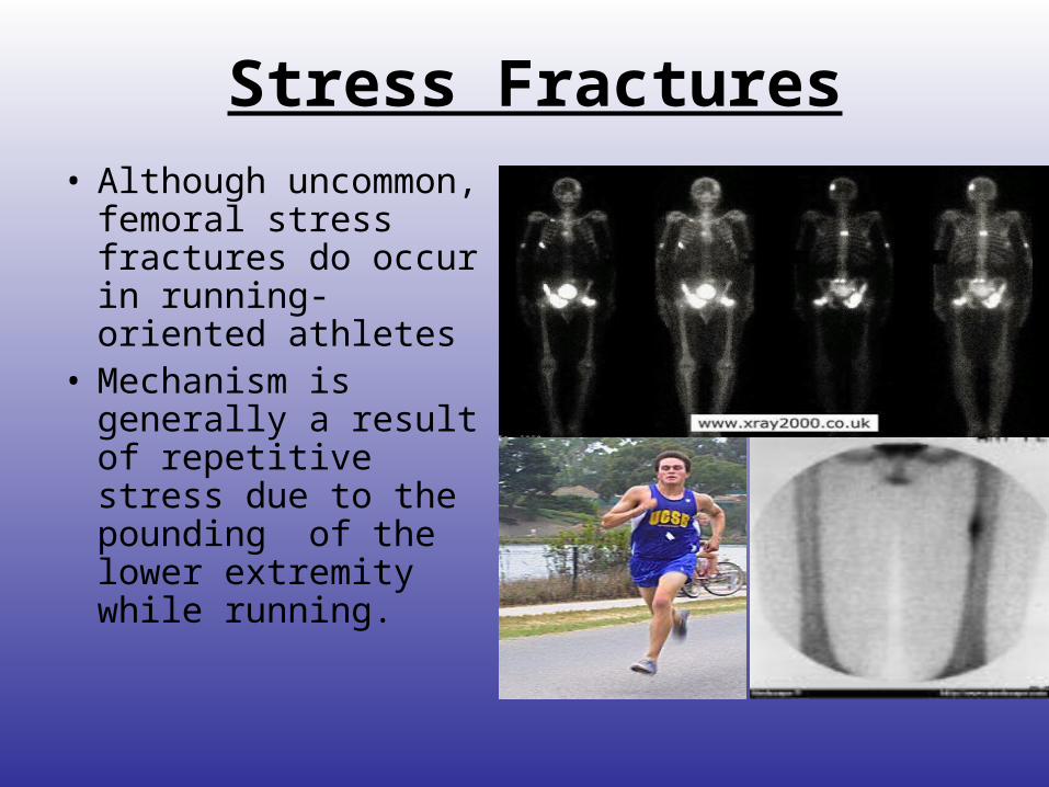
Stress Fractures
• Although uncommon, femoral stress fractures do occur in running-oriented athletes
• Mechanism is generally a result of repetitive stress due to the pounding of the lower extremity while running.
Stress Fx Treatment
• Athlete will generally complain of pain and discomfort.
• Treatment will involve referral to a physician. (Note: stress fracture may not show on an x-ray for 2-3 weeks)
• Rest and an alternate activity such as swimming (aquatic therapy).
• The rest period is generally 4-6 weeks.

Femur Fractures• The femur is the largest bone in the
body and requires a tremendous force to fracture it.
• Femur fracture signs & symptoms include:
– Severe pain– Loss of function– Internal bleeding– Swelling– Tearing of muscle, tendons, arteries,
and nerves– *Often causes the leg to externally
rotate*
• Femur fractures can be potentially life threatening due to the amount of internal bleeding.
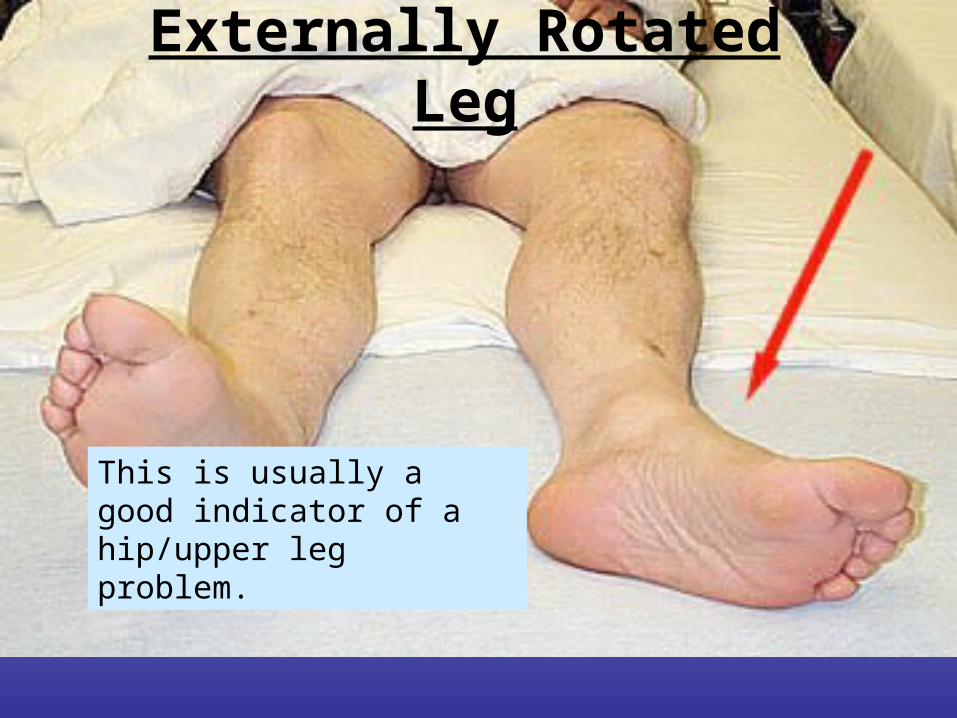
Externally Rotated Leg
This is usually a good indicator of a hip/upper leg problem.

Femur Fx Treatment
• Call 911 (medical emergency due to the fact that a lot of blood can be lost)
• Immobilize• EMS usually will use
a traction splint that gently pulls the femur, which helps reduce leg pain
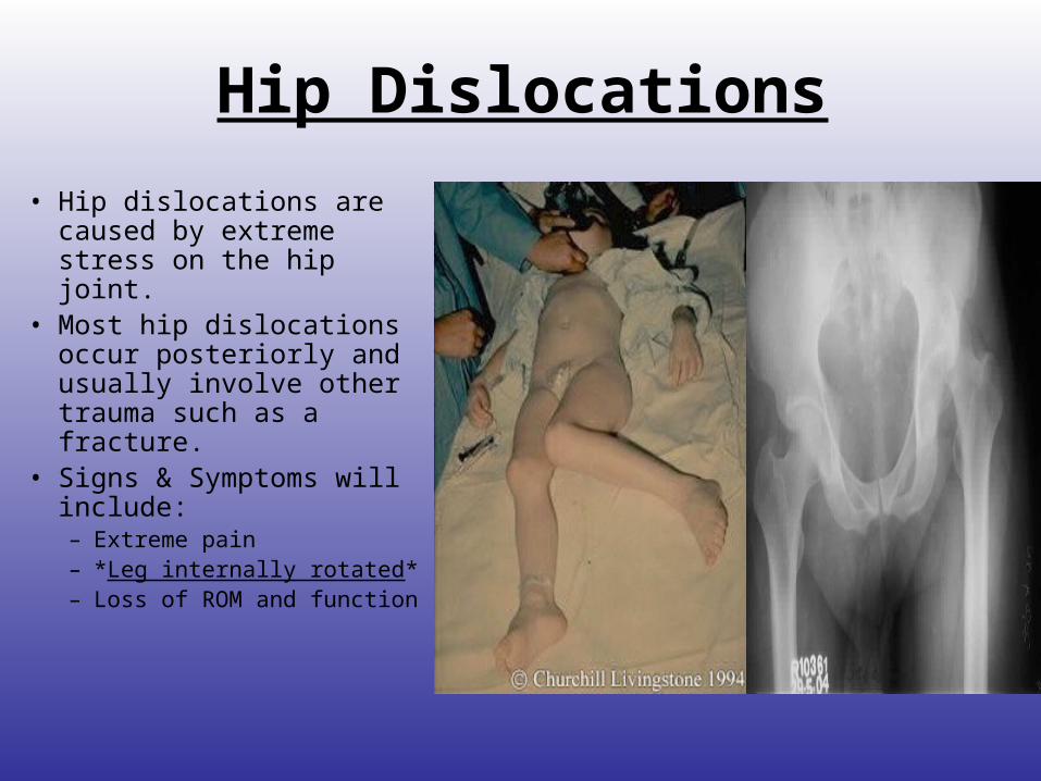
Hip Dislocations
• Hip dislocations are caused by extreme stress on the hip joint.
• Most hip dislocations occur posteriorly and usually involve other trauma such as a fracture.
• Signs & Symptoms will include:– Extreme pain– *Leg internally rotated*– Loss of ROM and function
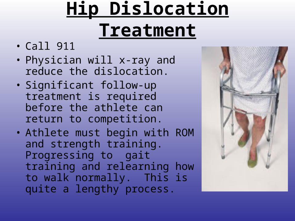
Hip Dislocation Treatment• Call 911• Physician will x-ray and reduce
the dislocation.• Significant follow-up treatment is
required before the athlete can return to competition.
• Athlete must begin with ROM and strength training. Progressing to gait training and relearning how to walk normally. This is quite a lengthy process.

Other Common Hip & Thigh Injuries
• Some other common injuries to the hip and thigh that we will discuss are:
• Hip and Thigh muscle contusions• Legg-Calve-Perthes Disease• Bursitis of the trochanter• Snapping hip phenomenon• Hip pointer (contusion)

Hip & Thigh Contusions
• “Hip Pointer” – iliac crest contusion, occurs most often in contact sports.
• Results from a blow to the inadequately protected iliac crest.
• The hip pointer is one of the most handicapping injuries in sports and is difficult to manage.
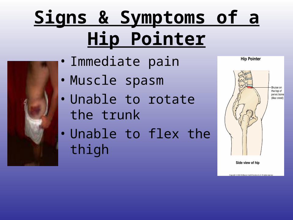
Signs & Symptoms of a Hip Pointer
• Immediate pain
• Muscle spasm
• Unable to rotate the trunk
• Unable to flex the thigh

Hip Pointer Treatment• Ice and pressure for at least 48 hours
• If severe bed rest for 1-2 days will help speed recovery.
• Referral to physician to rule out a fracture
• Ice massage
• Anti-inflammatory

Legg-Perthes Disease
• Legg-Perthes disease is an avascular necrosis of the femoral head.
• It occurs in children ages 3-12 and in boys more often than girls
• The reason for this condition is not clearly understood.
• Circulation becomes disrupted at the head of the femur, causing the articular cartilage to become necrotic and flattened
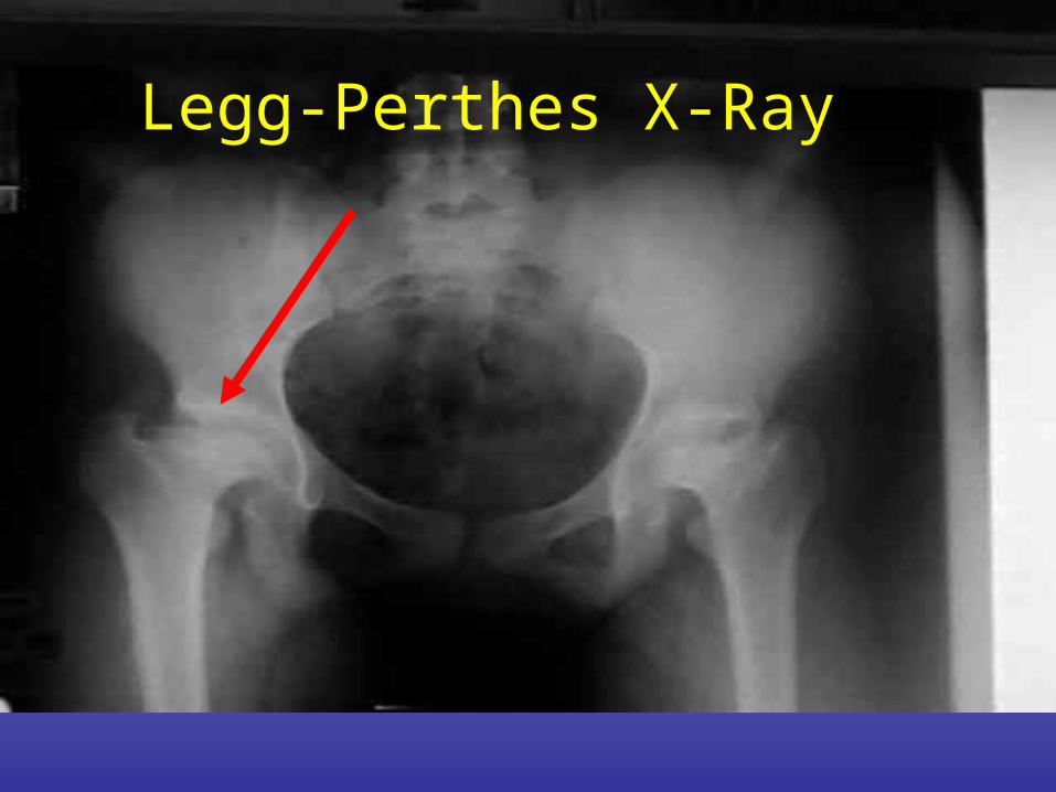
Legg-Perthes X-Ray

Legg-Perthes con’t.
• Signs & Symptoms– Complain of pain in the groin that is sometimes
referred to the abdomen or knee– Limping is typical
• Treatment– Bed rest– Special brace to avoid direct weight bearing
• If treated early enough the head of the femur will revascularize and regain its original shape

Bursitis of the Trochanter
• Trochanteric bursitis is a relatively common condition of the greater trochanter of the femur.
• Most common among women runners
• Treatment includes:– Elimination of running on inclined surfaces– Correction of any leg length discrepancies– Correct poor running form– Ice bags or Ice massage– Gentle stretching– Rest and anti-inflammatory

“Snapping Hip”• This injury is common among
dancers, gymnasts, and hurdlers.• It commonly occurs when the
athlete laterally rotates and flexes the hip joint repeatedly, causing the hip joint and associated soft tissues to become unstable.
• The athlete will complain of a snapping, mainly when balancing on one leg.
• Treatment includes:– Avoiding the action that causes the
snapping
– Stretching tight musculature
– Strengthening weak musculature
– Refer to physician if there is pain
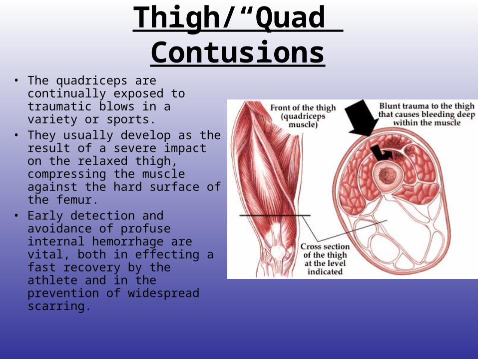
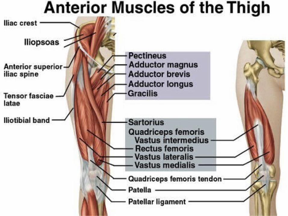
Thigh/“Quad” Contusions• The quadriceps are continually
exposed to traumatic blows in a variety or sports.
• They usually develop as the result of a severe impact on the relaxed thigh, compressing the muscle against the hard surface of the femur.
• Early detection and avoidance of profuse internal hemorrhage are vital, both in effecting a fast recovery by the athlete and in the prevention of widespread scarring.

Quad Contusions• Signs & symptoms (in general)
– Pain
– Temporary loss of function
– Immediate capillary effusion
• Quad contusions can be graded according to there severity.– First degree contusion
– Second degree contusion
– Third degree contusion

Quad Contusions Con’t
• First Degree Contusions– Creates a mild
hemorrhage– Little pain– No swelling– Mild point tenderness– No restriction of ROM

Quad Contusions Con’t.
• Second-Degree Contusion– Pain– Swelling– ROM knee flexion is less than 90
degrees– Obvious limp
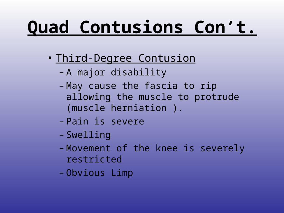
Quad Contusions Con’t.
• Third-Degree Contusion– A major disability– May cause the fascia to rip allowing the
muscle to protrude (muscle herniation ).– Pain is severe – Swelling – Movement of the knee is severely
restricted– Obvious Limp

Quad Contusion Care• Cold and Compression
can help control superficial hemorrhage
• Should be handled conservatively– RICE– Gentle static stretching– Crutches when limping is
present– Heat after the acute phase
has passed (48-72 hours)– Ace Wrap to give pressure
and provide support
Quad Contusion Care Con’t
• A severe blow or repeated blows to the thigh, usually the quadriceps muscle, can produce ectopic bone formation known as myositis ossification.
• Improper care of a thigh contusion can lead to myositis ossificans. Improper care may be:
• Attempting to run off a quad contusion• Too vigorous treatment of a contusion – for example,
massage directly over the contusion, ultrasound therapy, or superficial heat to the thigh.

Myositis Ossificans X-Ray

The End!
Questions????

What will you have to
know…


Are you ready?

Ilium
Illiac Crest
FemurIschial TuberosityPubic
Tubercle
FemoralHead
Sacrum
Coccyx
Ischium
Pubis
Sacroiliac JT
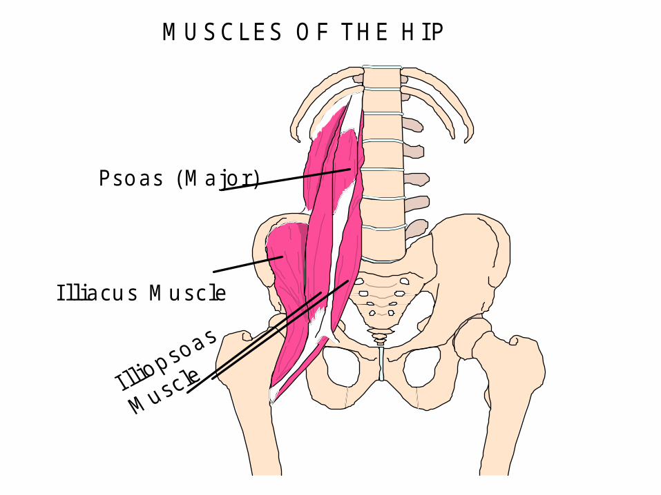
I lliacus Muscle
Psoas ( Major)
MUS CLES OF THE HI P
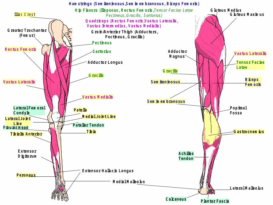
Rectus Femoris Sartorius
Illiac Crest
Vastus Lateralis
Vastus Medialis
Gracilis
Adductor Longus
Pectineus
Lateral FemoralCondyle
Tibia
Patellar Tendon
Patella
Tibialis Anterior
Peroneus
ExtensorDigitorum
Extensor Hallucis Longus
Fibular Head
Calcaneus
Gluteus MaximusGluteus Medius
AchillesTendon
Gastrocnemius
Semimembranosus
PopitealFossa
BicepsFemoris
Semiteninosus
Medial Malleolus
Lateral Malleolus
Gracilis
AdductorMagnus
Vastus Lateralis
Greater Trochanter(Femur)
Hamstrings (Semiteninosus,Semimembranosus, Biceps Femoris)
Hip Flexors (Illopsoas, Rectus Femoris,Tens or Fac iae LataePec tineus ,G rac ilis , Sartor ius )
Tensor FaciaeLatae
Quadriceps (Rectus Femoris,Vastus Lateralis,Vastus Intermedius, Vastus Medialis)
Lateral JointLine
Medial Joint Line
Groin/Anterior Thigh (Adductors,Pectineus, Gracilis)
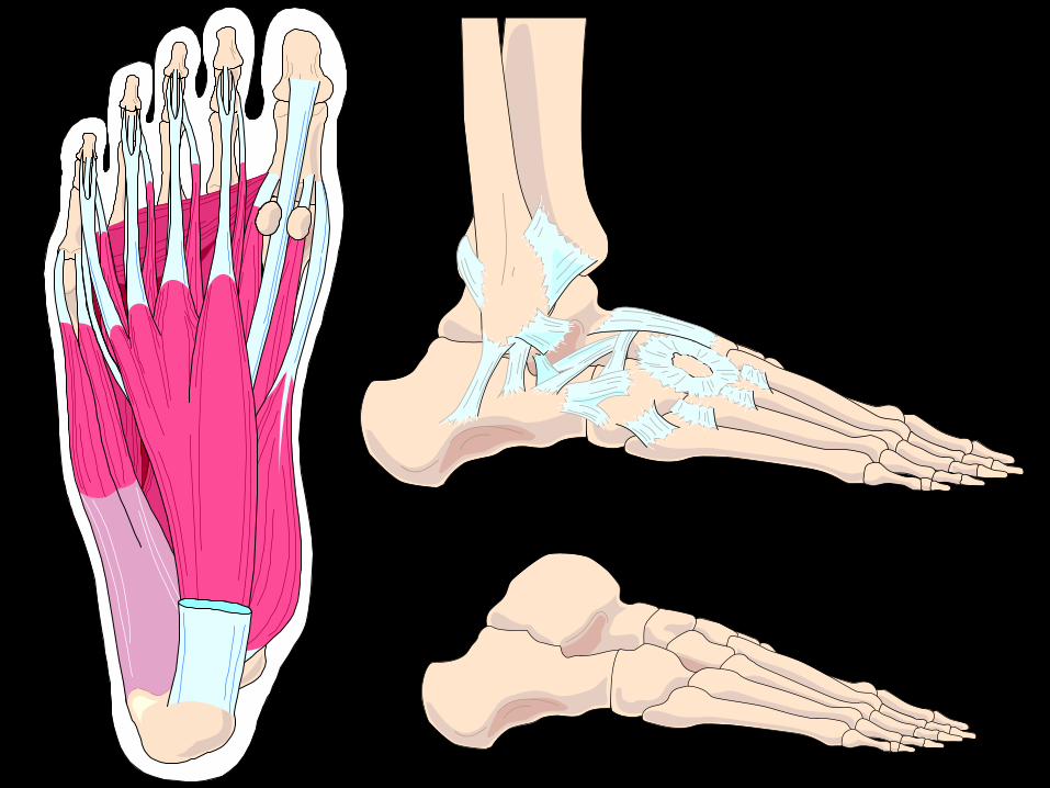
Plantar Fascia
ReviewAnkle, Lower leg and Foot

Talus
Calcaneus
Navicular
Cuboid
CuneiformBones (3)
Metatarsals
Phalanges

Tibia
Fibula
Anterior Tib-Fib Lig
Calcaneus
Talus
Navicular
Cuboid
PosteriorCalcano-Fibular
Anterior Talo-Fibular Lig.
Calcano-FibularLigament
Metatarsals
Phalanges
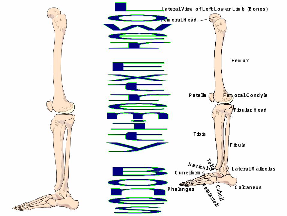
Lateral View of Left Lower Limb (Bones)
Femoral Head
Femur
Tibia
Fibula
Calcaneus
Fibular Head
Patella
Cuneiforms
Phalanges
Lateral Malleolus
Femoral Condyle
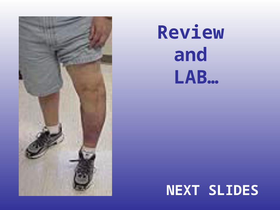
Review and
LAB…
NEXT SLIDES

Knee AnatomyLabel the structures
1. Femur2. Tibia3. Fibula4. MCL5. LCL6. ACL7. PCL8. Medial Meniscus9. Lateral Meniscus10. Name the action the Quadriceps
perform on the knee when contracted.________________
11. Name the action the Hamstrings perform on the knee when contracted._________________
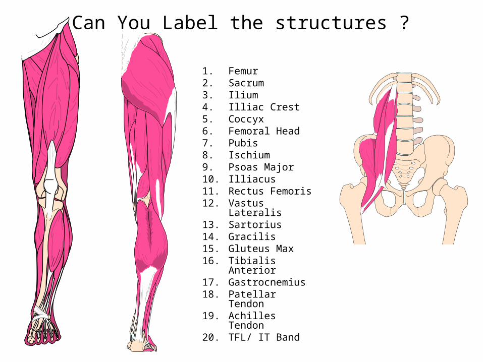
Can You Label the structures ?
1. Femur2. Sacrum3. Ilium4. Illiac Crest5. Coccyx6. Femoral Head7. Pubis8. Ischium9. Psoas Major10. Illiacus11. Rectus Femoris12. Vastus Lateralis13. Sartorius14. Gracilis15. Gluteus Max16. Tibialis Anterior17. Gastrocnemius18. Patellar Tendon19. Achilles Tendon20. TFL/ IT Band
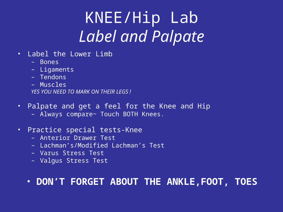
KNEE/Hip LabLabel and Palpate
• Label the Lower Limb– Bones– Ligaments– Tendons– MusclesYES YOU NEED TO MARK ON THEIR LEGS !
• Palpate and get a feel for the Knee and Hip– Always compare~ Touch BOTH Knees.
• Practice special tests-Knee– Anterior Drawer Test– Lachman’s/Modified Lachman’s Test– Varus Stress Test– Valgus Stress Test
• DON’T FORGET ABOUT THE ANKLE,FOOT, TOES