mucosal irritation potential of polyelectrolyte multilayer capsules
TRANSCRIPT

8/7/2019 Mucosal irritation potential of polyelectrolyte multilayer capsules
http://slidepdf.com/reader/full/mucosal-irritation-potential-of-polyelectrolyte-multilayer-capsules 1/11
Mucosal irritation potential of polyelectrolyte multilayer capsules
Liesbeth J. De Cock a, Joke Lenoir a, Stefaan D e Koker b, Vincent Vermeersch c, Andrei G. Skirtach d ,Peter Dubruel c, Els Adriaens a, Chris Vervaet a, Jean Paul Remon a, Bruno G. De Geest a , *
a Laboratory of Pharmaceutical Technology, Department of Pharmaceutics, Ghent University, Harelbekestraat 72, 9000 Ghent, Belgiumb Department of Molecular Biomedical Research, Ghent University, Technologiepark Zwijnaarde 927, 9052 Zwijnaarde, Belgiumc Polymer Chemistry and Biomaterials Research Group, Ghent University, Krijgslaan 281, 9000 Ghent, Belgiumd Max Planck Institute of Colloids and Interfaces, Am Mühlenberg 1, Golm, Germany
a r t i c l e i n f o
Article history:Received 15 October 2010Accepted 6 November 2010Available online 3 December 2010
Keywords:MucosaMicrocapsuleDrug deliveryHeparinBiocompatibility
a b s t r a c t
Polyelectrolyte multilayer capsules have recently gained interest as carriers for drug delivery. Whenenvisioning mucosal administration, one is focused with potential concerns such as tissue irritation andtissue damage, induced by the carrier itself. In this paper we demonstrate the use of a slug-based ( Arionlusitanicus ) assay to evaluate the mucosal irritation potential of different types of polyelectrolytes, theircomplexes and multilayer capsules. This assay allows to assess in a simple yet ef cient way mucosaltissue irritation without using large numbers of vertebrates such as mice, rabbits or non-humanprimates. We found that although single polyelectrolyte components do induce tissue irritation, thisresponse is dramatically reduced upon complexation with an oppositely charged polyelectrolyte,rendering fairly inert polyelectrolyte complexes. These ndings put polyelectrolyte multilayer capsulesfurther en route towards drug delivery applications.
Ó 2010 Elsevier Ltd. All rights reserved.
1. Introduction
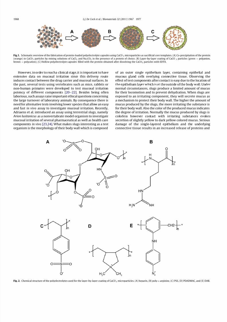
Polyelectrolyte multilayer capsules (PMLC) are a new type of carrier system which hold high potential for drug delivery andtissue engineering as they are well suited to ef ciently encapsulateand deliver antigens, growth factors, nucleic acids, anticancer drugsetc. [1e 3]. These capsules are fabricated by layer-by-layer (LbL)coating of a sacri cial template followed by dissolution of thistemplate, yielding hollow capsules. A very convenient method toencapsulate proteins intoPMLC was recently described by Volodkinet al. by co-precipitating a protein of choice with calcium chlorideand sodium carbonate forming calcium carbonate (CaCO 3) micro-
particles with the protein entrapped within its porous structure[4e 6]. Subsequently, the CaCO 3 microparticles are LbL coated withpolyelectrolytes of alternating charge and in a last step the CaCO 3cores are dissolved by extraction of the Ca 2þ ions with an aqueousEDTA solution ( Fig. 1). As the polyelectrolyte complex shell is semi-permeable, water, ions and low molecular weight species can freelydiffuse in and out but proteins remain entrapped. The major assetsof this type of capsule are (1) the mild conditions used for proteinencapsulation avoiding organic solvents, reactive chemistries or
high shear forces and (2) the ability to tailor the capsules ’ physico-chemical and biochemical properties by choosing from a virtuallyunlimited range of capsule components [7e 12] . Only incubation of in vitro cultured cells with extremely elevated concentrations of PMLC induced toxicity [13,14] .
Recently, a number of papers from our group and others havedemonstrated the potential of PMLC for vaccine delivery whereprotein antigens are encapsulated within the PMLC and ef cientlydelivered to antigen presenting cells in vitro as well as in vivo[1,14 e 17] . Formulating antigens into microparticles has a numberof distinct advantages compared to the delivery of soluble antigenincluding protection against degradation, enhanced uptake by
professional antigen presenting cells such as dendritic cells and theability to induce ‘ cross-presentation ’ of antigen to both CD4 andCD8 T cells, inducing a broad humoral as well as cellular immuneresponse [18,19] . As alternative to the parenteral route where thevaccine is administered through subcutaneous or intramuscularinjection there is an increased interest in mucosal vaccination fora number of reasons such as (1) the avoidance of needles, (2) thepossibility to store and apply the vaccine as dry powder, avoidingthe cold chain and (3) most importantly, the ability to vaccinate atthat speci c site where most infectious organisms enter the body,i.e. at mucosal surfaces, inducing a protective mucosal immuneresponse in addition to a systemic response.
* Corresponding author. Tel.: þ 32 9 264 81 89; fax: þ 32 9 222 82 36.E-mail address: [email protected] (B.G. De Geest).
Contents lists available at ScienceDirect
Biomaterials
j ou rna l homepage : www.e l sev i e r. com/ loca t e /b ioma te r i a l s
0142-9612/$ e see front matter Ó 2010 Elsevier Ltd. All rights reserved.
doi: 10.1016/j.biomaterials.2010.11.012
Biomaterials 32 (2011) 1967 e 1977
8/7/2019 Mucosal irritation potential of polyelectrolyte multilayer capsules
http://slidepdf.com/reader/full/mucosal-irritation-potential-of-polyelectrolyte-multilayer-capsules 2/11

8/7/2019 Mucosal irritation potential of polyelectrolyte multilayer capsules
http://slidepdf.com/reader/full/mucosal-irritation-potential-of-polyelectrolyte-multilayer-capsules 3/11
enzymes such as lactate dehydrogenase (LDH) and alkaline phos-phatase (ALP). LDH is a cytosolic enzyme and will be rst releasedin case of tissue damage. ALP is a membrane-bound enzyme andwill be released in case of severe tissue damage since it is onlypresent in the underlying connective tissue.
In this paper we assess the mucosal irritation potency of PMLCcomposed of different types of polyelectrolyte pairs as shown inFig. 2: (1) Heparin/poly- L -arginine (hep/pARG) as ‘ biopolymer ’ LbL lm, (2) polystyrene-4-sulfonate/poly(diallyldimethylammonium
chloride) (PSS/PDADMAC) as synthetic LbL lm and (3) poly-styrene-4-sulfonate/diazoresin (PSS/DAR) as covalently linkedsynthetic LbL lm. Hep/pARG and PDADMAC/PSS interact purelythrough electrostatics while under in uence of light the diazogroups of DAR react with the sulfonate groups of PSS forminga tough impermeable coating [25,26] . The single polyelectrolytecomponents were evaluated and compared with PMLC and withpolyelectrolyte complexes obtained by simply mixing the poly-electrolytes in solution. The degree of mucosal irritation wasanalyzed by evaluation of mucus production, the color of secretedmucus, release of proteins and enzymes, and slug mortality withina periodof 7 days after exposure. Thepresented methodology is notonly restricted to PMLC but could be widely applied to a largevariety of nano- and micro-scale drug delivery systems that are
currently under investigation.
2. Materials and methods
2.1. Materials
Branched poly(ethyleneimine) (PEI; 22 kDa), polystyrene-4-sulfonate (PSS;70 kDa), poly(diallyldimethylammonium chloride) (PDADMAC; 100 e 200 kDa) werepurchased from Sigma. Heparin (hep) was purchased from Diosynth biotechnology.Diazoresin (DAR) was purchased from Livingston Associates, P.C. Poly- L -arginine(pARG; 100 kDa) was a kind gift from Prof. Peter Dubruel (Polymer Chemistry andBiomaterials Research Group; Ghent University). Drum-dried waxy maize (DDWM)was obtained from National Starch.
2.2. UV/VIS analysis of layer-by-layer deposition
Quartz slides rinsed with piranha solution to render them more hydrophilicwere coated with PEI followed by deposition of layer-by-layer lms based onrespectively PSS/DAR, PSS/PDADMAC and hep/pARG. Deposition of each poly-electrolyte was performed by immersing the slide in a 1 mg/ml solution (containing0.5 M NaCl) of the respective polyelectrolyte, followed by a washing step in water toremove non-adsorbed polyelectrolyte. After each deposition step, the absorptionspectrum was recorded with a Shimadzu spectrophotometer.
2.3. Fabrication of polyelectrolyte capsules
Calcium carbonatemicroparticles(CaCO 3) coreswere fabricated by mixingequalvolumes of calcium chloride (CaCl 2) and sodium carbonate (Na 2CO3) solutions (1 M)under vigorously stirring. The obtained CaCO 3 core templates were subsequentlycoated by incubating them alternately into a 1 mg/ml polyelectrolyte solutioncontaining 0.5 M NaCl during 10 min. After the incubation period, the capsules werecentrifuged and washed twice with water to remove non-adsorbed polyelectrolyte.Two polyelectrolyte bilayers ((PSS/DAR) 2, (PSS/PDADMAC)2 and (hep/pARG) 2) weredeposited on the capsules and hollowcapsules were obtained after extraction of theCaCO3 core templates by addition of an aqueous 0.2 M EDTA solution.
2.4. Fabrication of polyelectrolyte complexes in solution
Complexes of PSS/DAR, PSS/PDADMAC and hep/pARG were made by mixingequal amounts (w/w) of the respective oppositely charged (20 mg/ml containing0.5 M NaCl) polyelectrolytes. The complexes were centrifuged and washed twicewith water to remove non-adsorbed polyelectrolyte.
2.5. Characterization of polyelectrolyte capsules and complexes
2.5.1. Scanning electron microscopyPolyelectrolyte complexes, polyelectrolyte-coated CaCO 3 microparticles and
hollow polyelectrolyte multilayer capsules obtained by dissolution of the CaCO 3cores were dried on a silicone wafer followed by sputtering with gold. SEM imageswere recorded with a Quanta200 FEGFEI scanning electronmicroscope operatingatan acceleration voltage of 5 kV.
2.5.2. Electrophoretic mobilityThe electrophoretic mobility during multilayer build-up on the CaCO 3 micro-
particles was measured using a Zetasizer Nano ZS (Malvern Instruments) equippedwith DispersionTechnology Software (DTS). The z-potentialwas calculated from theelectrophoretic mobility ( m) using the Smoluchowski relation: z ¼ mh/3 where h and3 are respectively the viscosity and permittivity of the solvent.
2.6. Elemental analysis
The composition of the respective polyelectrolyte complexes and capsules wasdetermined by elemental analysis (Vario EL, Elementar Analysensysteme).
2.7. Slug mucosal irritation test
Slugs were bred in the laboratory in plastic containers at 18 e 22 C and fed withlettuce, cucumber, carrots andproteinrichfood.Slugshaving a body weightof 3 e 6 gwere isolated 2 days before the start of the experiment, placed in a plastic boxcontaining a paper towel moistened with phosphate buffered saline and kept at
Table 1Amount of polyelectrolytes available in 10 mg hollow (PSS/PDADMAC) 2, (PSS/DAR)2 and (hep/pARG) 2 capsules.
(PSS/PDADMAC)2 capsulesPSS 4.8 mgPDADMAC 5.0 mg(PSS/DAR)2 capsulesPSS 3.8 mgDAR 6.1 mg(hep/pARG) 2 capsuleshep 5.7 mgpARG 4.3 mg
Fig. 3. Schematic overview of the slug mucosal irritation test.
L.J. De Cock et al. / Biomaterials 32 (2011) 1967 e 1977 1969

8/7/2019 Mucosal irritation potential of polyelectrolyte multilayer capsules
http://slidepdf.com/reader/full/mucosal-irritation-potential-of-polyelectrolyte-multilayer-capsules 4/11
18e 22 C. Only slugs without visible injuries on their foot surface and body wallwere used during the experiments.
The mucosal irritation potency was evaluated by placing the slugs on the testsubstances in a Petri dish during 1 h. Foreachtest substance5 slugs were used. Slugswere exposed to an amount of polyanion or polycation equal to the correspondingamount of that polyelectrolyte present in 10 mg capsules as determined byelemental analysis ( Table 1 ). The amount of polyelectrolyte was supplemented withdrum-dried waxy maize (DDWM) to obtain a total mass of 10 mg. As a negativecontrol 10 mg DDWM was used and as a positive control 10 mg of a mix of DDWMandsodium laurylsulfate(DDWM/SLS 4/1) wasused.In a next seriesof experiments,total amounts of 10 mg lyophilized hollow capsules, respectively lyophilized poly-electrolyte complexes were evaluated.
The mucus produced during the contact period of 1 h was measured by
weighing the Petri dishes with the test component before and after the contactperiod and was expressed as % (w/w) of the initial body weight. After the contactperiod with the test component, the slugs were placed on a new Petri dish con-taining 1 ml of phosphate buffered saline (PBS; pH 7.4) during 1 h. After this rst1 h period on PBS, the slugs were transferred to another Petri dish containing 1 mlPBS for the second 1 h period. The PBS samples of the two periods were collectedto quantify protein and enzyme release. A schematic overview of the slug mucosalirritation test is shown in Fig. 3.
Quanti cation of released proteins was performed by the NanoOrange Ò ProteinQuantitation Kit (Molecular Probes) and is based on protein binding to the Nano-Orange reagent at 95 C. The NanoOrange reagent as such is not uorescent butbecomes uorescentwithan excitation at 470 nmand emission at 590nm as soonasit binds to proteins. Bovine serum albumin was used as a standard and uorescencewas measured using a uorometer (Wallac 1420 multilabel counter, PerkinElmer).The protein concentration is expressed as mg/ml per g body weight.
Results of both mucus production and protein release (mean of the 2 samples)were log-transformed because of the inhomogeneity of the variances and subjectedto a one-way ANOVA combined with a post-hoc Scheffé test.
2.8. Determination of enzyme activity
2.8.1. Alkaline phosphatase (ALP)Alkaline phosphatase converts p-nitrophenyl phosphate into p-nitrophenol and
the enzyme activity was determined spectrophotometrically since p-nitrophenolabsorbs light at 405 nm in alkaline conditions. The amountof enzyme that catalyzesthe formation of 1 mmol/L of p-nitrophenol per minute under the assay conditions isde nedas oneunit of ALPactivity. Theenzyme activity, determined with an enzymekit (ALP CP, Horiba CPX) was expressed as U/L per g body weight and measured ona Cobas Mira Plus analyzer (ABX).
2.8.2. Lactate dehydrogenase (LDH)Lactate dehydrogenase catalyzes the conversion of pyruvate intolactate withthe
concomitant oxidation of NADH into NADþ
and the rate of decrease in absorbance at340 nm which is due to the oxidation of NADH is a measure for the LDH activity. Theamount of enzyme that catalyzes the formation of 1 mmol/L of NADþ per minuteunder the assay conditions is de ned as one unit of LDH activity. The enzymeactivity, determined with an enzyme kit (LDH CP, Horiba ABX) was expressed as U/L per g body weight and measured on a Cobas Mira Plus analyzer (ABX).
2.9. Assessment of slug mortality
Slugs were placed on Petri dishes containing protein rich food and a small papertowel moistened with PBS after exposure to test substances, and slug mortality wasdetermined at day 1, 2, 5 and 7.
2.10. Histological examination
After exposure, slugs were wrapped in aluminium foil and frozen in liquidnitrogen. Cryosections of 10 mm were cut, stained with hematoxylin and eosin, and
examined by light microscopy.
190 200 210 220 230 240 250 2600.0
0.1
0.2
0.3
0.4
0.5
Wavelength [nm]
0.0
0.2
0.4
0.6
0.8
1.0
Wavelength [nm]
0.0
0.1
0.2
0.3
0.4
0.5
Number of bilayers
0.00.20.40.60.81.01.21.41.61.82.0
A
bsorbance [a.u.]
Absorbance [a.u.]
Absorbance [a.u.]
Absorbance [a.u.]
Absorbance [a.u.]
A
bsorbance [a.u.]
Number of bilayers
0.0
0.1
0.2
0.3
0.4
0.5
Number of bilayers
200 250 300 350 400 450 500 550
6 8 10
0 2 4 6 8 10 0
0 2 4
2 4 6 8 10
190 200 210 220 230 240 250 2600.00.20.40.60.81.0
1.21.41.61.82.0
Wavelength [nm]
A B
C
Fig. 4. UV/VIS spectra of quartz slides coated with 10 bilayers of (A) hep/pARG, (B) PSS/PDADMAC and (C) PSS/DAR. The insets of graphs A, B and C show the absorbance of thequartz slides as a function of the number of deposited bilayers at a wavelength of respectively 190 nm, 196 nm and 380 nm.
L.J. De Cock et al. / Biomaterials 32 (2011) 1967 e 1977 1970
8/7/2019 Mucosal irritation potential of polyelectrolyte multilayer capsules
http://slidepdf.com/reader/full/mucosal-irritation-potential-of-polyelectrolyte-multilayer-capsules 5/11
3. Results and discussion
3.1. Characterization of polyelectrolyte complexation on planar surfaces, on sacri cial spherical templates and in solution
3.1.1. Formation of polyelectrolyte multilayer lms on planar surfaces
In rst instance the LbL lm forming ability of the differentpolyelectrolyte pairs was studied. Therefore quartz slides werechosen as planar substrates and after deposition of each polyanion/polycation bilayer the absorbance was measured by means of spectrophotometry. Ten bilayers heparin/pARG ( Fig. 4A), PSS/PDADMAC (Fig. 4B) and PSS/DAR (Fig. 4C) were deposited ontoa quartz slide and from the quasi linear increase of the absorbance
as a function of number of deposited bilayers, a successful multi-layer build-up could be concluded.
3.1.2. Formation of polyelectrolyte lms on sacri cial CaCO3templates
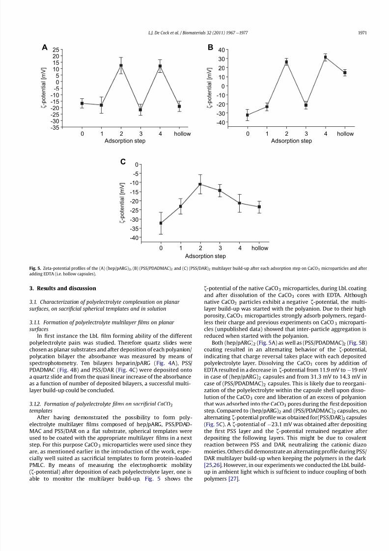
After having demonstrated the possibility to form poly-electrolyte multilayer lms composed of hep/pARG, PSS/PDAD-MAC and PSS/DAR on a at substrate, spherical templates wereused to be coated with the appropriate multilayer lms in a nextstep. For this purpose CaCO 3 microparticles were used since theyare, as mentioned earlier in the introduction of the work, espe-cially well suited as sacri cial templates to form protein-loadedPMLC. By means of measuring the electrophoretic mobility(z-potential) after deposition of each polyelectrolyte layer, one isable to monitor the multilayer build-up. Fig. 5 shows the
z-potential of the native CaCO 3 microparticles, during LbL coatingand after dissolution of the CaCO 3 cores with EDTA. Althoughnative CaCO 3 particles exhibit a negative z-potential, the multi-layer build-up was started with the polyanion. Due to their highporosity, CaCO 3 microparticles strongly adsorb polymers, regard-less their charge and previous experiments on CaCO 3 microparti-cles (unpublished data) showed that inter-particle aggregation isreduced when started with the polyanion.
Both (hep/pARG) 2 (Fig. 5A) as well as (PSS/PDADMAC) 2 (Fig. 5B)coating resulted in an alternating behavior of the z-potential,indicating that charge reversal takes place with each depositedpolyelectrolyte layer. Dissolving the CaCO 3 cores by addition of EDTA resulted in a decrease in z-potential from 11.9 mV to e 19 mV in case of (hep/pARG) 2 capsules and from 31.3 mV to 14.3 mV in
case of (PSS/PDADMAC) 2 capsules. This is likely due to reorgani-zation of the polyelectrolyte within the capsule shell upon disso-lution of the CaCO 3 core and liberation of an excess of polyanionthat was adsorbed into the CaCO 3 pores during the rst depositionstep. Compared to (hep/pARG) 2 and (PSS/PDADMAC) 2 capsules, noalternating z-potential pro le was obtained for(PSS/DAR) 2 capsules(Fig. 5C). A z-potential of À 23.1 mV was obtained after depositingthe rst PSS layer and the z-potential remained negative afterdepositing the following layers. This might be due to covalentreaction between PSS and DAR, neutralizing the cationic diazomoieties. Others did demonstrate an alternating pro le during PSS/DAR multilayer build-up when keeping the polymers in the dark[25,26] . However, in our experiments we conducted the LbL build-up in ambient light which is suf cient to induce coupling of bothpolymers [27] .
-35-30-25-20-15-10
-505
10152025
Adsorption step
-40
-35
-30
-25
-20-15
-10
-5
0
Adsorption step
A
0 1 2 3 4 hollow
0 1 2 3 4 hollow
0 1 2 3 4 hollow
-40
-30
-20-10
0
10
20
30
40
Adsorption step
B
C
Fig. 5. Zeta-potential pro les of the (A) (hep/pARG) 2, (B) (PSS/PDADMAC)2 and (C) (PSS/DAR)2 multilayer build-up after each adsorption step on CaCO 3 microparticles and afteradding EDTA (i.e. hollow capsules).
L.J. De Cock et al. / Biomaterials 32 (2011) 1967 e 1977 1971

8/7/2019 Mucosal irritation potential of polyelectrolyte multilayer capsules
http://slidepdf.com/reader/full/mucosal-irritation-potential-of-polyelectrolyte-multilayer-capsules 6/11

8/7/2019 Mucosal irritation potential of polyelectrolyte multilayer capsules
http://slidepdf.com/reader/full/mucosal-irritation-potential-of-polyelectrolyte-multilayer-capsules 7/11
The molar ratio of heparin to poly- L -arginine in capsules is 0.349indicating that a higher molar amount of poly- L -arginine is involvedin the layer-by-layer build-up on CaCO 3 microparticles. This can beeasily understood since poly- L -arginine in solution has a tertiarystructure compared to heparin having a more linear structure. Themolar ratio of simple complexes composed of heparin and poly- L -arginine is 0.351 which is remarkably similar to the value of PMLC.PSS and PDADMAC exhibit a molar ratio of 0.755 for PMLC and0.775 for simple polyelectrolyte complexes, also these values areremarkably close. Apparently, hep/pARG and PSS/PDADMACinteract in a similar fashion on a supported substrate as in solution.Also in case of PSS/PDADMAC there is an excess of polycation, butthis is less pronounced than in case of hep/pARG.
In contrast to hep/pARG and PSS/PDADMAC, a large discrepancyin the molar ratio of PSS/DAR capsules and simple complexes wasobserved. PMLC have a PSS/DAR ratio of 1.118 while simple PSS/DAR complexes have a ratio of 0.551. Thus, when depositing an LbL lmon CaCO3 microparticles, a higher molar amount of PSS is involvedwhile a higher molar amount of DAR is involved when formingcomplexes in solution. This could be due to a higher availability of sulfonate groups of PSS in solution whereby more DAR can interactwith PSS compared to the availability of sulfonate groups of adsorbed PSS on CaCO 3 microparticles.
3.2. Mucosal irritation potential of polyelectrolytes, polyelectrolytecomplexes and polyelectrolyte multilayer capsules
3.2.1. Mucus productionThe different polyelectrolytes, their complexes and multilayer
capsules characterized in the sections above were tested for their
mucosal irritation potency by a slug mucosal irritation assaydeveloped by Adriaens et al. [23,24] . This assay is a reproduciblequantitative method which does not require complex test equip-ment, sophisticated chemical analysis or a long experimental time.Furthermore this assay did not require vertebrates such as mice,rats and non-human primates resulting in a less laborious assay.
Slugs were exposed to the different samples in a dry powderstate obtained through lyophilization. The reason for this is thatdry powder is a common type of formulation for mucosal appli-cation which often holds problems concerning mucosal irritation.After 1 h exposure, we evaluated the mucus production by theslugs, the color of the produced mucus, release of enzymes andproteins, and slug mortality within a period of 7 days afterexposure. For PSS, 2 different amounts (denoted as PSS
∼
PSS/DAR andPSS∼ PSS/PDADMAC) were used according to the amount of PSSpresent in respectively hollow (PSS/DAR) 2 and (PSS/PDADMAC) 2capsules. The amount of a single polyelectrolyte componentavailable in 10 mg capsules could be calculated from the elementalanalysis results ( Table 1 ). To classify our test substances for theirdegree of irritation, the reactions of treated slugs were comparedto the reactions of slugs exposed to DDWM as negative andDDWM/SLS (4/1) as positive control since previous researchrevealed the non-irritating respectively irritating properties of these substances [23,28] .
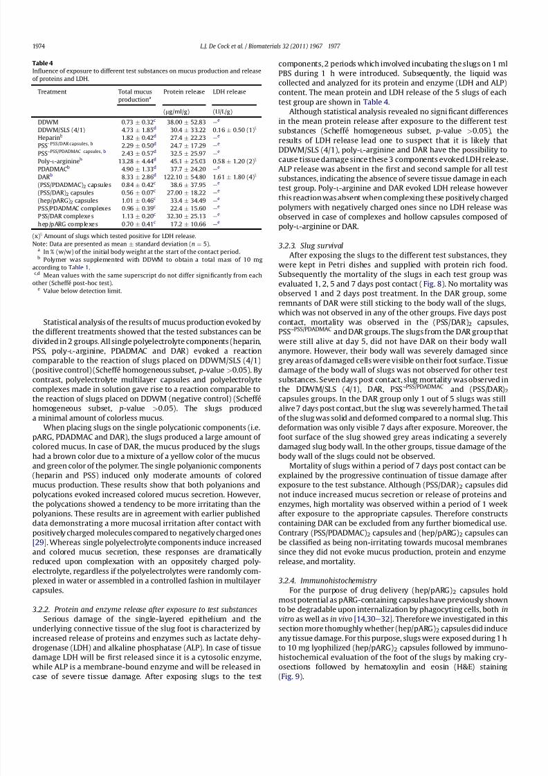
Table 4 summarizes the results of total mucus production(expressed as % (w/w) of the initial body weight) during exposureto the test substances. The negative control evoked a minimal
secretion of colorless mucus which was on average 0.7% of the bodyweight while the positive control evoked a high mucus secretion(4.7% of the body weight) which had a yellow color.
Fig. 7. Overview (1) and zoomed (2) scanning electron microscopy images of complexes composed of (A) hep/pARG, (B) PSS/PDADMAC and (C) PSS/DAR.
Table 2Elemental analysis of hollow (hep/pARG) 2, (PSS/DAR)2 and (PSS/PDADMAC) 2capsules and the corresponding complexes.
N(%) C(%) H(%) S(%)
Hollow (hep/pARG) 2 capsules 14.09 30.74 5.71 5.75Hollow (PSS/DAR) 2 capsules 6.88 53.76 4.76 5.87Hollow (PSS/PDADMAC) 2 capsules 4.33 48.87 7.31 7.49Hep/pARG complexes 15.72 32.98 6.00 6.51PSS/DAR complexes 8.67 62.89 4.68 3.38PSS/PDADMAC complexes 4.79 56.23 8.44 8.50
Table 3Molar ratio of polyanion to polycation in hollow capsules and complexescomposed of hep/pARG, PSS/DAR and PSS/PDADMAC.
Molar ratio
Hollow (hep/pARG) 2 capsules 0.349Hollow (PSS/DAR) 2 capsules 1.118Hollow (PSS/PDADMAC) 2 capsules 0.755Hep/pARG complexes 0.351PSS/DAR complexes 0.511PSS/PDADMAC complexes 0.775
L.J. De Cock et al. / Biomaterials 32 (2011) 1967 e 1977 1973
8/7/2019 Mucosal irritation potential of polyelectrolyte multilayer capsules
http://slidepdf.com/reader/full/mucosal-irritation-potential-of-polyelectrolyte-multilayer-capsules 8/11
Statistical analysis of the results of mucus production evoked bythe different treatments showed that the tested substances can bedivided in 2 groups. All single polyelectrolyte components (heparin,PSS, poly-L -arginine, PDADMAC and DAR) evoked a reactioncomparable to the reaction of slugs placed on DDWM/SLS (4/1)(positive control) (Scheffé homogeneous subset, p-value > 0.05). Bycontrast, polyelectrolyte multilayer capsules and polyelectrolytecomplexes made in solution gave rise to a reaction comparable tothe reaction of slugs placed on DDWM (negative control) (Scheffé
homogeneous subset, p-value > 0.05). The slugs produceda minimal amount of colorless mucus.
When placing slugs on the single polycationic components (i.e.pARG, PDADMAC and DAR), the slugs produced a large amount of colored mucus. In case of DAR, the mucus produced by the slugshad a brown color due to a mixture of a yellow color of the mucusand green color of the polymer. The single polyanionic components(heparin and PSS) induced only moderate amounts of coloredmucus production. These results show that both polyanions andpolycations evoked increased colored mucus secretion. However,the polycations showed a tendency to be more irritating than thepolyanions. These results are in agreement with earlier publisheddata demonstrating a more mucosal irritation after contact withpositively charged molecules compared to negatively charged ones
[29] . Whereas single polyelectrolyte components induce increasedand colored mucus secretion, these responses are dramaticallyreduced upon complexation with an oppositely charged poly-electrolyte, regardless if the polyelectrolytes were randomly com-plexed in water or assembled in a controlled fashion in multilayercapsules.
3.2.2. Protein and enzyme release after exposure to test substancesSerious damage of the single-layered epithelium and the
underlying connective tissue of the slug foot is characterized byincreased release of proteins and enzymes such as lactate dehy-drogenase (LDH) and alkaline phosphatase (ALP). In case of tissuedamage LDH will be rst released since it is a cytosolic enzyme,while ALP is a membrane-bound enzyme and will be released in
case of severe tissue damage. After exposing slugs to the test
components, 2 periods which involved incubating the slugs on 1 mlPBS during 1 h were introduced. Subsequently, the liquid wascollected and analyzed for its protein and enzyme (LDH and ALP)content. The mean protein and LDH release of the 5 slugs of eachtest group are shown in Table 4 .
Although statistical analysis revealed no signi cant differencesin the mean protein release after exposure to the different testsubstances (Scheffé homogeneous subset, p-value > 0.05), theresults of LDH release lead one to suspect that it is likely thatDDWM/SLS (4/1), poly- L -arginine and DAR have the possibility tocause tissue damage since these 3 components evoked LDH release.ALP release was absent in the rst and second sample for all testsubstances, indicating the absence of severe tissue damage in eachtest group. Poly- L -arginine and DAR evoked LDH release howeverthis reactionwas absent when complexing these positively chargedpolymers with negatively charged ones since no LDH release wasobserved in case of complexes and hollow capsules composed of poly- L -arginine or DAR.
3.2.3. Slug survivalAfter exposing the slugs to the different test substances, they
were kept in Petri dishes and supplied with protein rich food.Subsequently the mortality of the slugs in each test group wasevaluated 1, 2, 5 and 7 days post contact ( Fig. 8). No mortality wasobserved 1 and 2 days post treatment. In the DAR group, someremnants of DAR were still sticking to the body wall of the slugs,which was not observed in any of the other groups. Five days postcontact, mortality was observed in the (PSS/DAR) 2 capsules,PSS∼ PSS/PDADMAC and DAR groups. The slugs from the DAR group thatwere still alive at day 5, did not have DAR on their body wallanymore. However, their body wall was severely damaged sincegrey areas of damaged cells were visible on their foot surface. Tissuedamage of the body wall of slugs was not observed for other testsubstances. Seven days post contact, slug mortality was observed inthe DDWM/SLS (4/1), DAR, PSS ∼ PSS/PDADMAC and (PSS/DAR) 2capsules groups. In the DAR group only 1 out of 5 slugs was still
alive 7 days post contact, but the slug was severely harmed. The tailof the slug was solid and deformed compared to a normal slug. Thisdeformation was only visible 7 days after exposure. Moreover, thefoot surface of the slug showed grey areas indicating a severelydamaged slug body wall. In the other groups, tissue damage of thebody wall of the slugs could not be observed.
Mortality of slugs within a period of 7 days post contact can beexplained by the progressive continuation of tissue damage afterexposure to the test substance. Although (PSS/DAR) 2 capsules didnot induce increased mucus secretion or release of proteins andenzymes, high mortality was observed within a period of 1 weekafter exposure to the appropriate capsules. Therefore constructscontaining DAR can be excluded from any further biomedical use.Contrary (PSS/PDADMAC) 2 capsules and (hep/pARG) 2 capsules can
be classi ed as being non-irritating towards mucosal membranessince they did not evoke mucus production, protein and enzymerelease, and mortality.
3.2.4. ImmunohistochemistryFor the purpose of drug delivery (hep/pARG) 2 capsules hold
most potential as pARG-containing capsuleshave previously shownto be degradable upon internalization by phagocyting cells, both invitro as well as in vivo [14,30 e 32] . Therefore we investigated in thissection more thoroughly whether (hep/pARG) 2 capsules did induceany tissue damage. For this purpose, slugs were exposed during 1 hto 10 mg lyophilized (hep/pARG) 2 capsules followed by immuno-histochemical evaluation of the foot of the slugs by making cry-osections followed by hematoxylin and eosin (H&E) staining
(Fig. 9).
Table 4In uence of exposure to different test substances on mucus production and releaseof proteins and LDH.
Treatment Total mucusproduction a
Protein release LDH release
(mg/ml/g) (U/L/g)
DDWM 0.73 Æ 0.32 c 38.00 Æ 52.83 e e
DDWM/SLS (4/1) 4.73 Æ 1.85 d 30.4 Æ 33.22 0.16 Æ 0.50 (1) x
Heparin b 1.82 Æ 0.42 d 27.4 Æ 22.23 e e
PSS∼ PSS/DAR capsules, b 2.29 Æ 0.50 d 24.7 Æ 17.29 e e
PSS∼ PSS/PDADMAC capsules, b 2.43 Æ 0.57 d 32.5 Æ 25.97 e e
Poly- L -arginine b 13.28 Æ 4.44 d 45.1 Æ 25.03 0.58 Æ 1.20 (2) x
PDADMACb 4.90 Æ 1.33 d 37.7 Æ 24.20 e e
DAR b 8.33 Æ 2.86 d 122.10 Æ 54.80 1.61 Æ 1.80 (4) x
(PSS/PDADMAC)2 capsules 0.84 Æ 0.42 c 38.6 Æ 37.95 e e
(PSS/DAR)2 capsules 0.56 Æ 0.07 c 27.00 Æ 18.22 e e
(hep/pARG) 2 capsules 1.01 Æ 0.46 c 33.4 Æ 34.49 e e
PSS/PDADMAC complexes 0.96 Æ 0.39 c 22.4 Æ 15.60 e e
PSS/DAR complexes 1.13 Æ 0.20 c 32.30 Æ 25.13 e e
hep/pARG complexes 0.70 Æ 0.41 c 17.2 Æ 10.66 e e
(x) x Amount of slugs which tested positive for LDH release.Note: Data are presented as mean Æ standard deviation ( n ¼ 5).
a In % (w/w) of the initial body weight at the start of the contact period.b Polymer was supplemented with DDWM to obtain a total mass of 10 mg
according to Table 1 .c,d Mean values with the same superscript do not differ signi cantly from each
other (Scheffé post-hoc test).e Value below detection limit.
L.J. De Cock et al. / Biomaterials 32 (2011) 1967 e 1977 1974

8/7/2019 Mucosal irritation potential of polyelectrolyte multilayer capsules
http://slidepdf.com/reader/full/mucosal-irritation-potential-of-polyelectrolyte-multilayer-capsules 9/11
The foot of a slug consists of outer single-layered epitheliumoverlying connective tissue. Exposure of a slug to an irritatingcomponent may lead to severely damaged epithelium cells whichcan be observed through immunohistochemistry. Fig. 9A showsan H&E stained cryosection of the foot of a control slug which
was not exposed to any test substance. The outer single
epithelium layer is continuous indicating absence of tissuedamage. The foot of slugs exposed to (hep/pARG) 2 capsules(Fig. 9B) showed also an intact outer epithelium layer compa-rable to the control. These results further con rm on a micro-scopic scale the relatively inert character of (hep/pARG) 2
capsules regarding mucosal irritation.
Fig. 8. Survival curves of slugs exposed to (A) single polyelectrolytes, (B) hollow polyelectrolyte multilayer capsules and (C) polyelectrolyte complexes made in solution.
L.J. De Cock et al. / Biomaterials 32 (2011) 1967 e 1977 1975

8/7/2019 Mucosal irritation potential of polyelectrolyte multilayer capsules
http://slidepdf.com/reader/full/mucosal-irritation-potential-of-polyelectrolyte-multilayer-capsules 10/11
4. Conclusions
In this paper the mucosal irritation potency of several classes ebiopolymers,synthetic polyelectrolytesand reactive polyelectrolytese of oppositely charged polyelectrolytes, their complexes as well ashollow multilayer capsules were evaluated via a A. lusitanicus slug-based mucosal irritation assay. We found that notwithstanding thefact that single (bio)polyelectrolyte components do show a tendencyto induce tissue irritation, this irritation is abolished uponcomplexation with an oppositely charged polyelectrolyte, turningthe supramolecular structures into fairly inert species. In an erawhere there is a lot of controversy concerning safety and toxicityissues of nano- and micro-engineered materials, we demonstrate anef cient method to assess tissue irritation using an in vivo platformwhich does not involve vertebrates such as mice, rabbits or non-human primates. Taken together, these ndings put polyelectrolytecapsules further en route towards drug delivery applications.
Acknowledgments
LJDC wishes to express her gratitude to the Institute for thePromotionof Innovation by ScienceandTechnology inFlanders (IWT-Flanders) for their nancial support. SDK acknowledges GhentUniversity for a postdoctoral scholarship (BOF-GOA). BGDGacknowledges the FWO-Flanders for a postdoctoral scholarship. AGSacknowledges Prof. Helmut Möhwald for his continuous support.
Appendix
Figures with essential color discrimination. Figs. 1, 3, 8, and 9 inthis article are dif cult to interpret in black and white. The full colorimages can be found in the online version, at doi:10.1016/j.biomaterials.2010.11.012 .
References
[1] De Koker S, Naessens T, De Geest BG, Bogaert P, Demeester J, De Smedt S, et al.Biodegradable polyelectrolyte microcapsules: antigen delivery tools withTh17 skewing activity after pulmonary delivery. J Immunol 2010;184:203 e 11.
[2] Itoh Y, Matsusaki M, Kida T, Akashi M. Locally controlled release of basic broblast growth factor from multilayered capsules. Biomacromolecules2008;9:2202 e 6.
[3] Zhao QH, Zhang SA, Tong WJ, Gao CY, Shen JC. Polyelectrolyte microcapsules
templated on poly(styrene sulfonate)-doped CaCO 3 particles for loading and
sustained release of daunorubicin and doxorubicin. Eur Polym J 2006;42:3341 e 51.
[4] Volodkin DV, Larionova NI, Sukhorukov GB. Protein encapsulation via porousCaCO3 microparticles templating. Biomacromolecules 2004;5:1962 e 72.
[5] Volodkin DV, Petrov AI, Prevot M, Sukhorukov GB. Matrix polyelectrolytemicrocapsules: new system for macromolecule encapsulation. Langmuir2004;20:3398 e 406.
[6] Petrov AI, Volodkin DV, Sukhorukov GB. Protein-calcium carbonate copreci-pitation: a tool for protein encapsulation. Biotechnol Prog 2005;21:918 e 25.
[7] De Koker S, Lambrecht BN, Willart MA, Van Kooyk Y, Grooten J, Vervaet J, et al.Designing polymeric particles for antigen delivery. Chem Soc Rev doi:10.1039/b914943k
[8] De Cock LJ, De Koker S, De Geest BG, Grooten J, Vervaet C, Remon JP, et al.Polymeric multilayer capsules in drug delivery. Angew Chem Int Ed Engl2010;49:6954 e 73.
[9] Peyratout CS, Dahne L. Tailor-made polyelectrolyte microcapsules: frommultilayers to smart containers. Angew Chem Int Ed 2004;43:3762 e 83.
[10] Becker AL, Johnston AP, Caruso F. Layer-by-layer-assembled capsules and
lms for therapeutic delivery. Small 2010;6:1836 e 52.[11] De Geest BG, Sanders NN, Sukhorukov GB, Demeester J, De Smedt SC. Releasemechanisms for polyelectrolyte capsules. Chem Soc Rev 2007;36:636 e 49.
[12] Matsusaki M, Akashi M. Functional multilayered capsules for targeting andlocal drug delivery. Expert Opin Drug Del 2009;6:1207 e 17.
[13] Kirchner C, Javier AM, Susha AS, Rogach AL, Kreft O, Sukhorukov GB, et al.Cytotoxicity of nanoparticle-loaded polymer capsules. Talanta 2005;67:486 e 91.
[14] De Koker S, De Geest BG, Cuvelier C, Ferdinande L, Deckers W, Hennink WE,et al. In vivo cellular uptake, degradation, and biocompatibility of poly-electrolyte microcapsules. Adv Funct Mater 2007;17:3754 e 63.
[15] Chong SF, Sexton A, De Rose R, Kent SJ, Zelikin AN, Caruso F. A paradigm forpeptide vaccine delivery using viral epitopes encapsulated in degradablepolymer hydrogel capsules. Biomaterials 2009;30:5178 e 86.
[16] De Koker S, De Geest BG, Singh SK, De Rycke R, Naessens T, Van Kooyk Y, et al.Polyelectrolyte microcapsules as antigen delivery vehicles to dendritic cells:uptake, processing, and cross-presentation of encapsulated antigens. AngewChem Int Ed 2009;48:8485 e 9.
[17] De Rose R, Zelikin AN, Johnston APR, Sexton A, Chong SF, Cortez C, et al.Binding, internalization, and antigen presentation of vaccine-loaded nano-engineered capsules in blood. Adv Mater 2008;20:4698 e 703.
[18] Rock KL, Shen L. Cross-presentation: underlying mechanisms and role inimmune surveillance. Immunol Rev 2005;207:166 e 83.
[19] Reddy ST, Swartz MA, Hubbell JA. Targeting dendritic cells with biomate-rials: developing the next generation of vaccines. Trends Immunol 2006;27:573 e 9.
[20] Achilles SL, Shete PB, Whaley KJ, Moench TR, Cone RA. Microbicide ef cacyand toxicity tests in a mouse model for vaginal transmission of Chlamydiatrachomatis . Sex Transm Dis 2002;29:655 e 64.
[21] Chvapil M, Droegemueller W, Owen JA, Eskelson CD, Betts K. Studies of nonoxynol-9.1. The effect on the vaginas of rabbits and rats. Fertil Steril1980;33:445 e 50.
[22] Patton DL, Kidder GG, Sweeney YC, Rabe LK, Hillier SL. Effects of multipleapplications of benzalkonium chloride and nonoxynol 9 on the vaginalepithelium in the pigtailed macaque ( Macaca nemestrina ). Am J ObstetGynecol 1999;180:1080 e 7.
[23] Adriaens E, Remon JP. Evaluation of an alternative mucosal irritation test
using slugs. Toxicol Appl Pharmacol 2002;182:169e
75.
Fig. 9. Optical microscopy images of hematoxylin and eosin stained cryosections showing the foot of (A) a control, non-exposed, slug and (B) a slug exposed to (hep/pARG) 2
capsules.
L.J. De Cock et al. / Biomaterials 32 (2011) 1967 e 1977 1976

8/7/2019 Mucosal irritation potential of polyelectrolyte multilayer capsules
http://slidepdf.com/reader/full/mucosal-irritation-potential-of-polyelectrolyte-multilayer-capsules 11/11
[24] Adriaens E, Remon JP. Gastropods as an evaluation tool for screening the irri-tating potency of absorptionenhancersand drugs. PharmRes 1999;16:1240 e 4.
[25] Zhu HG, McShane MJ. Macromolecule encapsulation in diazoresin-basedhollow polyelectrolyte microcapsules. Langmuir 2005;21:424 e 30.
[26] Pastoriza-Santos I, Scholer B, Caruso F. Core-shell colloids and hollow poly-electrolyte capsules based on diazoresins. Adv Funct Mater 2001;11:122 e 8.
[27] De Geest BG, McShane MJ, Demeester J, De Smedt SC, Hennink WE. Micro-capsules ejecting nanosized species into the environment. J Am Chem Soc2008;130:14480 e 2.
[28] Callens C, Adriaens E, Dierckens K, Remon JP. Toxicological evaluation of
a bioadhesive nasal powder containing a starch and Carbopol (R) 974 P onrabbit nasal mucosa and slug mucosa. J Control Release 2001;76:81 e 91.
[29] Fichorova RN, Bajpai M, Chandra N, Hsiu JG, Spangler M, Ratnam V, et al.Interleukin IL-1, IL-6, and IL-8 predict mucosal toxicity of vaginal microbicidalcontraceptives. Biol Reprod 2004;71:761 e 9.
[30] De Geest BG, Vandenbroucke RE, Guenther AM, Sukhorukov GB, Hennink WE,Sanders NN, et al. Intracellularly degradable polyelectrolyte microcapsules.Adv Mater 2006;18:1005 e 9.
[31] Rivera-Gil P, De Koker S, De Geest BG, Parak WJ. Intracellular processing of proteins mediated by biodegradable polyelectrolyte capsules. Nano Lett2009;9:4398 e 402.
[32] Dierendonck M, De Koker S, Cuvelier C, Grooten J, Vervaet C, Remon JP, et al.
Facile two-step synthesis of porous antigen-loaded degradable polyelectrolytemicrospheres. Angew Chem Int Ed. doi:10.1002/anie.201001046 .
L.J. De Cock et al. / Biomaterials 32 (2011) 1967 e 1977 1977