multidrug-resistant candida haemulonii and c. auris tel ... · candida auris and c. haemulonii are...
TRANSCRIPT

Candida auris and C. haemulonii are closely related, mul-tidrug-resistant emerging fungal pathogens that are not readily distinguishable with phenotypic assays. We studied C. auris and C. haemulonii clinical isolates from 2 hospi-tals in central Israel. C. auris was isolated in 5 patients with nosocomial bloodstream infection, and C. haemulonii was found as a colonizer of leg wounds at a peripheral vascular disease clinic. Liberal use of topical miconazole and close
Multidrug-Resistant Candida haemulonii and C. auris,
Tel Aviv, IsraelRonen Ben-Ami, Judith Berman, Ana Novikov, Edna Bash, Yael Shachor-Meyouhas, Shiri Zakin,
Yasmin Maor, Jalal Tarabia, Vered Schechner, Amos Adler, Talya Finn
Emerging Infectious Diseases • www.cdc.gov/eid • Vol. 23, No. 2, February 2017 195
RESEARCH
Author affiliations: Tel Aviv University, Tel Aviv, Israel (R. Ben-Ami, J. Berman, S. Zakin, Y. Maor, V. Schechner, A. Adler); Tel Aviv Sourasky Medical Center, Tel Aviv (R. Ben-Ami, A. Novikov, E. Bash, J. Tarabia, V. Schechner, A. Adler, T. Finn); Ruth Rappaport Children’s Hospital, Haifa, Israel (Y. Shachor-Meyouhas); Wolfson Medical Center, Holon, Israel (Y. Maor).
DOI: http://dx.doi.org/10.3201/eid2302.161486
This activity has been planned and implemented through the joint providership of Medscape, LLC and Emerging Infectious Diseases. Medscape, LLC is accredited by the American Nurses Credentialing Center (ANCC), the Accreditation Council for Pharmacy Education (ACPE), and the Accreditation Council for Continuing Medical Education (ACCME), to provide continuing education for the healthcare team.
Medscape, LLC designates this Journal-based CME activity for a maximum of 1.00 AMA PRA Category 1 Credit(s)™. Physicians should claim only the credit commensurate with the extent of their participation in the activity.
All other clinicians completing this activity will be issued a certificate of participation. To participate in this journal CME activity: (1) review the learning objectives and author disclosures; (2) study the education content; (3) take the post-test with a 75% minimum passing score and complete the evaluation at http://www.medscape.org/journal/eid; and (4) view/print certificate. For CME questions, see page 375.
Release date: January 13, 2017; Expiration date: January 13, 2018
Learning Objectives
Upon completion of this activity, participants will be able to:
1. Assess the phylogenetic analysis of Candida auris and C. haemulonii in the current study 2. Analyze patient characteristics among infections with C. auris and C. haemulonii 3. Distinguish the pattern of antifungal resistance in infections with C. auris and C. haemulonii 4. Evaluate the pathologic potential of C. auris and C. haemulonii
CME Editor Karen L. Foster, MA, Technical Writer/Editor, Emerging Infectious Diseases. Disclosure: Karen L. Foster has disclosed no relevant financial relationships.
CME Author Charles P. Vega, MD, Clinical Professor of Family Medicine, University of California, Irvine. Disclosure: Charles P. Vega, MD, has disclosed the following financial relationships: served as an advisor or consultant for Allergan, Inc.; McNeil Consumer Healthcare; served as a speaker or a member of a speakers bureau for Shire Pharmaceuticals.
Authors Disclosures: Ronen Ben-Ami, MD, has disclosed the following relevant financial relationships: served as an advisor or consultant for Pfizer, Inc.; Merck & Co. Inc.; Neopharm; Teva Pharmaceuticals. Yasmin Maor, MD, has disclosed the following relevant financial relationships: served as an advisor or consultant for Novo Nordisk; served as a speaker or member of a speakers bureau for Pfizer; MSD. Judith Berman, PhD; Ana Novikov, MSc; Edna Bash, MSc; Yael Shachor-Meyouhas, MD; Shiri Zakin, PhD; Jalal Tarabia, PhD; Vered Schechner, MD, MSc; Amos Adler, MD; and Talya Finn, MBBS, have disclosed no relevant financial relationships.

RESEARCH
contact among patients were implicated in C. haemulonii transmission. C. auris exhibited higher thermotolerance, virulence in a mouse infection model, and ATP-dependent drug efflux activity than C. haemulonii. Comparison of ri-bosomal DNA sequences found that C. auris strains from Israel were phylogenetically distinct from isolates from East Asia, South Africa and Kuwait, whereas C. haemulo-nii strains from different countries were closely interrelated. Our findings highlight the pathogenicity of C. auris and un-derscore the need to limit its spread.
Candida species are leading causes of bloodstream infection (BSI) in hospitalized patients, particularly
those in intensive care units who are exposed to broad-spectrum antimicrobial drugs, indwelling vascular cath-eters, parenteral nutrition, abdominal surgery, and immu-nosuppressive agents (1,2). High rates of attributable death have been associated with delayed initiation of appropriate antifungal treatment (3,4). This problem is compounded by the emergence of drug-resistant Candida species, notably C. glabrata, in many hospitals (5).
C. auris is an emerging opportunistic pathogen, first reported in 2009 as an isolate from the external ear of an inpatient at a hospital in Japan (6). It has since been identi-fied as a cause of nosocomial BSI in numerous countries in East Asia, the Middle East, Africa, and Europe (7–11). C. auris might be resistant to multiple classes of antifungal agents and apparently has a potential for person-to-person transmission, challenging clinicians and infection control teams (12). C. auris often is misidentified by traditional mi-crobiological methods as C. haemulonii, a phylogenetically related drug-resistant Candida species that also is increas-ingly reported in healthcare facilities worldwide (13).
We report on the detection of multidrug-resistant C. auris and C. haemulonii in clinical specimens in Tel Aviv, Israel, and specifically on the emergence of C. auris as a cause of nosocomial BSI. We highlight distinct clinical and epidemiologic characteristics of these 2 species and present experimental evidence for differences in their virulence.
Materials and MethodsWe undertook this study after C. auris BSI was detected in 4 patients during May–October 2014 at the Tel Aviv Sourasky Medical Center (TASMC), a tertiary-level hospi-tal in Tel Aviv. An additional C. auris bloodstream isolate was recovered in April 2015 from a patient at the Wolfson Medical Center in Holon (southern Tel Aviv metropolitan area). No additional C. haemulonii or C. auris isolates were identified through inquiries at additional clinical microbiol-ogy laboratories in Israel.
The TASMC Institutional ethics committee approved this study. Need for informed consent was waived because of the observational and anonymous nature of the study.
Clinical Candida isolatesCandida isolates recovered from clinical specimens were identified at the TASMC Clinical Microbiology Labora-tory by growth characteristics on CHROMagar Candida (CHROMagar, Paris, France) and the Vitek 2 YST ID sys-tem (bioMérieux, Marcy-l’Étoile, France). The Vitek 2 data-base does not include C. auris, and this species is routinely misidentified as C. haemulonii (13). We therefore reviewed all isolates identified as C. haemulonii during January 2009–August 2015. Isolates recovered during May 2014–August 2015 were stored at -20°C and subjected to further analyses. We assessed thermotolerance by plating serial dilutions of yeast culture on Sabouraud dextrose agar (SDA) plates and assessing growth after 24 h incubation at 35°C–42°C.
Sequence-Based Species IdentificationCandida isolates were streaked on SDA plates to ensure pu-rity. We extracted DNA by using PrepMan Ultra solution (Applied Biosystems, Foster City, CA, USA) according to the manufacturer’s instructions and amplified and sequenced the internal transcribed spacer (ITS) and D1/D2 large sub-unit (LSU) ribosomal DNA segments by using primer pairs ITS1/ITS4 and LSU1/LSU2 (14), respectively. PCR was performed in 0.2-mL tubes with 0.4 µmol/L or 0.2 µmol/L of each primer for ITS and LSU, respectively; 10 μL Larova Red Load Taq Master Mix (5×) (Larova, Jena, Germany); and ≈25 ng of template. PCR conditions were 95°C for 4.5 min (denaturation), 40 cycles of 95°C for 30 s (denatur-ation), 55°C (ITS) or 48°C (LSU) for 30 s (annealing), 72°C for 1 min (extension), and a final extension stage of 72°C for 7 min. PCR products were resolved on 0.7% agarose gel and stained with SERVA DNA stain clear G (Tamar, Mev-aseret Zion, Israel). Products were cleaned with QIAquick PCR purification kit (QIAGEN, Hilden, Germany) and se-quenced at Hy-Labs (Rehovot, Israel). We then aligned ITS and LSU sequences with the matching type strain sequences for CBS5149T (C. haemulonii), CBS7798T (C. duobush-aemulonii), CNM-CL7239T (C. haemulonii var. vulnera), CBS10099T (C. pseudohaemulonii), and CBS10913T (C. auris). A similarity score of >98% in both ITS and LSU sequences was required for species-level identification. All new sequences were deposited in GenBank (Table 1, https://wwwnc.cdc.gov/EID/article/23/2/16-1486-T1.htm).
Phylogenetic AnalysesWe aligned ITS and LSU sequences of C. haemulonii and C. auris isolates by using MUSCLE (15) and generated phylogenetic trees with the neighbor-joining method (16), using the Kimura 2-parameter method to compute evolu-tionary distances (17). We tested phylogeny with the boot-strap method (500 replicates) and used Schizosaccharomy-ces pombe strains ATCC 38366 and CBS 356 as outgroups. Evolutionary analyses were performed in MEGA7 (18).
196 Emerging Infectious Diseases • www.cdc.gov/eid • Vol. 23, No. 2, February 2017
C. haemulonii and C. auris, Tel Aviv, Israel
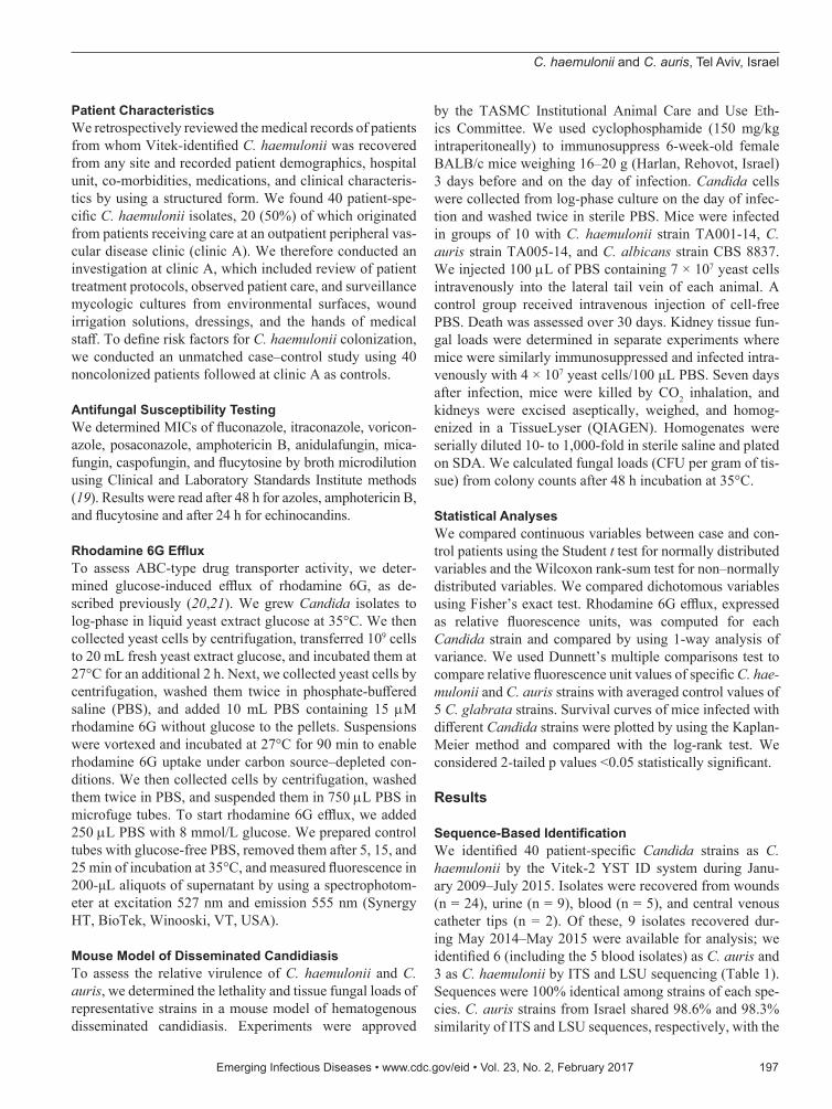
Patient CharacteristicsWe retrospectively reviewed the medical records of patients from whom Vitek-identified C. haemulonii was recovered from any site and recorded patient demographics, hospital unit, co-morbidities, medications, and clinical characteris-tics by using a structured form. We found 40 patient-spe-cific C. haemulonii isolates, 20 (50%) of which originated from patients receiving care at an outpatient peripheral vas-cular disease clinic (clinic A). We therefore conducted an investigation at clinic A, which included review of patient treatment protocols, observed patient care, and surveillance mycologic cultures from environmental surfaces, wound irrigation solutions, dressings, and the hands of medical staff. To define risk factors for C. haemulonii colonization, we conducted an unmatched case–control study using 40 noncolonized patients followed at clinic A as controls.
Antifungal Susceptibility TestingWe determined MICs of fluconazole, itraconazole, voricon-azole, posaconazole, amphotericin B, anidulafungin, mica-fungin, caspofungin, and flucytosine by broth microdilution using Clinical and Laboratory Standards Institute methods (19). Results were read after 48 h for azoles, amphotericin B, and flucytosine and after 24 h for echinocandins.
Rhodamine 6G EffluxTo assess ABC-type drug transporter activity, we deter-mined glucose-induced efflux of rhodamine 6G, as de-scribed previously (20,21). We grew Candida isolates to log-phase in liquid yeast extract glucose at 35°C. We then collected yeast cells by centrifugation, transferred 109 cells to 20 mL fresh yeast extract glucose, and incubated them at 27°C for an additional 2 h. Next, we collected yeast cells by centrifugation, washed them twice in phosphate-buffered saline (PBS), and added 10 mL PBS containing 15 µM rhodamine 6G without glucose to the pellets. Suspensions were vortexed and incubated at 27°C for 90 min to enable rhodamine 6G uptake under carbon source–depleted con-ditions. We then collected cells by centrifugation, washed them twice in PBS, and suspended them in 750 µL PBS in microfuge tubes. To start rhodamine 6G efflux, we added 250 µL PBS with 8 mmol/L glucose. We prepared control tubes with glucose-free PBS, removed them after 5, 15, and 25 min of incubation at 35°C, and measured fluorescence in 200-μL aliquots of supernatant by using a spectrophotom-eter at excitation 527 nm and emission 555 nm (Synergy HT, BioTek, Winooski, VT, USA).
Mouse Model of Disseminated CandidiasisTo assess the relative virulence of C. haemulonii and C. auris, we determined the lethality and tissue fungal loads of representative strains in a mouse model of hematogenous disseminated candidiasis. Experiments were approved
by the TASMC Institutional Animal Care and Use Eth-ics Committee. We used cyclophosphamide (150 mg/kg intraperitoneally) to immunosuppress 6-week-old female BALB/c mice weighing 16–20 g (Harlan, Rehovot, Israel) 3 days before and on the day of infection. Candida cells were collected from log-phase culture on the day of infec-tion and washed twice in sterile PBS. Mice were infected in groups of 10 with C. haemulonii strain TA001-14, C. auris strain TA005-14, and C. albicans strain CBS 8837. We injected 100 µL of PBS containing 7 × 10⁷ yeast cells intravenously into the lateral tail vein of each animal. A control group received intravenous injection of cell-free PBS. Death was assessed over 30 days. Kidney tissue fun-gal loads were determined in separate experiments where mice were similarly immunosuppressed and infected intra-venously with 4 × 10⁷ yeast cells/100 μL PBS. Seven days after infection, mice were killed by CO2 inhalation, and kidneys were excised aseptically, weighed, and homog-enized in a TissueLyser (QIAGEN). Homogenates were serially diluted 10- to 1,000-fold in sterile saline and plated on SDA. We calculated fungal loads (CFU per gram of tis-sue) from colony counts after 48 h incubation at 35°C.
Statistical AnalysesWe compared continuous variables between case and con-trol patients using the Student t test for normally distributed variables and the Wilcoxon rank-sum test for non–normally distributed variables. We compared dichotomous variables using Fisher’s exact test. Rhodamine 6G efflux, expressed as relative fluorescence units, was computed for each Candida strain and compared by using 1-way analysis of variance. We used Dunnett’s multiple comparisons test to compare relative fluorescence unit values of specific C. hae-mulonii and C. auris strains with averaged control values of 5 C. glabrata strains. Survival curves of mice infected with different Candida strains were plotted by using the Kaplan-Meier method and compared with the log-rank test. We considered 2-tailed p values <0.05 statistically significant.
Results
Sequence-Based IdentificationWe identified 40 patient-specific Candida strains as C. haemulonii by the Vitek-2 YST ID system during Janu-ary 2009–July 2015. Isolates were recovered from wounds (n = 24), urine (n = 9), blood (n = 5), and central venous catheter tips (n = 2). Of these, 9 isolates recovered dur-ing May 2014–May 2015 were available for analysis; we identified 6 (including the 5 blood isolates) as C. auris and 3 as C. haemulonii by ITS and LSU sequencing (Table 1). Sequences were 100% identical among strains of each spe-cies. C. auris strains from Israel shared 98.6% and 98.3% similarity of ITS and LSU sequences, respectively, with the
Emerging Infectious Diseases • www.cdc.gov/eid • Vol. 23, No. 2, February 2017 197

RESEARCH
C. auris type strain CBS10913T. C. haemulonii strains were 100% identical to C. haemulonii CBS5149T on the basis of ITS and LSU sequences.
Phylogenetic trees based on ITS and LSU sequences showed that the C. auris isolates from Tel Aviv are dis-tinct from other isolates from East Asia, Africa, and the Middle East. Specifically, isolates from Israel showed 98.6% similarity of ITS and LSU sequences with the India clone, represented by CBS12768, 96.2% similarity with the South Korea clone, and 96.7% similarity with strain
CH1 from Kuwait. In contrast, ITS and LSU sequences from Israel C. haemulonii strains were 100% homologous with C. haemulonii from South Korea, Brazil, and Kuwait, suggesting worldwide predominance of a single C. hae-mulonii clone (Figure 1).
Clinical FeaturesEight of 9 patients with sequence-validated isolates were hospitalized at TASMC (Table 1). An additional patient with C. auris infection was hospitalized at the Wolfson
198 Emerging Infectious Diseases • www.cdc.gov/eid • Vol. 23, No. 2, February 2017
Figure 1. Phylogenetic relationships of Candida auris and C. haemulonii strains isolated in Tel Aviv, Israel, compared with reference strains. Phylogenetic trees were generated from internal transcribed spacer (A) and D1/D2 domain of the ribosomal DNA large subunit sequences (B). The percentage of replicate trees in which the associated taxa clustered together in the bootstrap test (500 replicates) is shown next to each branch. Bold indicates strains from Tel Aviv. GenBank accession numbers are provided in parentheses. Scale bar indicates nucleotide substitutions per site.

C. haemulonii and C. auris, Tel Aviv, Israel
Medical Center, but was receiving regular care for HIV infection at TASMC. All 3 C. haemulonii isolates were re-covered from chronic leg ulcers of patients with peripheral vascular disease, 2 of whom were treated at vascular outpa-tient clinic A. Five of 6 C. auris isolates represented BSI: 3 patients had vascular catheter–related candidemia, and 2 had primary nosocomial candidemia of unclear origin. Two of 5 patients with C. auris BSI died during hospitalization.
We reviewed the medical records of 40 patients with Vitek-identified C. haemulonii cultures. Thirty-three (83%) were male. Median age was 74 years (range 37–91 years). Nineteen (48%) had peripheral vascular disease, 20 (50%) had diabetes mellitus, 22 (55%) had ischemic heart disease, and 11 (28%) had end-stage renal disease. Twenty patients (50%) were receiving regular care at clinic A, representing 8% (20/261) of all clinic patients. In all 20 patients, C. hae-mulonii had been recovered from chronic leg ulcers, and none had documented wound infection at the time of cul-ture. Cultures of environmental surfaces, medical devices, dressings, irrigation solutions, and hands of medical staff were negative for yeast. Compared with 40 control patients who were not carriers of C. haemulonii, carriers were older, had a lower glomerular filtration rate, and were more likely to be male and to have ischemic heart disease (Table 2). Observations revealed a practice among medical staff of routinely applying topical miconazole cream to chronic ul-cers without evidence of infection. Periodic wound cultures were obtained regularly, irrespective of signs of ulcer in-flammation or purulence. Multiple social interactions were noted among patients in a single room where wound care was performed.
Antifungal SusceptibilityAll C. haemulonii and C. auris isolates had fluconazole MICs >8 mg/L (range 16–64 mg/L; MIC50 32 mg/L). MICs of other azoles were also elevated: itraconazole, 0.25 to >16 mg/L (MIC50 0.5 mg/L); voriconazole, 0.25–1 mg/L (MIC50 0.5 mg/L); and posaconazole, 0.06 to >8 mg/L (MIC50 0.25 mg/L). Amphotericin B MIC ranged from 1 to 2 mg/L for C. auris isolates and from 2 to 8 mg/L for C. haemulonii isolates. All isolates appeared susceptible to
anidulafungin (MIC 0.03 mg/L) and all isolates except 1 C. haemulonii were susceptible to micafungin (MIC 0.12–0.5 mg/L; MIC50 0.12 mg/L). Caspofungin MIC was 0.5 mg/L for all isolates. All isolates except 1 C. auris were suscep-tible to flucytosine (Table 3).
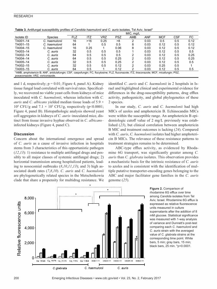
Rhodamine 6G EffluxRhodamine 6G is a substrate of ATP binding cassette (ABC) type efflux pumps responsible for multiazole resis-tance in C. glabrata. C. haemulonii and C. auris strains ex-hibited robust rhodamine 6G efflux activity when glucose (8 mM) was present in the medium, consistent with ABC-type transport. Rhodamine 6G efflux of C. auris strains was significantly greater than that of C. glabrata strains (14.4-, 10-, and 6.7-fold higher at 5, 15, and 25 min, respective-ly; p<0.0001) and C. haemulonii (3.8-, 3.8-, and 3.6-fold higher at 5, 15, and 25 min, respectively; p<0.0001). C. haemulonii showed greater rhodamine 6G efflux than C. glabrata (3.8-, 2.7-, and 1.9-fold higher at 5, 15, and 25 min, respectively (p<0.0001) (Figure 2).
ThermotoleranceSurvival and growth at physiologic temperature are prereq-uisites for microbial invasion and pathogenicity. C. haemu-lonii isolates grew well at 35°C, but growth at 37°C was poor or absent, and no growth occurred at 40°C and 42°C. In contrast, growth of C. auris isolates at 37°C and 40°C was similar to that of C. albicans, and 4 of 6 isolates grew at 42°C (Figure 3).
Virulence in a Mouse Model of Disseminated CandidiasisWe compared the virulence of C. auris and C. haemulonii isolates in a mouse model of hematogenous disseminated candidiasis. C. haemulonii was completely nonvirulent in this model; 100% of mice survived 12 days after inocula-tion with no visible signs of illness. In contrast, inoculation with C. auris resulted in rapid death and only 20% survival 5 days after infection (p = 0.0002, log-rank test). Death of mice infected with C. auris was significantly less rapid than that of mice infected with C. albicans (median survival 4 d
Emerging Infectious Diseases • www.cdc.gov/eid • Vol. 23, No. 2, February 2017 199
Table 2. Comparison of colonized and noncolonized patients with Candida haemulonii, clinic A, Tel Aviv Sourasky Medical Center, Tel Aviv, Israel*
p value Odds ratio (95% CI) Controls, n = 40 Cases, n = 20 Variable 0.015 NA 63.0 (43–94) 77.5 (44–91) Median age, y (range) 0.017 6.65 (1.26–65.0) 23 (57.5) 18 (90) Male sex 0.44 NA 48 (9–192) 40 (8–228) Median time in clinic A, mo (range) 0.022 NA 62.9 3.61 47.7 5.56 eGFR, mL/min/1.73m2, mean SEM 0.057 3.05 (0.88–11.2) 15 (37.5) 13 (65) Chronic kidney disease, stage 3–4 0.069 3.85 (0.76–21.0) 4 (10) 6 (30) Dialysis 0.003 5.5 (1.51–21.1) 10 (25) 13 (65) Ischemic heart disease 0.27 2.05 (0.59–7.37) 19 (47.5) 13 (65) Diabetes mellitus 1.00 0.80 (0.13–5.84) 35 (87.5) 17 (85) Peripheral vascular disease
*Values are no. (%) patients except as indicated. eGFR, glomerular filtration rate, estimated using the Modification of Diet in Renal Disease (MDRD) equation (22); NA, odds ratio is not applicable for continuous variables.
RESEARCH
and 1 d, respectively; p = 0.01; Figure 4, panel A). Kidney tissue fungal load correlated with survival rates. Specifical-ly, we recovered no viable yeast cells from kidneys of mice inoculated with C. haemulonii, whereas infection with C. auris and C. albicans yielded median tissue loads of 5.9 × 10⁴ CFU/g and 7.1 × 10⁵ CFU/g, respectively (p<0.0001; Figure 4, panel B). Histopathologic analysis showed yeast cell aggregates in kidneys of C. auris–inoculated mice, dis-tinct from tissue invasive hyphae observed in C. albicans–infected kidneys (Figure 4, panel C).
DiscussionConcern about the international emergence and spread of C. auris as a cause of invasive infection in hospitals stems from 3 characteristics of this opportunistic pathogen (12,13): 1) resistance to multiple antifungal drugs and pos-sibly to all major classes of systemic antifungal drugs; 2) horizontal transmission among hospitalized patients, lead-ing to nosocomial outbreaks (8,10,11,13); and 3) high as-sociated death rates (7,8,10). C. auris and C. haemulonii are phylogenetically related species in the Metschnikowia clade that share a propensity for multidrug resistance. We
identified C. auris and C. haemulonii in 2 hospitals in Is-rael and highlighted clinical and experimental evidence for differences in the drug-susceptibility patterns, drug efflux activity, pathogenicity, and global phylogenetics of these 2 species.
In our study, C. auris and C. haemulonii had high MICs of azoles and amphotericin B. Echinocandin MICs were within the susceptible range. An amphotericin B epi-demiologic cutoff value of 2 mg/L previously was estab-lished (23), but clinical correlation between amphotericin B MIC and treatment outcomes is lacking (24). Compared with C. auris, C. haemulonii isolates had higher amphoteri-cin B MICs. The relevance of these resistance patterns to treatment strategies remains to be determined.
ABC-type efflux activity, as evidenced by Rhoda-mine 6G transport, was significantly greater among C. auris than C. glabrata isolates. This observation provides a mechanistic basis for the intrinsic resistance of C. auris to azoles and is consistent with the identification of mul-tiple putative transporter-encoding genes belonging to the ABC and major facilitator gene families in the C. auris genome (25).
200 Emerging Infectious Diseases • www.cdc.gov/eid • Vol. 23, No. 2, February 2017
Table 3. Antifungal susceptibility profiles of Candida haemulonii and C. auris isolates, Tel Aviv, Israel*
Isolate ID Species MIC, mg/L
FLZ ITZ VRZ PSZ AMB ANF MCF CSF FC TA001–14 C. haemulonii 16 >16 0.25 >8 2 0.03 0.5 0.5 0.12 TA001–15 C. haemulonii 64 1 0.5 0.5 8 0.03 0.12 0.5 0.12 TA004–15 C. haemulonii 16 0.25 1 0.06 8 0.03 0.12 0.5 0.12 TA003–14 C. auris 32 0.5 0.5 0.5 1 0.03 0.12 0.5 0.5 TA002–14 C. auris 64 0.5 0.5 0.5 2 0.03 0.12 0.5 0.25 TA004–14 C. auris 64 0.5 0.5 0.25 2 0.03 0.12 0.5 0.25 TA005–14 C. auris 32 0.5 0.5 0.25 2 0.03 0.12 0.5 0.5 TA002–15 C. auris 32 0.5 0.5 0.12 2 0.03 0.25 0.5 1 TA003–15 C. auris 64 0.5 1 0.12 2 0.03 0.12 0.5 0.5 *AMB, amphotericin B; ANF, anidulafungin; CSF, caspofungin; FC, flucytosine; FLZ, fluconazole; ITZ, itraconazole; MCF, micafungin; PSZ, posaconazole; VRZ, voriconazole.
Figure 2. Comparison of rhodamine 6G efflux over time among Candida isolates from Tel Aviv, Israel. Rhodamine 6G efflux is expressed as relative fluorescence units measured in culture supernatants after the addition of 8 mM glucose. Statistical significance was measured with 1-way analysis of variance and Dunnett’s post-test comparing each C. haemulonii and C. auris strain with the averaged value of C. glabrata strains at the corresponding time point. White bars, 5 min; gray bars, 15 min; black bars, 25 min. *p<0.0001.

C. haemulonii and C. auris, Tel Aviv, Israel
Of Vitek-identified C. haemulonii isolates at TASMC, 50% were wound cultures from patients cared for at clinic A. That most of these isolates were not available for se-quencing is a limitation of our study. However, we iden-tified the 2 Candida isolates from clinic A patients that were available by ITS and LSU sequencing as C. haemu-lonii, and all 3 sequence-identified C. haemulonii isolates were recovered from leg ulcers of patients with peripheral vascular disease. Colonization of patients treated in close proximity in 1 room strongly suggests person-to-person transmission and supports interim guidelines for contact isolation (26). However, we were unable to identify an en-vironmental reservoir of C. haemulonii. We suggest that topical application of miconazole to wounds most likely caused selective pressure and facilitated the overgrowth of C. haemulonii. After this investigation and termina-tion of routine topical azole use, no additional cases of C. haemulonii were detected in clinic A during April 2015– July 2016.
We recovered 5 of 6 sequence-identified C. auris iso-lates from patients with nosocomial BSI. In contrast, all C. haemulonii isolates were cultured from superficial wounds. This observation reflects the global epidemiology of these
species. C. haemulonii has been isolated from chronic leg ulcers of patients in India and Brazil (27,28). C. auris has caused outbreaks of BSI in the United Kingdom (13), India (8), Kenya (11), South Africa (9), and South Korea (29), whereas reports of C. haemulonii as an agent of BSI have been infrequent (27,29–32). Moreover, C. auris funge-mia is associated with high death rates (8,10), contrasting with reports of patients surviving prolonged C. haemulo-nii fungemia (31). Fatal C. haemulonii fungemia, although rare, has been reported in neonates and in patients with can-cer and neutropenia (27,32).
In our study, C. auris, but not C. haemulonii, grew at 37°C–42°C and exhibited lethality and tissue invasion in a mouse model of invasive candidiasis only slightly less than those of C. albicans, the prototypical pathogenic Can-dida species. Both C. auris and C. haemulonii are unable to form hyphae, which contribute to virulence in C. albicans. Formation of large aggregates resulting from failure of budding yeast to separate has been noted in some C. auris isolates (33). We observed distinct yeast cell aggregates in the kidneys of mice with lethal C. auris infection, which suggests that aggregation might be a mode of immune evasion and persistence in tissue. The C. auris genome
Emerging Infectious Diseases • www.cdc.gov/eid • Vol. 23, No. 2, February 2017 201
Figure 3. Differing thermotolerance of Candida auris and C. haemulonii. A) Sabouraud dextrose agar plates showing growth of representative Candida strains after 24 h incubation at 35°C–42°C; B) Thermal growth range of Candida isolates from Tel Aviv, Israel.

RESEARCH
contains C. albicans gene orthologs, such as secreted pro-teinases and mannosyl transferases, which might have roles in pathogenesis (25). However, C. auris has a genome that is highly divergent from those of other Candida species, and most of its genes have not yet been characterized (25).
Ribosomal DNA sequences were identical among C. haemulonii strains from Israel, Kuwait, East Asia, South America, and the United States. In contrast, the global phy-logenetics of C. auris demonstrate distinct clones for each country, indicating greater genomic diversity for this spe-cies. Further study is needed to establish whether the di-vergence of C. auris clones translates into country-specific patterns of invasiveness, virulence, and drug resistance. Our findings affirm the need for intensified vigilance and mobilization of infection control measures to limit the spread of C. auris.
J.B. was supported by the European Research Council Advanced Award 340087 (RAPLODAPT).
Dr. Ben-Ami is head of the Infectious Diseases Unit and the National Center for Clinical Mycology at the Tel Aviv Medical Center, Israel. His research focuses on the epidemiology, drug resistance traits, and virulence determinants of medically important fungi.
References 1. Magill SS, Edwards JR, Bamberg W, Beldavs ZG, Dumyati G,
Kainer MA, et al.; Emerging Infections Program Healthcare- Associated Infections and Antimicrobial Use Prevalence Survey Team. Multistate point-prevalence survey of health care- associated infections. N Engl J Med. 2014;370:1198–208. http://dx.doi.org/10.1056/NEJMoa1306801
2. Kullberg BJ, Arendrup MC. Invasive candidiasis. N Engl J Med. 2015;373:1445–56. http://dx.doi.org/10.1056/ NEJMra1315399
3. Garey KW, Rege M, Pai MP, Mingo DE, Suda KJ, Turpin RS, et al. Time to initiation of fluconazole therapy impacts mortality in patients with candidemia: a multi-institutional study. Clin Infect Dis. 2006;43:25–31. http://dx.doi.org/10.1086/504810
4. Andes DR, Safdar N, Baddley JW, Playford G, Reboli AC, Rex JH, et al.; Mycoses Study Group. Impact of treatment strategy on outcomes in patients with candidemia and other forms of invasive candidiasis: a patient-level quantitative review of randomized trials. Clin Infect Dis. 2012;54:1110–22. http://dx.doi.org/10.1093/cid/cis021
5. Alexander BD, Johnson MD, Pfeiffer CD, Jiménez-Ortigosa C, Catania J, Booker R, et al. Increasing echinocandin resistance in Candida glabrata: clinical failure correlates with presence of FKS mutations and elevated minimum inhibitory concentrations. Clin Infect Dis. 2013;56:1724–32. http://dx.doi.org/10.1093/cid/cit136
6. Satoh K, Makimura K, Hasumi Y, Nishiyama Y, Uchida K, Yamaguchi H. Candida auris sp. nov., a novel ascomycetous yeast isolated from the external ear canal of an inpatient in a Japanese hospital. Microbiol Immunol. 2009;53:41–4. http://dx.doi.org/10.1111/j.1348-0421.2008.00083.x
202 Emerging Infectious Diseases • www.cdc.gov/eid • Vol. 23, No. 2, February 2017
Figure 4. Differing virulence of Candida auris and C. haemulonii assessed in a mouse model of hematogenous disseminated candidiasis. Virulence was assessed in immunosuppressed BALB/c mice after intravenous injection of yeast cell suspension. A) Survival curves showing significantly shorter survival of mice infected with C. albicans than C. auris and no death among mice infected with C. haemulonii. B) Kidney fungal load (CFU per gram of tissue) shown to be significantly higher in mice infected with C. albicans than in those infected with C. auris, whereas no viable yeast was cultured from kidneys of mice infected with C. haemulonii. C) In mouse kidneys, C. auris cells formed aggregates and no hyphae (top) whereas C. albicans formed extensive tissue-invasive hyphae (bottom); C. haemulonii was not detected in tissue sections (middle). Grocott methenamine silver staining, original magnification ×100 for panels, ×400 for insets.

C. haemulonii and C. auris, Tel Aviv, Israel
7. Lee WG, Shin JH, Uh Y, Kang MG, Kim SH, Park KH, et al. First three reported cases of nosocomial fungemia caused by Candida auris. J Clin Microbiol. 2011;49:3139–42. http://dx.doi.org/10.1128/JCM.00319-11
8. Chowdhary A, Sharma C, Duggal S, Agarwal K, Prakash A, Singh PK, et al. New clonal strain of Candida auris, Delhi, India. Emerg Infect Dis. 2013;19:1670–3. http://dx.doi.org/10.3201/eid1910.130393
9. Magobo RE, Corcoran C, Seetharam S, Govender NP. Candida auris–associated candidemia, South Africa. Emerg Infect Dis. 2014;20:1250–1. http://dx.doi.org/10.3201/eid2007.131765
10. Chowdhary A, Anil Kumar V, Sharma C, Prakash A, Agarwal K, Babu R, et al. Multidrug-resistant endemic clonal strain of Candida auris in India. Eur J Clin Microbiol Infect Dis. 2014;33:919–26. http://dx.doi.org/10.1007/s10096-013-2027-1
11. Okinda N, Kagotho E, Castanheira M, Njuguna A, Omuse G, Makau P, et al. Candidemia at a referral hospital in sub-Saharan Africa: emergence of Candida auris as a major pathogen. European Conference on Clinical Microbiology and Infectious Diseases; 2014 May 10–13; Barcelona, Spain.
12. Centers for Disease Control and Prevention. Global emergence of invasive infections caused by the multidrug-resistant yeast Candida auris [cited 2016 Jul 29]. http://www.cdc.gov/fungal/ diseases/candidiasis/candida-auris-alert.html
13. Public Health England. Candida auris identified in England [cited 2016 Jul 1]. https://www.gov.uk/government/publications/candida-auris-emergence-in-england/candida-auris-identified-in-england
14. Schoch CL, Seifert KA, Huhndorf S, Robert V, Spouge JL, Levesque CA, et al.; Fungal Barcoding Consortium. Nuclear ribosomal internal transcribed spacer (ITS) region as a universal DNA barcode marker for fungi. Proc Natl Acad Sci U S A. 2012;109:6241–6. http://dx.doi.org/10.1073/pnas.1117018109
15. Edgar RC. MUSCLE: multiple sequence alignment with high accuracy and high throughput. Nucleic Acids Res. 2004;32:1792–7. http://dx.doi.org/10.1093/nar/gkh340
16. Saitou N, Nei M. The neighbor-joining method: a new method for reconstructing phylogenetic trees. Mol Biol Evol. 1987;4:406–25.
17. Kimura M. A simple method for estimating evolutionary rates of base substitutions through comparative studies of nucleotide sequences. J Mol Evol. 1980;16:111–20. http://dx.doi.org/10.1007/BF01731581
18. Kumar S, Stecher G, Tamura K. MEGA7: Molecular Evolutionary Genetics Analysis version 7.0 for bigger datasets. Mol Biol Evol. 2016;33:1870–4. http://dx.doi.org/10.1093/molbev/msw054
19. Clinical and Laboratory Standards Institute. Reference method for broth dilution antifungal susceptibility testing of yeasts. Approved standard M27-A3. Wayne (PA): The Institute; 2008.
20. Maesaki S, Marichal P, Vanden Bossche H, Sanglard D, Kohno S. Rhodamine 6G efflux for the detection of CDR1-overexpressing azole-resistant Candida albicans strains. J Antimicrob Chemother. 1999;44:27–31. http://dx.doi.org/10.1093/jac/44.1.27
21. Kolaczkowski M, van der Rest M, Cybularz-Kolaczkowska A, Soumillion JP, Konings WN, Goffeau A. Anticancer drugs, ionophoric peptides, and steroids as substrates of the yeast multidrug transporter Pdr5p. J Biol Chem. 1996;271:31543–8. http://dx.doi.org/10.1074/jbc.271.49.31543
22. Levey AS, Coresh J, Greene T, Stevens LA, Zhang YL, Hendriksen S, et al.; Chronic Kidney Disease Epidemiology Collaboration. Using standardized serum creatinine values in the
modification of diet in renal disease study equation for estimating glomerular filtration rate. Ann Intern Med. 2006;145:247–54. http://dx.doi.org/10.7326/0003-4819-145-4-200608150-00004
23. Pfaller MA, Espinel-Ingroff A, Canton E, Castanheira M, Cuenca-Estrella M, Diekema DJ, et al. Wild-type MIC distributions and epidemiological cutoff values for amphotericin B, flucytosine, and itraconazole and Candida spp. as determined by CLSI broth microdilution. J Clin Microbiol. 2012;50:2040–6. http://dx.doi.org/10.1128/JCM.00248-12
24. Park BJ, Arthington-Skaggs BA, Hajjeh RA, Iqbal N, Ciblak MA, Lee-Yang W, et al. Evaluation of amphotericin B interpretive breakpoints for Candida bloodstream isolates by correlation with therapeutic outcome. Antimicrob Agents Chemother. 2006; 50:1287–92. http://dx.doi.org/10.1128/AAC.50.4.1287-1292.2006
25. Chatterjee S, Alampalli SV, Nageshan RK, Chettiar ST, Joshi S, Tatu US. Draft genome of a commonly misdiagnosed multidrug resistant pathogen Candida auris. BMC Genomics. 2015;16:686. http://dx.doi.org/10.1186/s12864-015-1863-z
26. Public Health England. Guidance for the laboratory investigation, management and infection prevention and control for cases of Candida auris [cited 2016 Jul 1]. https://www.gov.uk/government/uploads/system/uploads/attachment_data/file/534174/Guidance_Candida__auris.pdf
27. de Almeida JN Jr, Assy JG, Levin AS, Del Negro GM, Giudice MC, Tringoni MP, et al. Candida haemulonii complex species, Brazil, January 2010–March 2015. Emerg Infect Dis. 2016;22:561–3. http://dx.doi.org/10.3201/eid2203.151610
28. Kumar A, Prakash A, Singh A, Kumar H, Hagen F, Meis JF, et al. Candida haemulonii species complex: an emerging species in India and its genetic diversity assessed with multilocus sequence and amplified fragment-length polymorphism analyses. Emerg Microbes Infect. 2016;5:e49. http://dx.doi.org/10.1038/emi.2016.49
29. Kim MN, Shin JH, Sung H, Lee K, Kim EC, Ryoo N, et al. Candida haemulonii and closely related species at 5 university hospitals in Korea: identification, antifungal susceptibility, and clinical features. Clin Infect Dis. 2009;48:e57–61. http://dx.doi.org/10.1086/597108
30. Ramos LS, Figueiredo-Carvalho MH, Barbedo LS, Ziccardi M, Chaves AL, Zancopé-Oliveira RM, et al. Candida haemulonii complex: species identification and antifungal susceptibility profiles of clinical isolates from Brazil. J Antimicrob Chemother. 2015;70:111–5. http://dx.doi.org/10.1093/jac/dku321
31. Ruan SY, Kuo YW, Huang CT, Hsiue HC, Hsueh PR. Infections due to Candida haemulonii: species identification, antifungal susceptibility and outcomes. Int J Antimicrob Agents. 2010;35:85–8. http://dx.doi.org/10.1016/j.ijantimicag.2009.08.009
32. Khan ZU, Al-Sweih NA, Ahmad S, Al-Kazemi N, Khan S, Joseph L, et al. Outbreak of fungemia among neonates caused by Candida haemulonii resistant to amphotericin B, itraconazole, and fluconazole. J Clin Microbiol. 2007;45:2025–7. http://dx.doi.org/10.1128/JCM.00222-07
33. Borman AM, Szekely A, Johnson EM, Mitchell AP. Comparative pathogenicity of United Kingdom isolates of the emerging pathogen Candida auris and other key pathogenic Candida species. mSphere. 2016;1.pii:e00189-16.
Address for correspondence: Ronen Ben-Ami, Infectious Diseases Unit, Tel Aviv Sourasky Medical Center, 6 Weizmann St, Tel Aviv 64239, Israel; email: [email protected]
Emerging Infectious Diseases • www.cdc.gov/eid • Vol. 23, No. 2, February 2017 203