multiple neurovascular variations in a single cadaver report eissn 1308-4038 international journal...
TRANSCRIPT
Case Report
International Journal of Anatomical Variations (2012) 5: 81–84eISSN 1308-4038
Multiple neurovascular variations in a single cadaver
IntroductionThe knowledge of multiple variations of peripheral vessels and nerves has paramount importance in surgery and regional anesthesia. In the present case these variations were present in the superior and inferior extremities, thorax and abdomen. According to normal anatomy, the cephalic vein ascends on the lateral side of the biceps brachii muscle and drains into axillary vein after piercing the clavipectoral fascia. Medially, the basilic vein pierces the deep fascia at the lower border of the teres major, joins with the venae comitantes of the brachial artery to form the axillary vein [1].Deep artery of thigh arises from the lateral side of the femoral artery about 1.5–4 cm below the inguinal ligament and gives off the medial and lateral circumflex femoral arteries [1]. Sural nerve supplies the lateral side of dorsum of the foot and little toe, it communicates with the superficial peroneal nerve [2].In most of the cases azygos vein is situated on the right side of vertebral column. Hemiazygos and accessory azygos veins cross the vertebral column at the level of T7 and T8 vertebrae, respectively to end into the former [2].The lumbar plexus is formed in the psoas major muscle from the anterior rami of the upper four lumbar nerves.
The iliohypogastric and ilioinguinal (L1) nerves enter the abdominal wall from this plexus [1].At the porta hepatis, hepatic artery proper (arising from the common hepatic artery) divides into right and left branches before they run into the parenchyma of the liver [2].The renal veins open into the inferior vena cava almost at right angles [2].
Case ReportNeurovascular variations were found during routine dissection of a 65-year-old male cadaver in the Department of Anatomy, Calcutta National Medical College. Courses and branches of peripheral vessels and nerves were traced carefully. The psoas major muscle was retracted medially to expose the nerve roots of lumbar plexus. Different structures were identified and photographs were taken. Liver was dissected out along with all the structures of the porta hepatis. Saline solution mixed with anticoagulant was injected through different vessels at the porta hepatis and rinsed thoroughly. A radio-opaque dye was injected through the extra hepatic artery (after clamping other vessels) and radiograph was taken.
Bhattacharya SANTANU [1]
Majumdar SUDESHNA [1]
Chatterjee ARPITA [1]
Mazumdar ARDHENDU [2]
Department of Anatomy, Calcutta National Medical College [1], Department of Physiology, Institute of Prost Graduate Medical Education and Research (IPGMER) [2], Kolkata, INDIA.
Dr. Majumdar Sudeshna Associate Professor Department of Anatomy Calcutta National Medical College Kolkata, INDIA. +91 (943) 3007363 [email protected]
Received September 7th, 2011; accepted August 12th, 2012
AbstractNeurovascular variations are of interest to anatomists, anesthesiologists and surgeons. During routine dissection, multiple neurovascular variations were found in a 65-year-old male cadaver. In the left lower limb, medial circumflex femoral artery arose from the femoral artery; the sural nerve gave a communicating branch to superficial peroneal nerve. In abdomen an accessory hepatic artery supplied the right lobe of liver; the left kidney was drained by double renal veins. Ilioinguinal nerve arose from L2 instead of its normal origin from L1. Hemiazygos and accessory azygos veins formed a continuous venous line in thorax, which drained into the azygos vein by a single vein. In the both upper limbs axillary veins were formed at a higher level than usual with a single venous channel on the left side.
© Int J Anat Var (IJAV). 2012; 5: 81–84.
Key words [medial circumflex femoral artery] [hepatic artery] [renal vein] [veins of upper limb and thorax] [ilioinguinal nerve]
Published online November 9th, 2012 © http://www.ijav.org
82 Santanu et al.
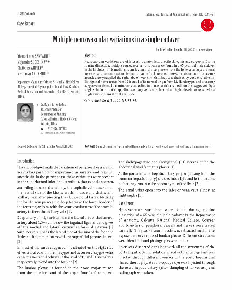
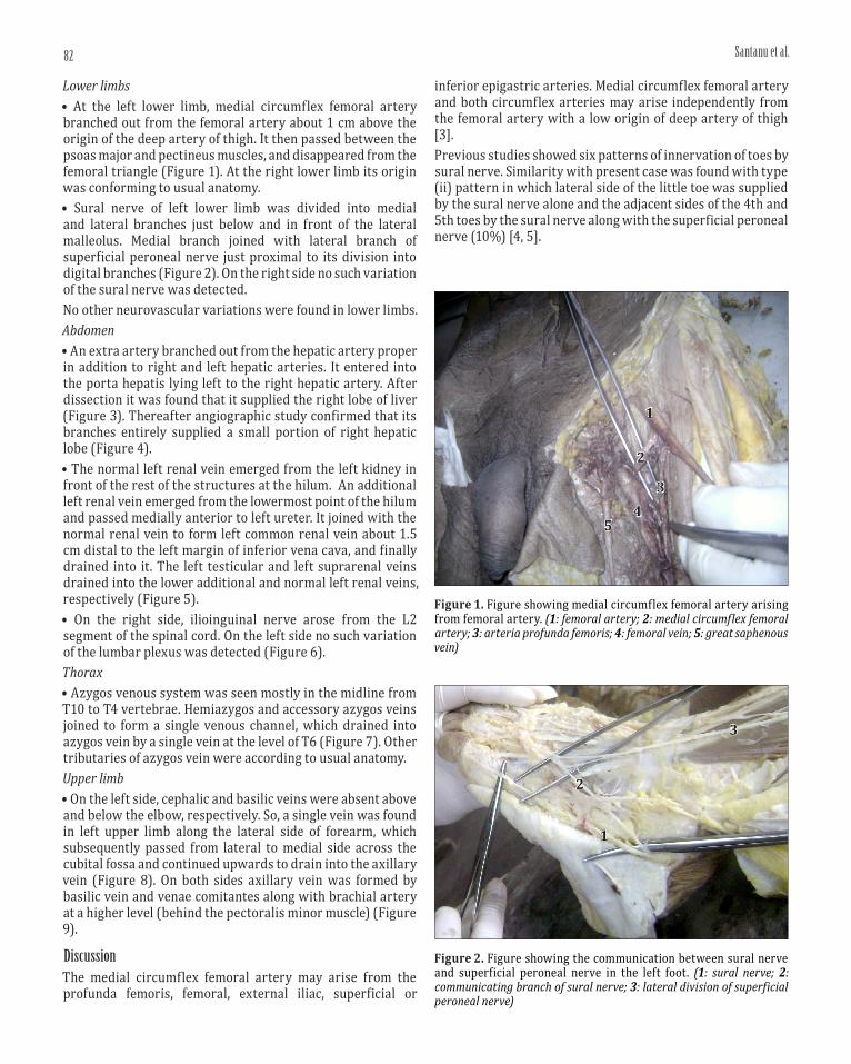
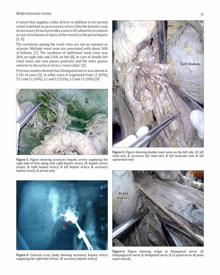
Lower limbs• At the left lower limb, medial circumflex femoral artery branched out from the femoral artery about 1 cm above the origin of the deep artery of thigh. It then passed between the psoas major and pectineus muscles, and disappeared from the femoral triangle (Figure 1). At the right lower limb its origin was conforming to usual anatomy. • Sural nerve of left lower limb was divided into medial and lateral branches just below and in front of the lateral malleolus. Medial branch joined with lateral branch of superficial peroneal nerve just proximal to its division into digital branches (Figure 2). On the right side no such variation of the sural nerve was detected.No other neurovascular variations were found in lower limbs. Abdomen• An extra artery branched out from the hepatic artery proper in addition to right and left hepatic arteries. It entered into the porta hepatis lying left to the right hepatic artery. After dissection it was found that it supplied the right lobe of liver (Figure 3). Thereafter angiographic study confirmed that its branches entirely supplied a small portion of right hepatic lobe (Figure 4).• The normal left renal vein emerged from the left kidney in front of the rest of the structures at the hilum. An additional left renal vein emerged from the lowermost point of the hilum and passed medially anterior to left ureter. It joined with the normal renal vein to form left common renal vein about 1.5 cm distal to the left margin of inferior vena cava, and finally drained into it. The left testicular and left suprarenal veins drained into the lower additional and normal left renal veins, respectively (Figure 5).• On the right side, ilioinguinal nerve arose from the L2 segment of the spinal cord. On the left side no such variation of the lumbar plexus was detected (Figure 6).Thorax• Azygos venous system was seen mostly in the midline from T10 to T4 vertebrae. Hemiazygos and accessory azygos veins joined to form a single venous channel, which drained into azygos vein by a single vein at the level of T6 (Figure 7). Other tributaries of azygos vein were according to usual anatomy.Upper limb• On the left side, cephalic and basilic veins were absent above and below the elbow, respectively. So, a single vein was found in left upper limb along the lateral side of forearm, which subsequently passed from lateral to medial side across the cubital fossa and continued upwards to drain into the axillary vein (Figure 8). On both sides axillary vein was formed by basilic vein and venae comitantes along with brachial artery at a higher level (behind the pectoralis minor muscle) (Figure 9).
DiscussionThe medial circumflex femoral artery may arise from the profunda femoris, femoral, external iliac, superficial or
1
2
34
5
Figure 1. Figure showing medial circumflex femoral artery arising from femoral artery. (1: femoral artery; 2: medial circumflex femoral artery; 3: arteria profunda femoris; 4: femoral vein; 5: great saphenous vein)
1
2
3
Figure 2. Figure showing the communication between sural nerve and superficial peroneal nerve in the left foot. (1: sural nerve; 2: communicating branch of sural nerve; 3: lateral division of superficial peroneal nerve)
inferior epigastric arteries. Medial circumflex femoral artery and both circumflex arteries may arise independently from the femoral artery with a low origin of deep artery of thigh [3]. Previous studies showed six patterns of innervation of toes by sural nerve. Similarity with present case was found with type (ii) pattern in which lateral side of the little toe was supplied by the sural nerve alone and the adjacent sides of the 4th and 5th toes by the sural nerve along with the superficial peroneal nerve (10%) [4, 5].
83Multiple neurovascular variations
1Left Kidney
2
3
4
Figure 5. Figure showing double renal veins on the left side. (1: left renal vein; 2: accessory left renal vein; 3: left testicular vein; 4: left suprarenal vein)
12
3
45
Figure 3. Figure showing accessory hepatic artery supplying the right lobe of liver along with right hepatic artery. (1: hepatic artery proper; 2: right hepatic artery; 3: left hepatic artery; 4: accessory hepatic artery; 5: portal vein)
1
Figure 4. Contrast x-ray study showing accessory hepatic artery supplying the right lobe of liver. (1: accessory hepatic artery)
1
2
3
4
Figure 6. Figure showing origin of ilioinguinal nerve. (1: iliohypogastric nerve; 2: ilioinguinal nerve; 3: L2 spinal nerve; 4: psoas major muscle)
Right Kidney
A vessel that supplies a lobe of liver in addition to its normal vessel is defined as an accessory artery (like the present case). An accessory branch provides a source of collateral circulation in case of occlusion or injury of the vessels in the porta hepatis [2, 6]. The variations among the renal veins are not as common as arteries. Multiple renal veins are associated with about 14% of kidneys [7]. The incidence of additional renal veins was 26% on right side and 2.6% on left [8]. In case of double left renal veins, one vein passes posterior and the other passes anterior to the aorta to form a ‘renal collar’ [2]. Previous studies showed that ilioinguinal nerve was absent in 2.5% of cases [3]. In other cases it originated from L1 (65%), T12 and L1 (14%), L1 and L2 (11%), L2 and L3 (10%) [9].

84 Santanu et al.
References
[1] Snell RS. Clinical Anatomy. 8th Ed., New Delhi, Lippincott, Williams and Wilkins. 2008; 278–279, 468–469, 581–582.
[2] Standring S, Gatzoulis MA, Collins P, Michael A, Shah P, Goldstraw P, Crossman AR, Johnson D, Mahadevan V, Healy JC, Borley NR, Wigley C, eds. Gray’s Anatomy, The Anatomical Basis of Clinical Practice. 40th Ed., Spain, Churchill Livingstone Elsevier. 2008; 939–940, 1073–1074, 1163–1175, 1225–1244, 1379–1380, 1427.
[3] Bergman RA, Afifi AK, Miyauchi R. Illustrated Encyclopedia of Human Anatomic Variations. http://www.anatomyatlases.org/AnatomicVariants/Cardiovascular/Directory/Region/ArteriesLowerLimb.shtml; http://www.anatomyatlases.org/AnatomicVariants/Cardiovascular/Directory/Region/VeinsUpperLimb.shtml; http://www.anatomyatlases.org/AnatomicVariants/NervousSystem/Regions/PlexusesNerves.shtml (accessed August 2011)
[4] Madhavi C, Isaac B, Antoniswamy B, Holla SJ. Anatomical variations of the cutaneous innervation patterns of the sural nerve on the dorsum of the foot. Clin Anat. 2005; 18: 206–209.
[5] Sankar DK, Bhanu SP, Susan PJ, Gajendra K. Variant formation of sural nerve and its distribution at the dorsum of the foot. Int J Anat Var (IJAV). 2009; 2: 33–34.
[6] Todo S, Makowka L, Tzakis AG, Marsh JW Jr, Karrer FM, Armany M, Miller C, Tallent MB, Esquivel CO, Gordon RD, Iwatsuki S, Starzl TE. Hepatic artery in liver transplantation. Transplant Proc. 1987; 19: 2406-2411.
[7] Pick JW, Anson BJ. The renal vascular pedicles. An anatomical study of 430 body-halves. J Urol. 1940; 44: 411–434.
[8] Satyapal KS, Rambiritch V, Pillai G. Additional renal veins: incidence and morphometry. Clin Anat. 1995; 8: 5–55.
[9] Klaassen Z, Marshall E, Tubbs RS, Louis RG Jr, Wartmann CT, Loukas M. Anatomy of the ilionguinal and ilihypogastric nerves with observation of their spinal nerve contributions. Clin Anat. 2011; 24: 454–461.
[10] Seib GA. The azygos system of veins in American whites and American Negroes, including observations on the inferior caval venous system. Am J Phys Anthropol. 1934; 19: 39–163.
1
2
34
Figure 7. Figure showing variant azygos system of veins. (1: hemiazygos vein; 2: accessory azygos vein; 3: azygos vein; 4: single communicating vein)
12
3
4
5
Figure 9. Figure showing different veins of right upper limb. (1: cephalic vein; 2: axillary vein; 3: basilic vein; 4: venae comitantes of brachial artery; 5: pectoralis minor muscle)
1
2
3
Figure 8. Figure showing cephalic vein and its course in the left side. (1: cephalic vein; 2: crossing of cephalic vein from lateral to medial side; 3: pectoralis minor muscle)
The accessory azygos vein may drain into the left brachiocephalic, hemiazygos or azygos vein. It can form a continuous channel along with left superior intercostal and hemiazygos veins [10]. Very occasionally independent left and right azygos veins may persist [2]. Absence of cephalic vein was found in 3% of male and 1% of female subjects. The delto-pectoral part of the cephalic vein may also be small or absent. With absence of cephalic vein, median antebrachial vein is usually enlarged to drain the area of cephalic vein [3]. In the present case, not only deltopectoral part of cephalic vein was absent but the basilic vein was also absent below the elbow in the left upper limb. It is an extremely rare finding.The findings of the previous studies having similarities with the present case are discussed here.
AcknowledgementWe express our heartiest gratitude to Professor Rita Roy, Dr. Hironmoy Roy, members of the Department of Anatomy, Calcutta National Medical College, Kolkata, for their cordial help to complete this case report.