muscular system ans 215 physiology and anatomy of domesticated animals · 2003-01-22 · muscular...
TRANSCRIPT
Muscular SystemANS 215
Physiology and Anatomy ofDomesticated Animals
I. Primary Functions (45 – 50% of body mass)A. Movement
1. Blood2. Ingesta/excreta3. Gametes/conceptus
B. SupportC. Generation of body heat
II. ClassificationsA. Smooth
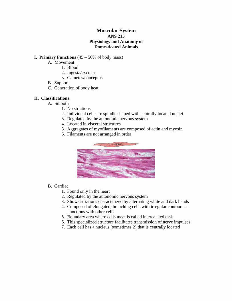
1. No striations2. Individual cells are spindle shaped with centrally located nuclei3. Regulated by the autonomic nervous system4. Located in visceral structures5. Aggregates of myofilaments are composed of actin and myosin6. Filaments are not arranged in order
B. Cardiac1. Found only in the heart2. Regulated by the autonomic nervous system3. Shows striations characterized by alternating white and dark bands4. Composed of elongated, branching cells with irregular contours at
junctions with other cells5. Boundary area where cells meet is called intercalated disk6. This specialized structure facilitates transmission of nerve impulses7. Each cell has a nucleus (sometimes 2) that is centrally located
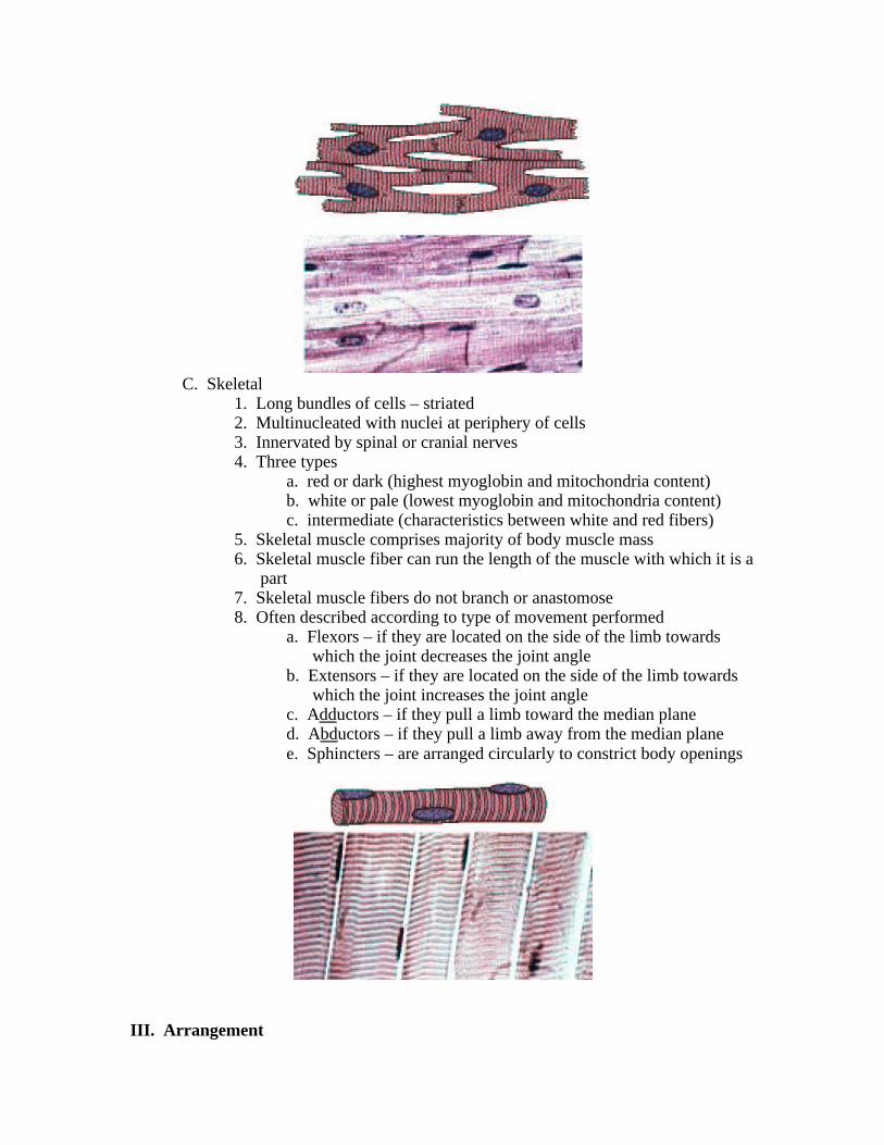
C. Skeletal1. Long bundles of cells – striated2. Multinucleated with nuclei at periphery of cells3. Innervated by spinal or cranial nerves4. Three types
a. red or dark (highest myoglobin and mitochondria content)b. white or pale (lowest myoglobin and mitochondria content)c. intermediate (characteristics between white and red fibers)
5. Skeletal muscle comprises majority of body muscle mass6. Skeletal muscle fiber can run the length of the muscle with which it is a
part7. Skeletal muscle fibers do not branch or anastomose8. Often described according to type of movement performed
a. Flexors – if they are located on the side of the limb towards which the joint decreases the joint angle
b. Extensors – if they are located on the side of the limb towardswhich the joint increases the joint angle
c. Adductors – if they pull a limb toward the median planed. Abductors – if they pull a limb away from the median planee. Sphincters – are arranged circularly to constrict body openings
III. Arrangement
A. Function of muscles is to contract or shorten and thereby move an objectB. Primary consideration for accomplishing this goal is arrangementC. Examples include:
1. Sheets2. Sheets rolled into tubes3. Bundles4. Rings5. Cones6. Discrete cells or clusters of cells
D. The effects of skeletal muscles (apart from sphincters) are noted somedistance from their location
1. This means the contraction must be transmitted2. One end must be anchored and the other attached directly to a tendon
or moveable part3. Accordingly, anatomic description of a skeletal muscle sometimes
refers to its origin and insertion
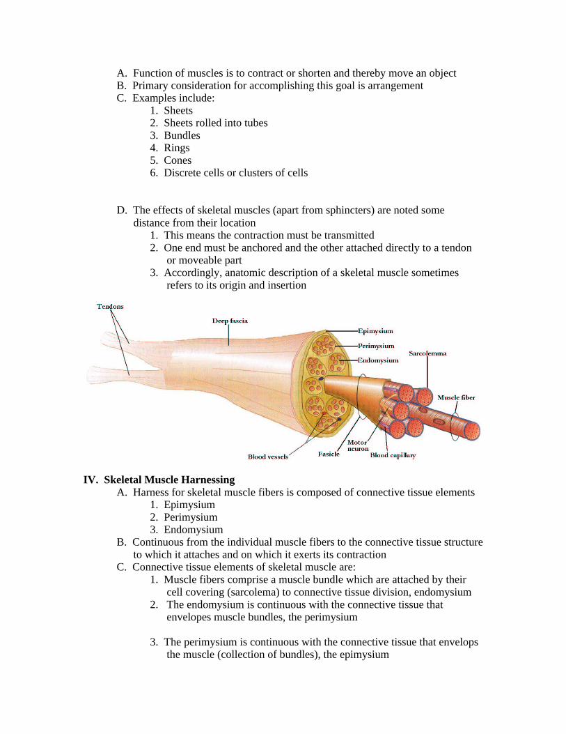
IV. Skeletal Muscle HarnessingA. Harness for skeletal muscle fibers is composed of connective tissue elements
1. Epimysium2. Perimysium3. Endomysium
B. Continuous from the individual muscle fibers to the connective tissue structureto which it attaches and on which it exerts its contraction
C. Connective tissue elements of skeletal muscle are:1. Muscle fibers comprise a muscle bundle which are attached by their
cell covering (sarcolema) to connective tissue division, endomysium2. The endomysium is continuous with the connective tissue that
envelopes muscle bundles, the perimysium
3. The perimysium is continuous with the connective tissue that envelops the muscle (collection of bundles), the epimysium
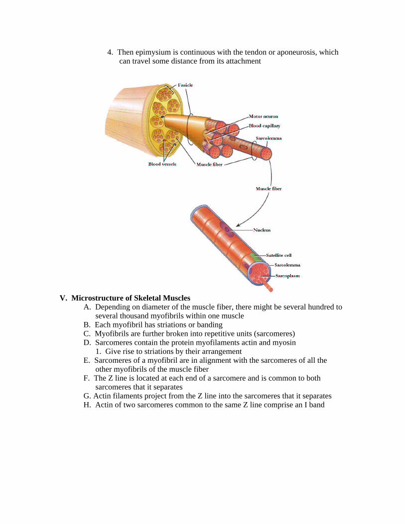
4. Then epimysium is continuous with the tendon or aponeurosis, which can travel some distance from its attachment
V. Microstructure of Skeletal MusclesA. Depending on diameter of the muscle fiber, there might be several hundred to
several thousand myofibrils within one muscleB. Each myofibril has striations or bandingC. Myofibrils are further broken into repetitive units (sarcomeres)D. Sarcomeres contain the protein myofilaments actin and myosin
1. Give rise to striations by their arrangementE. Sarcomeres of a myofibril are in alignment with the sarcomeres of all the
other myofibrils of the muscle fiberF. The Z line is located at each end of a sarcomere and is common to both
sarcomeres that it separatesG. Actin filaments project from the Z line into the sarcomeres that it separatesH. Actin of two sarcomeres common to the same Z line comprise an I band
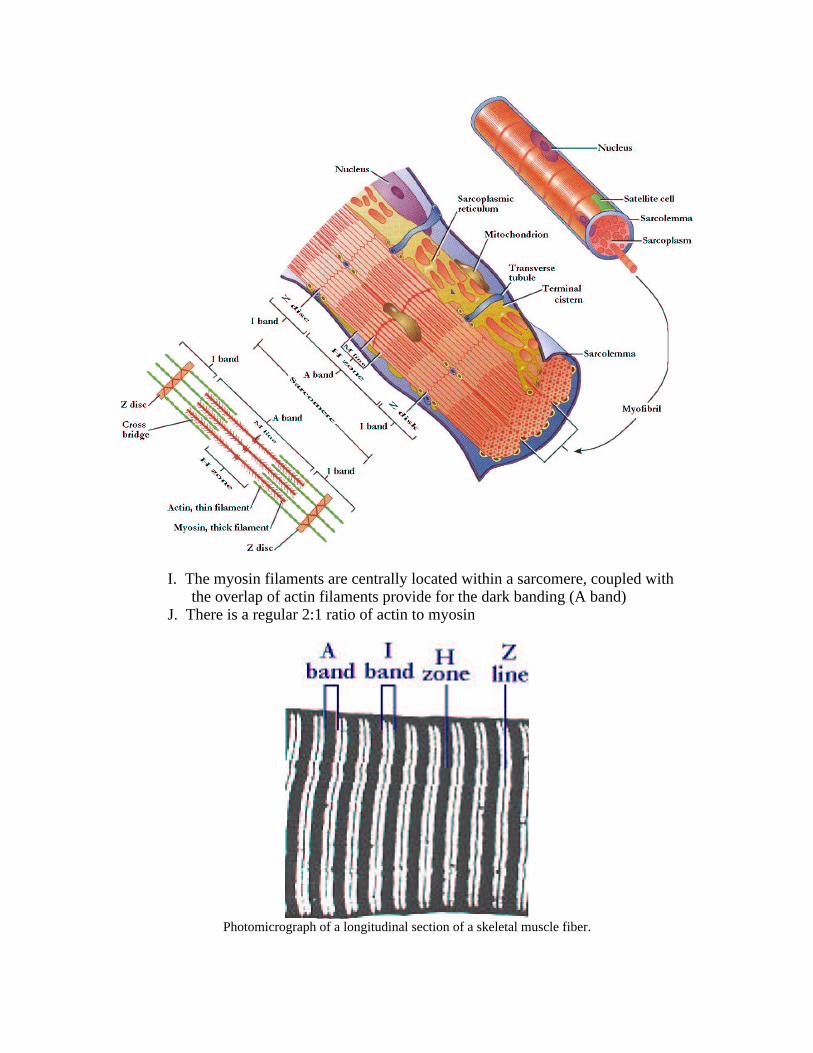
I. The myosin filaments are centrally located within a sarcomere, coupled with the overlap of actin filaments provide for the dark banding (A band)
J. There is a regular 2:1 ratio of actin to myosin
Photomicrograph of a longitudinal section of a skeletal muscle fiber.
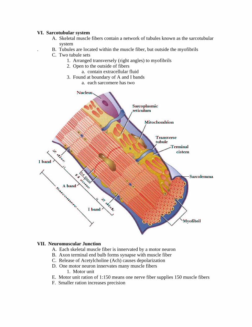
VI. Sarcotubular systemA. Skeletal muscle fibers contain a network of tubules known as the sarcotubular
system. B. Tubules are located within the muscle fiber, but outside the myofibrils
C. Two tubule sets1. Arranged transversely (right angles) to myofibrils2. Open to the outside of fibers
a. contain extracellular fluid3. Found at boundary of A and I bands
a. each sarcomere has two
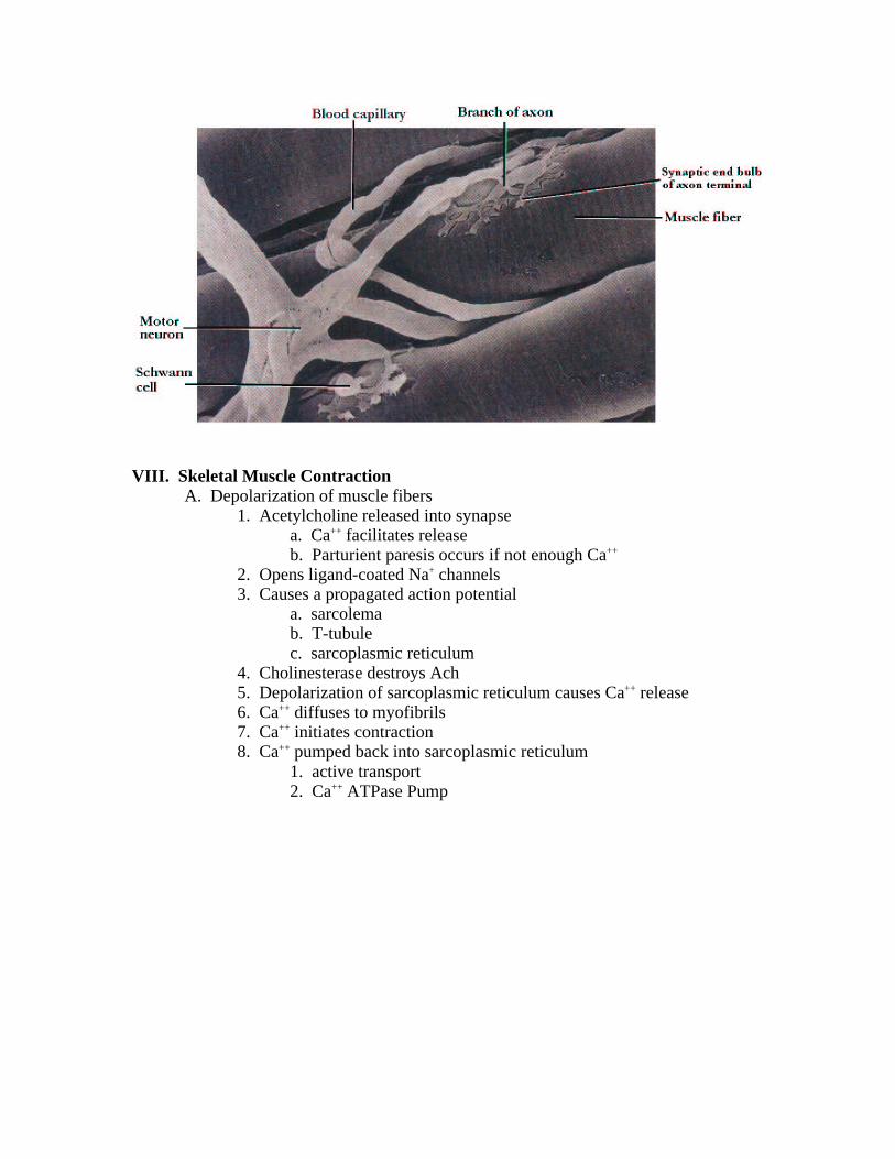
VII. Neuromuscular JunctionA. Each skeletal muscle fiber is innervated by a motor neuronB. Axon terminal end bulb forms synapse with muscle fiberC. Release of Acetylcholine (Ach) causes depolarizationD. One motor neuron innervates many muscle fibers
1. Motor unitE. Motor unit ration of 1:150 means one nerve fiber supplies 150 muscle fibersF. Smaller ration increases precision
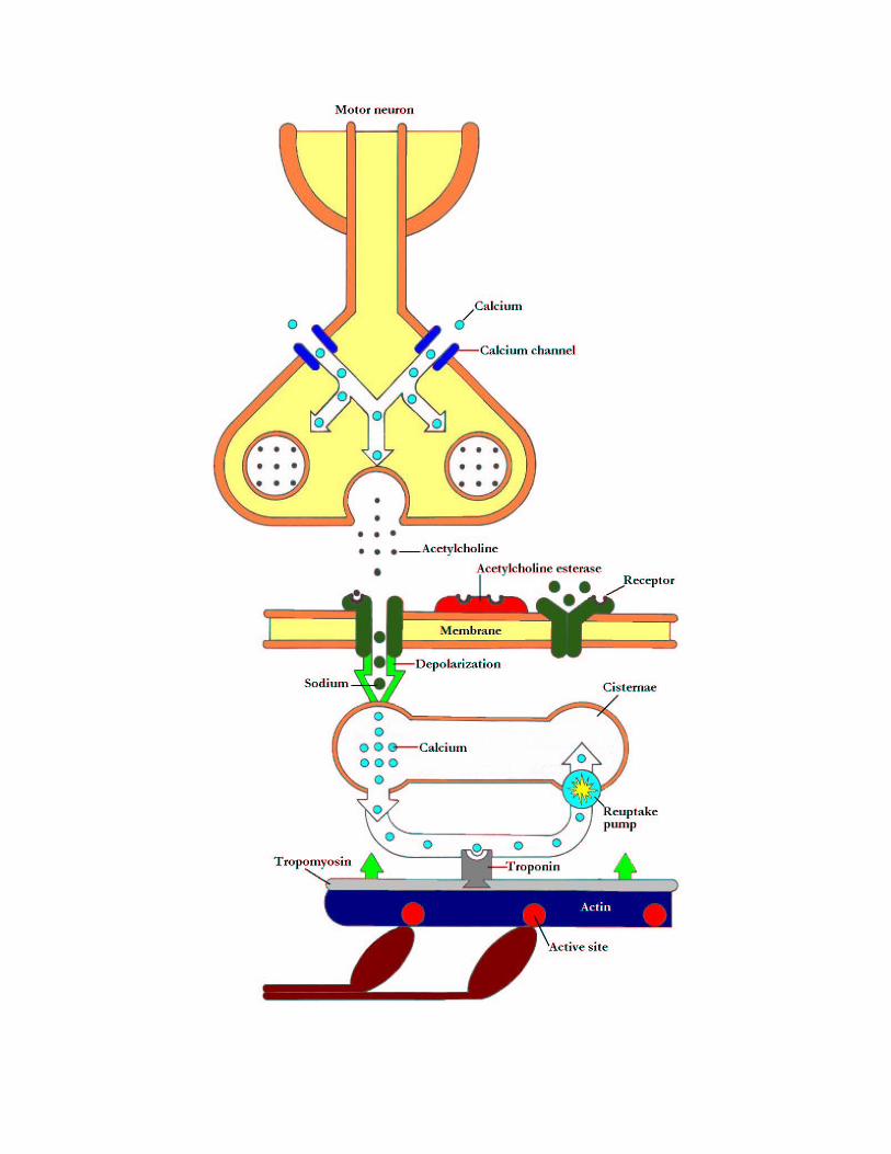
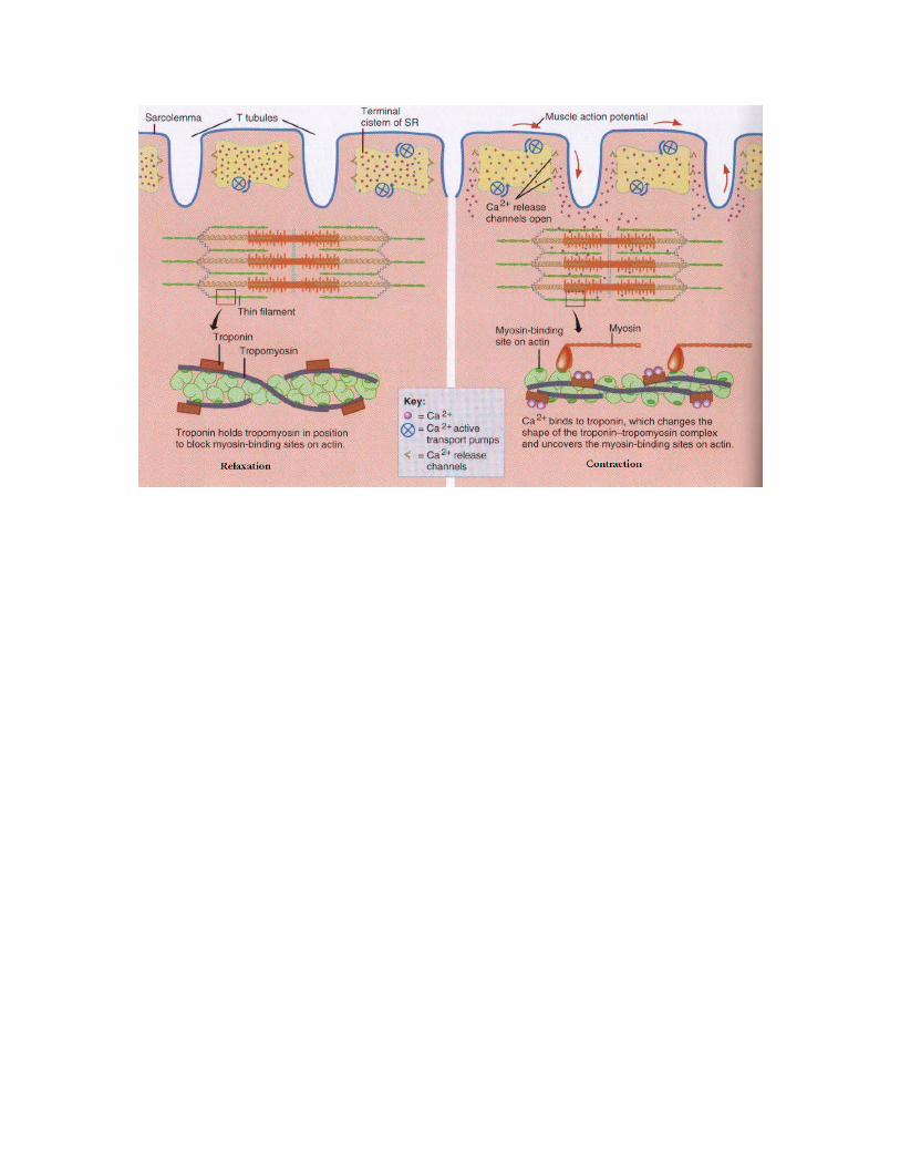
VIII. Skeletal Muscle ContractionA. Depolarization of muscle fibers
1. Acetylcholine released into synapsea. Ca++ facilitates releaseb. Parturient paresis occurs if not enough Ca++
2. Opens ligand-coated Na+ channels3. Causes a propagated action potential
a. sarcolemab. T-tubulec. sarcoplasmic reticulum
4. Cholinesterase destroys Ach5. Depolarization of sarcoplasmic reticulum causes Ca++ release6. Ca++ diffuses to myofibrils7. Ca++ initiates contraction8. Ca++ pumped back into sarcoplasmic reticulum
1. active transport2. Ca++ ATPase Pump