mutations in cenpe define a novel kinetochore-centromeric ...cmm.ucsd.edu › cleveland › linked...
TRANSCRIPT

1 3
Hum GenetDOI 10.1007/s00439-014-1443-3
OrIGInal InvestIGatIOn
Mutations in CENPE define a novel kinetochore-centromeric mechanism for microcephalic primordial dwarfism
Ghayda M. Mirzaa · Benjamin Vitre · Gillian Carpenter · Iga Abramowicz · Joseph G. Gleeson · Alex R. Paciorkowski · Don W. Cleveland · William B. Dobyns · Mark O’Driscoll
received: 20 May 2013 / accepted: 31 March 2014 © springer-verlag Berlin Heidelberg 2014
structure nucleates microtubule attachments onto mitotic chromosomes. Here, we described novel compound het-erozygous variants in CENPE in two siblings who exhibit a profound MPD associated with developmental delay, simplified gyri and other isolated abnormalities. CENPE encodes centromere-associated protein e (CenP-e), a core kinetochore component functioning to mediate chro-mosome congression initially of misaligned chromosomes and in subsequent spindle microtubule capture during mito-sis. Firstly, we present a comprehensive clinical descrip-tion of these patients. then, using patient cells we docu-ment abnormalities in spindle microtubule organization, mitotic progression and segregation, before modeling the cellular pathogenicity of these variants in an independent cell system. Our cellular analysis shows that a pathogenic defect in CenP-e, a kinetochore-core protein, largely phe-nocopies PCNT-mutated microcephalic osteodysplastic primordial dwarfism-type II patient cells. PCNT encodes
Abstract Defects in centrosome, centrosomal-associated and spindle-associated proteins are the most frequent cause of primary microcephaly (PM) and microcephalic pri-mordial dwarfism (MPD) syndromes in humans. Mitotic progression and segregation defects, microtubule spindle abnormalities and impaired Dna damage-induced G2-M cell cycle checkpoint proficiency have been documented in cell lines from these patients. this suggests that impaired mitotic entry, progression and exit strongly contribute to PM and MPD. Considering the vast protein networks involved in coordinating this cell cycle stage, the list of potential target genes that could underlie novel develop-mental disorders is large. One such complex network, with a direct microtubule-mediated physical connection to the centrosome, is the kinetochore. this centromeric-associated
Electronic supplementary material the online version of this article (doi:10.1007/s00439-014-1443-3) contains supplementary material, which is available to authorized users.
G. M. Mirzaa · W. B. Dobyns Division of Genetic Medicine, Department of Pediatrics, Center for Integrative Brain research, seattle Children’s research Institute, University of Washington, seattle, Wa, Usa
B. vitre · D. W. Cleveland Department of Cellular and Molecular Medicine, ludwig Institute for Cancer research, University of California, san Diego, la Jolla, Ca, Usa
G. Carpenter · I. abramowicz · M. O’Driscoll (*) Human Dna Damage response Disorders Group, Genome Damage and stability Centre, University of sussex, Falmer, Brighton Bn1 9rQ, UKe-mail: [email protected]
J. G. Gleeson Department of neurosciences and Pediatrics, University of California, san Diego, la Jolla, Ca, Usa
a. r. Paciorkowski Department of neurology, Center for neural Development and Disease, University of rochester Medical Center, rochester, nY, Usa
a. r. Paciorkowski Department of Pediatrics, Center for neural Development and Disease, University of rochester Medical Center, rochester, nY, Usa
a. r. Paciorkowski Department of Biomedical Genetics, Center for neural Development and Disease, University of rochester Medical Center, rochester, nY, Usa

Hum Genet
1 3
a centrosome-associated protein. these results highlight a common underlying pathomechanism. Our findings pro-vide the first evidence for a kinetochore-based route to MPD in humans.
Introduction
Microcephalic primordial dwarfism (MPD) is the collective term for a group of clinically overlapping syndromes whose underlying genetic basis has started to emerge within the last decade or so. specific examples include seckel syn-drome, microcephalic primordial dwarfism-type II (MOP-Dii) and Meier-Gorlin syndrome (Gorlin et al. 1975; Hall et al. 2004; Majewski et al. 1982b; seckel 1960). For these specific disorders, causative defects have been identified in central components of the atr-dependent Dna damage response (DDr), in a centrosome-associated protein and in components of the Dna replication licensing machinery, respectively (Bicknell et al. 2011a, b; Griffith et al. 2008; Guernsey et al. 2011; O’Driscoll et al. 2003; Ogi et al. 2012; Qvist et al. 2011; rauch et al. 2008). Defects in Dna double strand break repair and other genome stability path-ways can also cause MPD (Buck et al. 2006; Ijspeert et al. 2013; Murray et al. 2014; O’Driscoll et al. 2001; shaheen et al. 2014; shamseldin et al. 2012).
Primary microcephaly (PM) with and without overt short stature is commonly caused by defects in centro-some and spindle pole-associated proteins (Mahmood et al. 2011; Poirier et al. 2013; thornton and Woods 2009). these PM and MPD-associated defects collectively under-score the requirement for optimal cell cycle progression and checkpoint control for normal human development, including growth and neurogenesis. Currently, mutations in genes encoding core centrosome and centrosome-asso-ciated proteins represent the most frequent causative defect identified so far in MPDs. the centrosome is an important microtubule-organizing centre essential for coordinating G2-M progression, normal mitotic cell division and even cilia formation. Interestingly, defects in the atr-dependent DDr and in Dna replication licensing machinery are also associated with centrosome abnormalities, and vice versa, suggestive of a multifactorial but interconnected underlying pathobiology of MPDs (alderton et al. 2004, 2006; Griffith et al. 2008; stiff et al. 2013).
the centromeric kinetochore of mitotic chromosomes is the attachment site for spindle microtubules, ensuring their amphitelic attachments onto all of the chromosomes, thereby enabling their alignment at metaphase (Hori and Fukagawa 2012; santaguida and Musacchio 2009). this facilitates accurate and timely segregation. Unat-tached kinetochores activate the mitotic spindle assembly
checkpoint (saC) (Cleveland et al. 2003). saC activation delays mitosis until the kinetochores of each chromosome have properly attached to spindle microtubules (Musac-chio 2011; Musacchio and salmon 2007). Congeni-tal defects in the saC gene BUB1B which encodes the saC kinase Bubr1 cause mosaic variegated aneuploidy (Mva): an MPD associated with aneuploidy and elevated cancer incidence (Hanks et al. 2004; shinya Matsuura et al. 2006).
In contrast to the centrosome-spindle pole, pathogenic defects in core kinetochore components are currently notably under-represented as a cause of MPD. Using an exome sequencing strategy, we describe novel compound heterozygous variants in CENPE in two siblings charac-terized by a profound MPD with severe developmental delay, simplified gyri and various isolated abnormalities. Centromere-associated protein-e (CenP-e) is a large (>300 kD) kinetochore-associated kinesin-like motor pro-tein required for spindle microtubule capture and attach-ment at the kinetochore (abrieu et al. 2000; Yao et al. 2000). Unsurprisingly, Cenpe deletion in mice is early embryonic lethal (Putkey et al. 2002). Conditional Cenpe deletion in mouse embryonic fibroblasts and an adult regenerating liver system resulted in cells with profound mitotic defects including chromosome misalignment and segregation failure (Putkey et al. 2002; Weaver et al. 2003). Firstly, we detail the clinical presentation and progression of both siblings. then, using patient-derived lymphoblas-toid cell lines (lCls), we catalog a series of mitotic abnor-malities including aberrant spindle microtubule organiza-tion, delayed mitotic progression and elevated levels of binucleate cells. the latter phenotype, in particular, is sug-gestive of an impaired ability to exit mitosis effectively. We also find that the nuclei of these binucleates are often of unequal size indicative of impaired chromosome segre-gation during mitosis and subsequent cytokinesis failure. to further characterize and consolidate the cellular path-ogenicity of the CENPE variants we have identified, we model each individually and in combination using Flip-In technology (Invitrogen) coupled with sirna-mediated knockdown of the endogenous CenP-e in an independent cell system.
Interestingly, we find that many of these cellular phe-notypes observed in the patient lCls are also observed in lCls from a patient with a pathogenic defect in PCnt; the centrosome-associated protein implicated in MOPDii (Griffith et al. 2008; rauch et al. 2008). therefore, using MPD patient-derived material, we show that a novel kine-tochore-associated defect in CenP-e shares overlapping phenotypes of abnormal spindle microtubule structure and mitotic progression to that of a pathogenic defect in a cen-trosome-associated protein.

Hum Genet
1 3
Methods
Dna extraction
Genomic Dna was extracted from peripheral blood samples using either the Puregene kit®, Magnapure® or autogen® systems following the manufacturers’ recommendations.
Whole exome sequencing and analysis
We performed whole exome sequencing (Wes) of peripheral blood Dna from subject lr05-054a1. We used the nimbel-gen whole exome capture kit, and sequence was generated on an Illumina GaII machine. sequence was aligned to hg19 using BWa 0.6.2 and single nucleotide variants and indels were called using GatK 2.3.9 UnifiedGenotyper. Mean cov-erage was calculated using GatK 2.3.9 Depth Of Coverage Walker. annotation of variants, including identification of variants present in dbsnP, 1,000 Genomes, and the nHlBI exome variant server (evs), was performed with anno-var (Wang et al. 2010). Pedigree-specific nonsynonymous variants, including potential compound heterozygotes, were identified using sOlve-Brain. variants in genes associated with known PM and seckel-related syndromes, as well as other known developmental brain disorders, were identified using sOlve to filter against the contents of the Develop-mental Brain Disorders Database 1.2 (DBDB). Final annota-tion of biological function and pathway membership of can-didate genes with nonsynonymous compound heterozygous variants were performed using lynx (sulakhe et al. 2014).
Weblinks:BWa: http://bio-bwa.sourceforge.net/bwa.shtml.GatK: http://www.broadinstitute.org/gsa/wiki/index.php/
Home_Page.annovar: http://www.openbioinformatics.org/annovar/.sOlve: https://github.com/alex-paciorkowski/sOlve.DBDB: https://www.dbdb.urmc.rochester.edu/associations/
list.lynx: http://lynx.ci.uchicago.edu:8080/.
sanger sequencing
PCr amplification was performed with 50 ng of genomic Dna using taq Dna polymerase (applied Biosystems®). exons 23 and 29 of CENPE (nM_001813.2) were targeted for mutation analysis by sanger sequencing using standard methods.
Cell lines
eBv transformed lymphoblastoid cell lines (lCls) were made from both patients (lr05-054a1 and lr05-054a2)
using lymphocytes fractionated from whole blood. Wild-type (Wt; aG09387) cells were obtained from Coriell Cell repository (new Jersey, Usa). PCNT-mutated MOPDii lCls were from patient Cv1559 and carry a homozygous mutation in PCNT resulting in p.e220X; Family 1 from Griffith et al. (2008).
antibodies
anti-53BP1 was the mouse monoclonal from Bethyl. anti-CenP-e was mouse monoclonal [1H12] from abcam used for patient lCl indirect immunofluorescence whilst anti-CenP-e (aKIn04) from cytoskeleton was used for Western blotting. anti-BUBr1 was a mouse monoclo-nal (ab4637) from abcam. anti-α-tubulin was a mouse monoclonal (clone B512) from sigma-aldrich and anti-β-tubulin was a rabbit polyclonal (H-235) from santa Cruz. anti-ParP (MCs 1522G) was from abD serotec and anti-p85-ParP (cleaved-ParP: G7341) was obtained from Promega.
Indirect immunofluorescence
Following various treatments (see below), lCls were pelleted, swollen in 75 mM KCl (10 min), fixed in 4 % paraformaldehyde (10 min) prior to cytospinning onto poly-l-lysine coated slides. lCls were then permeabi-lized (0.1 % triton X-100 in 5 % Bsa-PBs for 2 min) and blocked (5 % Bsa-PBs for 10 min) prior to incu-bation with primary and secondary antibodies diluted in 5 % Bsa-PBs. slides were counter-stained with 4′,6-diamidino-2-phenylindole (DaPI) and mounted in vectashield anti-fade medium. Images were cap-tured using simplePCI software on the Zeiss axioplan platform.
lCl treatments
HU‑induced 53BP1 foci
lCls were treated with 5 mM HU for 2 h prior to harvest-ing and processing for indirect immunofluorescence stain-ing using anti-53BP1.
Microtubule spindle stabilization
spindles were stabilized by treating exponentially grow-ing lCls with taxol (10 μM) and the proteasome inhibi-tor MG132 (10 μM) for 1 h in complete medium at 37 °C before shifting the cells to packed ice for 1.5 h. lCls were then pelleted and processed for indirect immunofluores-cence using anti-α-tubulin.
Hum Genet
1 3
BubR1 phosphorylation
exponentially growing lCls were treated with 0.2 μg/ml colcemid for 2 h prior to pelleting and whole cell extracts made using urea buffer (9 M urea, 50 mM tris–HCl at pH 7.5 and 10 mM 2-mercaptoethanol) and sonication (15 s at 30 % amplitude using a micro-tip; sigma-aldrich).
Colcemid block and release into cytochalasin B
lCls were treated with colcemid (0.2 μg/ml for 4 h), pel-leted, washed in complete medium before release into com-plete medium supplemented with cytochalasin B (5 μg/ml). Cells were incubated for 24 h and processed for indirect immunofluorescence using anti-β-tubulin.
G2‑M checkpoint analysis
lCls were irradiated with 3 Gy Ir in complete medium or 7 J/m2 in PBs before seeding into complete medium sup-plemented with 0.2 μg/ml colcemid and incubated for 4 h. lCls were pelleted, swollen in 75 mM KCl (10 min) and fixed in 4 % paraformaldehyde (10 min) prior to cytospin-ning onto poly-l-lysine coated slides. Cells were counter-stained with DaPI and mounted in vectashield anti-fade prior to analysis on the Zeiss axioplan system.
Flp-In system and variant modeling
the Flp-In (Invitrogen) system employed here for CENPE in DlD-1 cells has been described previously (Kim et al. 2010). the cDna and sirna for CENPE used here are identical to those of Kim et al. (2010). lipofectamine rnai MaX (Invitrogen) was used for CENPE sirna (5′-CCaCUaGaGUUGaaaGaUa-3′) with doxycycline used at 1 μg/ml for induction of sirna-resistant CENPE construct. For the live cell imaging, an rFP-histone H2B expressing DlD-1 line was used to visualize chromosomes, as described in Kim et al. (2010).
Results
Clinical presentation and progression of siblings lr05-054a1 and lr05-054a2
the features of both siblings are summarized in table 1. this family is a non-consanguineous family of european ancestry. the first affected, a boy (lr05-054a1), was born at 34 weeks of gestation to 33-year-old mother, who had a history of one miscarriage. Pregnancy was complicated by intrauterine growth restriction detected at 6 and 10 weeks of gestation; biparietal diameter, head circumference,
femoral length, abdominal circumference and estimated fetal weight were all less than 10th percentile for gesta-tional age. at 34 weeks of gestation, prenatal ultrasound was performed due to maternal hypertension and showed a severely small fetus for gestational age with decreased fetal activity, so delivery was induced at that time. at birth, lr05-054a1 exhibited severe congenital microcephaly, with a birth OFC of 24.5 cm (−5 sD), growth deficiency with a weight of 1.5 kg (−3 to −4 sD) and length 40.5 cm (−2 to −3 sD). Facial features included a sloping fore-head, prominent nose, and mild micrognathia. this boy had a single seizure at 7 months of age which consisted of generalized body stiffening, extension of the arms and legs with breath holding. electroencephalogram was abnormal showing generalized rhythmic fast activity followed by generalized slowing, and multifocal sharp waves. He had a few additional seizures between 7 and 10 months of age that were subsequently well controlled on phenobarbital.
at 4 months of age, he had a viral upper respiratory ill-ness and, because of worsening respiratory symptoms, was admitted and underwent a cardiac echocardiogram which showed abnormal tricuspid and mitral valve inflow patterns with increased early and late atrial (e/a) ratios, suggestive of diastolic dysfunction, and decreased deceleration times varying with the respiratory cycles, indicative of increased flow reversal in the pulmonary veins with atrial contrac-tion. additional echo findings include trivial tricuspid and mitral regurgitation, biatrial enlargement with paradoxial septal wall motion suggesting elevated right ventricular systolic pressure, a trabeculated and hypertrophied apex of the left ventricle but with normal dimensions and good systolic function. Overall, these features were consistent with congenital restrictive cardiomyopathy, for which he was treated with digoxin, lasix and aspirin. Other medical complications include poor weight gain, partly due to the underlying syndrome and partly due to gastroesophageal reflux, hyperopia requiring glasses, and mild congenital subglottic stenosis of no clinical significance.
On last examination at 5 years, lr05-054a1 exhibited the following growth measurements: weight 9.14 kg (−5 sD), length 83.7 cm (−7 sD), and OFC 37.0 cm (−9 sD). His facial appearance was significant for striking micro-cephaly with a very low forehead, round face, prominent nose, mild micrognathia and decreased subcutaneous fat (Fig. 1a–c). His head appeared disproportionately smaller than his body whilst his nose and ears disproportionately large. His hands and feet were mildly small. even though lr05-054a1 did not exhibit the nasal preponderance with downward curved nasal tip classically associated with seckel syndrome, his overall features appeared compatible with profound MPD of this type (Hall et al. 2004; Majew-ski et al. 1982a, b). On neurological exam, he was alert but did not follow commands or communicate. eye movements
Hum Genet
1 3
were full, pupils normal and facial movements symmetric. Motor exam showed mild spasticity in the hip adductors and ankles. Deep tendon reflexes were normal (2+) and symmetric. Developmentally, this child made very slow but steady progress albeit remaining severely handicapped. He began to pull himself to stand only briefly at 3.5 years of age, and crawled at 4 years. On assessment at 5 years, he had no communication skills besides vocalizing without signs, words or pointing. He used a rake and pincer grasp. He was fed a soft diet by mouth. His general health includ-ing his cardiac course deteriorated over time; he developed pericardial effusion on a few occasions requiring percu-taneous drainage and also had several episodes of pneu-monia, with progressive worsening of his oxygen satura-tion levels. a final episode of pneumonia at 8 years of age proved fatal.
the pregnancy of the second affected child of the fam-ily, a girl (lr05-054a2), was also complicated by micro-cephaly and intrauterine growth restriction prenatally detected on ultrasound. she was born at 37 weeks of gesta-tion with birth weight 1.76 kg (−3 sD) and length 41.5 cm (−3 sD). she was noticed to have the same facial features as her brother’s at birth. she exhibited frequent episodes of otitis media requiring myringotomy tube placement, and minor foot deformities consisting of tight heel cords
and medially deviated halluces, for which she underwent heel cord lengthening and tenotomy of great toes for better alignment. Unlike her brother, she did not have episodes of pneumonia or other severe illnesses requiring hospitaliza-tion. echocardiogram and electrocardiogram were normal. she did not have seizures, and was described as a generally happy child.
On last exam at 3 years, her weight was 9.1 kg (−3 to −4 sD), length 83 cm (−2 to −3 sD), and OFC 37.5 cm (−7 sD). Her palms, fingers and toes measured less than −2 sD. Her overall appearance was significant for a small head, small round face with a low forehead and proportion-ately large ears and nose (Fig. 1d). the helix of the right ear was mildly under-folded, and great toes medially deviated. neurological exam revealed an alert child with brief visual tracking and a strong cry. tone was mildly increased and deep tendon reflexes minimally brisk (2++). eye move-ments were full with intermittent nystagmus. Facial move-ments were symmetrical. Developmentally, this girl sat alone at 1–1.5 years, pulled to stand, cruised and climbed on furniture at 2–2.5 years, and crawled at 2.5 years. at 2.5 years, her language was limited to a few words such as “hi” and “bye”, along with mimicking speech sounds. she picked small objects but did not point or draw. she attended preschool at 3 years of age. she was able to chew solids
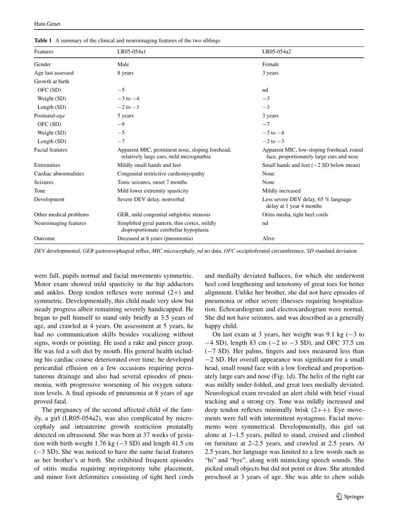
Table 1 a summary of the clinical and neuroimaging features of the two siblings
DEV developmental, GER gastroesophageal reflux, MIC microcephaly, nd no data, OFC occipitofrontal circumference, SD standard deviation
Features lr05-054a1 lr05-054a2
Gender Male Female
age last assessed 8 years 3 years
Growth at birth
OFC (sD) −5 nd
Weight (sD) −3 to −4 −3
length (sD) −2 to −3 −3
Postnatal-age 5 years 3 years
OFC (sD) −9 −7
Weight (sD) −5 −3 to −4
length (sD) −7 −2 to −3
Facial features apparent MIC, prominent nose, sloping forehead, relatively large ears, mild micrognathia
apparent MIC, low-sloping forehead, round face, proportionately large ears and nose
extremities Mildly small hands and feet small hands and feet (−2 sD below mean)
Cardiac abnormalities Congenital restrictive cardiomyopathy none
seizures tonic seizures, onset 7 months none
tone Mild lower extremity spasticity Mildly increased
Development severe Dev delay, nonverbal less severe Dev delay, 65 % language delay at 1 year 4 months
Other medical problems Ger, mild congenital subglottic stenosis Otitis media, tight heel cords
neuroimaging features simplified gyral pattern, thin cortex, mildly disproportionate cerebellar hypoplasia
nd
Outcome Deceased at 8 years (pneumonia) alive

Hum Genet
1 3
with some feeding difficulties, such as choking occasion-ally, requiring a softer diet.
Clinical work-up of siblings lr05-054a1 and lr05-054a2
Clinical workup performed on lr05-054a1 with inconclu-sive results includes a karyotype, a chromosomal microar-ray (signature Genomics), chromosome breakage studies, metabolic tests and tOrCH titers. a skeletal survey dem-onstrated apparent microcephaly with no evidence of crani-osynostosis, subtle widening of the ribs and possible meta-physeal areas of sclerosis in the distal femurs and proximal tibias. the hands appeared osteopenic and metacarpals were relatively short. Overall, these findings were not char-acteristic of a specific bone dysplasia. Brain MrI scan of lr05-054a1 at 17 days of life showed extreme microceph-aly with a low forehead and a diffuse severely simplified gyral pattern with virtually no gyri over the frontal lobe, a thin 2–3 mm cortex and immature white matter (Fig. 1e, f). additional findings include partial agenesis of the cor-pus callosum and cerebellar hypoplasia that appeared
disproportionate to the degree of microcephaly (Fig. 1e, f). notably, neither sibling had a history of blood dyscrasias or other childhood cancers.
Molecular-genetic analysis of family lr05-054
although lr05-054a1 did not present with the skeletal features characteristic of MOPDii (e.g., coxa vara, small iliac wings, v-shaped distal femoral epiphysis, metaphy-seal flaring of radius and ulna), these features can take time to manifest (Hall et al. 2004). since defects in the centro-some-associated protein pericentrin, encoded by PCNT, are currently one of the most commonly identified defects associated with profound MPD in humans (MOPDii) (Bober et al. 2012), we sequenced PCNT and did not find any pathogenic mutations in this family.
Wes was then performed using peripheral blood from subject lr05-054a1. Mean depth of coverage across the targeted exome was 99.5x. the mean depth of coverage across CENPE was 45.5x. sOlve identified 4,322 genes with two or more heterozygous nonsynonymous variants.
Fig. 1 Images of the affected siblings lr05-054a1 and lr05-054a2. a–c Patient lr05-054a1 at 16 months of age. d Patient lr05-054a2 at 2 years of age showing microcephaly, low-sloping forehead, pro-portionately large ears and nose, a prominent nose, micrognathia, and severe growth deficiency. e t1-weighted mid-sagittal and f
t2-weighted axial brain MrI images of lr05-054a1 at 17 days of life show severe microcephaly, with low-sloping forehead, severely simplified gyral pattern with virtually no gyri in the frontal region, mildly increased extra-axial space, and disproportionately small cer-ebellar vermis
Hum Genet
1 3
However, most of these variants were in genes not likely contributory to the phenotype (DBDB, dbsnP and evs). there were no novel variants detected in other known PM, MPD or seckel syndrome genes.
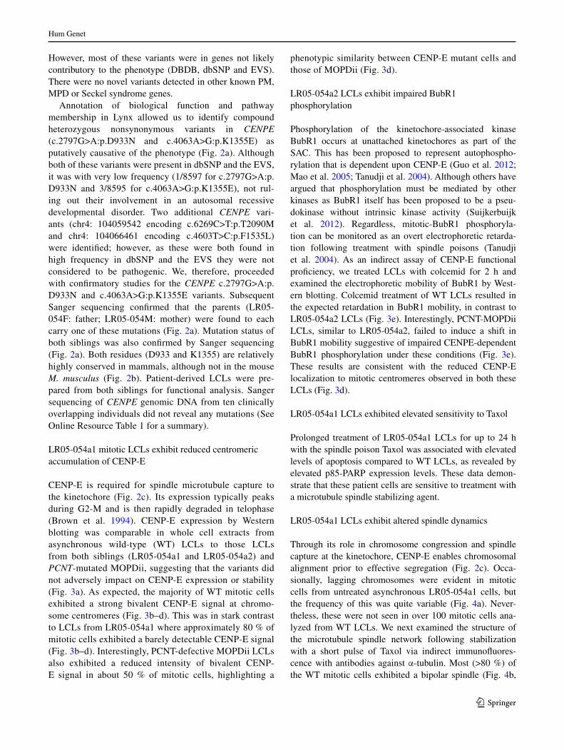
annotation of biological function and pathway membership in lynx allowed us to identify compound heterozygous nonsynonymous variants in CENPE (c.2797G>a:p.D933n and c.4063a>G:p.K1355e) as putatively causative of the phenotype (Fig. 2a). although both of these variants were present in dbsnP and the evs, it was with very low frequency (1/8597 for c.2797G>a:p.D933n and 3/8595 for c.4063a>G:p.K1355e), not rul-ing out their involvement in an autosomal recessive developmental disorder. two additional CENPE vari-ants (chr4: 104059542 encoding c.6269C>t:p.t2090M and chr4: 104066461 encoding c.4603t>C:p.F1535l) were identified; however, as these were both found in high frequency in dbsnP and the evs they were not considered to be pathogenic. We, therefore, proceeded with confirmatory studies for the CENPE c.2797G>a:p.D933n and c.4063a>G:p.K1355e variants. subsequent sanger sequencing confirmed that the parents (lr05-054F: father; lr05-054M: mother) were found to each carry one of these mutations (Fig. 2a). Mutation status of both siblings was also confirmed by sanger sequencing (Fig. 2a). Both residues (D933 and K1355) are relatively highly conserved in mammals, although not in the mouse M. musculus (Fig. 2b). Patient-derived lCls were pre-pared from both siblings for functional analysis. sanger sequencing of CENPE genomic Dna from ten clinically overlapping individuals did not reveal any mutations (see Online resource table 1 for a summary).
lr05-054a1 mitotic lCls exhibit reduced centromeric accumulation of CenP-e
CenP-e is required for spindle microtubule capture to the kinetochore (Fig. 2c). Its expression typically peaks during G2-M and is then rapidly degraded in telophase (Brown et al. 1994). CenP-e expression by Western blotting was comparable in whole cell extracts from asynchronous wild-type (Wt) lCls to those lCls from both siblings (lr05-054a1 and lr05-054a2) and PCNT-mutated MOPDii, suggesting that the variants did not adversely impact on CenP-e expression or stability (Fig. 3a). as expected, the majority of Wt mitotic cells exhibited a strong bivalent CenP-e signal at chromo-some centromeres (Fig. 3b–d). this was in stark contrast to lCls from lr05-054a1 where approximately 80 % of mitotic cells exhibited a barely detectable CenP-e signal (Fig. 3b–d). Interestingly, PCnt-defective MOPDii lCls also exhibited a reduced intensity of bivalent CenP-e signal in about 50 % of mitotic cells, highlighting a
phenotypic similarity between CenP-e mutant cells and those of MOPDii (Fig. 3d).
lr05-054a2 lCls exhibit impaired Bubr1 phosphorylation
Phosphorylation of the kinetochore-associated kinase Bubr1 occurs at unattached kinetochores as part of the saC. this has been proposed to represent autophospho-rylation that is dependent upon CenP-e (Guo et al. 2012; Mao et al. 2005; tanudji et al. 2004). although others have argued that phosphorylation must be mediated by other kinases as Bubr1 itself has been proposed to be a pseu-dokinase without intrinsic kinase activity (suijkerbuijk et al. 2012). regardless, mitotic-Bubr1 phosphoryla-tion can be monitored as an overt electrophoretic retarda-tion following treatment with spindle poisons (tanudji et al. 2004). as an indirect assay of CenP-e functional proficiency, we treated lCls with colcemid for 2 h and examined the electrophoretic mobility of Bubr1 by West-ern blotting. Colcemid treatment of Wt lCls resulted in the expected retardation in Bubr1 mobility, in contrast to lr05-054a2 lCls (Fig. 3e). Interestingly, PCnt-MOPDii lCls, similar to lr05-054a2, failed to induce a shift in Bubr1 mobility suggestive of impaired CenPe-dependent Bubr1 phosphorylation under these conditions (Fig. 3e). these results are consistent with the reduced CenP-e localization to mitotic centromeres observed in both these lCls (Fig. 3d).
lr05-054a1 lCls exhibited elevated sensitivity to taxol
Prolonged treatment of lr05-054a1 lCls for up to 24 h with the spindle poison taxol was associated with elevated levels of apoptosis compared to Wt lCls, as revealed by elevated p85-ParP expression levels. these data demon-strate that these patient cells are sensitive to treatment with a microtubule spindle stabilizing agent.
lr05-054a1 lCls exhibit altered spindle dynamics
through its role in chromosome congression and spindle capture at the kinetochore, CenP-e enables chromosomal alignment prior to effective segregation (Fig. 2c). Occa-sionally, lagging chromosomes were evident in mitotic cells from untreated asynchronous lr05-054a1 cells, but the frequency of this was quite variable (Fig. 4a). never-theless, these were not seen in over 100 mitotic cells ana-lyzed from Wt lCls. We next examined the structure of the microtubule spindle network following stabilization with a short pulse of taxol via indirect immunofluores-cence with antibodies against α-tubulin. Most (>80 %) of the Wt mitotic cells exhibited a bipolar spindle (Fig. 4b,
Hum Genet
1 3
c). In direct contrast, approximately 80 % of mitotic cells from PCnt-MOPDii (PCnt) and lr05-054a1 exhibited multipolar spindles suggestive of massively disorganized spindle network in these cells (Fig. 4b, c). abnormal spin-dle structure has been reported for PCnt-MOPDii previ-ously (rauch et al. 2008). Interestingly, again we observe similar cellular phenotypes between a centrosomal defect (PCnt) and that of CENPE-mutated lr05-054a1 lCls.
lr05-054a2 lCls exhibit elevated levels of binucleates with features consistent with altered chromosome segregation
CenP-e deficiency is associated with elevated frequency of polar chromosomes and mis-segregation (Guo et al. 2012; Putkey et al. 2002; tanudji et al. 2004; Weaver et al. 2003). We did not observe a significantly elevated
Fig. 2 Family lr05-054 have compound heterozygous variants in CENPE. a representative sequence chromatographs identify-ing mutations in CENPE (nM001813.2) showing the two mis-sense mutations identified in both siblings (c.2797G>a, p.D933n and c.4063a>G, p.K1355e; a-b), as well as parental carrier status: p.D933n is paternally inherited (lr05-054F; father) and p.K1355e is maternally inherited (lr05-054M; mother). b Multiple sequence alignments of the regions around amino acids 933 and 1,355 were assembled using MUsCle (version 3.6) via nCBI. Identical sequences are identified in gray, divergent in black and the target residues in red and by the red asterisk. Both D933 and K1355 are relatively well conserved in mammals. although interestingly, neither residue is conserved in mice, M. musculus. In fact, the mouse con-tains n933, one of the variants identified in our patients. It is nota-ble that CenP-e protein identity between H. sapien and Cenpe of M. musculus is only 64.4 % (for R. noregicus 56 %). this is in marked contrast to chimpanzee P. troglodytes (98.8 %), rhesus monkey M. mulatta (95.8 %) or cattle B. taraus (79 %). In fact, the mouse (M. musculus) Cenpe is only 2,471 amino acids in length compared to
2,701 amino acids of the human CenP-e. Whilst both proteins pos-sess the kinesin motor domain atPase, only the human protein pos-sesses the sMC (C0G1196) chromosome segregation atPase and sMC_n(pFam02463) recF/recn/sMC n-terminal domain, high-lighting the significant sequence and domain disparity between mouse and human herein. c schematic representation of a mitotic cell, showing the microtubule nucleating centrosome (purple), the microtubule spindles (blue), mitotic chromosomes (gray) and their centromeric kinetochores (green). Known genetic defects in centro-somal and centrosome-associated proteins are shown in purple and spindle pole components in blue. the dashed box indicates a magni-fied schematic of the kinetochore showing CenP-e capturing micro-tubules. CenP-e is characterized by a large 230-nm flexible coiled-coil domain that can move about to enable microtubule capture by the globular head regions which are composed of microtubule interac-tion, kinesin motor and atP-binding domains. the CENPE mutations identified here encode residues within this coiled-coil (color figure online)

Hum Genet
1 3
spontaneous frequency of micronuclei in PCnt or CenP-e lCls (lr05-054a2), although we did detect an approxi-mately two-fold increase in the level of bi-nucleated cells here, compared to Wt (Fig. 4d). Binucleates are the prod-uct of impaired and/or delayed cytokinesis which can be a consequence of a failure to coordinately transit mitosis
effectively. strikingly, approximately 50 % of binucleates detected in the lr05-054a2 lCls exhibited nuclei of une-qual size, suggestive of unequal segregation during mito-sis and impaired cytokinesis (Fig. 4e). We did not observe this phenotype in the binucleates from the PCnt-MOPDii lCls.
Fig. 3 CENPE- and PCNT-mutated patient lCls exhibit reduced kinetochore localization of CenP-e and reduced Bubr1 phospho-rylation. a CenP-e expression is comparable in whole cell extracts (WCe) from wild-type (lCls) and lCls from both siblings (lr05-054a1 and lr05-054a2) and from PCNT-mutated MOPDii lCls. the left‑hand panels show 25 μg WCe and the right‑hand panel 50 μg of WCe. b Indirect immunofluoresence (IF) showing CenP-e focalization onto kinetochores of mitotic cells in wild-type (Wt), PCNT-mutated MOPDii (PCnt) and CENPE-mutated patient derived lCls (lr05-054a1). Cells were treated with taxol (10 μM) and MG132 (10 μM) for 1 h followed by 1.5 h on ice to stabilize spindle microtubules. Both PCnt and lr05-054a1 mitotic cells exhibit a clear under-representation of defined CenP-e foci. all images were acquired under identical exposure conditions (scale bar; 10 μm). c Close-up image of CenP-e focalization in a mitotic cell from wild-type (Wt) and CENPE-mutated patient derived lCls (lr05-054a1). It has been estimated that only approximately 50 CenP-e dimers occupy each normal human kinetochore (Brown et al. 1994) (scale bar; 10 μm). d the distribution of mitotic cells exhibiting strong bivalent CenP-e foci and those under-stained for
CenP-e foci in wild-type (Wt), PCNT-mutated MOPDii (PCnt) and CENPE-mutated patient derived lCls (lr05-054a1). Between 30 and 50 mitotic cells were scored for each cell line under identi-cal conditions as a. strong bivalent CenP-e staining is under-repre-sented in PCnt and lr05-054a1 lCls compared to Wt. e lCls, from wild-type (Wt), PCNT-mutated MOPDii (PCnt) and the female CENPE-mutated patient sib (lr05-054a2) were treated with colcemid (0.2 μg/ml) for 2 h and phosphorylation status of Bubr1 was determined by electrophoretic shift following Western blotting. In contrast to Wt, colcemid treatment did not induce significant Bubr1 retardation (phosphorylation) in PCnt or lr05-054a2 lCls under these conditions. Interestingly, following a more prolonged treatment with colcemid (≥4 h), Bubr1 retardation is evident in both these lCls suggestive of a leaky/hypomorphic defect. the blot was re-probed for MCM2 expression as a loading control. f lCls were treated with 2.5 μM taxol and incubated for the times indicated. apoptosis induction and levels were assessed by blotting for cleaved form of ParP; p85-ParP. the p85-ParP was detected at 16 h post-treatment in lr05-054a1 lCls and further increasing at 24 h. Under these conditions, no apoptosis was evident in the Wt lCls

Hum Genet
1 3
expression of the D933n and K1355e variants of CenP-e induce mitotic spindle abnormalities and polar chromosomes
We used Flp-In (Invitrogen) recombination to intro-duce sirna-resistant p.D933n and p.K1355e variants of CENPE, individually and in-cis (Double mut; dou-ble mutant), into DlD-1 cells under doxycycline (Dox) conditional regulation, as described in detail by Kim et al. (2010). Following sirna-mediated silencing of the
endogenous CENPE, Dox treatment (8 h) resulted in strong expression of these variants and cells were fixed for immu-nofluorescence imaging. this allowed us to interrogate the impacts on mitotic spindle structure and chromosome segregation when each variant was expressed individually and when engineered together into CenP-e. the induction scheme used is shown in Fig. 5a and comparable CenP-e expression levels shown in Fig. 5b. In this independent model cell system, we found that p.D933n and p.K1355e individually or when in-cis caused an elevated frequency
Fig. 4 CENPE-mutated patient lCls exhibit abnormal spindle microtubule organization and evidence of aberrant segregation and delayed progression through mitosis. a an image of a lagging chro-mosome in an anaphase cells from an asynchronous culture of lr05-054a1. b the lower panels show IF images of α-tubulin staining microtubule network (green) from wild-type (Wt) and lr05-054a1 lCls following treatment with taxol (10 μM) and MG132 (10 μM) for 1 h then 1.5 h on ice to stabilize spindle microtubules. the upper panels are the corresponding DaPI-staining nuclei. a normal bipolar spindle is seen in Wt mitotic cell whilst multipolar spindles are fre-quently seen in mitotics from lr05-054a1. c the distribution of spin-dle abnormalities noted in mitotic cells in wild-type (Wt), PCNT-mutated MOPDii (PCnt) and CENPE-mutated patient derived lCls (lr05-054a1). Between 40 and 50 mitotic cells were scored for each
cell line. Multipolar spindles are over-represented in mitotic cells from PCnt and lr05-054a1 lCls compared to Wt. d the spon-taneous levels of micronuclei (Mn) are not statistically significantly elevated (ns not significant student t test) in PCNT-mutated MOP-Dii (PCnt) or CENPE-mutated patient derived lCls (lr05-054a2) compared to Wt. In contrast, binucleates (Bn) levels are approxi-mately twofold higher in these lCls compared to Wt potentially suggestive of elevated rates of cytokinesis failure (*p < 0.05 student t test). e DaPI (upper panel) and their corresponding IF-mediated β-tubulin images (lower panel) of binucleates show that the binucle-ates (circled) from lr05-054a2 lCls frequently exhibit nuclei of different sizes. the β-tubulin staining facilitates the identification of true binucleate cells. Differing sized nuclei within a binucleate are indicative of unequal segregation during mitosis (color figure online)

Hum Genet
1 3
of mitotic cells with polar chromosomes (Fig. 5c, d), along with the elevated levels of mitotic cells with abnormal spin-dles (monopolar and multipolar mitotic spindles; Fig. 5c and e). these findings independently show that both these patient-derived CenP-e variants can adversely impact on normal mitotic spindle formation and chromosome segre-gation, phenotypes consistent with our observations derived from patient lCls.
lr05-054a2 lCls exhibit impaired mitotic progression
to examine the proficiency of mitotic progression in patient lCls in more detail, we performed a block-release-block experiment using colcemid coupled with cytochalasin B treatments. specifically, lCls were briefly treated with colcemid (4 h) to synchronize a sub-population of cells in mitosis. the colcemid was removed by washing and cells
Fig. 5 expression of the D933n and K1355e variants of CenP-e results in mitotic spindle abnormalities and polar chromosomes. a DlD-1 cells engineered by Flp-In recombination for the condi-tional (doxycycline-induced) expression of sirna-resistant CENPE and CENPE patient variants were plated and 24 h later subjected to CENPE sirna targeting endogenous CenP-e only. after 40 h approx., the cells were treated with doxycycline (Dox; 1 μg/ml) for 8 h to induce the expression of the sirna-resistant CENPE vari-ants. Cells were then fixed and processed for immunofluorescence. b expression levels of the various CenP-e variants including a construct with both variants engineered in-cis (Double mut; double mutant) were compared to that of wild-type (Wt) CenP-e following 8 h in doxycycline. c Immunofluoresence images following 8 h treat-ment with doxycycline for wild-type (Wt), D933n alone, K1355e
alone and the double mutant (Double mut) with both D933n and K1355e in-cis co-stained with anti-CenP-e and anti-α-tubulin. Polar chromosomes not aligned properly stain strongly for CenP-e under these conditions as seen in D933n alone, K1355e alone and the dou-ble mutant (Scale bar 5 μm). d the frequency of mitotic cells with ≥5 unaligned polar chromosomes is elevated following the expres-sion of D933n alone, K1355e alone or following the expression of both variants in-cis (Double mut). Between 70 and 90 mitotic cells were evaluated in total for each construct. e expression of D933n alone, K1355e alone or both variants in-cis (Double mut) resulted in an increased frequency of mitotic cells with abnormal spindle struc-ture (monopolar plus multipolar spindles) compared to the expression of wild-type (Wt) CenP-e in this system (n the number of mitotic cells scored)

Hum Genet
1 3
then released into cytochalasin B, a cytokinesis inhibitor, for 24 h. the frequency of cytochalasin B-induced binucle-ate cells was compared to untreated cells for each of the Wt, PCnt-MOPDii and lr05-054a2 lCls. the ratio of binucleate cells, that is the number of binucleates following colcemid block and release into cytochalasin B, over the level of binucleates in untreated (Unt) lCls, reflects the progression through mitosis following release from the col-cemid block. We observed a decreased ratio of binucleate cells following this block-release-block procedure in PCNT and CENPE (lr05-054a2)-mutated lCls compared to Wt consistent with a delay in progression through mitosis upon release from colcemid (Fig. 6a).
expression of the D933n and K1355e variants of CenP-e are associated with delayed mitotic progression
Using our Flp-In (Invitrogen) CENPE system in DlD-1 cells engineered to stably co-express rFP-histone H2B to enable chromosome visualization, we employed live cell imaging to determine the kinetics of mitotic progression (Fig. 6b, c). Whilst we found that expression of p.D933n alone or expression of p.K1355e alone, on average, was associated with a slower progression through mitosis com-pared to Wt CenP-e, these delays were not statistically significant (Fig. 6c). the mean mitotic transit time for Wt CenP-e was 117 min compared to 175 min for D933n and 147 min for K1355e. But when both variants were engi-neered in-cis into CenP-e, this resulted in a statistically significant delay in mitosis (205 min) compared to Wt CenP-e (117 min) (Fig. 6b, c). these findings show that expression of a CENPE cDna engineered to carry both patient-derived variants (p.D933n and p.K1355e) in-cis resulted in a significantly delayed mitosis. these findings are consistent with the block-release-block findings using lr05-054a2 lCls (Fig. 6a).
lr05-054a1 lCls exhibit a functional atr-dependent Dna damage response
seckel syndrome and MOPDii patient-derived lCls exhibit defects in the atr-dependent DDr, including impaired Uv-induced G2-M cell cycle checkpoint acti-vation and compromised atr-DDr dependent signal-ing (alderton et al. 2004; Griffith et al. 2008; O’Driscoll et al. 2003; Ogi et al. 2012; Qvist et al. 2011). to deter-mine whether lCls from patient lr05-054a1 exhibited an impaired atr-dependent DDr, we treated the cells with different forms of radiation: Uv to activate the atr-DDr and Ir to activate the atM-DDr, and then assessed mitotic index (MI). G2-M cell cycle checkpoint activa-tion is indicated by a decrease in MI due to the cell cycle arrest in G2 phase following radiation. similar to Wt
lCls, lr05-054a1 exhibited a decrease in MI follow-ing Uv indicative of a functional atr-dependent check-point (Fig. 7a). this is in contrast to lCls derived from a patient mutated in ATR (atr-s; atr-seckel syndrome) or in PCNT-mutated MOPDii lCls (PCnt). all patient-derived lCls demonstrated a drop in MI following Ir indicative of a functional atM-dependent G2-M check-point (Fig. 7a).
Following stalling of Dna replication forks with hydroxyurea (HU), 53BP1 forms microscopically detect-able foci in an atr- and CHK1-dependent manner (sen-gupta et al. 2004). seckel syndrome and PCNT-mutated MOPDii patient-derived lCls exhibit impaired HU-induced 53BP1 foci formation indicative of an impaired atr-DDr (Griffith et al. 2008; Ogi et al. 2012). Consist-ent with the normal Uv-induced G2-M checkpoint acti-vation in lr05-054a1, these cells were proficient in HU-induced 53BP1 foci formation, in contrast to atr-s and PCnt-defective MOPDii lCls (Fig. 7b). Collectively, these results suggest that lr05-054a1 does not exhibit an impaired atr-DDr.
Discussion
the developing embryonic neuroepithelium is exquisitely sensitive to perturbations in cell cycle kinetics as this system is characterized by a temporally restricted rapid expansion of stem cells (rakic 1995). It is also sensitive to perturbations in spindle orientation, as alterations in the cleavage plane can disrupt symmetric-asymmetric division, migration and differentiation (Fish et al. 2006). Further-more, defects in microtubule dynamics and microtubule interacting proteins are associated with a range of neu-ronal migration disorders (e.g., lissencephaly, pachygyria, polymicrogyria) coupled with microcephaly (Jaglin and Chelly 2009; Poirier et al. 2013).
Collectively, disruption of centrosome-spindle pro-cesses can result in an overlapping set of human patholo-gies ranging from PM, PM with short stature to overt MPD (al-Dosari et al. 2010; Bond et al. 2002, 2005; Guernsey et al. 2010; Kalay et al. 2011; sir et al. 2011). Whilst path-ogenic defects have been identified in centrosome, microtu-bule spindle and spindle-associated components, defects in core kinetochore components underlying MPD with corti-cal malformation have not yet been reported. recently, a rare homozygous mutation in CASC5 was reported in three consanguineous families from the same isolated geographi-cal region; one of whom was the original Moroccan fam-ily used to define the MCPH4 locus (Genin et al. 2012; Jamieson et al. 1999). CasC5 is a component of the KMn complex (Knl1-Mis12 complex-ndc80 complex), a kine-tochore localized network implicated in several functions

Hum Genet
1 3
including microtubule stabilization and saC silencing (Bolanos-Garcia et al. 2009; Kiyomitsu et al. 2007, 2011. In contrast to the CenP-e patients described here, the CASC5-mutated individuals were characterized by PM (age-related OFC ranging from −4 to −7 sD), normal stat-ure, normal motor milestones without evidence of epilepsy or neurological deficits and an otherwise unremarkable
medical history with the oldest patients in their thirties (Genin et al. 2012; Jamieson et al. 1999). Interestingly, detailed analysis did not yield any spontaneous mitotic and/or cell cycle defect or evidence of genomic instability in cells from these patients, again in contrast to the CenP-e defect described here (Genin et al. 2012). the authors pos-tulated that this may perhaps be due to the restriction of
Fig. 6 CENPE-mutated patient lCls exhibit delayed mitotic pro-gression which is also observed upon conditional expression of CenP-e carrying D933n and K1355e in-cis. a the ratio of binucle-ates following colcemid block and release into cytochalasin B com-pared to untreated (Unt) cells is lower in PCNT-mutated MOPDii (PCnt) or CENPE-mutated patient derived lCls (lr05-054a2), compared to Wt, consistent with a slower progression through mito-sis under these conditions in these patient cells. b DlD-1 cells engi-neered by Flp-In recombination for the conditional (doxycycline-induced) expression of sirna-resistant CENPE and CENPE patient variants were plated and 24 h later subjected to CENPE sirna tar-geting endogenous CenP-e only as described in Fig. 5a. these cells also contain a rFP-tagged histone H2B to enable real-time analy-sis of chromosome dynamics during mitosis by live cell imaging. approximately 40 h post-sirna, the cells were treated with doxycy-cline (Dox; 1 mg/ml) to induce the expression of the sirna-resistant CENPE variants and cells analyzed by live cell imaging. still images
from the live cell movies following mitosis when wild-type (Wt) CenP-e was expressed compared to the double mutant (Double mut) expressing D933n and K1355e in-cis. the time (in minutes) is indicated in the various panels showing onset of the transition from prophase–metaphase–anaphase–telophase under each conditional expression condition. c the time from the onset of nuclear envelope breakdown (neB) to anaphase was calculated for each CenP-e variant when expressed individually or in-cis (Double mut; double mutant) and represented in box‑whisker plots. the average time of the onset of neB to anaphase is shown in red for each variant. expres-sion of D933n alone, or K1355e alone, did not result in a statistically significant delay compared to expression of wild-type (Wt) CenPe (ns not significant by student t test), although the average transition for each variant was slightly longer compared to Wt. *expression of both variants in-cis did result in a statistically significant delay from neB to anaphase onset compared to Wt (p < 0.05 student t test) (color figure online)

Hum Genet
1 3
such anticipated cellular phenotypes to the neuronal system (Genin et al. 2012).
Different defects even within the same centrosomal pro-teins have previously been found to exhibit a spectrum of growth and neurodevelopmental phenotypes, the basis of which is unclear (al-Dosari et al. 2010; Bond et al. 2005; Guernsey et al. 2010; Kalay et al. 2011). an important implication of our findings is that hypomorphic defects in other core kinetochore and kinetochore-associated fac-tors (aside from CenP-e) may also underlie MPD in humans. this should be considered when designing tar-geted sequencing strategies and/or analyzing Wes data-sets. Our work also illustrates a useful platform to model the functional impacts of candidate gene variants (Flip-In recombination of sirna-resistant variant carrying cDna). this is particularly relevant in our situation as one of the patient variant changes reflected the amino acid at the equivalent position in mouse Cenpe. no doubt this variant would likely have been excluded based on standard filter-ing approaches if used in isolation. Modeling variant patho-genicity in an independent cell system when possible repre-sents a powerful approach when used in combination with patient cell-based functional analysis.
another important aspect of our findings is that they pro-vide a wider clinical picture for how a hypomorphic defect in a core kinetochore component presents. nevertheless, with regard to the congenital heart defect (CHD) observed here specifically in lr05-054a1, we cannot be absolutely sure that impaired CenP-e is a causative and/or a con-tributing factor. Interestingly, CHDs have been reported in
Mva patients, although specifically in those with CEP57 mutations (CEP57 encodes a centrosome-associated pro-tein), rather than BUB1B. But, the underlying pathomecha-nism of CHDs in this context has not been described (snape et al. 2011). as part of our exome analysis of lr05-054a1, we examined 78 genes with known associations with CHDs for potential pathogenic variants but did not find any strong candidates (Online resource tables 4 & 5).
the functional analysis of patient lCls outlined here also demonstrates that defects in spatially distinct proteins that play different roles in mitosis can manifest with simi-lar cellular phenotypes (Online resource tables 3). We find that impaired PCnt function mimics impaired CenP-e function with respect to CenP-e focalization, CenP-e-dependent Bubr1 phosphorylation, mitotic spindle organi-zation and mitotic progression. the basis of this is unclear but may be related to the abnormal spindles characteristic of PCnt-MOPDii cells (rauch et al. 2008). nevertheless, we also did observe some divergence in cellular phenotypes between CenP-e and PCnt (Fig. 4e). additionally, we did not detect an impaired atr-dependent DDr in CenP-e lCls, in contrast to PCnt-MOPDii lCls (Fig. 7a, b). this is likely due to the fact that the centrosome appears to be an important site of localized modification and/or inter-action for many DDr factors, including CHK1, a direct substrate of atr (Kramer et al. 2004; tibelius et al. 2009; Zhang et al. 2007).
at 2,701 amino acids in length, human CenP-e is one of the largest fibrous extensions emanating from the kinetochore. structurally, CenP-e is a dimeric kinesin
Fig. 7 CENPE-mutated patient lCls exhibit a functional atr-dependent Dna damage response in contrast to PCnt-MOPDii and atr-seckel syndrome patient cells. a lCls from wild-type (Wt), ATR-mutated seckel syndrome (atr-s), PCNT-mutated MOPDii (PCnt) and the CENPE-mutated male sib (lr05-054a1) were either un-irradiated (Unt), irradiated with ultraviolet light (Uv) or ion-izing radiation (Ir) and their mitotic index (MI % mitosis) enumer-ated at 4 h. Uv-irradiation specifically activates the atr-dependent G2-M cell cycle checkpoint whilst Ir activates the atM-dependent checkpoint. Both Wt and lr05-054a1 lCls demonstrate a robust
decrease in MI under both conditions. this is in contrast to lCls from ATR or PCNT-mutated patients who exhibit a specific defect in the atr-mediated G2-M cell cycle checkpoint arrest, as demon-strated previously. b Hydroxyurea (HU)-induced 53BP1 foci for-mation is an atr and CHK1-dependent process. Cells were either untreated (Unt) or treated with 5 mM HU and 53BP1 foci positive cells enumerated after 2 h via IF. Both Wt and lr05-054a1 lCls demonstrate a similarly increased frequency of cells with 53BP1 foci under these conditions, in contrast to lCls from ATR or PCNT-mutated patients
Hum Genet
1 3
with a large 230 nm flexible α-helical coiled-coil region (D336-a2471) separating a pair of the head domains that constitute the kinesin motor, atP binding and microtubule interacting regions (M1-K327), from the C-terminal kine-tochore binding domain (Q2472-Q2663) (Fig. 2c) (Kim et al. 2008). Interestingly, defects in other kinesin fam-ily proteins with roles in microtubule dynamics and traf-ficking have been identified in patients with overt corti-cal structural abnormalities including MIC (Poirier et al. 2013). Both CENPE mutations identified here localize to the α-helical coiled-coil region of CenP-e. a noted fea-ture of the structure of CenP-e is the inherent flexibility of the coiled-coil region which is thought to be essen-tial for searching out microtubules (Fig. 2c) (Kim et al. 2008). It is difficult to accurately postulate how together both of the amino acid changes identified here (D933n and K1355e) could impact on CenP-e function. But, the K1355e does represent a change in both charge and size of the side chain (Online resource table 2). Our find-ings provide the basis for further modeling and analysis of these changes at the structural level, as conditional expres-sion of either variant in a model cell system does appear to adversely impact on aspects of normal mitotic progression (Figs. 5, 6).
In summary, we identify rare variants in CENPE in two siblings with a profound MPD, structural brain malforma-tions and isolated abnormalities. We catalog various abnor-malities in mitotic spindle organization, mitotic progres-sion and segregation in lCls from these patients, as well as using a model cell system following conditional expres-sion of each CENPE variant, alone and together in-cis. In demonstrating significant phenotypic overlap between CENPE-mutated lCls and those from a PCNT-mutated MOPDii patient, our findings are consistent with a model wherein aberrant mitotic spindle structure, chromosome mis-segregation and delayed mitotic progression repre-sent a significant underlying pathomechanism for MPD in humans. Our findings demonstrate the first example of a core kinetochore defect in contributing to this pathomech-anism. this has implications for our understanding of the molecular basis of normal brain and body development and potential for novel genetic defect identification.
Acknowledgments We wish to thank the family and the referring physicians for their contribution to this study. the O’Driscoll labo-ratory is funded by Cancer research UK, leukaemia lymphoma research (UK) and the Medical research Council (UK). the Dobyns laboratory is funded by the Us national Institutes of Health under nInDs grant ns058721. the Paciorkowski laboratory is funded by the Us national Institutes of Health under nInDs grant ns078054. thanks also to the Center for Integrated research Computing at the University of rochester Medical Center for the computational resources for data analysis. the Cleveland laboratory is supported by the ludwig Cancer Institute and nIH (r01-GM29513). B.v is a Human Frontiers science Program postdoctoral fellow.
Conflict of interest the authors declare they have no conflict of interest.
References
abrieu a, Kahana Ja, Wood KW, Cleveland DW (2000) CenP-e as an essential component of the mitotic checkpoint in vitro. Cell 102:817–826. doi:10.1016/s0092-8674(00)00070-2
alderton GK, Joenje H, varon r, Borglum aD, Jeggo Pa, O’Driscoll M (2004) seckel syndrome exhibits cellular features demon-strating defects in the atr signalling pathway. Hum Mol Genet 13:3127–3138
alderton GK, Galbiati l, Griffith e, surinya KH, neitzel H, Jackson aP, Jeggo Pa, O’Driscoll M (2006) regulation of mitotic entry by microcephalin and its overlap with atr signalling. nat Cell Biol 8:725–733
al-Dosari Ms, shaheen r, Colak D, alkuraya Fs (2010) novel CenPJ mutation causes seckel syndrome. J Med Genet 47:411–414. doi:10.1136/jmg.2009.076646
Bicknell ls, Bongers eMHF, leitch a, Brown s, schoots J, Harley Me, aftimos s, al-aama JY, Bober M, Brown PaJ, van Bok-hoven H, Dean J, edrees aY, Feingold M, Fryer a, Hoefsloot lH, Kau n, Knoers nvaM, MacKenzie J, Opitz JM, sarda P, ross a, temple IK, toutain a, Wise Ca, Wright M, Jackson aP (2011a) Mutations in the pre-replication complex cause Meier-Gorlin syndrome. nat Genet 43:356–359. http://www.nature.com/ng/journal/v43/n4/abs/ng.775.html#supplementary-information
Bicknell ls, Walker s, Klingseisen a, stiff t, leitch a, Kerzendor-fer C, Martin C-a, Yeyati P, al sanna n, Bober M, Johnson D, Wise C, Jackson aP, O’Driscoll M, Jeggo Pa (2011b) Muta-tions in OrC1, encoding the largest subunit of the origin rec-ognition complex, cause microcephalic primordial dwarfism resembling Meier-Gorlin syndrome. nat Genet 43:350–355. http://www.nature.com/ng/journal/v43/n4/abs/ng.776.html# supplementary-information
Bober MB, niiler t, Duker al, Murray Je, Ketterer t, Harley Me, alvi s, Flora C, rustad C, Bongers eMHF, Bicknell ls, Wise C, Jackson aP (2012) Growth in individuals with Majewski osteod-ysplastic primordial dwarfism type II caused by pericentrin muta-tions. am J Med Genet Part a 158a:2719–2725. doi:10.1002/ ajmg.a.35447
Bolanos-Garcia vM, Kiyomitsu t, D’arcy s, Chirgadze DY, Gross-mann JG, Matak-vinkovic D, venkitaraman ar, Yanagida M, robinson Cv, Blundell tl (2009) the crystal structure of the n-terminal region of BUB1 provides insight into the mechanism of BUB1 recruitment to kinetochores. structure 17:105–116. doi:10.1016/j.str.2008.10.015
Bond Jre, Mochida GH, Hampshire DJ, scott s, askham JM, springell K, Mahadevan M, Crow YJ, Markham aF, Walsh Ca, Woods CG (2002) asPM is a major determinant of cerebral cor-tical size. nat Genet 32:316–320
Bond J, roberts e, springell K, lizarraga sB, scott s, Higgins J, Hampshire DJ, Morrison ee, leal GF, silva eO, Costa sM, Baralle D, raponi M, Karbani G, rashid Y, Jafri H, Bennett C, Corry P, Walsh Ca, Woods CG (2005) a centrosomal mechanism involving CDK5raP2 and CenPJ controls brain size. nat Genet 37:353–355
Brown KD, Coulson rM, Yen tJ, Cleveland DW (1994) Cyclin-like accumulation and loss of the putative kinetochore motor CenP-e results from coupling continuous synthesis with specific degrada-tion at the end of mitosis. J Cell Biol 125:1303–1312. doi:10.1083/ jcb.125.6.1303
Buck D, Malivert l, de Chasseval r, Barraud a, Fondaneche M-C, sanal O, Plebani a, stephan J-l, Hufnagel M, le Deist F, Fischer

Hum Genet
1 3
a, Durandy a, de villartay J-P, revy P (2006) Cernunnos, a novel nonhomologous end-joining factor, is mutated in human immunodeficiency with microcephaly. Cell 124:287–299
Cleveland DW, Mao Y, sullivan KF (2003) Centromeres and kine-tochores: from epigenetics to mitotic checkpoint signaling. Cell 112:407–421. doi:10.1016/s0092-8674(03)00115-6
Fish Jl, Kosodo Y, enard W, Pääbo s, Huttner WB (2006) aspm spe-cifically maintains symmetric proliferative divisions of neuroepi-thelial cells. Proc natl acad sci 103:10438–10443. doi:10.1073/pnas.0604066103
Genin a, Desir J, lambert n, Biervliet M, van Der aa n, Pierquin G, Killian a, tosi M, Urbina M, lefort a, libert F, Pirson I, abramowicz M (2012) Kinetochore KMn network gene CasC5 mutated in primary microcephaly. Hum Mol Genet 21:5306–5317. doi:10.1093/hmg/dds386
Gorlin r, Cervenka J, Moller K, Horrobin M, Witkop CJ (1975) Mal-formation syndromes. a selected miscellany. Birth Defects Orig artic ser 11:39–50
Griffith e, Walker s, Martin C-a, vagnarelli P, stiff t, vernay B, sanna na, saggar a, Hamel B, earnshaw WC, Jeggo Pa, Jack-son aP, O’Driscoll M (2008) Mutations in pericentrin cause seckel syndrome with defective atr-dependent Dna damage signaling. nat Genet 40:232–236
Guernsey Dl, Jiang H, Hussin J, arnold M, Bouyakdan K, Perry s, Babineau-sturk t, Beis J, Dumas n, evans sC, Fergu-son M, Matsuoka M, Macgillivray C, nightingale M, Patry l, rideout al, thomas a, Orr a, Hoffmann I, Michaud Jl, awadalla P, Meek DC, ludman M, samuels Me (2010) Muta-tions in centrosomal protein CeP152 in primary microcephaly families linked to MCPH4. am J Human Genet 87:40–51. doi:10.1016/j.ajhg.2010.06.003
Guernsey Dl, Matsuoka M, Jiang H, evans s, Macgillivray C, night-ingale M, Perry s, Ferguson M, leBlanc M, Paquette J, Patry l, rideout al, thomas a, Orr a, McMaster Cr, Michaud Jl, Deal C, langlois s, superneau DW, Parkash s, ludman M, skidmore Dl, samuels Me (2011) Mutations in origin recognition complex gene OrC4 cause Meier-Gorlin syndrome. nat Genet 43:360–364. http://www.nature.com/ng/journal/v43/n4/abs/ng.777.html# supplementary-information
Guo Y, Kim C, ahmad s, Zhang J, Mao Y (2012) CenP-e-dependent Bubr1 autophosphorylation enhances chromosome alignment and the mitotic checkpoint. J Cell Biol 198:205–217. doi:10.1083/ jcb.201202152
Hall JG, Flora C, scott CI Jr, Pauli rM, tanaka KI (2004) Majewski osteodysplastic primordial dwarfism type II (MOPD II): natural history and clinical findings. am J Med Genet Part a 130a:55–72
Hanks s, Coleman K, reid s, Plaja a, Firth H, Fitzpatrick D, Kidd a, Mehes K, nash r, robin n, shannon n, tolmie J, swansbury J, Irrthum a, Douglas J, rahman n (2004) Constitutional ane-uploidy and cancer predisposition caused by biallelic mutations in BUB1B. nat Genet 36:1159–1161
Hori t, Fukagawa t (2012) establishment of the vertebrate kine-tochore. Chromosome res 20:547–561
Ijspeert H, Warris a, van der Flier M, reisli I, Keles s, Chishimba s, van Dongen JJM, van Gent DC, van der Burg M (2013) Clinical spectrum of lIG4 deficiency is broadened with severe dysmatu-rity, primordial dwarfism, and neurological abnormalities. Hum Mutat 34:1611–1614. doi:10.1002/humu.22436
Jaglin XH, Chelly J (2009) tubulin-related cortical dysgeneses: microtubule dysfunction underlying neuronal migration defects. trends Genet 25:555–566
Jamieson Cr, Govaerts C, abramowicz MJ (1999) Primary autoso-mal recessive microcephaly: homozygosity mapping of MCPH4 to chromosome 15. am J Human Genet 65:1465–1469. doi: 10.1086/302640
Kalay e, Yigit G, aslan Y, Brown Ke, Pohl e, Bicknell ls, Kay-serili H, li Y, tuysuz B, nurnberg G, Kiess W, Koegl M, Bae-ssmann I, Buruk K, toraman B, Kayipmaz s, Kul s, Ikbal M, turner DJ, taylor Ms, aerts J, scott C, Milstein K, Dollfus H, Wieczorek D, Brunner HG, Hurles M, Jackson aP, rauch a, nurnberg P, Karaguzel a, Wollnik B (2011) CeP152 is a genome maintenance protein disrupted in seckel syndrome. nat Genet 43:23–26. http://www.nature.com/ng/journal/v43/n1/abs/ng.725.html#supplementary-information
Kim Y, Heuser Je, Waterman CM, Cleveland DW (2008) CenP-e combines a slow, processive motor and a flexible coiled coil to produce an essential motile kinetochore tether. J Cell Biol 181:411–419. doi:10.1083/jcb.200802189
Kim Y, Holland aJ, lan W, Cleveland DW (2010) aurora kinases and protein phosphatase 1 mediate chromosome congression through regulation of CenP-e. Cell 142:444–455. doi:10.1016/j.cell.2010.06.039
Kiyomitsu t, Obuse C, Yanagida M (2007) Human blinkin/aF15q14 is required for chromosome alignment and the mitotic check-point through direct interaction with Bub1 and Bubr1. Dev Cell 13:663–676. doi:10.1016/j.devcel.2007.09.005
Kiyomitsu t, Murakami H, Yanagida M (2011) Protein interaction domain mapping of human kinetochore protein blinkin reveals a consensus motif for binding of spindle assembly checkpoint pro-teins Bub1 and Bubr1. Mol Cell Biol 31:998–1011. doi:10.1128/ mcb.00815-10
Kramer aM, lukas n, syljuasen C, Wilkinson rG, nigg CJ, Bar-tek ea, lukas J (2004) Centrosome-associated Chk1 prevents premature activation of cyclin-B-Cdk1 kinase. nat Cell Biol 6:884–891
Mahmood s, ahmad W, Hassan M (2011) autosomal recessive pri-mary microcephaly (MCPH): clinical manifestations, genetic het-erogeneity and mutation continuum. Orphanet J rare Dis 6:39
Majewski F, Goecke t, Opitz JM (1982a) studies of microce-phalic primordial dwarfism I: approach to a delineation of the seckel syndrome. am J Med Genet 12:7–21. doi:10.1002/ ajmg.1320120103
Majewski F, ranke M, schinzel a, Opitz JM (1982b) studies of microcephalic primordial dwarfism II: the osteodysplastic type II of primordial dwarfism. am J Med Genet 12:23–35
Mao Y, Desai a, Cleveland DW (2005) Microtubule capture by CenP-e silences Bubr1-dependent mitotic checkpoint signal-ing. J Cell Biol 170:873–880. doi:10.1083/jcb.200505040
Murray Je, Bicknell ls, Yigit G, Duker al, van Kogelenberg M, Haghayegh s, Wieczorek D, Kayserili H, albert MH, Wise Ca, Brandon J, Kleefstra t, Warris a, van der Flier M, Bamforth Js, Doonanco K, adès l, Ma a, Field M, Johnson D, shackley F, Firth H, Woods CG, nürnberg P, Gatti ra, Hurles M, Bober MB, Wollnik B, Jackson aP (2014) extreme growth failure is a com-mon presentation of ligase Iv deficiency. Hum Mutat 35:76–85. doi:10.1002/humu.22461
Musacchio a (2011) spindle assembly checkpoint: the third decade. Phil trans roy soc B Biol sci 366:3595–3604. doi:10.1098/ rstb.2011.0072
Musacchio a, salmon eD (2007) the spindle-assembly checkpoint in space and time. nat rev Mol Cell Biol 8:379–393
O’Driscoll M, Cerosaletti KM, Girard P-M, Dai Y, stumm M, Kysela B, Hirsch B, Gennery a, Palmer se, seidel J, Gatti ra, varon r, Oettinger Ma, sperling K, Jeggo Pa, Concannon P (2001) Dna ligase Iv mutations identified in patients exhibiting development delay and immunodeficiency. Mol Cell 8:1175–1185
O’Driscoll M, ruiz-Perez vl, Woods CG, Jeggo Pa, Goodship Ja (2003) a splicing mutation affecting expression of ataxia-telan-giectasia and rad3-related protein (atr) results in seckel syn-drome. nat Genet 33:497–501

Hum Genet
1 3
Ogi t, Walker s, stiff t, Hobson e, limsirichaikul s, Prescott K, suri M, Byrd PJ, Matsuse M, Mitsutake n, nakazawa Y, vasudevan P, Barrow M, stewart Gs, taylor aMr, O’Driscoll M, Jeggo Pa (2012) Identification of the first atrIP-deficient patient and novel mutations in atr define a clinical spectrum for atr-atrIP seckel syndrome. Plos Genet 8:e1002945
Matsuura s, Matsumoto Y, Morishima K, Izumi H, Matsumoto H, Ito e, tsutsui K, Kobayashi J, tauchi H, Kajiwara Y, Hama s, Kurisu K, tahara H, Oshimura M, Komatsu K, Ikeuchi t, Kajii t (2006) Monoallelic BUB1B mutations and defective mitotic-spindle checkpoint in seven families with premature chromatid separa-tion (PCs) syndrome. am J Med Genet Part a 140a:358–367
Poirier K, lebrun n, Broix l, tian G, saillour Y, Boscheron C, Par-rini e, valence s, Pierre Bs, Oger M, lacombe D, Genevieve D, Fontana e, Darra F, Cances C, Barth M, Bonneau D, Bernadina BD, n’Guyen s, Gitiaux C, Parent P, des Portes v, Pedespan JM, legrez v, Castelnau-Ptakine l, nitschke P, Hieu t, Mas-son C, Zelenika D, andrieux a, Francis F, Guerrini r, Cowan nJ, Bahi-Buisson n, Chelly J (2013) Mutations in tUBG1, DYnC1H1, KIF5C and KIF2a cause malformations of corti-cal development and microcephaly. nat Genet 45:639–647. doi:10.1038/ng.2613. http://www.nature.com/ng/journal/v45/n6/abs/ng.2613.html#supplementary-information
Putkey Fr, Cramer t, Morphew MK, silk aD, Johnson rs, McIntosh Jr, Cleveland DW (2002) Unstable kinetochore-microtubule cap-ture and chromosomal instability following deletion of CenP-e. Dev Cell 3:351–365. doi:10.1016/s1534-5807(02)00255-1
Qvist P, Huertas P, Jimeno s, nyegaard M, Hassan MJ, Jack-son sP, Børglum aD (2011) CtIP mutations cause seckel and jawad syndromes. Plos Genet 7:e1002310. doi:10.1371/journal.pgen.1002310
rakic P (1995) a small step for the cell, a giant leap for mankind: a hypothesis of neocortical expansion during evolution. trends neurosci 18:383–388. doi:10.1016/0166-2236(95)93934-p
rauch a, thiel Ct, schindler D, Wick U, Crow YJ, ekici aB, van essen aJ, Goecke tO, al-Gazali l, Chrzanowska KH, Zweier C, Brunner HG, Becker K, Curry CJ, Dallapiccola B, Devriendt K, Dorfler a, Kinning e, Megarbane a, Meinecke P, semple rK, spranger s, toutain a, trembath rC, voss e, Wilson l, Hen-nekam r, de Zegher F, Dorr H-G, reis a (2008) Mutations in the pericentrin (PCnt) gene cause primordial dwarfism. science 319:816–819. doi:10.1126/science.1151174
santaguida s, Musacchio a (2009) the life and miracles of kineto-chores. eMBO J 28:2511–2531. http://www.nature.com/emboj/journal/v28/n17/suppinfo/emboj2009173a_s1.html
seckel HPG (1960) Bird-headed dwarfs: studies in developmental anthropology including human proportions. In: thomas Ct (ed) Bird-headed dwarfs: studies in developmental anthropology including human proportions. s.Karger, Basel
sengupta s, robles aI, linke sP, sinogeeva nI, Zhang r, Pedeux r, Ward IM, Celeste a, nussenzweig a, Chen J, Halazonetis tD, Harris CC (2004) Functional interaction between BlM helicase and 53BP1 in a Chk1-mediated pathway during s-phase arrest. J Cell Biol 166:801–813
shaheen r, Faqeih e, ansari s, abdel-salam G, al-Hassnan Zn, al-shidi t, alomar r, sogaty s, alkuraya Fs (2014) Genomic anal-ysis of primordial dwarfism reveals novel disease genes. Genome res. doi:10.1101/gr.160572.113
shamseldin He, elfaki M, alkuraya Fs (2012) exome sequencing reveals a novel Fanconi group defined by XrCC2 mutation. J Med Genet 49:184–186. doi:10.1136/jmedgenet-2011-100585
sir J-H, Barr ar, nicholas aK, Carvalho OP, Khurshid M, sossick a, reichelt s, D’santos C, Woods CG, Gergely F (2011) a pri-mary microcephaly protein complex forms a ring around parental centrioles. nat Genet 43:1147–1153. http://www.nature.com/ng/journal/v43/n11/abs/ng.971.html#supplementary-information
snape K, Hanks s, ruark e, Barros-nunez P, elliott a, Murray a, lane aH, shannon n, Callier P, Chitayat D, Clayton-smith J, FitzPatrick Dr, Gisselsson D, Jacquemont s, asakura-Hay K, Micale Ma, tolmie J, turnpenny PD, Wright M, Douglas J, rah-man n (2011) Mutations in CeP57 cause mosaic variegated aneu-ploidy syndrome. nat Genet 43:527–529. http://www.nature.com/ng/journal/v43/n6/abs/ng.822.html#supplementary-information
stiff t, alagoz M, alcantara a, Outwin e, Brunner HG, Bongers eMHF, O’Driscoll M, Jeggo Pa (2013) Deficiency in origin licensing proteins impairs cilia formation: implications for the aetiology of Meier-Gorlin syndrome. Plos Genet 9(3):e1003360
suijkerbuijk sJe, van Dam teunis JP, Karagöz Ge, von Castelmur e, Hubner nina C, Duarte afonso Ms, vleugel M, Perrakis a, rüdiger stefan GD, snel B, Kops Geert JPl (2012) the verte-brate mitotic checkpoint protein BUBr1 is an unusual pseudoki-nase. Dev Cell 22:1321–1329. doi:10.1016/j.devcel.2012.03.009
sulakhe D, Balasubramanian s, Xie B, Feng B, taylor a, Wang s, Berrocal e, Dave U, Xu J, Börnigen D, Gilliam tC, Maltsev n (2014) lynx: a database and knowledge extraction engine for integrative medicine. nucleic acids res 42:D1007–D1012. doi:10.1093/nar/gkt1166
tanudji M, shoemaker J, l’Italien l, russell l, Chin G, schebye XM (2004) Gene silencing of CenP-e by small interfering rna in Hela cells leads to missegregation of chromosomes after a mitotic delay. Mol Biol Cell 15:3771–3781. doi:10.1091/mbc.e03-07-0482
thornton GK, Woods CG (2009) Primary microcephaly: do all roads lead to rome? trends Genet 25:501–510. doi:10.1016/j.tig.2009.09.011
tibelius a, Marhold J, Zentgraf H, Heilig Ce, neitzel H, Ducommun B, rauch a, Ho aD, Bartek J, Krämer a (2009) Microcephalin and pericentrin regulate mitotic entry via centrosome-associated Chk1. J Cell Biol 185:1149–1157. doi:10.1083/jcb.200810159
Wang K, li M, Hakonarson H (2010) annOvar: functional anno-tation of genetic variants from high-throughput sequencing data. nucleic acids res 38:e164. doi:10.1093/nar/gkq603
Weaver Baa, Bonday ZQ, Putkey Fr, Kops GJPl, silk aD, Cleve-land DW (2003) Centromere-associated protein-e is essential for the mammalian mitotic checkpoint to prevent aneuploidy due to single chromosome loss. J Cell Biol 162:551–563. doi:10.1083/jcb.200303167
Yao X, abrieu a, Zheng Y, sullivan KF, Cleveland DW (2000) CenP-e forms a link between attachment of spindle microtu-bules to kinetochores and the mitotic checkpoint. nat Cell Biol 2:484–491
Zhang s, Hemmerich P, Grosse F (2007) Centrosomal localization of Dna damage checkpoint proteins. J Cell Biochem 101:451–465