mycobacterium tuberculosis and tlr2 agonists … tuberculosis and tlr2 agonists inhibit induction of...
TRANSCRIPT

of May 18, 2018.This information is current as
TLR9Class I MHC Antigen Cross Processing byAgonists Inhibit Induction of Type I IFN and
and TLR2Mycobacterium tuberculosis
Alex Huang, W. Henry Boom and Clifford V. HardingDaimon P. Simmons, David H. Canaday, Yi Liu, Qing Li,
http://www.jimmunol.org/content/185/4/2405doi: 10.4049/jimmunol.0904005July 2010;
2010; 185:2405-2415; Prepublished online 21J Immunol
Referenceshttp://www.jimmunol.org/content/185/4/2405.full#ref-list-1
, 42 of which you can access for free at: cites 60 articlesThis article
average*
4 weeks from acceptance to publicationFast Publication! •
Every submission reviewed by practicing scientistsNo Triage! •
from submission to initial decisionRapid Reviews! 30 days* •
Submit online. ?The JIWhy
Subscriptionhttp://jimmunol.org/subscription
is online at: The Journal of ImmunologyInformation about subscribing to
Permissionshttp://www.aai.org/About/Publications/JI/copyright.htmlSubmit copyright permission requests at:
Email Alertshttp://jimmunol.org/alertsReceive free email-alerts when new articles cite this article. Sign up at:
Print ISSN: 0022-1767 Online ISSN: 1550-6606. Immunologists, Inc. All rights reserved.Copyright © 2010 by The American Association of1451 Rockville Pike, Suite 650, Rockville, MD 20852The American Association of Immunologists, Inc.,
is published twice each month byThe Journal of Immunology
by guest on May 18, 2018
http://ww
w.jim
munol.org/
Dow
nloaded from
by guest on May 18, 2018
http://ww
w.jim
munol.org/
Dow
nloaded from

The Journal of Immunology
Mycobacterium tuberculosis and TLR2 Agonists InhibitInduction of Type I IFN and Class I MHC Antigen CrossProcessing by TLR9
Daimon P. Simmons,* David H. Canaday,†,‡,x Yi Liu,* Qing Li,*,† Alex Huang,*,{
W. Henry Boom,†,‡,1 and Clifford V. Harding*,‡,1
Dendritic cells (DCs) cross process exogenous Ags and present them by class I MHC (MHC-I) molecules to CD8+ T cells specific for
Ags from viruses and bacteria such as Mycobacterium tuberculosis. Unmethylated CpG DNA signals through TLR9 to induce type
I IFN (IFN-a/b), which enhances MHC-I Ag cross processing, but lipoproteins that signal through TLR2 do not induce IFN-a/b.
In these studies we observed that M. tuberculosis, which expresses agonists of both TLR9 and TLR2, did not induce production of
IFN-a/b or cross processing by murine DCs. Furthermore,M. tuberculosis and TLR2 agonists inhibited induction of IFN-a/b and
DC cross processing by CpG DNA. Exogenous IFN-a/b effectively enhanced cross processing ofM. bovis bacillus Calmette-Guerin
expressing OVA, bypassing the inhibition of induction of endogenous IFN-a/b. In addition, inhibition of TLR9-induced cross
processing of M. bovis bacillus Calmette-Guerin expressing OVA could be circumvented by pretreating cells with CpG DNA to
induce IFN-a/b and MHC-I cross processing before inhibitory mycobacterial TLR2 agonists were present. Inhibition of the
response to one TLR by another may affect the ultimate response to pathogens like M. tuberculosis that express agonists of
multiple TLRs, including TLR2 and TLR9. This mechanism may contribute to immune evasion and explain why IFN-a/b
provides little contribution to host immunity to M. tuberculosis. However, downregulation of certain TLR responses may
benefit the host by preventing detrimental excessive inflammation that may occur in the presence of persistent infection. The
Journal of Immunology, 2010, 185: 2405–2415.
After aerosol infection with Mycobacterium tuberculosis,T cells play a critical role in limiting the growth of bac-teria in host lungs (1, 2). Although CD4+ T cells are
critical for host survival, CD8+ T cells are also important for pro-tection (3–9). Mice are more susceptible to mycobacterial infectionif they lack the ability to produce a class I MHC (MHC-I)-restrictedT cell response (1, 6, 10, 11). Ag-specific CD8+ T cells can be foundin both mice (7, 12, 13) and humans (9, 14) infected with M.tuberculosis, resulting in the eventual development of central mem-ory T cells (15). Adoptive transfer of CD8+ T cells improves hostresponses to M. tuberculosis infection (16).
Mycobacteria evadehost immune responses inmanyways, andonemechanism to counteract the protective effects of T cells is byinhibiting Ag processing and limiting T cell activation. M. tuber-culosis inhibits the class II MHC Ag processing pathway and ex-pression of related molecules, including class II MHC moleculesthemselves (17–23). Some data indicate that MHC-I cross process-ing, which enables the presentation of exogenous or vacuolar Ags byMHC-I molecules, is inhibited byM. tuberculosis (24), but relativelylittle information is available regarding the mechanisms by whichM. tuberculosis may inhibit MHC-I Ag processing mechanisms.Multiple molecules expressed by mycobacteria signal through
innate immune receptors to induce a variety of cytokines, includingtype I IFN (IFN-a/b) (25), which promotes priming of CD8+ T cellresponses (26). Unmethylated mycobacterial DNA is immuno-stimulatory (27) due to recognition of unmethylated CpG motifsby TLR9; consequent TLR9 signaling induces IFN-a/b and othercytokines. M. tuberculosis also can induce IFN-a/b throughintracellular innate immune receptors (28). TLR9-induced IFN-a/b enhances cross priming and phenotypic maturation of CD8+
T cells in vivo (29, 30). IFN-a/b induces cross processing indendritic cells (DCs) (31, 32) and activates cytolytic CD8+ T cells(33). In contrast, mycobacterial lipoproteins are agonists of TLR2(34–36), a receptor that does not generally induce IFN-a/b (37, 38),althoughwith certain cell types and ligands TLR2may participate ininduction of IFN-a/b (39). Mice deficient in both TLR2 and TLR9have diminished survival relative to that of either single knockoutwhen infected with M. tuberculosis (40), indicating that TLR2 andTLR9 play nonredundant roles in host protection.IFN-a/b signaling may have both beneficial and detrimental
effects for the host in mycobacterial infection. Although somestudies have suggested that treatment with IFN-a/b may beclinically useful (41–43), others contend that it may be deleterious
*Department of Pathology, †Division of Infectious Diseases, ‡Center for AIDS Re-search, and {Department of Pediatrics, Case Western Reserve University/UniversityHospitals Case Medical Center; and xGeriatric Research, Education and ClinicalCenter, Cleveland Veterans Affairs Medical Center, Cleveland, OH 44106
1H.B. and C.V.H. are cosenior authors.
Received for publication December 15, 2009. Accepted for publication June 4, 2010.
This work was supported by National Institutes of Health Grants AI034343,AI035726, AI069085 (to C.V.H.), AI027243, HL055967 (to W.H.B.), AI073217,and AI077056 (to D.H.C.). D.P.S. received partial support from National Institutesof Health Grants HL083823 and GM007250.
Address correspondence and reprint requests to Dr. Clifford V. Harding, Departmentof Pathology, Case Western Reserve University, Wolstein Research Building 6534,10900 Euclid Avenue, Cleveland, OH 44106. E-mail address: [email protected]
Abbreviations used in this paper: BCG,Mycobacterium bovis bacillus Calmette-Guerin;BCG-OVA, a strain of recombinant M. bovis bacillus Calmette-Guerin that expressesa C-terminal fragment of ovalbumin (aa 230–359); DC, dendritic cell; IFN-a/b, type IIFN; mDC, myeloid dendritic cell;MHC-I, class IMHC;MOI,multiplicity of infection;Mtb, M. tuberculosis; ODN, oligodeoxynucleotide; Pam3CSK4, synthetic triacylatedlipopeptide N-palmitoyl-S-[2,3-bis(palmitoyloxy)-(2RS)-propyl]-[R]-cysteinyl-[S]-seryl-[S]-lysyl-[S]-lysyl-[S]-lysyl-[S]-lysine; pDC, plasmacytoid dendritic cell.
Copyright� 2010 by The American Association of Immunologists, Inc. 0022-1767/10/$16.00
www.jimmunol.org/cgi/doi/10.4049/jimmunol.0904005
by guest on May 18, 2018
http://ww
w.jim
munol.org/
Dow
nloaded from
(44), and clinical utility is not established. Strains ofM. tuberculosisthat are associated with high IFN-a/b production are particularlyvirulent in mice (45). In murine aerosol infection with M. bovisbacillus Calmette-Guerin (BCG), IFN-a/b mediates partial innateimmune control of early infection, although it does not have a majorinfluence on later outcome of infection (46). The complexity ofresponses to IFN-a/b in M. tuberculosis infection may be due todifferent effects of IFN-a/b on different cells. IFN-a/b limits theability of M. tuberculosis-infected monocytes to control bacterialgrowth (47), yet it may have beneficial effects in priming CD8+
T cell responses to M. tuberculosis. Although IFN-a/b may havea mixed role during M. tuberculosis infection, it enhances theimmune response of DCs in conjunction with BCG (48), and itsimportance in CD8+ T cell priming may make it a crucial part ofvaccine efforts (49).M. tuberculosis has been reported to alter responsiveness of
cells to IFN-a/b (50–52), but there is little known about whetherM. tuberculosis alters production of IFN-a/b. Patients with tuber-culosis have decreased levels of circulating plasmacytoid DCs(pDCs) and myeloid DCs (mDCs), and the DCs present have im-paired IFN-a production ex vivo (53), but the mechanisms for thisare unclear. The studies presented in this paper demonstrate thatM. tuberculosis inhibits induction of IFN-a/b in response todefined CpG oligodeoxynucleotide (ODN) TLR9 agonists or en-dogenous agonists expressed by M. tuberculosis. The ability ofM. tuberculosis to inhibit IFN-a/b induction may alter host re-sponses to M. tuberculosis infection, and these mechanisms maycontribute to either immune evasion by the pathogen or host-beneficial control of immune responses.
Materials and MethodsCells and media
Incubations were carried out at 37˚C with 5% CO2. Medium for DC growthwas RPMI 1640 (Hyclone, Logan, UT) with L-glutamine and glucose,supplemented with 10% heat-inactivated FCS, 50 mM 2-ME, 1 mM sodiumpyruvate, 10 mM HEPES buffer, and 1% penicillin/streptomycin (Hyclone).Medium for Ag processing assays was DMEM with L-glutamine and glu-cose (Hyclone), supplemented with 10% heat-inactivated FCS, 50 mM 2-ME, 1 mM sodium pyruvate, 10 mM HEPES buffer, and no antibiotics.Supplemented DMEM containing 1% penicillin/streptomycin was used forIL-2 bioassays with CTLL-2 cells. The CD8OVA 1.3 T hybridoma cell line,specific for SIINFEKL peptide (OVA257–264):K
b, was used to detect MHC-I:peptide complexes in Ag processing assays.
Reagents and animals
C57BL/6 mice were obtained from The Jackson Laboratory (Bar Harbor,ME). TLR92/2, TLR22/2, and MyD882/2 mice (on C57BL/6 back-ground) were provided by Shizuo Akira (Osaka, Japan). IFN-a/bR2/2
mice (on 129/SvEv background) were from B&K Universal (Grimston,U.K.); 129/SvEv mice, congenic B6.SJL-Ptprca/BoyAiTac mice, andOT-I RAG2/2 mice were from Taconic Farms (Germantown, NY). Allof the mice were housed under specific pathogen-free conditions. Latex-OVA was made by conjugating OVA (Sigma-Aldrich, St. Louis MO) to2-mm polystyrene beads (Polysciences, Warrington, PA) in 0.2 M sodiumcitrate buffer (pH 4.2; Fisher, Pittsburgh, PA). Endotoxin-free OVA wasobtained from Hyglos (Regensburg, Germany). CpG-AODN 2336 (59-ggGGAC GAC GTC GTG ggg ggG-39) and non–CpG-A ODN 2243 (59-ggGGGA GCATGC TGg ggg gG-39) were from Coley Pharmaceutical Group(Wellesley, MA) for in vitro experiments or InvivoGen (San Diego, CA)for immunization experiments. Synthetic triacylated lipopeptide N-palmitoyl-S-[2,3-bis(palmitoyloxy)-(2RS)-propyl]-[R]-cysteinyl-[S]-seryl-[S]-lysyl-[S]-lysyl-[S]-lysyl-[S]-lysine (Pam3CSK4) was from InvivoGen.Triacylated LpqH lipopeptide containing 15 aa of the N-terminal se-quence of M. tuberculosis LpqH (19-kDa lipoprotein) was from EMCMicrocollections (Tubingen, Germany). rIFN-a4 and rIFN-b were fromPBL InterferonSource (Piscataway, NJ).
Cloning and expression of 6x His-tagged proteins
M. tuberculosis LprAwas cloned previously (34).M. tuberculosis LprG wascloned as described (35) by amplification from M. tuberculosis H37Rv
genomic DNA by PCR. The gene was ligated into the shuttle vector pVV16(provided by J. Belisle, Colorado State University, Fort Collins, CO) behindthe constitutively active heat shock protein 60 promoter and in-frame witha C-terminal 6x His tag. M. smegmatis strain MC2 1-2C (R. Wilkinson,Imperial College London, London, U.K.) was transformed and grown inMiddlebrook 7H9 broth (Difco, Lawrence, KS), supplemented with 1%casamino acids (Fisher), 0.2% glycerol, 0.2% glucose (Fisher), 0.05%Tween80, and 30 mg/ml kanamycin until bacteria were in late log-phase growth.Bacteria were isolated by centrifugation at 6000 3 g for 20 min at 4˚C.
Purification of 6x His-tagged proteins
LprA and LprG were expressed in M. smegmatis and purified as described(34). Cells were resuspended for 15 min at 37˚C in lysis buffer (2.5 ml perliter of bacterial culture) consisting of 50 mM NaH2PO4, 300 mM NaCl,20 mM imidazole (pH 8), 2.5% protease inhibitor mixture (P8849; Sigma-Aldrich), 75 U/ml benzonase (70664-3; Novagen, Madison, WI), and2.5 mg lysozyme (L-3790; Sigma-Aldrich), and bacteria were passed fourtimes through a French press (2000 pounds per square inch). Insolublematerial was removed by ultracentrifugation at 100,000 3 g for 1 h at4˚C. Ni beads (1018244; Qiagen, Valencia, CA) were added to superna-tants for 2–4 h at 4˚C, then transferred to polypropylene columns, andwashed three times with 25 volumes of wash buffer (50 mM NaH2PO4,1 M NaCl, 20 mM imidazole, and 10% glycerol [pH 8]). Bound proteinwas dissociated with elution buffer (50 mM NaH2PO4, 300 mM NaCl, and450 mM imidazole [pH 8]) and desalted into 20 mM Tris (pH 8) usingPD-10 columns (17-085-01; GE Healthcare, Uppsala, Sweden). Proteinswere purified further by anion-exchange chromatography using quaternaryammonium columns (17-5053-01; GE Healthcare), eluted by stepwiseaddition of 50, 150, 200, and 1000 mM NaCl, and concentrated using10-kDa cutoff Centricon units (UFC801008; Millipore, Bedford, MA).Material eluted at 50–200 mM NaCl was used for all of the experiments.Protein purity was verified by SDS-PAGE with silver stain and anti-His6Western blot. Protein concentrations were determined by bicinchoninicacid protein assay (23225; Pierce, Rockford, IL).
DC culture
Bonemarrowcellswere isolated frommouse femursand tibias, andRBCswerelysed with ACK lysis buffer (BioWhittaker, Walkersville, MD). Marrow cellswere cultured in enriched RPMI 1640 medium containing 1 mg/ml Flt3ligand-Ig fusion protein (Bioexpress, West Lebanon, NH) for 8–10 d in a six-well plate with 10 ml per well to produce a DC culture containing mDCs andpDCs. On days 3 and 6, half of the medium was removed and replaced withmedium containing 2 mg/ml Flt3 ligand. On day 8, nonadherent cells werecollected.
Bacteria
M. tuberculosis (strains H37Ra and H37Rv) and BCG were from theAmerican Type Culture Collection (Manassas, VA). BCG-OVA, a strain ofrecombinant M. bovis BCG that expresses a C-terminal fragment of OVA(aa 230–359), was a kind gift from Dr. Subash Sad (University of Ottawa,Ottawa, Ontario, Canada) (54) and was kindly provided by Dr. KevinUrdahl (University of Washington, Seattle, WA). M. tuberculosis, BCG,and BCG-OVA were grown in 7H9 medium (Difco), supplemented with1% glycerol, 0.05% Tween 80 (Sigma-Aldrich), and 10% albumin/dextrose/catalase (BD Biosciences, Franklin Lakes, NJ) with 40 mg/mlkanamycin sulfate (Sigma-Aldrich) as selection for BCG-OVA. Bacteriawere cultured with shaking at 37˚C to mid-log phase (∼1 wk) and har-vested by centrifugation at 5000 3 g for 30 min at 4˚C. M. tuberculosisH37Ra and BCG were washed in RPMI 1640 medium, resuspended inRPMI 1640 medium supplemented with 10% FCS and 6% glycerol, andflash-frozen in a dry ice/ethanol bath. M. tuberculosis H37Rv was centri-fuged at 2000 3 g for 20 min, collected into 50-ml conical tubes (5 ml pertube) with 3-mm glass beads (Fisher), vortexed at high speed for 5 min todeclump bacteria, centrifuged at 85 3 g for 10 min, and frozen in 1-mlaliquots in bacterial medium. Prior to use in experiments, all of the bacteriawere thawed at 37˚C for 30–60 min. M. tuberculosis H37Ra and BCG-OVA were declumped with 10 passes through a 23-gauge needle andcentrifuged at 200 3 g for 2 min to pellet large clumps. M. tuberculosisH37Rv was declumped by adding 3-mm glass beads, vortexing for 5 min,and centrifuging for 5 min at 325 3 g to remove large clumps. Theresulting bacteria in suspension were used for experiments. BacterialCFU values were quantified by performing serial dilutions of declumpedbacteria in PBS supplemented with 0.05% Tween 80. Three spots of 10 mlper dilution were placed on 7H11 plates (7H11 agar [Difco], 5% glycerol,and 10% oleic acid, albumin, dextrose, catalase solution [BD Biosciences])and allowed to grow at 37˚C for quantification of colonies.
2406 M. tuberculosis INHIBITS IFN AND Ag CROSS PROCESSING
by guest on May 18, 2018
http://ww
w.jim
munol.org/
Dow
nloaded from

Ag presentation assay
DCs were placed into flat-bottom 96-well plates at 7.53 104–303 104 cellsper well. Cells were incubated with Pam3CSK4, non–CpG-A ODN 2243,CpG-AODN2336, orM. tuberculosisH37Ra for 16–18 h inmediumwithoutantibiotics, washed with fresh medium without antibiotics, and incubatedwith latex-OVA or BCG-OVA that was spun onto the plates by centrifugationat 9003 g for 10min at 37˚C. CD8OVA 1.3 T hybridoma cells (105 per well)were added and incubated for 24 h. Supernatants (100 ml) were harvested,frozen, thawed, and used in a CTLL-2 bioassay for IL-2. CTLL-2 cells (53103 per well) were added and incubated for 24 h. Alamar blue (Invitrogen,Carlsbad, CA) was added (15 ml) for an additional 24 h. Alamar blue re-duction was analyzed by reading the difference between OD at 570 nm andOD at 595 nm on a plate reader (model 680; Bio-Rad, Hercules, CA).
DC cytokine release
DCs (7.53 104–203 104 cells per well) were incubated in flat-bottom 96-well plates with Pam3CSK4, non–CpG-A ODN 2243, CpG-A ODN 2336,BCG, or M. tuberculosis H37Ra in medium without antibiotics for 24 h.Supernatants were frozen, thawed, and assessed by ELISA kits for IFN-aor IFN-b (PBL InterferonSource), IL-10 (BD Biosciences), IL-12, orTNF-a (R&D Systems, Minneapolis, MN).
Mouse immunization
To obtain OT-I T cells, spleens were disrupted with the plunger of a 3-mlsyringe, and splenocytes were passed over a 70-mm filter to producea single-cell suspension. RBCs were lysed with ACK lysis buffer, and cellswere washed twice with 10 ml PBS. Cells were labeled in 2 mMCFSE/DA (Invitrogen) in PBS at 2 3 106–5 3 106 cells per milliliter atroom temperature for 15 min. Labeling was quenched in twice the labelingvolume of ice-cold PBS supplemented with 10% FCS. Cells were washedtwice with 10 ml ice-cold PBS and resuspended in ice-cold HBSS. A totalof 1 3 106–5 3 106 labeled OT-I cells were injected i.v. retro-orbitally intocongenic B6.SJL mice (200 ml per mouse). After 3 d, mice were injectedbilaterally in the dorsum of the foot with 20 mg endotoxin-free OVA in PBSwith or without CpG-A 2336 (25 mg), Pam3CSK4 (25 mg), or a combinationof CpG-A 2336 (25 mg) and Pam3CSK4 (25 mg). After an additional 2 d,popliteal lymph nodes were harvested and up to 107 cells were washed inPBS with 0.1% BSA (Sigma-Aldrich) and incubated for 15 min on ice in Fcblock (anti-CD16 and anti-CD32; BD Biosciences) at a dilution of 1:50in 250 ml PBS with 0.1% BSA. An additional 250 ml allophycocyanin-conjugated anti-CD45.2 (BD Biosciences) was added at a final dilution of1:100 for 30 min on ice. Cells were washed three times with PBS with 0.1%BSA, fixed in 400 ml PBS with 0.1% BSA and 1% paraformaldehyde(Polysciences), and analyzed on a BD LSR-II flow cytometer (BD Bio-sciences, San Jose, CA). Data were analyzed using FlowJo, version 8.8.6(Tree Star, Ashland, OR), and proliferation was determined using theFlowJo proliferation calculator.
ResultsIFN-a/b–dependent MHC-I cross processing is induced byTLR9 agonists but not TLR2 agonists
M. tuberculosis expresses agonists of both TLR9 and TLR2, so westudied the effects of TLR2 and TLR9 stimulation on cross pro-cessing. For TLR9 stimulation, we used CpG-A ODN 2336, anunmethylated type A CpG ODN that is known to induce IFN-a/b(55). Non–CpG-AODN 2243 was used as a negative control. Con-sistent with prior data (32, 55), CpG-A ODN 2336 enhancedMHC-I cross processing by DCs in a manner dependent on IFN-a/b signaling, and this effect was lost in DCs frommice lacking theIFN-a/bR (Fig. 1A). The latter observation also indicates that IFN-a/b regulation of the T hybridoma cells was not the cause of theeffect. These data demonstrate that induction of cross processingand presentation by CpG ODN are dependent on sensitivity of DCsto CpG-induced IFN-a/b. In contrast, MHC-I cross processing wasnot induced by the synthetic lipopeptide TLR2 agonist Pam3CSK4
(Fig. 1B). Although M. tuberculosis expresses TLR9 agonist CpGDNA, it also failed to induce MHC-I cross processing (Fig. 1B).We performed ELISAs to determine whether functional differ-
ences in Ag processing corresponded to differences in productionof IFN-a/b by DCs. In early studies, results with IFN-a and IFN-bwere similar (data not shown), and IFN-b was chosen as the
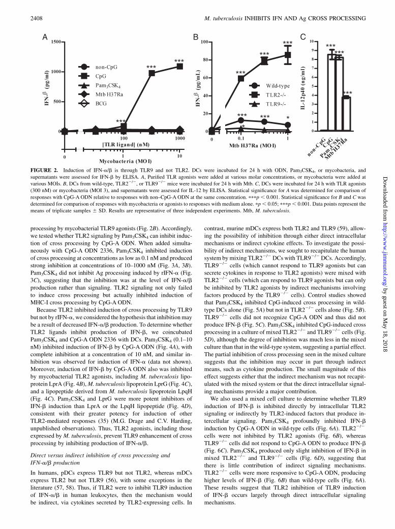
primary target for analysis. CpG-A ODN 2336 induced productionof high levels of IFN-b, whereas Pam3CSK4 did not (Fig. 2A).Mycobacteria (M. tuberculosis H37Ra or BCG) induced only lowlevels of IFN-b in wild-type DCs (Fig. 2A, 2B). M. tuberculosisTLR9 agonists were capable of inducing TLR9 signaling, becausethe IFN-b that mycobacteria induced was dependent on TLR9(absent in TLR92/2 cells; Fig. 2B) and MyD88 (absent inMyD882/2 cells; data not shown). M. tuberculosis-induced IFN-bwas increased notably in TLR22/2 DCs, suggesting that TLR2signaling by M. tuberculosis suppresses induction of IFN-a/b byM. tuberculosis TLR9 agonists. Although M. tuberculosis inducedless IFN-b than CpG-A ODN in wild-type DCs, the IFN-b levelsinduced in TLR22/2 cells (Fig. 2B) were similar to those inducedby lower-efficacy CpG-B ODN and CpG-C ODN (data not shown).Both IL-12p40 (Fig. 2C) and TNF-a (data not shown) were highlyinduced by CpG, Pam3CSK4, and mycobacteria. Thus, mycobac-teria induce TLR9 signaling, but simultaneous induction of TLR2signaling interferes with TLR9 induction of IFN-a/b. Overall,agonists that enhanced cross processing also strongly induced pro-duction of IFN-b.
TLR2 agonists inhibit TLR9 induction of IFN-a/b andMHC-I cross processing
We considered the hypothesis that mycobacterial TLR2 agonistsinhibit induction of IFN-a/b and IFN-a/b–dependent MHC-I cross
FIGURE 1. TLR9signaling inducesMHC-I cross processing through IFN-
a/b, but TLR2 signaling does not. DCs were incubated with or without
agonists for 16–18 h, washed, and incubated for 24 h with latex-OVA and
CD8OVA 1.3 T hybridoma cells. A, Induction of cross processing by CpG-A
ODN 2336 (300 nM) in wild-type versus IFN-a/bR2/2 DCs. B, DC cross
processing of latex-OVA after incubation with non–CpG-A ODN 2243 (300
nM), CpG-A ODN 2336 (300 nM), Pam3CSK4 (10 nM), or M. tuberculosis
(MOI 1). Statistical significance was determined for comparison of responses
with CpG-A ODN relative to responses by wild-type cells without CpG-A
ODN or with non–CpG-A ODN at the same Ag concentration. ppp , 0.01;
pppp , 0.001. Data points represent the means of triplicate samples 6 SD.
Results are representative of three or more independent experiments.
The Journal of Immunology 2407
by guest on May 18, 2018
http://ww
w.jim
munol.org/
Dow
nloaded from
processing by mycobacterial TLR9 agonists (Fig. 2B). Accordingly,we tested whether TLR2 signaling by Pam3CSK4 can inhibit induc-tion of cross processing by CpG-A ODN. When added simulta-neously with CpG-A ODN 2336, Pam3CSK4 inhibited inductionof cross processing at concentrations as low as 0.1 nM and producedstrong inhibition at concentrations of 10–1000 nM (Fig. 3A, 3B).Pam3CSK4 did not inhibit Ag processing induced by rIFN-a (Fig.3C), suggesting that the inhibition was at the level of IFN-a/bproduction rather than signaling. TLR2 signaling not only failedto induce cross processing but actually inhibited induction ofMHC-I cross processing by CpG-A ODN.Because TLR2 inhibited induction of cross processing by TLR9
but not by rIFN-a, we considered the hypothesis that inhibitionmaybe a result of decreased IFN-a/b production. To determine whetherTLR2 ligands inhibit production of IFN-b, we coincubatedPam3CSK4 and CpG-A ODN 2336 with DCs. Pam3CSK4 (0.1–10nM) inhibited induction of IFN-b by CpG-A ODN (Fig. 4A), withcomplete inhibition at a concentration of 10 nM, and similar in-hibition was observed for induction of IFN-a (data not shown).Moreover, induction of IFN-b by CpG-A ODN also was inhibitedby mycobacterial TLR2 agonists, including M. tuberculosis lipo-protein LprA (Fig. 4B),M. tuberculosis lipoprotein LprG (Fig. 4C),and a lipopeptide derived from M. tuberculosis lipoprotein LpqH(Fig. 4C). Pam3CSK4 and LprG were more potent inhibitors ofIFN-b induction than LprA or the LpqH lipopeptide (Fig. 4D),consistent with their greater potency for induction of otherTLR2-mediated responses (35) (M.G. Drage and C.V. Harding,unpublished observations). Thus, TLR2 agonists, including thoseexpressed byM. tuberculosis, prevent TLR9 enhancement of crossprocessing by inhibiting production of IFN-a/b.
Direct versus indirect inhibition of cross processing andIFN-a/b production
In humans, pDCs express TLR9 but not TLR2, whereas mDCsexpress TLR2 but not TLR9 (56), with some exceptions in theliterature (57, 58). Thus, if TLR2 were to inhibit TLR9 inductionof IFN-a/b in human leukocytes, then the mechanism wouldbe indirect, via cytokines secreted by TLR2-expressing cells. In
contrast, murine mDCs express both TLR2 and TLR9 (59), allow-ing the possibility of inhibition through either direct intracellularmechanisms or indirect cytokine effects. To investigate the possi-bility of indirect mechanisms, we sought to recapitulate the humansystem bymixing TLR22/2DCs with TLR92/2DCs. Accordingly,TLR92/2 cells (which cannot respond to TLR9 agonists but cansecrete cytokines in response to TLR2 agonists) were mixed withTLR22/2 cells (which can respond to TLR9 agonists but can onlybe inhibited by TLR2 agonists by indirect mechanisms involvingfactors produced by the TLR92/2 cells). Control studies showedthat Pam3CSK4 inhibited CpG-induced cross processing in wild-type DCs alone (Fig. 5A) but not in TLR22/2 cells alone (Fig. 5B).TLR92/2 cells did not recognize CpG-A ODN and thus did notproduce IFN-b (Fig. 5C). Pam3CSK4 inhibited CpG-induced crossprocessing in a culture of mixed TLR22/2 and TLR92/2 cells (Fig.5D), although the degree of inhibition was much less in the mixedculture than that in the wild-type system, suggesting a partial effect.The partial inhibition of cross processing seen in the mixed culturesuggests that the inhibition may occur in part through indirectmeans, such as cytokine production. The small magnitude of thiseffect suggests either that the indirect mechanism was not recapit-ulated with the mixed system or that the direct intracellular signal-ing mechanisms provide a major contribution.We also used a mixed cell culture to determine whether TLR9
induction of IFN-b is inhibited directly by intracellular TLR2signaling or indirectly by TLR2-induced factors that produce in-tercellular signaling. Pam3CSK4 profoundly inhibited IFN-binduction by CpG-A ODN in wild-type cells (Fig. 6A). TLR22/2
cells were not inhibited by TLR2 agonists (Fig. 6B), whereasTLR92/2 cells did not respond to CpG-A ODN to produce IFN-b(Fig. 6C). Pam3CSK4 produced only slight inhibition of IFN-b inmixed TLR22/2 and TLR92/2 cells (Fig. 6D), suggesting thatthere is little contribution of indirect signaling mechanisms.TLR22/2 cells were more responsive to CpG-A ODN, producinghigher levels of IFN-b (Fig. 6B) than wild-type cells (Fig. 6A).These results suggest that TLR2 inhibition of TLR9 inductionof IFN-b occurs largely through direct intracellular signalingmechanisms.
C
FIGURE 2. Induction of IFN-a/b is through TLR9 and not TLR2. DCs were incubated for 24 h with ODN, Pam3CSK4, or mycobacteria, and
supernatants were assessed for IFN-b by ELISA. A, Purified TLR agonists were added at various molar concentrations, or mycobacteria were added at
various MOIs. B, DCs from wild-type, TLR22/2, or TLR92/2 mice were incubated for 24 h with Mtb. C, DCs were incubated for 24 h with TLR agonists
(300 nM) or mycobacteria (MOI 3), and supernatants were assessed for IL-12 by ELISA. Statistical significance for A was determined for comparison of
responses with CpG-A ODN relative to responses with non–CpG-A ODN at the same concentration. pppp , 0.001. Statistical significance for B and C was
determined for comparison of responses with mycobacteria or agonists to responses with medium alone. pp, 0.05; pppp, 0.001. Data points represent the
means of triplicate samples 6 SD. Results are representative of three independent experiments. Mtb, M. tuberculosis.
2408 M. tuberculosis INHIBITS IFN AND Ag CROSS PROCESSING
by guest on May 18, 2018
http://ww
w.jim
munol.org/
Dow
nloaded from

M. tuberculosis inhibits TLR9 induction of IFN-a/b andMHC-I cross processing
Because M. tuberculosis expresses TLR2 agonists, we tested itseffect on induction of MHC-I cross processing and IFN-a/b. Inwild-type cells, CpG-A ODN induction of MHC-I cross processing
was inhibited by M. tuberculosis H37Ra (multiplicity of infection[MOI] of 1; Fig. 7A).M. tuberculosis inhibition of cross processingwas TLR2-dependent, because it was reversed in TLR22/2 cells(Fig. 7B), although the degree of TLR2 dependence was not com-plete in all of the experiments. The level of inhibition in wild-type cells depended on the dose of M. tuberculosis (MOI 0.01–1;Fig. 7C). Incubation of wild-type DCs with M. tuberculosis H37Ra(MOI 0.3–3) inhibited IFN-b production (Fig. 8A, 8B). M.tuberculosis H37Rv also inhibited IFN-b production in wild-type
FIGURE 3. TLR2 agonists inhibit TLR9 induction of cross processing.
DCswere treated as in Fig. 1 with CpG-AODN2336 (300 nM) or rIFN-a4 (1
ng/ml; 137.5 U/ml) and various doses of Pam3CSK4. A, Induction of cross
processing after incubation with CpG-A ODN 2336 (300 nM) or inhibition
with Pam3CSK4. T cell response is shown as a function of Ag dose with
different lines for each Pam3CSK4 condition. Medium and CpG are repre-
sented with solid lines, and conditions with Pam3CSK4 are represented with
dotted lines. B, T cell response is shown as a function of Pam3CSK4 concen-
tration with different lines for each concentration of latex-OVA (drawn from
the same data set as A).C, Induction of cross processing after incubation with
IFN-a4 (1 ng/ml) and Pam3CSK4 (10 nM). Data points represent the means
of triplicate samples6 SD. The addition of Pam3CSK4 produced a significant
inhibition of MHC-I cross processing relative to CpG-A ODN alone. p ,0.0001 for all of the values with 100 nM Pam3CSK4; p, 0.001 for all of the
values with 10 nM Pam3CSK4; p , 0.005 for all of the values with 1 nM
Pam3CSK4; p, 0.05 for all of the values with 0.1 nM Pam3CSK4, except at
316 ng/ml latex-OVA. Comparison of responses between cells treated with
IFN-a4 in the presence or absence of Pam3CSK4 resulted in a difference that
was not significant. Data points represent the means of triplicate samples 6SD. Results are representative of three independent experiments.
FIGURE 4. TLR2 agonists inhibit TLR9 induction of IFN-a/b. DCs were
treated as in Fig. 2.A, Various doses ofCpG-AODN2336or Pam3CSK4were
added to DCs. B, Various doses of CpG-A ODN 2336 or LprAwere added
to DCs. C, Various doses of CpG-A ODN were combined with medium,
LpqH lipopeptide, LprA, or LprG (TLR2 agonists all at concentrations of
10 nM). D, Various doses of Pam3CSK4, LpqH lipopeptide, LprA, or LprG
were combined with 1 mM CpG-A ODN. Data points represent the means
of triplicate samples6 SD. For A–C, the addition of lipoproteins produced a
significant inhibition of IFN-b relative to that of CpG-A ODN alone. pp ,0.05 for all of the values; ppp, 0.01 for all of the values; pp, 0.05 for 0.1
nM Pam3CSK4 at 100 and 316 nM CpG only. For D, the addition of lipo-
proteins produced a significant inhibition of IFN-b relative to that of CpG-A
ODNalone (0 nM lipoprotein). p, 0.01 for LprA for 3.16–100 nM; p, 0.01
for Pam3CSK4, LpqH lipopeptide, and LprG at all of the values. Results are
representative of four or more independent experiments for A and two or
more independent experiments for each lipoprotein for B–D.
The Journal of Immunology 2409
by guest on May 18, 2018
http://ww
w.jim
munol.org/
Dow
nloaded from

DCs (Fig. 8C), and this effect was reversed in TLR22/2 cells (Fig.8D), confirming that this effect occurs with virulentM. tuberculosis.These results indicate that M. tuberculosis signaling through TLR2inhibits induction of IFN-a/b, decreasing MHC-I cross processing,whichmay reduce priming ofM. tuberculosis-specific CD8+ T cells.
Exogenous IFN-a/b or prior induction of IFN-a/b canovercome M. tuberculosis inhibition and induce MHC-I crossprocessing of mycobacterial Ag
Because mycobacterial TLR2 agonists inhibit the ability of TLR9agonists from M. tuberculosis to induce IFN-a/b, the potential forM. tuberculosis-induced TLR9 signaling to activate MHC-I crossprocessing of mycobacterial Ag may be diminished in the course of
infection. We tested whether other sources of IFN-a/b may still beeffective for induction of MHC-I cross processing by mycobacteria-infected cells. We assessed MHC-I cross processing of BCG-OVAfor presentation to CD8OVA T hybridoma cells (Fig. 9); in thisscenario, BCG-OVA is both providing the Ag for cross processingand potentially regulating induction of endogenous IFN-a/b thatcan promote cross processing activity. Incubation of DCs withBCG-OVA alone produced only a low level of cross presentation,but addition of exogenous IFN-a at the same time or prior toaddition of mycobacteria resulted in strong cross processing (Fig.9A). Similar results were achieved with exogenous IFN-b (data notshown). Although addition of mycobacteria at the same time asCpG-A ODN inhibited induction of MHC-I cross processing (Figs.
FIGURE 5. TLR2 inhibition of TLR9-induced cross processing is par-
tially indirect. DCs were treated as in Fig. 1 with CpG-A ODN 2336
(300 nM) and Pam3CSK4 (10 nM). A, Wild-type DCs (105 per well). B,
TLR22/2 DCs (105 per well). C, TLR92/2 DCs (105 per well). D,
TLR22/2 DCs and TLR92/2 DCs (5 3 104 of each per well). Statistical
significance was determined for comparison of responses between cells
treated with CpG with or without Pam3CSK4. ppp , 0.01; pppp ,0.001. Data points represent the means of triplicate samples 6 SD. Results
are representative of three independent experiments.
FIGURE 6. TLR2 inhibition of TLR9-induced IFN-a/b production is
partially indirect. DCs were treated as in Fig. 2 with various doses of
CpG-A ODN 2336 and Pam3CSK4 (10 nM). A, Wild-type DCs (105 per
well). B, TLR22/2 DCs (105 per well). C, TLR92/2 DCs (105 per well). D,
TLR22/2 DCs and TLR92/2 DCs (105 of each per well). Statistical signif-
icance was determined for comparison of responses between cells treated
with CpG-A ODN with or without Pam3CSK4. ppp , 0.01; pppp , 0.001.
Data points represent the means of triplicate samples 6 SD. Results are
representative of three independent experiments.
2410 M. tuberculosis INHIBITS IFN AND Ag CROSS PROCESSING
by guest on May 18, 2018
http://ww
w.jim
munol.org/
Dow
nloaded from

8, 9B), addition of CpG-A ODN prior to addition of mycobacteriaenabled MHC-I cross processing of BCG-OVA, and longer preincu-bations with CpG-AODN resulted inmore efficient cross processing(Fig. 9B). When incubated without any IFN-a/b stimulus other thanthat endogenously induced by BCG-OVA, we found that TLR22/2
DCs processed mycobacterial Ag more efficiently than wild-type orTLR92/2 DCs (Fig 9C). Although M. tuberculosis inhibits endog-enous production of IFN-a/b that can induce MHC-I Ag cross
A
B
C
FIGURE 7. M. tuberculosis inhibits TLR9 induction of cross process-
ing. DCs were treated as in Fig. 1 with CpG-A ODN 2336 (300 nM) and
various doses of Mtb H37Ra. A, Induction of cross processing in wild-
type DCs by CpG-A ODN 2336 and inhibition by Mtb (MOI 1). B, In-
duction of cross processing in TLR22/2 DCs by CpG-A ODN 2336 and
lack of inhibition by Mtb (MOI 1). C, Induction of cross processing by
CpG-A ODN 2336 in wild-type DCs and inhibition by various doses of
M. tuberculosis. For A and B, statistical significance was determined for
comparison of CpG-treated cells with or without M. tuberculosis at the
same Ag concentration. ppp , 0.01. For C, statistical significance was
determined for comparison of cells treated with Mtb H37Ra at various
MOIs to cells without Mtb H37Ra (MOI 0). pp , 0.05; ppp , 0.01;
pppp , 0.001. Data points represent the means of triplicate samples 6SD. Results are representative of three independent experiments. Mtb,
M. tuberculosis.FIGURE 8. M. tuberculosis inhibits TLR9 induction of IFN-a/b. DCs
were incubated with CpG ODN and M. tuberculosis as in Fig. 2. A and B,
Induction of IFN-b by CpG-A ODN 2336 is inhibited by M. tuberculosis
H37Ra. C, Induction of IFN-b by CpG-A ODN 2336 is inhibited by M.
tuberculosisH37Rv (MOI 1). D,M. tuberculosis H37Rv inhibition of IFN-b
is dependent on TLR2. TLR22/2 DCs were incubated with CpG-A ODN
2336with or withoutM. tuberculosisH37Rv (MOI 1). Statistical significance
was determined for comparison of cells treated withM. tuberculosis H37Ra
or H37Rv at various MOI to cells withoutM. tuberculosis at the same dose of
CpG-A ODN. pppp , 0.001. Data points represent the means of triplicate
samples 6 SD. Results are representative of three or more independent
experiments for A and B and two independent experiments for C and D.
The Journal of Immunology 2411
by guest on May 18, 2018
http://ww
w.jim
munol.org/
Dow
nloaded from

processing, IFN-a/b from other sources or prior induction ofendogenous IFN-a/b can overcome mycobacterial inhibition andinduce MHC-I cross processing of Ag expressed in mycobacteria.
TLR2 agonists in an adjuvant model inhibit CpG induction ofcross priming in vivo
We examined a potential role for interactions between TLR2 andTLR9 signaling in the regulation of vaccine responses in vivo.CFSE-labeled OT-I cells (which express CD45.2) were adoptivelytransferred (1 3 106–5 3 106 cells) into B6.SJL mice (which ex-press CD45.1). The recipient animals then were immunized withOVAwith or without CpG-A 2336, Pam3CSK4, or a combination ofCpG-A and Pam3CSK4. Although TLR2 agonists induced a lowlevel of OT-I T cell proliferation, they effectively inhibited induc-tion of higher levels of OT-I proliferation by TLR9 agonists (Fig.10). These results indicate that TLR2 agonists may regulateresponses to CpG ODN in vivo, particularly induction of CD8+
T cell responses. Reduced CD8+ T cell responses may result fromTLR2 inhibition of TLR9-induced IFN-a/b–dependent MHC-Icross processing. Moreover, these results indicate that the inclusionof TLR2 agonists in vaccine adjuvants or the presence of TLR2agonists in vaccine vectors (e.g., BCG) may have an importantinfluence on the nature of vaccine-induced T cell immunity.
DiscussionPathogens may express agonists of more than one TLR or innateimmune receptor, and innate immune recognition by multiplereceptorsmay produce a distinct response pattern.M. tuberculosis isa human pathogen that expresses agonists of both TLR9 (DNApresent in the bacterial cytoplasm) and TLR2 (lipoproteins andglycolipids present in the cell wall, some of which are shed from thebacteria). Our studies demonstrate that TLR9 agonists induce IFN-a/b and MHC-I Ag cross processing, whereas TLR2 agonists andwhole mycobacteria do not. Furthermore, TLR2 agonists inhibitTLR9 induction of both IFN-a/b and MHC-I cross processing.Infection of DCs with mycobacteria resulted in TLR2-dependentinhibition of IFN-a/b expression, resulting in decreased inductionof IFN-a/b–dependentMHC-I cross processing. Thus, even thoughM. tuberculosis expresses TLR9 agonists, it may prevent TLR9induction of IFN-a/b and MHC-I cross processing, potentially re-ducing induction of CD8 T cell responses. Although the host mayoften benefit from integration of signaling by multiple TLRs toproduce responses unique to the pattern of TLR stimulation, somepatterns of stimulation may produce signaling outcomes that aredetrimental to host responses to certain pathogens. Successfulpathogens like M. tuberculosis may use such mechanisms to pro-mote persistence in the host. However, the host may have evolvedsuch mechanisms to provide feedback inhibition to prevent induc-tion of excessive inflammation. In the absence of this feedbackinhibition, inflammation may be increased in a way that is ineffec-tive or even detrimental to the host in the presence of persistentinfection (60).M. tuberculosis inhibits Ag processing through many mecha-
nisms, and TLR2 may be one of many pathways exploited by thebacteria to inhibit MHC-I cross processing and presentation toCD8+ T cells. M. tuberculosis and purified TLR2 agonists inhibitboth cross processing and IFN-a/b production, but inhibition byM.tuberculosis is only partially reversed in TLR22/2 cells, suggestingthat M. tuberculosis inhibits IFN-a/b through other pathways inaddition to TLR2. Mycobacteria express a variety of molecules thatdrive host responses through different innate immune receptors,some of which may regulate induction of IFN-a/b in a mannersimilar to TLR2.Murine mDCs express both TLR2 and TLR9, allowing for the
possibility that inhibition may occur directly through intracellularsignaling by TLR2 and TLR9 in the same cell or indirectly (e.g.,through cytokines induced by TLR2 in one cell that act upon
FIGURE 9. Exogenous IFN-a/b or prior induction of IFN-a/b can
overcome M. tuberculosis inhibition and induce MHC-I cross processing
of mycobacterial Ag. DCs were incubated with medium, rIFN-a4 (1 ng/
ml, 137.5 U/ml), rIFN-b (1 ng/ml; 28.75 U/ml), or CpG-AODN 2336 (300
nM) for various periods of preincubation. BCG-OVA and CD8OVA 1.3 T
hybridoma cells were added for 24 h (in continued presence of IFN or
ODN at half of the prior concentration). A, Induction of cross processing of
mycobacterial Ag by IFN-a4. At MOIs of 10 and 31.6, p, 0.005 for all of
the points with IFN-a4 relative to medium. B, Induction of cross
processing of mycobacterial Ag by preincubation with CpG-A ODN
2336. For comparison with the non-CpG condition, p , 0.01 for MOIs
10 and 31.6 at 0 h CpG ODN preincubation; p , 0.005 for MOI 3.16 and
p , 0.0005 at 2.5 h; p , 0.0005 for MOIs 3.16, 10, and 31.6 at 8 h; p ,0.05 for MOI 1 at 18 h). C, Induction of cross processing in 3 3 105 wild-
type, TLR22/2, or TLR92/2 DCs that were incubated 24 h in V-bottom
96-well plates with BCG-OVA (MOI 0.1–10) and CD8OVA 1.3 T hybrid-
oma cells. For C, statistical significance was determined for comparison of
induction of cross processing in wild-type and TLR22/2 cells. ppp, 0.01;
pppp, 0.001. Data points represent the means of triplicate samples6 SD.
Results are representative of at least three independent experiments.
2412 M. tuberculosis INHIBITS IFN AND Ag CROSS PROCESSING
by guest on May 18, 2018
http://ww
w.jim
munol.org/
Dow
nloaded from

another cell). In contrast, humanmDCs express TLR2 but not TLR9(56), with some exceptions in the literature (57, 58), and humanpDCs express TLR9 but not TLR2, suggesting that TLR2 inhibitionof TLR9-induced responses may occur through indirect mecha-nisms in humans. For example, in human PBMCs hepatitis C viruscore protein inhibits TLR9 induction of IFN-a/b through TLR2signaling that produces inhibitory cytokines (61). We investigatedindirect mechanisms in the murine system by mixing TLR22/2
DCs with TLR92/2 DCs. Any inhibition observed in this systemmust be derived indirectly by one set of cells responding to TLR2
agonists and inhibiting other cells that respond to TLR9 agonists.We found that inhibition occurs in this system, but it is less efficientthan that in wild-type murine DCs. This suggests that a secretedfactor may be responsible for at least part of the inhibition by TLR2in a manner similar to that of human PBMCs. The small magnitudeof inhibition in this system, however, suggests that inhibition mayoccur partly or largely through intracellular signaling in wild-type cells. Alternatively, the mechanism in wild-type cells mayinvolve cytokines in a manner that is incompletely recapitulatedby mixing TLR22/2 and TLR92/2 cells. This may be due to highlyefficient autocrine signaling in the wild-type cells that is replacedby less efficient paracrine signaling in the mixed cell system. Be-cause we add TLR2 and TLR9 agonists simultaneously, there maybe insufficient time for production of cytokines to produce a strongindirect inhibitory effect, and the magnitude of inhibition in wild-type cells despite the simultaneous addition of both signals suggeststhat direct signaling may be an important factor.During mycobacterial infection in vivo, IFN-a/b may produce
a balance of beneficial effects (e.g., by enhancing priming of CD8+
T cells) and harmful effects [e.g., increasing bacterial proliferationin infected cells (47) and organism burden in vivo (45) ordecreasing induction of Th1 immunity (45)]. In M. tuberculosisinfection, it is unclear whether inhibition of the IFN-a/b pathwayis of greater benefit to the pathogen (as a means of immune evasion)or the host (as a means to limit pathogen-beneficial effects or host-detrimental excessive inflammation). Some virulent M. tuberculo-sis strains have been reported to induce higher levels of IFN-a/bthan avirulent mycobacterial strains (45), suggesting that IFN-a/bmay promote progression of infection with these strains. This couldresult from greater expression of IFN-a/b-inducing molecules bythese virulent M. tuberculosis strains. Alternatively, our resultssuggest that these strains may express lower levels of TLR2 agonistactivity, resulting in reduced inhibition of IFN-a/b induction andgreater levels of IFN-a/b expression.In the setting of vaccination, however, the strategic balance of
IFN-a/b effects may be different, because innate immune effects ofIFN-a/b that increase organism burden during M. tuberculosisinfection may not be of significance during vaccination, and thedominant mechanisms may be those that affect T cell responses tovaccination. We observed that prior induction of IFN-a/b signalingallows DCs to effectively cross process and present Ags expressedby mycobacteria in vitro, and this mechanism could enhance T cellresponses to mycobacterial vaccine strains. Our vaccination studiesfurther substantiate that TLR2 responses can inhibit TLR9induction of CD8+ T cell responses in vivo, a mechanism ofpotential significance for responses to bacterial infection andbacterial vaccine strains. The BCG vaccine uses an organism thatactivates an immune response but inhibits production of IFN-a/b;strategies that enhance IFN-a/b effects might enhance induction ofT cell immunity by BCG. Another possibility is vaccination witha TLR9 agonist andM. tuberculosis Ags to prime strong CD4+ andCD8+ responses that could be further enhanced by subsequent BCGvaccination. Our studies indicate the importance of consideringoutcomes that are determined by combinations of signals fromdifferent receptors that differentially regulate immune responses(e.g., production of IFN-a/b). Furthermore, the complexity ofsuch mechanisms may influence responses to infection andvaccination in different ways.
AcknowledgmentsWe thank Shizuo Akira for MyD882/2, TLR22/2, and TLR92/2 mice and
Subash Sad and Kevin Urdahl for recombinant BCG-OVA. Kitty Daniel
and Supriya Shukla provided lipoprotein preparations.
A B
C D
E
FIGURE 10. TLR2 agonist inhibits TLR9-induced priming of CD8+
T cells invivo.A–D, CFSE-labeledOT-I splenocytes (expressingCD45.2)were
adoptively transferred into a congenic B6.SJL mouse (expressing CD45.1).
After 72 h, mice were immunized in the dorsum of the foot with OVA
(20mg) with or without CpG-A 2336 (25mg), Pam3CSK4 (25mg), or a combi-
nation of CpG-A 2336 (25 mg) and Pam3CSK4 (25 mg). Two days after immu-
nization, popliteal lymph nodes were collected and analyzed separately for
each mouse. Cells were stained with allophycocyanin-conjugated anti-
CD45.2, and gating for OT-I cells was based on CD45.2 expression. Prolifera-
tion was assessed by CFSE dye dilution.E, Percentage of cells that dividedwas
determined by FlowJo software. Student t test was performed to compare T cell
responses between groups of mice as indicated. In addition, analysis of com-
bined data from three independent experiments revealed a significant enhance-
ment of responses of mice immunized with OVA and CpG (n = 12) relative to
that ofmice immunizedwith OVA alone (n = 11, p, 0.0001), and responses of
mice immunized with OVA, CpG, and Pam3CSK4 (n = 12) were significantly
lower than those of mice immunized with OVA and CpG (p = 0.0116).
The Journal of Immunology 2413
by guest on May 18, 2018
http://ww
w.jim
munol.org/
Dow
nloaded from

DisclosuresThe authors have no financial conflicts of interest.
References1. Mogues, T., M. E. Goodrich, L. Ryan, R. LaCourse, and R. J. North. 2001. The
relative importance of T cell subsets in immunity and immunopathology ofairborne Mycobacterium tuberculosis infection in mice. J. Exp. Med. 193: 271–280.
2. Muller, I., S. P. Cobbold, H. Waldmann, and S. H. Kaufmann. 1987. Impairedresistance to Mycobacterium tuberculosis infection after selective in vivo de-pletion of L3T4+ and Lyt-2+ T cells. Infect. Immun. 55: 2037–2041.
3. van Pinxteren, L. A. H., J. P. Cassidy, B. H. C. Smedegaard, E. M. Agger, andP. Andersen. 2000. Control of latent Mycobacterium tuberculosis infection isdependent on CD8 T cells. Eur. J. Immunol. 30: 3689–3698.
4. Flynn, J. L., and J. Chan. 2001. Immunology of tuberculosis. Annu. Rev.Immunol. 19: 93–129.
5. Flynn, J. L., and J. D. Ernst. 2000. Immune responses in tuberculosis. Curr.Opin. Immunol. 12: 432–436.
6. Flynn, J. L., M. M. Goldstein, K. J. Triebold, B. Koller, and B. R. Bloom. 1992.Major histocompatibility complex class I-restricted T cells are required for re-sistance toMycobacterium tuberculosis infection. Proc. Natl. Acad. Sci. USA 89:12013–12017.
7. Kamath, A. B., J. Woodworth, X. Xiong, C. Taylor, Y. Weng, and S. M. Behar.2004. Cytolytic CD8+ T cells recognizing CFP10 are recruited to the lung afterMycobacterium tuberculosis infection. J. Exp. Med. 200: 1479–1489.
8. Lewinsohn, D. A., E. Winata, G. M. Swarbrick, K. E. Tanner, M. S. Cook,M. D. Null, M. E. Cansler, A. Sette, J. Sidney, and D. M. Lewinsohn. 2007.Immunodominant tuberculosis CD8 antigens preferentially restricted by HLA-B.PLoS Pathog. 3: 1240–1249.
9. Lewinsohn, D. M., M. R. Alderson, A. L. Briden, S. R. Riddell, S. G. Reed, andK. H. Grabstein. 1998. Characterization of human CD8+ T cells reactive withMycobacterium tuberculosis-infected antigen-presenting cells. J. Exp. Med. 187:1633–1640.
10. Urdahl, K. B., D. Liggitt, and M. J. Bevan. 2003. CD8+ T cells accumulate in thelungs ofMycobacterium tuberculosis-infected Kb-/-Db-/- mice, but provide min-imal protection. J. Immunol. 170: 1987–1994.
11. Sousa, A. O., R. J. Mazzaccaro, R. G. Russell, F. K. Lee, O. C. Turner, S. Hong,L. Van Kaer, and B. R. Bloom. 2000. Relative contributions of distinct MHCclass I-dependent cell populations in protection to tuberculosis infection in mice.Proc. Natl. Acad. Sci. USA 97: 4204–4208.
12. Irwin, S. M., A. A. Izzo, S. W. Dow, Y. A. Skeiky, S. G. Reed, M. R. Alderson,and I. M. Orme. 2005. Tracking antigen-specific CD8 T lymphocytes in thelungs of mice vaccinated with the M. tuberculosis72F polyprotein. Infect.Immun. 73: 5809–5816.
13. Billeskov, R., C. Vingsbo-Lundberg, P. Andersen, and J. Dietrich. 2007.Induction of CD8 T cells against a novel epitope in TB10.4: correlation withmycobacterial virulence and the presence of a functional region of difference-1.J. Immunol. 179: 3973–3981.
14. Lewinsohn, D. M., L. Zhu, V. J. Madison, D. C. Dillon, S. P. Fling, S. G. Reed,K. H. Grabstein, and M. R. Alderson. 2001. Classically restricted human CD8+T lymphocytes derived from Mycobacterium tuberculosis-infected cells: defini-tion of antigenic specificity. J. Immunol. 166: 439–446.
15. Kamath, A., J. S. Woodworth, and S. M. Behar. 2006. Antigen-specific CD8+T cells and the development of central memory during Mycobacterium tubercu-losis infection. J. Immunol. 177: 6361–6369.
16. Orme, I. M., and F. M. Collins. 1983. Protection against Mycobacterium tu-berculosis infection by adoptive immunotherapy. Requirement for T cell-deficient recipients. J. Exp. Med. 158: 74–83.
17. Hmama, Z., R. Gabathuler, W. A. Jefferies, G. de Jong, and N. E. Reiner. 1998.Attenuation of HLA-DR expression by mononuclear phagocytes infected withMycobacterium tuberculosis is related to intracellular sequestration of immatureclass II heterodimers. J. Immunol. 161: 4882–4893.
18. Noss, E. H., R. K. Pai, T. J. Sellati, J. D. Radolf, J. Belisle, D. T. Golenbock,W. H. Boom, and C. V. Harding. 2001. Toll-like receptor 2-dependent inhibitionof macrophage class II MHC expression and antigen processing by 19-kDalipoprotein of Mycobacterium tuberculosis. J. Immunol. 167: 910–918.
19. Pancholi, P., A. Mirza, N. Bhardwaj, and R. M. Steinman. 1993. Sequestrationfrom immune CD4+ T cells of mycobacteria growing in human macrophages.Science 260: 984–986.
20. Wojciechowski, W., J. DeSanctis, E. Skamene, and D. Radzioch. 1999. Atten-uation of MHC class II expression in macrophages infected with Mycobacteriumbovis bacillus Calmette-Guerin involves class II transactivator and depends onthe Nramp1 gene. J. Immunol. 163: 2688–2696.
21. Kincaid, E. Z., and J. D. Ernst. 2003. Mycobacterium tuberculosis exerts gene-selective inhibition of transcriptional responses to IFN-gamma without inhibitingSTAT1 function. J. Immunol. 171: 2042–2049.
22. Pai, R. K., M. Convery, T. A. Hamilton, W. H. Boom, and C. V. Harding. 2003.Inhibition of IFN-gamma-induced class II transactivator expression by a 19-kDalipoprotein from Mycobacterium tuberculosis: a potential mechanism for im-mune evasion. J. Immunol. 171: 175–184.
23. Pai, R. K., M. E. Pennini, A. A. Tobian, D. H. Canaday, W. H. Boom, andC. V. Harding. 2004. Prolonged Toll-like receptor signaling by Mycobacteriumtuberculosis and its 19-kilodalton lipoprotein inhibits gamma interferon-inducedregulation of selected genes in macrophages. Infect. Immun. 72: 6603–6614.
24. Tobian, A. A., N. S. Potter, L. Ramachandra, R. K. Pai, M. Convery,W. H. Boom, and C. V. Harding. 2003. Alternate class I MHC antigen processing
is inhibited by Toll-like receptor signaling pathogen-associated molecular pat-terns: Mycobacterium tuberculosis 19-kDa lipoprotein, CpG DNA, and lipopoly-saccharide. J. Immunol. 171: 1413–1422.
25. Remoli, M. E., E. Giacomini, G. Lutfalla, E. Dondi, G. Orefici, A. Battistini,G. Uze, S. Pellegrini, and E. M. Coccia. 2002. Selective expression of type I IFNgenes in human dendritic cells infected with Mycobacterium tuberculosis. J.Immunol. 169: 366–374.
26. Cho, H. J., T. Hayashi, S. K. Datta, K. Takabayashi, J. H. Van Uden, A. Horner,M. Corr, and E. Raz. 2002. IFN-alpha beta promote priming of antigen-specificCD8+ and CD4+ T lymphocytes by immunostimulatory DNA-based vaccines. J.Immunol. 168: 4907–4913.
27. Tokunaga, T., H. Yamamoto, S. Shimada, H. Abe, T. Fukuda, Y. Fujisawa,Y. Furutani, O. Yano, T. Kataoka, T. Sudo, et al. 1984. Antitumor activity ofdeoxyribonucleic acid fraction from Mycobacterium bovis BCG. I. Isolation,physicochemical characterization, and antitumor activity. J. Natl. Cancer Inst.72: 955–962.
28. Pandey, A. K., Y. Yang, Z. Jiang, S. M. Fortune, F. Coulombe, M. A. Behr,K. A. Fitzgerald, C. M. Sassetti, and M. A. Kelliher. 2009. NOD2, RIP2 andIRF5 play a critical role in the type I interferon response to Mycobacteriumtuberculosis. PLoS Pathog. 5: e1000500.
29. Durand, V., S. Y. Wong, D. F. Tough, and A. Le Bon. 2004. Shaping of adaptiveimmune responses to soluble proteins by TLR agonists: a role for IFN-alpha/beta. Immunol. Cell Biol. 82: 596–602.
30. Kamath, A. T., C. E. Sheasby, and D. F. Tough. 2005. Dendritic cells andNK cells stimulate bystander T cell activation in response to TLR agoniststhrough secretion of IFN-alpha beta and IFN-gamma. J. Immunol. 174: 767–776.
31. Datta, S. K., V. Redecke, K. R. Prilliman, K. Takabayashi, M. Corr, T. Tallant,J. DiDonato, R. Dziarski, S. Akira, S. P. Schoenberger, and E. Raz. 2003. Asubset of Toll-like receptor ligands induces cross-presentation by bone marrow-derived dendritic cells. J. Immunol. 170: 4102–4110.
32. Kuchtey, J., P. J. Chefalo, R. C. Gray, L. Ramachandra, and C. V. Harding. 2005.Enhancement of dendritic cell antigen cross-presentation by CpG DNA involvestype I IFN and stabilization of class I MHC mRNA. J. Immunol. 175: 2244–2251.
33. Thornley, T. B., N. E. Phillips, B. C. Beaudette-Zlatanova, T. G. Markees, K. Bahl,M. A. Brehm, L. D. Shultz, E. A. Kurt-Jones, J. P. Mordes, R. M. Welsh, et al.2007. Type 1 IFN mediates cross-talk between innate and adaptive immunity thatabrogates transplantation tolerance. J. Immunol. 179: 6620–6629.
34. Pecora, N. D., A. J. Gehring, D. H. Canaday, W. H. Boom, and C. V. Harding.2006. Mycobacterium tuberculosis LprA is a lipoprotein agonist of TLR2 thatregulates innate immunity and APC function. J. Immunol. 177: 422–429.
35. Drage, M. G., H.-C. Tsai, N. D. Pecora, T.-Y. Cheng, A. R. Arida, S. Shukla,R. E. Rojas, D. B. Moody, W. H. Boom, J. C. Sacchettini, and C. V. Harding.2010. Mycobacterium tuberculosis lipoprotein LprG (Rv1411c) binds triacylatedglycolipid agonists of Toll-like receptor 2. Nat. Struct. Mol. Biol. In press.
36. Thoma-Uszynski, S., S. Stenger, O. Takeuchi, M. T. Ochoa, M. Engele,P. A. Sieling, P. F. Barnes, M. Rollinghoff, P. L. Bolcskei, M. Wagner, et al.2001. Induction of direct antimicrobial activity through mammalian Toll-likereceptors. Science 291: 1544–1547.
37. Toshchakov, V., B. W. Jones, P. Y. Perera, K. Thomas, M. J. Cody, S. Zhang,B. R. Williams, J. Major, T. A. Hamilton, M. J. Fenton, and S. N. Vogel. 2002.TLR4, but not TLR2, mediates IFN-beta-induced STAT1alpha/beta-dependentgene expression in macrophages. Nat. Immunol. 3: 392–398.
38. Kaisho, T., and S. Akira. 2003. Regulation of dendritic cell function throughToll-like receptors. Curr. Mol. Med. 3: 373–385.
39. Barbalat, R., L. Lau, R. M. Locksley, and G. M. Barton. 2009. Toll-like receptor2 on inflammatory monocytes induces type I interferon in response to viral butnot bacterial ligands. Nat. Immunol. 10: 1200–1207.
40. Bafica, A., C. A. Scanga, C. G. Feng, C. Leifer, A. Cheever, and A. Sher. 2005.TLR9 regulates Th1 responses and cooperates with TLR2 in mediating optimalresistance to Mycobacterium tuberculosis. J. Exp. Med. 202: 1715–1724.
41. Giosue, S., M. Casarini, L. Alemanno, G. Galluccio, P. Mattia, G. Pedicelli,L. Rebek, A. Bisetti, and F. Ameglio. 1998. Effects of aerosolized interferon-alpha in patients with pulmonary tuberculosis. Am. J. Respir. Crit. Care Med.158: 1156–1162.
42. Palmero, D., K. Eiguchi, P. Rendo, L. Castro Zorrilla, E. Abbate, andL. J. Gonzalez Montaner. 1999. Phase II trial of recombinant interferon-alpha2bin patients with advanced intractable multidrug-resistant pulmonary tuberculo-sis: long-term follow-up. Int. J. Tuberc. Lung Dis. 3: 214–218.
43. Ward, C. M., H. Jyonouchi, S. V. Kotenko, S. V. Smirnov, R. Patel, H. Aguila,G. McSherry, B. Dashefsky, and S. M. Holland. 2007. Adjunctive treatment ofdisseminated Mycobacterium avium complex infection with interferon alpha-2bin a patient with complete interferon-gamma receptor R1 deficiency. Eur. J.Pediatr. 166: 981–985.
44. Telesca, C., M. Angelico, P. Piccolo, L. Nosotti, A. Morrone, C. Longhi,M. Carbone, and L. Baiocchi. 2007. Interferon-alpha treatment of hepatitis Dinduces tuberculosis exacerbation in an immigrant. J. Infect. 54: e223–e226.
45. Manca, C., L. Tsenova, A. Bergtold, S. Freeman, M. Tovey, J. M. Musser,C. E. Barry, III, V. H. Freedman, and G. Kaplan. 2001. Virulence of a Myco-bacterium tuberculosis clinical isolate in mice is determined by failure to induceTh1 type immunity and is associated with induction of IFN-alpha /beta. Proc.Natl. Acad. Sci. USA 98: 5752–5757.
46. Kuchtey, J., S. A. Fulton, S. M. Reba, C. V. Harding, and W. H. Boom. 2006.Interferon-alphabeta mediates partial control of early pulmonary Mycobacteriumbovis bacillus Calmette-Guerin infection. Immunology 118: 39–49.
47. Bouchonnet, F., N. Boechat, M. Bonay, and A. J. Hance. 2002.Alpha/beta interferon impairs the ability of human macrophages to controlgrowth of Mycobacterium bovis BCG. Infect. Immun. 70: 3020–3025.
2414 M. tuberculosis INHIBITS IFN AND Ag CROSS PROCESSING
by guest on May 18, 2018
http://ww
w.jim
munol.org/
Dow
nloaded from

48. Giacomini, E., M. E. Remoli, V. Gafa, M. Pardini, L. Fattorini, and E. M. Coccia.2009. IFN-beta improves BCG immunogenicity by acting on DC maturation.J. Leukoc. Biol. 85: 462–468.
49. Sikora, A. G., N. Jaffarzad, Y. Hailemichael, A. Gelbard, S. W. Stonier,K. S. Schluns, L. Frasca, Y. Lou, C. Liu, H. A. Andersson, et al. 2009. IFN-alphaenhances peptide vaccine-induced CD8+ T cell numbers, effector function, andantitumor activity. J. Immunol. 182: 7398–7407.
50. Prabhakar, S., Y. Qiao, Y. Hoshino, M. Weiden, A. Canova, E. Giacomini,E. Coccia, and R. Pine. 2003. Inhibition of response to alpha interferon byMycobacterium tuberculosis. Infect. Immun. 71: 2487–2497.
51. Prabhakar, S., Y. Qiao, A. Canova, D. B. Tse, and R. Pine. 2005. IFN-alpha betasecreted during infection is necessary but not sufficient for negative feedbackregulation of IFN-alpha beta signaling by Mycobacterium tuberculosis. J. Immu-nol. 174: 1003–1012.
52. Mariotti, S., R. Teloni,E. Iona,L.Fattorini,G.Romagnoli,M.C.Gagliardi,G.Orefici,and R. Nisini. 2004. Mycobacterium tuberculosis diverts alpha interferon-inducedmonocyte differentiation from dendritic cells into immunoprivileged macrophage-like host cells. Infect. Immun. 72: 4385–4392.
53. Lichtner,M.,R. Rossi, F.Mengoni, S. Vignoli, B. Colacchia, A. P.Massetti, I. Kamga,A. Hosmalin, V. Vullo, and C. M. Mastroianni. 2006. Circulating dendritic cells andinterferon-alpha production in patients with tuberculosis: correlation with clinicaloutcome and treatment response. Clin. Exp. Immunol. 143: 329–337.
54. Dudani, R., Y. Chapdelaine, H. Faassen Hv, D. K. Smith, H. Shen, L. Krishnan,and S. Sad. 2002. Multiple mechanisms compensate to enhance tumor-protectiveCD8(+) T cell response in the long-term despite poor CD8(+) T cell priminginitially: comparison between an acute versus a chronic intracellular bacteriumexpressing a model antigen. J. Immunol. 168: 5737–5745.
55. Gray, R. C., J. Kuchtey, and C. V. Harding. 2007. CpG-B ODNs potently inducelow levels of IFN-alphabeta and induce IFN-alphabeta-dependent MHC-I cross-presentation in DCs as effectively as CpG-A and CpG-C ODNs. J. Leukoc. Biol.81: 1075–1085.
56. Kadowaki, N., S. Ho, S. Antonenko, R. W. Malefyt, R. A. Kastelein, F. Bazan,and Y. J. Liu. 2001. Subsets of human dendritic cell precursors express differentToll-like receptors and respond to different microbial antigens. J. Exp. Med. 194:863–869.
57. Hoene, V., M. Peiser, and R. Wanner. 2006. Human monocyte-deriveddendritic cells express TLR9 and react directly to the CpG-A oligonucleotideD19. J. Leukoc. Biol. 80: 1328–1336.
58. Semnani, R. T., P. G. Venugopal, C. A. Leifer, S. Mostbock, H. Sabzevari, andT. B. Nutman. 2008. Inhibition of TLR3 and TLR4 function and expression inhuman dendritic cells by helminth parasites. Blood 112: 1290–1298.
59. Edwards, A. D., S. S. Diebold, E. M. Slack, H. Tomizawa, H. Hemmi, T. Kaisho,S. Akira, and C. Reis e Sousa. 2003. Toll-like receptor expression in murine DCsubsets: lack of TLR7 expression by CD8 alpha+ DC correlates with unrespon-siveness to imidazoquinolines. Eur. J. Immunol. 33: 827–833.
60. Drennan, M. B., D. Nicolle, V. J. Quesniaux, M. Jacobs, N. Allie, J. Mpagi,C. Fremond, H. Wagner, C. Kirschning, and B. Ryffel. 2004. Toll-like receptor2-deficient mice succumb to Mycobacterium tuberculosis infection. Am. J.Pathol. 164: 49–57.
61. Dolganiuc, A., S. Chang, K. Kodys, P. Mandrekar, G. Bakis, M. Cormier, andG. Szabo. 2006. Hepatitis C virus (HCV) core protein-induced, monocyte-mediated mechanisms of reduced IFN-alpha and plasmacytoid dendritic cell lossin chronic HCV infection. J. Immunol. 177: 6758–6768.
The Journal of Immunology 2415
by guest on May 18, 2018
http://ww
w.jim
munol.org/
Dow
nloaded from