mycology terminology
TRANSCRIPT
8/2/2019 Mycology Terminology
http://slidepdf.com/reader/full/mycology-terminology 1/15
MYCOLOGY TERMINOLOGY
Clinical mycology remains more of a descriptive art than an analytical science. Youmay find that the identification of fungi requires a greater development of your visualacuity than was necessary in bacteriology. There are also fewer biochemical tests
available to aid in the differential identification of fungi. As a result you will spendconsiderable time in the laboratory visually examining fungal cultures. You will identifycharacteristic fungal structures by observing colonial growth both macroscopically andmicroscopically.
A thorough understanding of correct fungal terminology is of critical importance. Youmay find that the terms used to describe fungi are unusual, at times redundant, andoften very confusing. We have attempted to simplify the jumbled jargon by providingyou with the following list of terms that will be used most often in the laboratory sectionof this course. Although most of these terms have already been introduced in thelecture notes, they have been included in the laboratory manual as well.
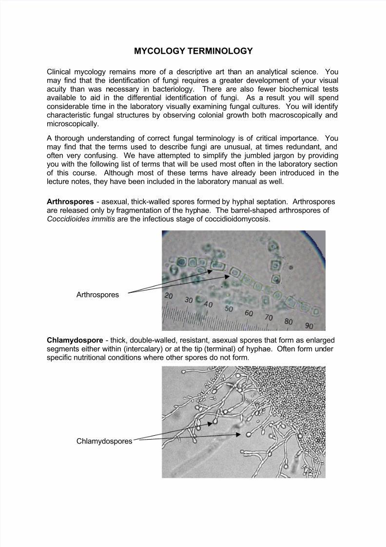
Arthrospores - asexual, thick-walled spores formed by hyphal septation. Arthrosporesare released only by fragmentation of the hyphae. The barrel-shaped arthrospores of Coccidioides immitis are the infectious stage of coccidioidomycosis.
Chlamydospore - thick, double-walled, resistant, asexual spores that form as enlargedsegments either within (intercalary) or at the tip (terminal) of hyphae. Often form under specific nutritional conditions where other spores do not form.
Arthrospores
Chlamydospores
8/2/2019 Mycology Terminology
http://slidepdf.com/reader/full/mycology-terminology 2/15
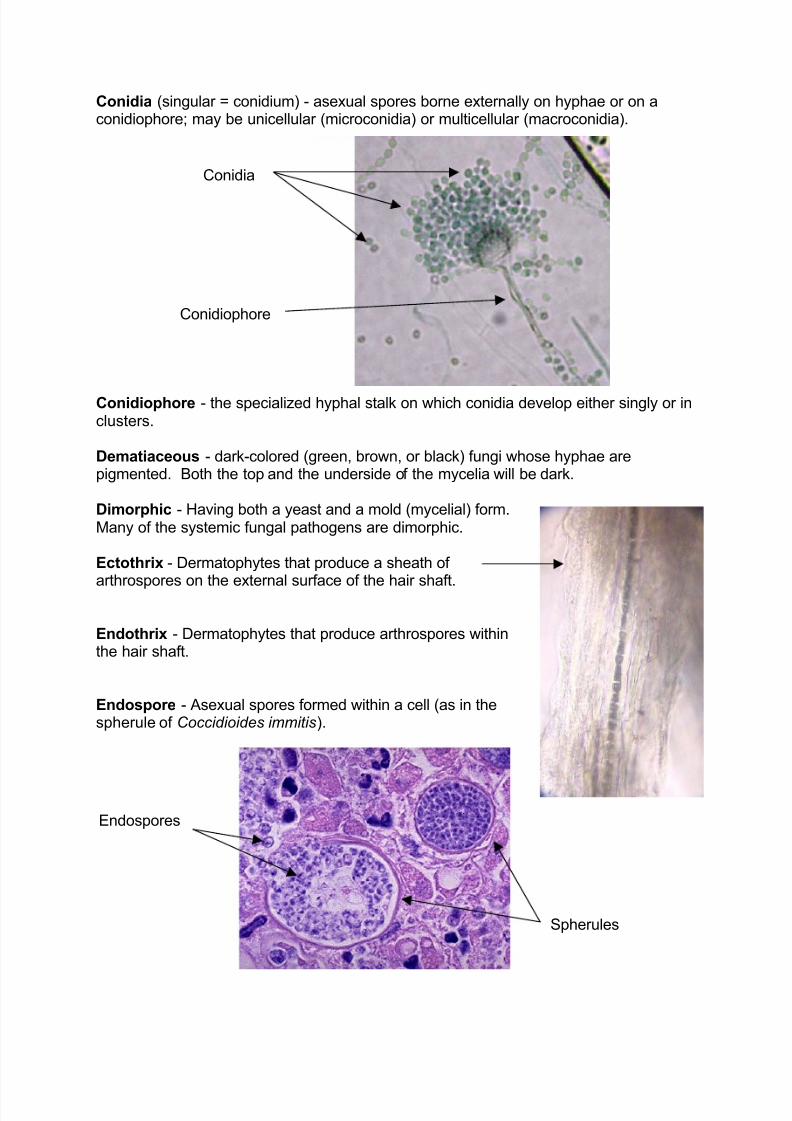
Conidia (singular = conidium) - asexual spores borne externally on hyphae or on aconidiophore; may be unicellular (microconidia) or multicellular (macroconidia).
Conidiophore - the specialized hyphal stalk on which conidia develop either singly or inclusters.
Dematiaceous - dark-colored (green, brown, or black) fungi whose hyphae arepigmented. Both the top and the underside of the mycelia will be dark.
Dimorphic - Having both a yeast and a mold (mycelial) form.Many of the systemic fungal pathogens are dimorphic.
Ectothrix - Dermatophytes that produce a sheath of arthrospores on the external surface of the hair shaft.
Endothrix - Dermatophytes that produce arthrospores withinthe hair shaft.
Endospore - Asexual spores formed within a cell (as in thespherule of Coccidioides immitis).
Conidiophore
Conidia
Endospores
Spherules

8/2/2019 Mycology Terminology
http://slidepdf.com/reader/full/mycology-terminology 3/15
Geophilic - Refers to fungi whose natural habitat is thesoil. Use of this term is generally restricted to certainDermatophytes (e.g. Microsporum gypseum).
Germ-tube - The initial hyphal outgrowth of a
germinating spore or yeast; especially important for identification of Candida albicans.
Mycelium - the intertwined mass of hyphae that formsthe mold colony. The vegetative mycelium is composedof those hyphae that adhere to the substrate andabsorbs nutrients. The aerial mycelium is composed of those hyphae that grow up from the surface and support the spores.
Pseudohyphae - chains of successively buddingyeast cells that have complete cell walls, but havenot detached from one another.
Rhizoid - rootlike branched hyphae which anchor themycelium to the substrate; characteristic of certainZygomycetes (Rhizopus and Absidium).
Septate - Cross-walls (septae) that divide hyphaeinto segments. If there are few or no cross-walls thehyphae are considered to be aseptate.
Sporangia (singular = sporangium)-spherical sack within which asexual
Germ tube
Pseudohyphae
Rhizoid
Septae
8/2/2019 Mycology Terminology
http://slidepdf.com/reader/full/mycology-terminology 4/15
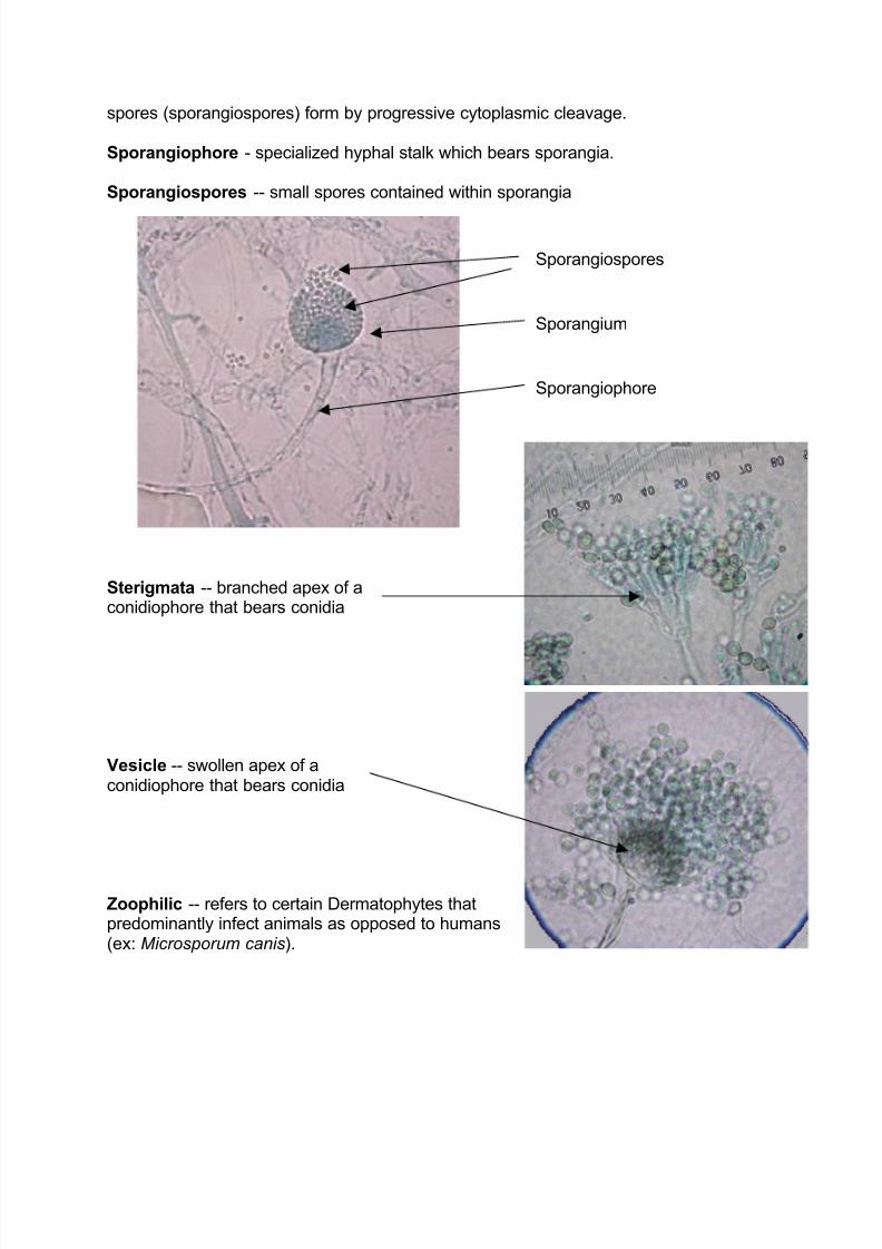
spores (sporangiospores) form by progressive cytoplasmic cleavage.
Sporangiophore - specialized hyphal stalk which bears sporangia.
Sporangiospores -- small spores contained within sporangia
Sterigmata -- branched apex of aconidiophore that bears conidia
Vesicle -- swollen apex of aconidiophore that bears conidia
Zoophilic -- refers to certain Dermatophytes thatpredominantly infect animals as opposed to humans(ex: Microsporum canis).
Sporangium
Sporangiospores
Sporangiophore
8/2/2019 Mycology Terminology
http://slidepdf.com/reader/full/mycology-terminology 5/15
IDENTIFICATION OF FUNGAL ORGANISMS
ZYGOMYCETES
Zygomycetes are rapidly growing molds that typically produce abundant hyphae that
look like cotton candy and fill the air space in the tube or plate. They grow very rapidly(perhaps within 1-2 days) both at room temperature and at 37°C (thermotolerant). The
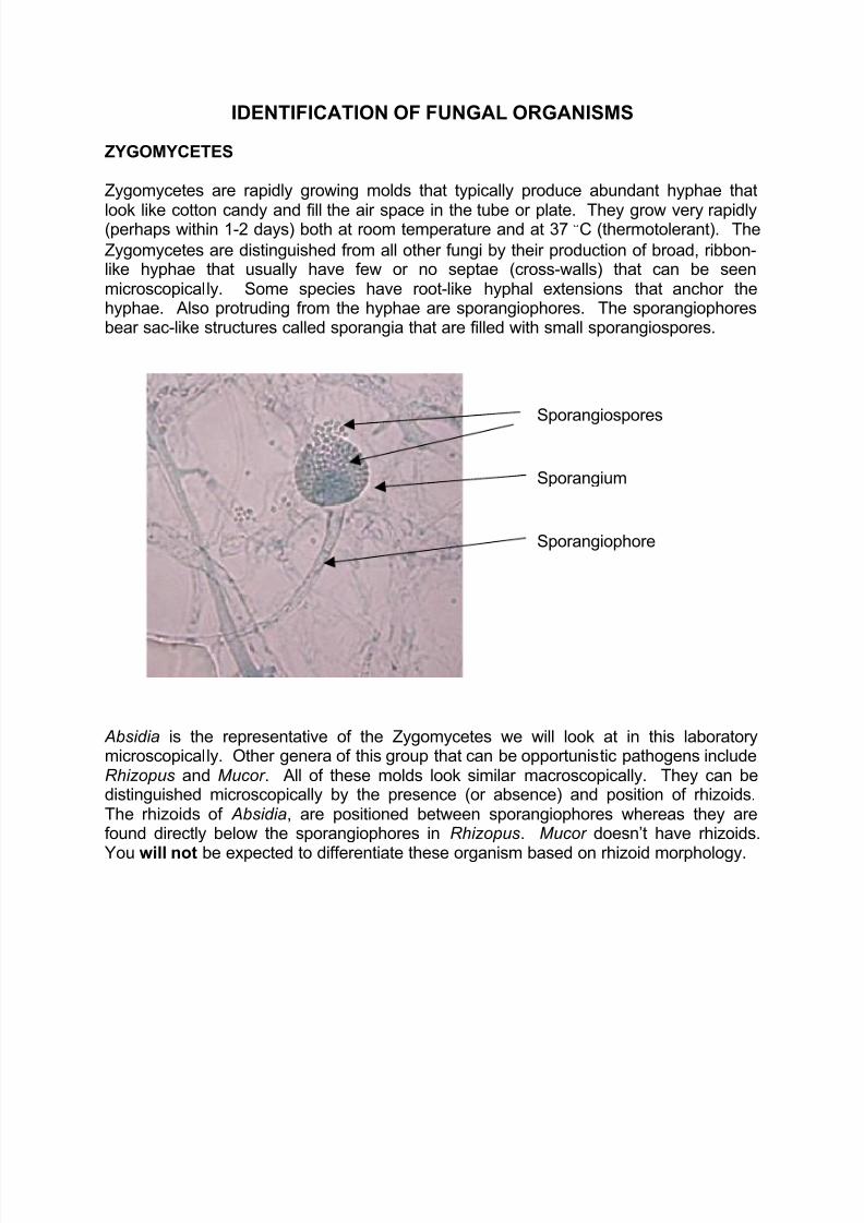
Zygomycetes are distinguished from all other fungi by their production of broad, ribbon-like hyphae that usually have few or no septae (cross-walls) that can be seenmicroscopically. Some species have root-like hyphal extensions that anchor thehyphae. Also protruding from the hyphae are sporangiophores. The sporangiophoresbear sac-like structures called sporangia that are filled with small sporangiospores.
Absidia is the representative of the Zygomycetes we will look at in this laboratorymicroscopically. Other genera of this group that can be opportunistic pathogens includeRhizopus and Mucor . All of these molds look similar macroscopically. They can bedistinguished microscopically by the presence (or absence) and position of rhizoids.The rhizoids of Absidia, are positioned between sporangiophores whereas they arefound directly below the sporangiophores in Rhizopus. Mucor doesn’t have rhizoids.
You will not be expected to differentiate these organism based on rhizoid morphology.
Sporangium
Sporangiospores
Sporangiophore

8/2/2019 Mycology Terminology
http://slidepdf.com/reader/full/mycology-terminology 6/15
DEMATIACEOUS MOLDS
The term dematiaceous refers to molds whose hyphae are pigmented. Because of thepigment, the colonies of these fungi will appear dark green, brown, or black on both thetop and reverse (underside). We will provide you with an isolate of Alternaria as an
example of this group of fungi. Although it is hardly ever associated with infection, it is avery common contaminant, especially in specimens collected in the barn. Note the“hand-grenade” shape of the pigmented macroconidia. Other opportunistic pathogensof the dematiaceous group include Cladosporium, Phialophora, and Drechslera.

8/2/2019 Mycology Terminology
http://slidepdf.com/reader/full/mycology-terminology 7/15
HYALINE MOLDS
Hyaline molds are fast-growing fungi that are common contaminants and occasionalopportunistic pathogens. Some of these fungi can be relatively common and virulentpathogens (for example Aspergillus fumigatus in birds). Examples of opportunistic
pathogens in this group include Pseudallescheria boydii , Aspergillus sp., Penicillium sp.,and Fusarium sp.
Macroscopically, the colonies of these molds are lightly pigmented or non-pigmented.They may be white, light grey, green or light brown. The color is seen chiefly on top of the colony and is due to pigment in the many spores that are produced, not the hyphae.The hyphae are colorless (hyaline) and are septate. These organisms have hyphal
projections called conidiophores. Some conidiophores terminate in either a swollenvesicle or a branched sterigmata. The vesicle and sterigmata bear abundant sporescalled conidia. These structures as a unit are sometimes referred to as “fruiting heads”or “fruiting bodies.” Other conidiophores bear conidia singly or in small clusters.
Most colonies of Penicillium sp.are someshade of green, but grey, yellow, orange,pink, or white colors may be
encountered. The surface of thecolonies is powdery because of the hugenumbers of conidia. The septate hyphaeof Penicillium sp. produce conidiophores which terminate inbranched sterigmata that bear conidia.
Aspergillus colonies are often white,grey, or green. They may also beyellow, tan, or brown. The colonies of
Aspergillus niger are black. Theseptate hyphae of Aspergillus sp.produce conidiophores whichterminate in swollen vesicles that
bear chains of conidia.

8/2/2019 Mycology Terminology
http://slidepdf.com/reader/full/mycology-terminology 8/15
The colonies of Fusarium sp. are cottony and have a distinctive lavender color. Their hyphae are septate and produce both macroconidia and microconidia.The microconidiaare oval. The macroconidia are banana-shaped and multi-cellular . Typically, themacroconidia are produced in small clusters.
The colonies of Pseudallescheria boydii have a low, cottony surface that appearswithin 2-6 days. Unlike many hyaline molds, it will grow on mycobiotic agar. Theseptate hyphae produce single conidia that resemble lollipops. These lollipop conidia are similar to those produced by an important fungal pathogen, Blastomycesdermatitidis. Fortunately, Blastomyces can be converted to a yeast at 37° (we’ll
discuss this later) while P. boydii cannot.

8/2/2019 Mycology Terminology
http://slidepdf.com/reader/full/mycology-terminology 9/15
YEASTS
The colonial and microscopic appearance of yeasts differs greatly from that of molds.They grow somewhat more rapidly and may prefer to grow at warmer temperatures (35-37°C) than room temperature. Unlike molds, they do not have a fuzzy colonial
appearance because they do not produce hyphae. Yeast colonies are flat and creamyin consistency, similar to many bacterial colonies. They grow a little more slowly, maybecome a little larger, and may have a more dense convex colonial appearance thanbacterial colonies.
Microscopically, yeasts are larger than bacteria and can be visualized easily at 400xmagnification (40x objective). Yeasts reproduce by budding -- the cell wall projectsoutward to form a new daughter cell that eventually pinches off from the mother cell.Some may not separate entirely while budding, forming chains of unseparated yeastcells called pseudohyphae.
Cryptococcus neoformans producesmucoid colonies on Sabouraud agar and brain-heart infusion agar.Microscopically, these organisms areseen as budding yeasts with a narrowneck connecting the mother anddaughter yeast cells. The productionof a large, mucoid capsule is a uniquefeature of Cryptococcus among
fungal organisms.
Candida albicans colonies have abuttery-like consistency on mycobiotic
agar. Microscopically, Candida iscomposed principally of yeast cells,with occasional strings of unseparated budding yeast cells(pseudohyphae), and short structuresresembling hyphae.

8/2/2019 Mycology Terminology
http://slidepdf.com/reader/full/mycology-terminology 10/15
Malassezia pachydermatis (also calledPityrosporum) is a very common cause of otitis externa in dogs, especially those withheavy, floppy ears. We don’t often culture it inour clinical laboratory because it is so easy todiagnose from clinical specimens. Ears
infected with this organism have acharacteristic bad smell. Microscopically, theorganisms are oval or shaped like snowshoesor bowling pins. Somewhat surprisingly, thereis rarely an inflammatory reaction in thesecases, although a lot of brown discharge isproduced that contains yeast organisms,squamous epithelial cells, and cerumen.
Other yeasts, like Saccharomyces --
worshipped by many veterinarymedical students for its role in theproduction of beer -- are non-pathogenic. They produce white totan colonies that have a smooth,buttery appearance. Microscopically,they consist of single or budding
yeasts. Prepare a Gram stain of Saccharomyces and compare itsmicroscopic morphology to that of

8/2/2019 Mycology Terminology
http://slidepdf.com/reader/full/mycology-terminology 11/15
DIMORPHIC FUNGI
This class of fungi is characterized by two forms of growth:1. Growth as a mold with septate hyphae in their natural reservoir (e.g.soil) or when
incubated at 25° C. on conventional fungal media (Sabouraud dextrose or potato
dextrose agars).2. Growth as a yeast in the tissues of an animal/person or when incubated at 37° on
enriched media (Brain heart infusion agar).
Compare the macroscopic colonial appearances of the yeast and mold forms of theorganisms. The yeast forms have more of a moist appearance than the mold forms.The distinction is not always obvious, however, as the mold forms of these pathogensdo not usually produce abundant aerial hyphae and therefore may lack the fuzzyappearance of common molds. In addition, conversion to the yeast form is oftenincomplete, resulting in cultures that have characteristics of both the mold and yeastforms of the organism. We will only provide the mold form of C. immitis because it is
rather difficult to induce the yeast form of Coccidioides immitis in the laboratory becauseit requires increased levels of CO2.
Although the colonial morphology of the dimorphs is rather nondescript, the microscopicappearance of these organisms, especially in clinical specimens, is much more useful inidentifying them. The following pages include descriptions of the microscopicappearance of these dimorphic organisms.

8/2/2019 Mycology Terminology
http://slidepdf.com/reader/full/mycology-terminology 12/15
Blastomyces dermatitidis
The mold form of Blastomyces dermatitidis produces single "lollipop" conidia directlyfrom the septate hyphae or delicate conidiophores. The yeast forms from clinicalspecimens are big, broad-based budding yeast cells with thick, double walls.
Histoplasma capsulatum
The mold form of Histoplasma capsulatum produces spiny, tuberculate macroconidia that form on narrow conidiophores. They resemble the steering wheel of an old-fashioned
ship like the Mayflower or an expensive yacht. Microconidia are rare. The yeast forms seen in clinical specimens are small, single-celled or budding yeasts present withinmacrophages.

8/2/2019 Mycology Terminology
http://slidepdf.com/reader/full/mycology-terminology 13/15
Coccidioides immitis
The mold form of Coccidioides immitis produces barrel-shaped arthrospores which areformed by fragmentation of the hyphae. The arthrospores are the infectious form of theorganism. The yeast forms seen in clinical specimens are huge spherules that containnumerous small endospores.
Sporothrix schenckii
The mold form of Sporothrix schenckii produces thin, branching hyphae with delicateconidiopheres that bear clusters of small teardrop-shaped conidia at their tips. Theyresemble flowers on delicate stems. The yeast forms seen in clinical specimens are cigar-shaped cells that are also known as “cigar bodies.” The inflammatory reaction sometimesseen in the body is called an asteroid body.

8/2/2019 Mycology Terminology
http://slidepdf.com/reader/full/mycology-terminology 14/15
DIMORPHIC FUNGI
Microsporum canis
The septate hyphae of Microsporum canis produce large, thick-walled, multi-septate,
canoe-shaped macroconidia. Single macroconidia are borne directly from thehyphae or by attachment to a specialized cell. Microconidiospores are sparse if presentat all.
Microsporum gypseum
The septate hyphae of Microsporum gypseum produce large macroconidia that aresimilar to those of Microsporum canis except that they are rowboat-shaped with morerounded ends. Microconidia are conspicuously absent.

8/2/2019 Mycology Terminology
http://slidepdf.com/reader/full/mycology-terminology 15/15
Trichophyton sp.
The septate hyphae of Trichophyton produce numerous small microconidia that formalong the sides of the hyphae in clusters. Macroconidia are similar to those of Microsporum sp. except that they are cigar-shaped and have thinner, smoother walls.
They are also quite rare, so don’t spend a lot of time looking for them. If you are luckyenough to find one, let the rest of us know!