n -functionalized blue luminescent guanosines by 2,2...
TRANSCRIPT
N2-Functionalized Blue Luminescent Guanosines by 2,2′-Dipyridylaminoand 2-(2′-Pyridyl)benzimidazolyl Chelate Groups and Their Interactionswith Zn(II) Ions
Sanela Martic, Gang Wu,* and Suning Wang*
Department of Chemistry, Queen’s UniVersity, Kingston, Ontario K7L 3N6, Canada
Received May 16, 2008
The syntheses of new blue luminescent N2-modified guanosine derivatives with chromophores p-4,4′-biphenyl-NPh2 (1a), p-4,4′-biphenyl-N(2-py)2 (1b), and p-4,4′-biphenyl-2-(2′-pyridyl)benzimidazolyl (1c), respectively, havebeen achieved. These new N2-guanosines are moderate blue emitters with λmax ) 395 nm (1a), 370 nm (1b), and403 nm (1c) and Φ ) 0.13, 0.07, and 0.10 in tetrahydrofuran, respectively. Spectroscopic studies and density-functional theory calculations established that the guanine moiety and the new chromophore in all three moleculesare involved in the luminescent process. We have also established that guanosines 1a-1c can interact with metalions such as Zn(II). The interactions of Zn(II) ions with the three guanosines were examined via absorption,fluorescence, circular dichroism (CD), and NMR spectroscopic analyses. We have found that these guanosinesdisplay a distinct fluorescent response toward Zn(II) ions which can be attributed to the presence of the chelatechromophore N(2-py)2 in 1b and 2-py-benzimidazolyl in 1c. For 1a and 1b, the addition of Zn(II) ions causesstraight fluorescent quenching while for 1c the addition of Zn(II) ions causes quenching initially, which is followedby a distinct spectral red shift and the intensity enhancement of the new emission peak. NMR and CD studiesdemonstrated that the Zn(II) ions bind preferentially to the guanine moiety in 1a and 1b but to the2-(2′-py)benzimidazolyl chelate site in 1c. Moreover, the anion-dependent CD response of 1a-1c toward Zn(II)salts points to the possible involvement of intramolecular hydrogen bonding between the acetate bound to theZn(II) ion and the hydroxyl groups of the guanosine.
Introduction
Interactions between metal ions and nucleobases are ofgreat importance because along with hydrogen bonding theyplay important roles in DNA/RNA chemistry and in theirapplications,1 including the development of potential metalcomplexes-based anticancer drugs2 and DNA-based biosen-sors for metal ions.3 As a result, many metal-nucleoside
complexes have been investigated and reported previously.4
Our group has been interested in the development of guanine/guanosine based ligand systems for binding to metal ionsbecause among nucleobases, guanine displays the mostversatile hydrogen bonding patterns, including the biologi-cally important G-quartet motif,5 which may allow us toachieve the assembly of extended systems by taking advan-tage of both hydrogen bonds and metal-ligand bonds.
* To whom correspondence should be addressed. E-mail: [email protected].(1) (a) Pyle, A. M. J. Biol. Inorg. Chem. 2002, 7, 679–690. (b) Li, J.;
Zheng, W.; Kwon, A. H.; Lu, Y. Nucleic Acids Res. 2000, 28, 481–488.
(2) (a) Jiang, P.; Guo, Z. Coord. Chem. ReV. 2004, 248, 205–229. (b)Breaker, R. R.; Joyce, G. F. Chem. Biol. 1995, 2, 655–660. (c)Jamieson, E. R.; Lippard, S. J. Chem. ReV. 1999, 99, 2467–2498. (d)Monchaud, D.; Fichou, M.-P. T. Org. Biomol. Chem. 2008, 6, 627–636.
(3) (a) Clever, G. H.; Kaul, C.; Carell, T. Angew. Chem., Int. Ed. 2007,46, 6226–6236. (b) Lee, A. H. F.; Kool, E. T. J. Am. Chem. Soc.2006, 128, 9219–9230.
(4) (a) Purohit, C. S.; Mishra, A. K.; Verma, S. Inorg. Chem. 2007, 46,8493–8495. (b) Sigel, H.; Massoud, S. S.; Corfu, N. A. J. Am. Chem.Soc. 1994, 116, 2958–2971. (c) Anorbe, M. G.; Welzel, T.; Lippert,B. Inorg. Chem. 2007, 46, 8222–8227. (d) Melchart, M.; Habtemariam,A.; Novakova, O.; Moggach, S. A.; Fabbiani, F. P. A.; Parson, S.;Brabec, V.; Sadler, P. J. Inorg. Chem. 2007, 46, 8950–8962. (e) Vrabel,M.; Pohl, R.; Klepetarova, B.; Votruba, I.; Hocek, M. Org. Biomol.Chem. 2007, 5, 2849–2857. (f) Lippert, B. Coord. Chem. ReV. 2000,200-202, 487–516. (g) Shipman, M. A.; Price, C.; Gibson, A. E.;Elsegood, M. R. J.; Clegg, W.; Houlton, A. Chem.sEur. J. 2000, 6,4371–4378. (h) Song, H.; Li, X.; Long, Y.; Schatte, G.; Kraatz, H.-B.Dalton Trans. 2006, 4696–4701.
(5) Davis, J. T. Angew. Chem., Int. Ed. 2004, 43, 668–698.
Inorg. Chem. 2008, 47, 8315-8323
10.1021/ic800899b CCC: $40.75 2008 American Chemical Society Inorganic Chemistry, Vol. 47, No. 18, 2008 8315Published on Web 08/19/2008
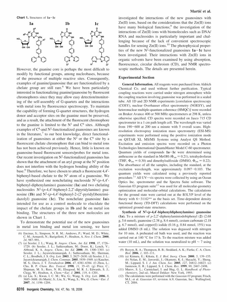
However, the guanine core is perhaps the most difficult tomodify by functional groups, among nucleobases, becauseof the presence of multiple reactive sites. Consequently,examples of guanine/guanosine that are functionalized by achelate group are still rare.6 We have been particularlyinterested in functionalizing guanine/guanosine by fluorescentchromophores since they may allow easy detection/monitor-ing of the self-assembly of G-quartets and the interactionswith metal ions by fluorescence spectroscopy. To maintainthe capability of forming G-quartet structures, the hydrogendonor and acceptor sites on the guanine must be preserved,and as a result, the attachment of the fluorescent chromophoreto the guanine is limited to the N2 and C8 sites. Althoughexamples of C8-and N2-functionalized guanosines are knownin the literature,7 to our best knowledge, direct functional-ization of guanosines at either the N2 or the C8 site by afluorescent chelate chromophore that can bind to metal ionshas not been achieved previously. Hence, little is known onguanosine-based fluorescent sensors/probes for metal ions.Our recent investigation on N2-functionalized guanosines hasshown that the attachment of an aryl group at the N2 positiondoes not interfere with the hydrogen bonding ability of thebase.8 Therefore, we have chosen to attach a fluorescent 4,4′-biphenyl-based chelate to the N2 atom of a guanosine. Wehave synthesized one nonchelating nucleoside N2-(p-4,4′-biphenyl-diphenylamino) guanosine (1a) and two chelatingnucleosides N2-(p-4,4′-biphenyl-2,2′-dipyridylamino) gua-nosine (1b) and N2-(p-4,4′-biphenyl-2-(2′-pyridyl)benzimi-dazolyl) guanosine (1c). The nonchelate guanosine 1aisintended for use as a control molecule to elucidate theimpact of the chelate groups in 1b and 1c on metal ionbinding. The structures of the three new molecules areshown in Chart 1.
To demonstrate the potential use of the new guanosinesin metal ion binding and metal ion sensing, we have
investigated the interactions of the new guanosines withZn(II) ions, based on the considerations that the Zn(II) ionshave many biological functions;9 the investigation of theinteractions of Zn(II) ions with biomolecules such as DNA/RNA and nucleosides is particularly important and chal-lenging because of the lack of convenient spectroscopichandles for sensing Zn(II) ions.10 The photophysical proper-ties of the new N2-functionalized guanosines 1a-1c havebeen investigated. Their interactions with Zn(II) ions inorganic solvents have been examined by using absorption,fluorescence, circular dichroism (CD), and NMR spectro-scopic methods. The details are presented herein.
Experimental Section
General Information. All reagents were purchased from AldrichChemical Co. and used without further purification. Typicalcoupling reactions were carried under nitrogen atmosphere whilethe coupling reaction involving guanosine was performed in a sealedtube. All 1D and 2D NMR experiments [correlation spectroscopy(COSY), nuclear Overhauser effect spectrometry (NOESY), andheteronuclear multiple-quantum coherence (HMQC)] were recordedon Bruker Avance 400 or 500 MHz spectrometers at 298 K, unlessotherwise specified. CD spectra were recorded on Jasco 715 CDspectrometer in a 1 cm path length cell. The wavelength was variedfrom 190-600 at 200 nm a minute with 5 overall scans. High-resolution electrospray ionization mass spectrometry (ESI-MS)experiments were performed using the positive ionization modeon QSTAR XL MS/MS Systems using Analyst QS Method.Excitation and emission spectra were recorded on a PhotonTechnologies International QuantaMaster Model C-60 spectrometer.Quantum yields of compounds 1a-1c were determined usinganthracene as the standard in MeOH (Φem ) 0.21), tetrahydrofuran(THF, Φem ) 0.30) and dimethylsulfoxide (DMSO, Φem ) 0.22).The absorbance of all the samples, including the standard, at theexcitation wavelength, was approximately 0.097-0.103. Thequantum yields were calculated using a previously reportedprocedure.11 All UV-vis spectra were collected by using an OceanOptics Inc. spectrometer and the Spectra Suite software. TheGaussian 03 program suite12 was used for all molecular-geometryoptimization and molecular-orbital calculations. The calculationsfor the ground state were carried out at the B3LYP level of thetheory with 6-311G** as the basis set. Time-dependent densityfunctional theory (TD-DFT) calculations were performed on theoptimized ground-state structures.
Synthesis of N2-(p-4,4′-biphenyldiphenylamino) guanosine(1a). To a mixture of p-2,2′-diphenylaminiodobiphenyl (2) (2.68g, 5.6 mmol), guanosine (2.38 g, 8.4 mmol), cesium carbonate (2.18g, 6.7 mmol), and copper(I) iodide (0.16 g, 0.84 mmol, 15%) wasadded DMSO (8 mL). The solution was degassed with nitrogenfor 10 min. A preheated oil bath was used, and the reaction wascarried out at 140 °C for 17 h. To the reaction mixture was addedwater (10 mL), and the solution was neutralized to pH ∼ 7 using
(6) Encinas, S.; Simpson, N. R. M.; Andrews, P.; Ward, M. D.; White,C. M.; Armaroli, N.; Barigelletti, F.; Houlton, A. New J. Chem. 2000,24, 987–991.
(7) (a) Sessler, J. L.; Wang, R. Angew. Chem., Int. Ed. 1998, 37, 1726–1729. (b) Sessler, J. L.; Sathiosatham, M.; Doerr, K.; Lynch, V.;Abboud, K. A. Angew. Chem., Int. Ed. 2000, 39, 1300–1303. (c)Sessler, J. L.; Jayawickramarajah, J.; Sathiosatham, M.; Sherman,C. L.; Brodbelt, J. S. Org. Lett. 2003, 5, 2627–2630. (d) Sessler, J. L.;Jayawickramarajah, J. Chem. Commun. 2005, 1939–1949. (e) Kaucher,M. S.; Davis, J. T. Tetrahedron 2006, 47, 6381–6384. (f) Zhou, L.;Cho, B. P. Chem. Res. Toxicol. 1998, 11, 35–43. (g) Price, C.;Shipman, M. S.; Rees, N. H.; Elsegood, M. R. J.; Edwards, S. J.;Clegg, W.; Houlton, A. Chem.sEur. J. 2001, 119, 4–1201.
(8) (a) Liu, X.; Kwan, I. C. M.; Wu, G.; Wang, S. Org. Lett. 2006, 8,3685–3688. (b) Martic, S.; Liu, X.; Wang, S.; Wu, G. Chem.sEur. J.2007, 14, 1196–1204.
(9) Bozym, R. A.; Thompson, R. B.; Stoddard, A. K.; Fierke, C. A. Chem.Biol. 2006, 1, 103–111.
(10) (a) Kimura, E.; Kikuta, E. J. Biol. Inorg. Chem. 2000, 5, 139–155.(b) Nolan, E. M.; Jaworski, J.; Okamoto, K.-I.; Hayachi, Y.; Sheng,M.; Lippard, S. J. J. Am. Chem. Soc. 2005, 127, 16812–16823. (c)Jamieson, E. R.; Lippard, S. J. Chem. ReV. 1999, 99, 2467–2498.
(11) Murov, S. L.; Carmichael, I. and Hug, G. L. Handbook of Photo-chemistry, 2nd ed.; Marcel Dekker: New York, 1993.
(12) The calculations were performed with the Gaussian 03 program; Frisch,M.J.; et al. Gaussian 03, revision A.9; Gaussian, Inc.: Wallingford,CT, 2004.
Chart 1. Structures of 1a-1c
Martic et al.
8316 Inorganic Chemistry, Vol. 47, No. 18, 2008
aqueous HCl. Further addition of water (30 mL) led to precipitationof the product as a beige solid. The solid was washed further withwater to remove unreacted guanosine. The crude solid was furtherpurified using a reverse phase silica gel column with CH2Cl2,followed by MeOH/H2O (4/1) as eluent to give 1a as a white solid(1.14 g, 35%). Mp >299 °C. 1H NMR (400 MHz, DMSO-d6) δ11.8 (broad, s, 1H, N1H), 10.1 (broad, s, 1H, N2H), 8.04 (s, 1H,H8), 7.8 (d, J ) 7.4 Hz, 2H), 7.61 (q, J ) 2.8, 8.4 Hz, 4H), 7.32(t, J ) 7.7 Hz, 4H), 7.03 (m, 8H), 5.80 (d, J ) 5.5 Hz, 1H, H1′),5.47 (d, J ) 5.8 Hz, 1H, 2-OH), 5.17 (d, J ) 4.3 Hz, 1H, 3-OH),5.00 (t, J ) 5.2 Hz, 1H, 5-OH), 4.55 (d, J ) 5.0 Hz, 1H, H2′), 4.11(d, J ) 3.6 Hz, 1H, H3′), 3.89 (d, J ) 3.9 Hz, 1H, H4′), 3.63 (m,J ) 4.5, 7.3 Hz, 1H, H5′), 3.52 (m, J ) 4.9, 7.4 Hz, 1H, H5′′). 13CNMR (400 MHz, DMSO-d6) δ 151.1, 147.9, 146.8, 139.8, 137.3,134.9, 133.7, 130.3 (4C), 127.8 (2C), 127.2 (2C), 125.1, 124.7 (4C),124.5 (2C), 123.8 (2C), 120.1 (2C), 119.2, 115.9, 114.7, 87.8 (C1),85.9 (C4), 74.4 (C2), 71.1 (C3), 62.2 (C5); ESI-MS m/z 603.1907[M + H]+. HRMS ESI m/z calcd for C34H30N6O5 ·H+ [M + H]+
603.23559, found 603.23565.
Synthesis of N2-(p-4,4′-biphenyl-2,2′-dipyridylamino) gua-nosine (1b). Compound 1b was obtained as a colorless solid usingthe same procedure as described for 1a by using compound 3 asthe starting material (2.98 g, 6.63 mmol) and guanosine (2.21 g,7.81 mmol) in 29% yield (1.16 g). Mp 226-228 °C. 1H NMR (400MHz, DMSO-d6) δ 10.64 (s, 1H, N1H), 8.96 (s, 1H, N2H), 8.23(d, J ) 3.85 Hz, 2H), 8.07 (s, 1H, H8), 7.67 (m, J ) 8.27 Hz, 8H),7.14 (d, J ) 8.39 Hz, 2H), 7.03 (m, J ) 5.06, 7.16, 8.40 Hz, 4H),5.78 (d, J ) 5.57 Hz, 1H, H1′), 5.46 (d, J ) 6.04 Hz, 1H, 2-OH),5.17 (d, J ) 4.99 Hz, 1H, 3-OH), 4.98 (m, J ) 5.25 Hz, 1H, 5-OH),4.50 (q, J ) 5.47 Hz, 1H, H2′), 4.11 (d, J ) 4.21 Hz, 1H, H3′),3.89 (d, J ) 3.81 Hz, 1H, H4′), 3.65 (m, J ) 4.18, 4.89, 22.31 Hz,1H, H5′), 3.52 (m, J ) 4.67, 6.96, 22.31 Hz, 1H, H5′′). 13C NMR(400 MHz, DMSO-d6) δ 158.4, 158.3 (2C), 157.3, 150.6, 150.3,149.9, 148.9 (2C), 146.3, 147.8, 144.5, 138.7 (2C), 137.2, 134.5,128.1 (2C), 128.0 (2C), 127.7 (2C), 120.3, 119.2 (2C), 117.6 (2C),88.1 (C1), 86.1 (C4), 74.6 (C2), 71.0 (C3), 62.1 (C5); ESI-MS m/z[M + H]+ 605.2189, [M + Na]+ 627.3245; HRMS ESI m/z calcdfor C32H28N8O5 ·H+ [M + H]+ 605.2255, found 605.2212.
Synthesis of N2-(p-4,4′-biphenyl-2-(2′-pyridyl)benzymidazolyl)guanosine (1c). Compound 1c was obtained as a colorless solidusing the same procedure as described for 1a by using compound4 as the starting material (3.51 g, 7.39 mmol) and guanosine (1.90g, 6.72 mmol) in 11% yield (0.53 g). Mp 253-256 °C. 1H NMR(500 MHz, DMSO-d6) δ 10.68 (s, 1H, N1H), 9.01 (s, 1H, N2H),8.39 (d, J ) 4.2 Hz, 1H, Hpy), 8.21 (d, J ) 7.88 Hz, 1H, HBn),8.09 (s, 1H, H8), 7.97 (t, J ) 7.69 Hz, 1H, HBn), 7.84 (d, J ) 7.46Hz, 2H), 7.73 (m, 4H), 7.46 (d, J ) 8.31 Hz, 2H), 7.37 (m, J )1.06, 6.53, 7.78 Hz, 5H), 5.81 (d, J ) 5.54 Hz, 1H, H1′), 5.46 (d,J ) 6.05 Hz, 1H, 1-OH), 5.18 (d, J ) 4.89 Hz, 1H, 2-OH), 4.99(d, J ) 5.30 Hz, 1H, 3-OH), 4.54 (d, J ) 5.41 Hz, 1H, H2′), 4.14(d, J ) 4.34 Hz, 1H, H3′), 3.93 (d, J ) 3.89 Hz, 1H, H4′), 3.66 (m,J ) 6.88, 11.83 Hz, 1H, H5′), 3.64 (m, J ) 6.86, 11.83 Hz, 1H,H5”); 13C NMR (500 MHz, DMSO-d6) δ 151.3, 150.7, 149.9, 149.6,143.2, 139.9, 137.9 (2C), 137.8, 137.7, 128.5, 128.4 (2C), 128.0(2C), 127.9 (2C), 127.7 (2C), 125.5, 125.1 (2C), 124.8, 123.8, 120.8,120.4 (2C), 119.4, 111.8, 88.1 (C1), 86.2 (C4), 74.7 (C2), 71.1(C3), 62.3 (C5); ESI-MS m/z [M + H]+ 629.2216; HRMS ESIm/z calcd for C34H28N8O5 ·H+ [M + H]+ 629.2260, found 629.2263.
Results and Discussion
Syntheses of Guanosines 1a-1c. To retain the biologicalactivity of guanosines, the hydrogen-bonding sites and the
ribose unit must remain intact, which is the key concern inN2-functionalization. Other considerations that have influ-enced the design of N2-functionalized guanosines are fluo-rescence and the chelating ability of the functional groups.Our earlier investigation has shown that diarylamines suchas 2,2′-dipyridylamino and heterocyclic groups such as 2-(2′-pyridyl)benzimidazolyl are highly emissive when attachedto an aryl group such as phenyl or biphenyl13 and are ableto chelate to a variety of metal ions, including Zn(II), readily,producing fluorescent or phosphorescent metal complexes.14
On the basis of these considerations, we decided to incor-porate p-4,4′-biphenyl-N(2-py)2 and p-4,4′-biphenyl-2-(2′-py)benzimidazolyl groups at the N2-site of guanosine toproduce the new guanosines 1b and 1c. For comparisonpurpose, the nonchelating group p-4,4′-biphenyl-NPh2 func-tionalized guanosine at the N2 site, 1a, was also synthesized.
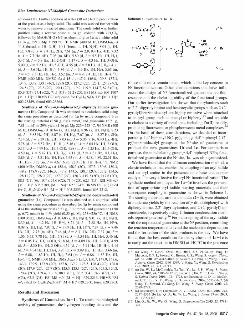
We have found that the Ullmann condensation method, aclassic technique that enables the coupling of an aryl halideand an aryl amine in the presence of a base and coppercatalyst,15 is very effective for aryl N2-functionalization. Thesynthetic method employed for 1a-1c involves the prepara-tion of appropriate aryl iodide starting materials and theirsubsequent coupling to guanosine as shown in Scheme 1.The starting materials, aromatic iodides (2-4), were obtainedin moderate yields by the reaction of p-diiodobiphenyl withdiphenylamine, 2,2′-dipyridylamine, and 2-(2′-pyridyl)ben-zimidazole, respectively using Ullmann condensation meth-ods reported previously.16 For the coupling of the aryl iodidewith the unprotected guanosine at the N2 position, controllingthe reaction temperature to avoid the nucleoside depurinationand the formation of the side products is the key. We havefound that the best condition for the synthesis of 1a-1c isto carry out the reaction in DMSO at 140 °C in the presence
(13) (a) Wang, S. Coord. Chem. ReV. 2001, 215, 79–98. (b) Pang, J.;Marcotte, E. P. J.; Seward, C.; Brown, R. S.; Wang, S. Angew. Chem.,Int. Ed. 2001, 40, 4042–4045. (c) Seward, C.; Pang, J.; Wang, S. Eur.J. Inorg. Chem. 2002, 1390–1399. (d) Kang, Y.; Wang, S. TetrahedronLett. 2002, 43, 3711–3713.
(14) (a) Jia, W. L.; McCormick, T.; Tao, Y.; Lu, J.-P.; Wang, S. Inorg.Chem. 2005, 44, 5706–5712. (b) Jia, W. L.; Hu, Y. F.; Gao, J.; Wang,S. Dalton Trans. 2006, 1721–1728. (c) Durantaye, L. D. L.; McCor-mick, T.; Liu, X. Y.; Wang, S. Dalton Trans. 2006, 5675–5682. (d)Kang, Y.; Seward, C.; Song, D.; Wang, S. Inorg. Chem. 2003, 42,2789–2797.
(15) (a) Beletskaya, I. P.; Cheprakov, A. V. Coord. Chem. ReV. 2004, 248,2337–2364. (b) Liu, Q. D.; Jia, W. L.; Wang, S. Inorg. Chem. 2005,44, 1332–1343.
(16) Liu, Q.; Jia, W.; Wu, G.; Wang, S. Organometallics 2003, 22, 3781–3791.
Scheme 1. Synthesis of 1a-1c
Blue Luminescent N2-Modified Guanosine DeriWatiWes
Inorganic Chemistry, Vol. 47, No. 18, 2008 8317
of CuI and Cs2CO3. When this simple procedure was used,the selective arylation at the exocyclic amine without theN1, N3 and/or N7-arylated side products, as determined byNMR, was achieved. Compounds 1a-1c were isolated inmoderate yields (11-37%) and were fully characterizedusing high-resolution mass spectrometry, 1H, 13 C, COSY,and HMQC NMR spectroscopy (see Supporting Informa-tion).
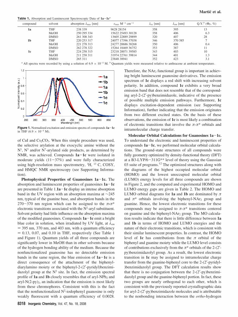
Photophysical Properties of Guanosines 1a-1c. Theabsorption and luminescent properties of guanosines 1a-1care presented in Table 1.1a-1c display an intense absorptionband in the UV region with an absorption maxima at ∼245nm, typical of the guanine base, and absorption bands in the270-370 nm region which can be assigned to the π-π*electronic transitions associated with the N2-aryl substituent.Solvent polarity had little influence on the absorption maximaof the modified guanosines. Compounds 1a-1c emit a brightblue color in solution, when irradiated by UV light, at λem
) 395 nm, 370 nm, and 403 nm, with a quantum efficiency) 0.13, 0.07, and 0.10 in THF, respectively (See Table 1and Figure 1). Quantum yields of all three compounds aresignificantly lower in MeOH than in other solvents becauseof the hydrogen bonding ability of the medium. Because thenonfunctionalized guanosine has no detectable emissionbands in the same region, the blue emission of 1a-1c is adirect consequence of the attachment of the biphenyl-diarylamino moiety or the biphenyl-2-(2′-pyridyl)benzimi-dazolyl group at the N2 site. In fact, the emission spectralprofile of 1a and 1b closely resembles that of aryl-NPh2 andaryl-N(2-py)2, an indication that the emission is most likelyfrom these chromophores. Consistent with this is the factthat the nonfunctionalized N2-iodophenyl guanosine is onlyweakly fluorescent with a quantum efficiency of 0.0028.
Therefore, the NAr2 functional group is important in achiev-ing bright luminescent guanosine derivatives. The emissionspectrum of 1c displays a red shift with increasing solventpolarity. In addition, compound 1c exhibits a very broademission band that does not resemble that of the correspond-ing aryl-2-(2′-py)benzimidazole, indicative of the presenceof possible multiple emission pathways. Furthermore, 1cdisplays excitation-dependent emission (see SupportingInformation), further indicating that the emission originatesfrom two different excited states. On the basis of theseobservations, the emission of 1c is most likely a combinationof electronic transitions that involve the π-π* orbitals andintramolecular charge transfer.
Molecular Orbital Calculations for Guanosines 1a-1c.To understand the electronic and luminescent properties ofcompounds 1a-1c, we performed molecular orbital calcula-tions. The ground-state structures of all compounds werefully geometry optimized by density-functional theory (DFT)at a B3-LYP/6-311G** level of theory using the Gaussian03 suite of programs.12 The optimized structures along withthe diagrams of the highest occupied molecular orbital(HOMO) and the lowest unoccupied molecular orbital(LUMO) energy levels for all three compounds are shownin Figure 2, and the computed and experimental HOMO andLUMO energy gaps are given in Table 2. The HOMO andLUMO orbital diagrams for 1a and 1b are dominated by πand π* orbitals involving the biphenyl-NAr2 group andguanine. Hence, the lowest electronic transitions for thesecompounds may be assigned to π-π* transitions centeredon guanine and the biphenyl-NAr2 group. The MO calcula-tion results indicate that there is little difference between 1aand 1b in terms of HOMO and LUMO energies and thenature of their electronic transitions, which is consistent withtheir similar luminescent properties. In contrast, the HOMOlevel of 1c has contributions from the π orbital of thebiphenyl and guanine moiety while the LUMO level consistsof contributions exclusively from the π* orbitals of the 2-(2′-py)benzimidazolyl group. As a result, the lowest electronictransition in 1c may be assigned to intramolecular chargetransfer from the guanine-biphenyl core to the 2-(2′-pyridyl-)benzimidazolyl group. The DFT calculation results showthat there is no conjugation between the 2-(2′-py)benzimi-dazolyl group and the guanine-biphenyl portion. In fact, thesetwo groups are nearly orthogonal to each other, which isconsistent with the previously reported crystallographic datafor aryl-2-(2′-py)benzimidazole molecules and is attributableto the nonbonding interaction between the ortho-hydrogen
Table 1. Absorption and Luminescent Spectroscopic Data of 1a-1ca
compound solvent absorption λmax [nm] εmax M-1 cm-1 λex [nm] λem [nm] Q.Y.b (ΦF, %)
1a THF 238 339 9628 28154 358 395 13MeOH 250 295 334 15622 25493 30128 358 406 6.3DMSO 261 308 343 13605 22889 29899 320 407 28
1b THF 220 253 317 24077 17396 37038 346 370 385 7MeOH 221 278 313 24277 20686 38268 350 406 0.4DMSO 262 278 322 15264 18469 36752 353 387 11
1c THF 224 258 315 23218 20071 39585 343 403 10MeOH 211 258 311 31974 22761 39814 344 401 1.3DMSO 265 311 23848 38941 352 423 3.1
a All spectra were recorded by using a solution of 6.9 × 10⁻⁵ M. b Quantum yields were measured relative to anthracene at ambient temperature.
Figure 1. Normalized excitation and emission spectra of compounds 1a-1cin THF (6.9 × 10⁻⁵ M).
Martic et al.
8318 Inorganic Chemistry, Vol. 47, No. 18, 2008
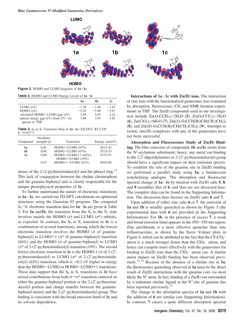
atoms of the 2-(2-py)benzimidazolyl and the phenyl ring.13
This lack of conjugation between the chelate chromophoreand the guanine-biphenyl unit is clearly responsible for theunique photophysical properties of 1c.
To further understand the nature of electronic transitionsin 1a-1c, we carried out TD-DFT calculations on optimizedstructures using the Gaussian 03 program. The computedS0-S1 electronic transition data for 1a-1c are given in Table3. For 1a and1b, the transition from the S0 to the S1 stateinvolves mainly the HOMO (π) and LUMO (π*) orbitals,as expected. In contrast, the S0 to S1 transition in 1c is acombination of several transitions, among which the lowestelectronic transition involves the HOMO (π of guanine-biphenyl) to LUMO+1 (π* of guanine-biphenyl) transition(64%) and the HOMO (π of guanine-biphenyl) to LUMO(π* of 2-(2′-py)benzimidazolyl) transition (19%). The secondlowest electronic transition in 1c is the HOMO-1 (π of 2-(2′-py)benzimidazolyl) to LUMO (π* of 2-(2′-py)benzimida-zolyl) (63%) transition, which is ∼0.2 eV higher in energythan the HOMO-LUMO or HOMO-LUMO+1 transitions.These data support that the S0 to S1 transitions in 1c havemixed contributions from both πfπ* transition centered oneither the guanine-biphenyl portion or the 2-(2′-py)benzimi-dazolyl portion and charge transfer between the guanine-biphenyl moiety and the 2-(2′-py)benzimidazolyl group. Thisfinding is consistent with the broad emission band of 1c andits solvent dependence.
Interactions of 1a-1c with Zn(II) ions. The interactionof zinc ions with the functionalized guanosines was examinedby absorption, fluorescence, CD, and NMR titration experi-ments in THF. The Zn(II) compounds used in our investiga-tion include Zn(O2CCH3)2 ·2H2O (5), Zn(O2CCF3)2 ·3H2O(6), Zn(ClO4)2 ·6H2O (7), Zn[(S)-O2CCH(Br)CH(CH3)CH3]2
(8), and Zn[(R)-O2CCH(Br)CH(CH3)CH3]2 (9). Attempts toisolate zinc(II) complexes with any of the guanosines havenot been successful.
Absorption and Fluorescence Study of Zn(II) Bind-ing. The blue emission of compounds 1b and1c stems fromthe N2-arylamine substituent; hence, any metal ion bindingto the 2,2′-dipyridylamino or 2-(2′-py)benzimidazolyl groupshould have a significant impact on their emission spectra.To establish the role of the guanine site in Zn(II) bindingwe performed a parallel study using 1a, a luminescentnonchelating analogue. The absorption and fluorescentspectral change of 1a-1c by titration with Zn(II) salts 5, 8,and 9 resembles that of 6, and thus are not discussed here.The complete data can be found in the Supporting Informa-tion. The discussion here focuses on Zn(II) salts 6 and 7.
Upon addition of either zinc salts 6 or 7, the emission of1a and 1b is steadily quenched as shown by Figure 3 (theexperimental data with 6 are provided in the SupportingInformation). For 1b, in the presence of excess 7, a weakand broad emission band appears in the 450-550 nm region.Zinc perchlorate is a more effective quencher than zinctrifluoroacetate, as shown by the Stern-Volmer plots inFigure 4, which can be attributed to the fact that the CF3CO2
-
anion is a much stronger donor than the ClO4- anion, and
hence can compete more effectively with the guanosines forbinding to Zn(II) ions than the ClO4
- anion can. A similaranion impact on Zn(II) binding has been observed previ-ously.13,14 Because of the absence of a chelate site in 1a,the fluorescence quenching observed in 1a must be the directresult of Zn(II) interactions with the guanine core via mostlikely the N7 atom. In fact, binding of a Zn(II) ion surroundedby a tridentate chelate ligand at the N7-site of guanine hasbeen reported previously.17
The change in the absorption spectra of 1a and 1b withthe addition of 6 are similar (see Supporting Information).In contrast, 7 causes a quite different absorption spectral
Figure 2. HOMO and LUMO diagrams of 1a-1c.
Table 2. HOMO and LUMO Energy Levels of 1a-1c
1a 1b 1c
LUMO (eV) -1.36 -1.46 -1.65HOMO (eV) -5.25 -5.49 -5.87calculated HOMO-LUMO gap (eV) 3.89 4.02 4.22optical energy gap (eV) from UV-vis
spectra in THF3.69 3.91 3.92
Table 3. S0 to S1 Transition Data of 1a-1c (TD-DFT, B3-LYP/6-311G**)
CompoundOscillator
strength (f) Transition Energy (nm/eV)
1a 0.81 HOMOfLUMO (67%) 361/3.431b 0.88 HOMOfLUMO (67%) 351/3.531c 0.60 HOMOfLUMO+1 (64%),
HOMOfLUMO (19%)317/3.91
0.57 HOMO-1fLUMO (63%) 304/4.08
Blue Luminescent N2-Modified Guanosine DeriWatiWes
Inorganic Chemistry, Vol. 47, No. 18, 2008 8319
change of 1a and 1b: for 1a, the absorption band at 317 nmexperiences some decrease while for 1b, the same absorptionband decreases in intensity and a weak absorption bandappears in the 350-450 nm region, an indication that 7 maybe interacting with both guanine and the dipyridylaminogroups in 1b, which is consistent with the appearance of abroad weak emission band in the fluorescent titration spectrawith 7. In fact, it has been shown previously that the bindingof a Zn(II) ion to a dipyridylamino ligand causes quenchingand an emission spectral red shift.13,14 We also confirmedthis by titrating the model compound 4-iodo-biphenyl-N(2-Py)2 (3) with 7 that showed the appearance of a broad weakemission band with the addition of Zn(II) ions (see Sup-porting Information).
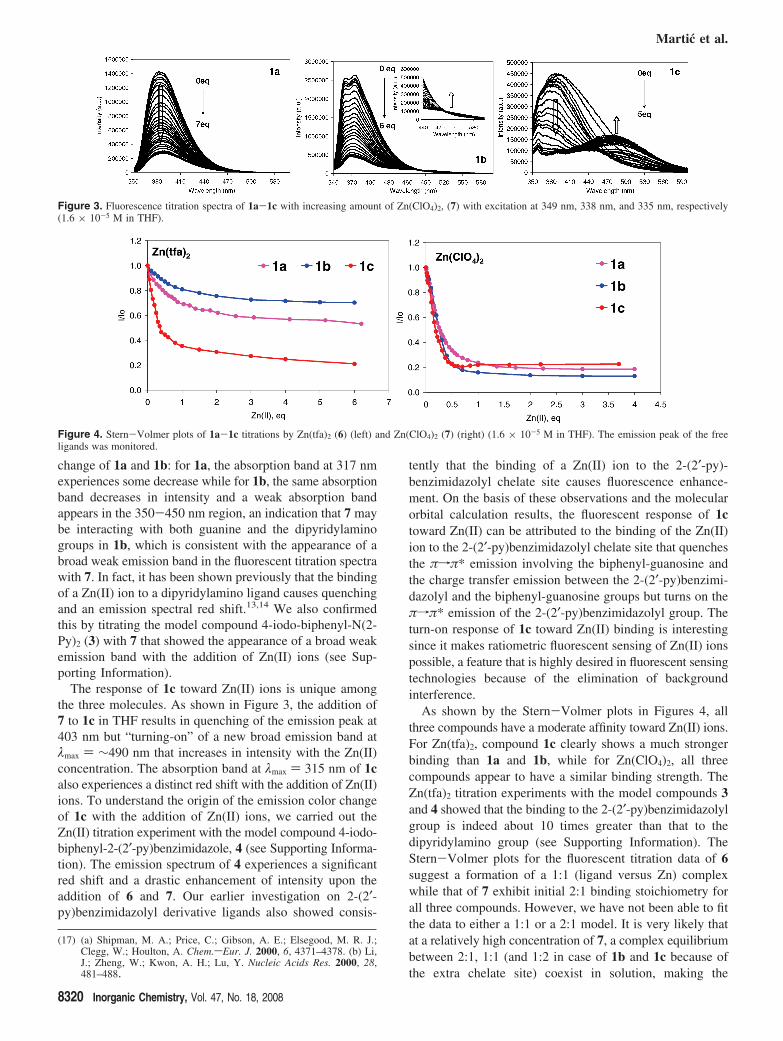
The response of 1c toward Zn(II) ions is unique amongthe three molecules. As shown in Figure 3, the addition of7 to 1c in THF results in quenching of the emission peak at403 nm but “turning-on” of a new broad emission band atλmax ) ∼490 nm that increases in intensity with the Zn(II)concentration. The absorption band at λmax ) 315 nm of 1calso experiences a distinct red shift with the addition of Zn(II)ions. To understand the origin of the emission color changeof 1c with the addition of Zn(II) ions, we carried out theZn(II) titration experiment with the model compound 4-iodo-biphenyl-2-(2′-py)benzimidazole, 4 (see Supporting Informa-tion). The emission spectrum of 4 experiences a significantred shift and a drastic enhancement of intensity upon theaddition of 6 and 7. Our earlier investigation on 2-(2′-py)benzimidazolyl derivative ligands also showed consis-
tently that the binding of a Zn(II) ion to the 2-(2′-py)-benzimidazolyl chelate site causes fluorescence enhance-ment. On the basis of these observations and the molecularorbital calculation results, the fluorescent response of 1ctoward Zn(II) can be attributed to the binding of the Zn(II)ion to the 2-(2′-py)benzimidazolyl chelate site that quenchesthe πfπ* emission involving the biphenyl-guanosine andthe charge transfer emission between the 2-(2′-py)benzimi-dazolyl and the biphenyl-guanosine groups but turns on theπfπ* emission of the 2-(2′-py)benzimidazolyl group. Theturn-on response of 1c toward Zn(II) binding is interestingsince it makes ratiometric fluorescent sensing of Zn(II) ionspossible, a feature that is highly desired in fluorescent sensingtechnologies because of the elimination of backgroundinterference.
As shown by the Stern-Volmer plots in Figures 4, allthree compounds have a moderate affinity toward Zn(II) ions.For Zn(tfa)2, compound 1c clearly shows a much strongerbinding than 1a and 1b, while for Zn(ClO4)2, all threecompounds appear to have a similar binding strength. TheZn(tfa)2 titration experiments with the model compounds 3and 4 showed that the binding to the 2-(2′-py)benzimidazolylgroup is indeed about 10 times greater than that to thedipyridylamino group (see Supporting Information). TheStern-Volmer plots for the fluorescent titration data of 6suggest a formation of a 1:1 (ligand versus Zn) complexwhile that of 7 exhibit initial 2:1 binding stoichiometry forall three compounds. However, we have not been able to fitthe data to either a 1:1 or a 2:1 model. It is very likely thatat a relatively high concentration of 7, a complex equilibriumbetween 2:1, 1:1 (and 1:2 in case of 1b and 1c because ofthe extra chelate site) coexist in solution, making the
(17) (a) Shipman, M. A.; Price, C.; Gibson, A. E.; Elsegood, M. R. J.;Clegg, W.; Houlton, A. Chem.sEur. J. 2000, 6, 4371–4378. (b) Li,J.; Zheng, W.; Kwon, A. H.; Lu, Y. Nucleic Acids Res. 2000, 28,481–488.
Figure 3. Fluorescence titration spectra of 1a-1c with increasing amount of Zn(ClO4)2, (7) with excitation at 349 nm, 338 nm, and 335 nm, respectively(1.6 × 10⁻⁵ M in THF).
Figure 4. Stern-Volmer plots of 1a-1c titrations by Zn(tfa)2 (6) (left) and Zn(ClO4)2 (7) (right) (1.6 × 10⁻⁵ M in THF). The emission peak of the freeligands was monitored.
Martic et al.
8320 Inorganic Chemistry, Vol. 47, No. 18, 2008
extraction of the binding constant challenging. In fact, the1:1 and 2:1 complexes of 1a with Zn(II) ions from thesolution of 1a and 7 in DMSO have been positively identifiedin the ESI-MS spectra (see Supporting Information).
For comparison, we also investigated the fluorescent responseof the new guanosines toward Cd(ClO4)2 ·6H2O (10) andHg(ClO4)2 ·3H2O (11). Mercury(II) salt 11 causes completequenching of the emission of 1a and 1b and about 90%quenching of the emission of 1c without the appearance of newemission peaks, which is attributable to the strong heavy atomeffects of Hg(II) ion. The response of 10 toward all threecompounds is similar to that of 7 except that it is much moresensitive than 7 toward 1b and 1c because of the strongerbinding of Cd(II) ions toward the dipyridylamino group andthe 2-(2-py)benzimidazolyl group (see Supporting Information),a trend that is consistent with previous reports in the literature.14
CD Study of Zn (II) Binding. One advantage of usingfunctionalized guanosines as ligands for metal ions is thatthey are inherently chiral, thus making it possible to examinemetal ion binding by CD spectroscopy. In fact, CD has beenused frequently as a sensitive tool for the investigation ofself-assembly and conformational change of guanosine andits simple derivatives.18 Monitoring Zn(II) ions binding tonucleotides by CD has also been reported in the literature.19
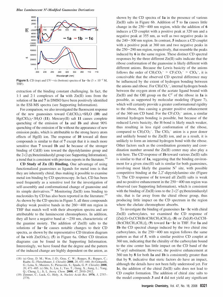
As shown by the CD spectra in Figure 5, all three compoundsdisplay weak positive bands in the 260-400 nm region inTHF that match well with their absorption spectra and areattributable to the luminescent chromophores. In addition,they all have a negative band at ∼250 nm, characteristic ofthe guanine moiety. The addition of Zn(II) salts to thesolutions of 1a-1c causes notable changes to their CDspectra, as shown by the representative CD titration diagramof 1a with Zn(OAc)2 (5) in Figure 6a. Other CD titrationdiagrams can be found in the Supporting Information.Interestingly, we have found that the degree and the patternof the induced change are highly dependent on the anion, as
shown by the CD spectra of 1a in the presence of variousZn(II) salts in Figure 6b. Addition of 7 to 1a causes littlechange in the 250-400 nm region, while the addition of 6induces a CD couplet with a positive peak at 320 nm and anegative peak at 355 nm, as well as two negative peaks inthe 240-300 nm region. In contrast, 5 induces a CD coupletwith a positive peak at 360 nm and two negative peaks inthe 250-290 nm region, respectively, that resemble the peaksinduced by 6 in the same region. These distinct CD spectralresponses by the three different Zn(II) salts indicate that theribose conformation of the guanosine is likely different withdifferent anions. Because the Lewis basicity of the anionsfollows the order of CH3CO2
- > CF3CO2- > ClO4
-, it isconceivable that the observed CD spectral difference maybe influenced by the extent of hydrogen bonding betweenthe anions and ribose. For CH3CO2
-, internal hydrogen bondsbetween the oxygen atom of the acetate ligand bound withZn(II) and the OH group on the C5′ of the ribose in 1a ispossible, as supported by molecular modeling (Figure 7),which will certainly provide a greater conformational rigidityto the ribose, thus causing a greater positive enhancementof the 360 nm CD band. For the CF3CO2
- anion, a similarinternal hydrogen bonding is possible, but because of thereduced Lewis basicity, the H-bond is likely much weaker,thus resulting in less rigid conformation of the ribose,compared to CH3CO2
-. The ClO4- anion is a poor donor
and unlikely bound to the Zn(II) ion, and as a result, it isunlikely to form an internal hydrogen bond with the ribose.Other factors such as the coordination geometry and coor-dination number around the Zn(II) center may also play arole here. The CD response of 1b toward various Zn(II) saltsis similar to that of 1a, suggesting that the binding environ-ment for a given zinc(II) salt is similar for both guanosines,involving most likely the guanine N7 site, with possiblecompetitive binding at the 2,2′-dipyridylamino site (Figure7). The CD response of 1c toward all Zn(II) salts is weakand no positive enhancement in the 250-400 nm region wasobserved (see Supporting Information), which is consistentwith the binding of Zn(II) ions to the 2-(2′-py)benzimidazolylsite, that is far away from the chiral ribose group, thusproducing little impact on the CD spectrum in the regionwhere the chelate chromophore absorbs.
To investigate the binding of guanosines 1a-1c with chiralZn(II) carboxylates, we examined the CD response of[Zn[(S)-O2CCH(Br)CH(CH3)CH3)]2 (8) or Zn[(R)-O2CCH-(Br)CH(CH3)CH3)]2 (9) toward 1a-1c in THF. For 1a and1b the CD spectral change induced by the two chiral zinccarboxylates, in the 250-400 nm region follows the samepattern as that of 5, with a similar positive CD couplet at360 nm, indicating that the chirality of the carboxylate boundto the zinc center has little impact on the CD band of theN2-aryl substituent. However, the positive enhancement at360 nm by 8 for both 1a and 1b is consistently greater thanthat by 9, indicative that steric factors do have an impact,although the details have not been fully understood yet. For1c, the addition of the chiral Zn(II) salts does not lead toCD couplet formation. The addition of chiral zinc salts tothe model compounds 3 and 4 did not yield any significant
(18) (a) Gray, D. M.; Wen, J.-D.; Gray, C. W.; Repges, R.; Repges, C.;Raabe, G.; Fleischhauer, J. Chirality 2008, 20, 431–440. (b) Gottarelli,G.; Lena, S.; Masiero, S.; Pieraccini, S.; Spada, G. P. Chirality 2008,20, 471–485. (c) Shi, S.; Liu, J.; Yao, T.; Geng, X.; Jiang, L.; Yang,Q.; Cheng, L.; Ji, L. Inorg. Chem. 2008, 47, 2910–2912.
(19) Zimmer, C.; Luck, G.; Holy, A. Nucleic Acids Res. 1976, 3, 2757–2770.
Figure 5. CD (top) and UV-vis (bottom) spectra of 1a-1c (3 × 10⁻⁵ M,THF).
Blue Luminescent N2-Modified Guanosine DeriWatiWes
Inorganic Chemistry, Vol. 47, No. 18, 2008 8321

CD signals in the 250-400 nm region, as expected. Thecovalent attachment of the chromophores to the chiralguanosine is clearly important for achieving the distinct CD
spectral change of 1a-1c upon binding to Zn(II) ions. Thefull understanding of the CD spectral response by 1a-1crequires extensive experimental and theoretical work whichis beyond the scope of this report.
1H NMR Study of Zn(II) Binding. To determine thebinding site of the Zn(II) ions in 1a-1c, 1H NMR titrationexperiments were performed with Zn(II) salts 6 and 7 inTHF-d8. The partial 1H NMR spectra of titration by 7 areshown in Figure 8, while the NMR titration data bycompound 6 can be found in the Supporting Information.Upon the addition of 7 to the solution of 1a, a well resolved1H NMR spectrum with a dramatic downfield shift of theH8 resonance (∼1.0 ppm) was observed, supporting that theZn(II) ion interacts with the N7 site of the guanine base. Asimilar downfield shifting and broadening of the proton peakadjacent to the N7 binding site in guanine, upon Zn(II) ioncoordination, has been reported by Houlton et al.17 Similarly,the addition of 7 to the solution of 1b results in a downfieldshift of the H8 resonance by ∼0.1 ppm. Moreover, the pyridylH1
Py resonance at 8.2 ppm in 1b broadens and experiencesa small downfield shift upon the addition of zinc(II),consistent with the binding of zinc(II) ions at both thedipyridylamino site and the guanine N7 site. The addition ofup to 0.2 equiv of 7 to the solution of 1c (This is themaximum amount that can be added before precipitationoccurs because of the poor solubility of 1c.) results in theimmediate broadening of the H1
Py peak of the pyridyl ringand the H1
Bn peak of the benzimidazolyl group, while theH8 resonance of the guanine remains unaffected.
The 1H NMR response of 1a-1c to 6 follows the similarpattern as that of 7 in terms of the coordination sites involved.The addition of more than 1 equiv of 6 to 1a results in thebroadening of all signals because of a slow exchange betweenthe bound and free Zn(II) ions, attributable to the relativelystrong donor ability of the tfa anion that can compete forbinding to Zn(II) with 1a. The addition of 6 (up to 2.2 eq)to 1b results in broadening of all pyridyl protons because ofthe dynamic equilibrium in solution between the bound andfree Zn(II) ions, which was confirmed by variable temper-ature NMR spectra at 190-308 K. At 190 K, a complexpattern with new broad peaks was observed for the spectrumof 1b with 6, indicative of the presence of multiple species.Moreover, the broad peaks at δ > 10 ppm attributable to
Figure 6. (a) Left: CD titration spectra of 1a by 5. b) Right: CD spectra of 1a in the presence of 5 equiv of various Zn(II) salts (5-9) (THF, [1a]) 3.0 ×10⁻⁵ M).
Figure 7. Molecular models showing the binding site of Zn(OAc)2 with1a-1c. Top: the binding model for 1a showing intramolecular H-bondinginteractions between C5′-OH of ribose and one O atom of Zn(OAc)2. Thisis also one of the binding modes for 1b. Middle: the 2nd binding mode for1b showing the coordination of Zn(OAc)2 at the dipyridylamino site.Bottom: the binding model for 1c showing the coordination of Zn(OAc)2
at the 2-(2-py)benzimidazolyl site.
Martic et al.
8322 Inorganic Chemistry, Vol. 47, No. 18, 2008

the imino and amino protons of guanosine involved inhydrogen bonding were also observed, an indication thatthe complex low temperature NMR spectrum of 1b in thepresence of Zn(tfa)2 likely has contributions from both theZn(II) complex and oligomeric species formed by hydrogenbonding between guanine units. Similar to 1b, the 1H NMRspectrum of 1c-6 complex is dominated by broad signals overa large temperature range (220-298 K), which can be againattributed to dynamic exchange between the bound and freeZn(II) ions and hydrogen bonding among guanine units atlow temperatures. Hydrogen bonds involving the ribose OHgroups could not be resolved in 1H NMR spectra because ofthe poor solubility of the complexes.
Conclusions
On the basis of the fluorescent, CD, and NMR titrationdata, we can conclude that the preferred binding site for theZn(II) ions in 1a and 1b is the guanine N7 site. Thepreference for the N7 site in a guanine/guanosine by a zinc(II)ion is previously known.17 For 1b, the dipyridylamino group,albeit a weaker binding site than the guanosine, is likelycompeting for the Zn(II) binding. For 1c, the preferredbinding site is the 2-(2′-py)benzimidazolyl chelate becauseof its high binding affinity for Zn(II) ions. Because of thepoor donor capability of the perchlorate anion, Zn(ClO4)2
may form a 2:1 complex (L/Zn) with the guanosine via eitherthe guanine site or the N2-functionalized chelate site. In fact,the 2:1 binding mode involving two 2-(2′-py)benzimidazolylderivative ligands and one Zn(II) ion has been observedpreviously.20
In summary, three new N2-functionalized luminescentguanosines have been synthesized. We have shown that theseluminescent guanosines can bind to metal ions such as Zn(II)ions. The new highly emissive functional groups attachedto the guanosine are very useful for probing metal-guanosineinteractions via both fluorescent and CD modes. In thefluorescent mode, compound 1c has a unique turn-onresponse toward Zn(II) or Cd(II) ions. In the CD mode, both1a and 1b are highly responsive to Zn(II) acetate and thederivatives. While N2-fluorescent nucleosides are not capableof effective chiral discrimination in CD, they are efficientin distinguishing different anions associated with the zinc(II)ion. Investigations of the interactions between functionalizedguanosines 1a-1c and other metal ions and their impact onthe self-assembly of guanosines such as G-quartet formationare underway, and the results will be reported in due course.
Acknowledgment. We thank the Natural Sciences andEngineering Research Council of Canada for financialsupport. We are in debt to Dr. Yimin She for his assistancein ESI-MS experiments and Theresa McCormick for her helpin CD experiments.
Supporting Information Available: The complete absorption,fluorescence and CD titration data of 1a-1c with 5-11, 1D and2D NMR data for 1a-1c, ESI-MS spectra for 1a/7, and 1H NMRtitration data of 1a-1c with 6 and 7. This material is availablefree of charge via the Internet at http://pubs.acs.org.
IC800899B
(20) Liu, S.-F.; Wu, Q.; Schmider, H. L.; Aziz, H.; Hu, N.-X.; Popovic,Z.; Wang, S. J. Am. Chem. Soc. 2000, 122, 3671–3678.
Figure 8. Partial 1H NMR spectra of 1a-1c in the presence of various amounts of Zn(ClO4)2 ([1a] ) 1.9 × 10⁻⁴ M, [1b] ) 1.4 × 10⁻⁴ M, [1c]) 1.5 ×10⁻⁴ M, 298 K, THF-d8).
Blue Luminescent N2-Modified Guanosine DeriWatiWes
Inorganic Chemistry, Vol. 47, No. 18, 2008 8323