naosite: nagasaki university's academic output...
TRANSCRIPT

This document is downloaded at: 2018-06-07T08:25:53Z
TitleIntraoperative pancreatography using an endoscopic naso-pancreaticdrainage tube for the prevention of pancreatic fistula after local pancreaticresection
Author(s) Kuroki, Tamotsu; Tajima, Yoshitsugu; Tsuneoka, Noritsugu; Adachi,Tomohiko; Kanematsu, Takashi
Citation Hepato-Gastroenterology, 56(90), pp.535-537; 2009
Issue Date 2009-03
URL http://hdl.handle.net/10069/23100
Right © H.G.E. Update Medical Publishing.
NAOSITE: Nagasaki University's Academic Output SITE
http://naosite.lb.nagasaki-u.ac.jp
T. Kuroki et al.
1
Original Article
Intraoperative pancreatography using an endoscopic
naso-pancreatic drainage tube for the prevention of pancreatic
fistula after local pancreatic resection
Tamotsu Kuroki, Yoshitsugu Tajima, Noritsugu Tsuneoka, Tomohiko
Adachi, Takashi Kanematsu
Department of Surgery, Nagasaki University, Graduate School of
Biomedical Sciences, 1-7-1 Sakamoto, Nagasaki 852-8501, Japan.
Running title: Pancreatic local resection using ENPD tube
Offprint requests to: T. Kuroki
FAX: 81-95819-7319
e-mail: [email protected].
T. Kuroki et al.
2
Abstract
Background: Local pancreatic resections can avoid the unnecessary resection
of the normal pancreatic parenchyma in comparison with standard pancreatic
resection. However, the incidence of pancreatic fistula after local pancreatic
resection is high, and still responsible for most morbidity and mortality.
Methods: We reviewed 6 patients who underwent intraoperative pancreatography
using an endoscopic naso-pancreatic drainage (ENPD) tube during local
pancreatic resection for the prevention of postoperative pancreatic fistula.
Results: One patient had injury to the main pancreatic duct during surgery,
and transient pancreatic fistula of grade B occurred. In this patient, ENPD
tube was left in place for the management the pancreatic fistula, resulted
in a favorable outcome. Other 5 patients showed no postoperative complications
including pancreatic fistula.
Conclusions: Intraoperative pancreatography using ENPD tube is a simple
technique and useful for the prevention of pancreatic fistula after local
pancreatic resection.
Keywords: Pancreatic fistula; pancreatic local resection; ENPD;
pancreatography
T. Kuroki et al.
3
Introduction
Recent improvements in surgical devices and advances in knowledge
of the pancreatic anatomy have allowed surgeons to apply a variety
of novel surgical procedures when performing local pancreatic
resections. Local pancreatic resections can avoid the unnecessary
resection of the normal pancreatic parenchyma in comparison with
standard pancreatic resection such as pancreaticoduodenectomy
and distal pancreatectomy, consequently preserve the endocrine
and exocrine functions of the pancreas. In local pancreatic
resection, the main pancreatic duct and common bile duct are able
to be preserved without any dissection or reconstruction of the
pancreaticobiliary tract. In addition, many benign and low-grade
malignant pancreatic lesions, including intraductal papillary
mucinous neoplasms (IPMNs) and mucinous cystic neoplasms, have
recently been detected by the improved diagnostic modalities. Such
pancreatic lesions would be a good indication for performing a
local pancreatic resection. However, pancreatic fistula occurs
in a high incidence after local pancreatic resection and is still
responsible for most morbidity and mortality after pancreatic
surgery, possibly leading to a lethal bleeding [1-3]. To prevent
T. Kuroki et al.
4
pancreatic fistula after pancreatic surgery, many surgical
techniques and instruments have been proposed, such as pancreatic
transection using an ultrasonic dissector, an automatic stapler,
fibrin glue sealing of the pancreatic stump, and administration
of somatostatin analogue [4-10]. However, these attempts failed
to completely avoid the occurrence of postoperative pancreatic
fistula. In this report, we describe the efficacy of preoperative
endoscopic naso-pancreatic drainage (ENPD) tube technique to
avoid the injury to the main pancreatic duct in performing local
pancreatic resection.
T. Kuroki et al.
5
Patients and Methods
Six patients underwent a local pancreatic resection in the
pancreatic head with a use of ENPD tube between April 2005 and
June 2007. There were 3 men and 3 women with a mean age of 73 years
(range, 56-84 years). Diagnoses included branch-type IPMN of the
pancreas (n=5) and insulinoma (n=1). All patients had a soft
pancreas without fibrosis. Under general anesthesia, we placed
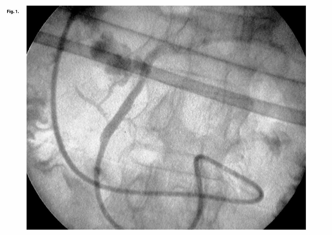
a 5F-size ENPD tube (Olympus, Tokyo Japan) for intraoperative
pancreatography prior to laparotomy (Fig. 1). The ENPD tube was
inserted deeply into the main pancreatic duct for approximately
10cm through the orifice. During the pancreatic transection, the
main pancreatic duct was carefully evaluated by repeated
intraoperative US and pancreatography via the ENPD tube. Five
patients underwent local resection in the superior pancreatic head
region. Two patients underwent local resection in the uncinate
process of the pancreas. After a completion of local pancreatic
resection, pancreatography via the ENPD tube was performed to
check out the injury to the main pancreatic duct or leakage from
the transected pancreatic branch ducts at the cut surface of the
pancreas. A mixture of indigocarmine and contrast material was
T. Kuroki et al.
6
used for the pancreatography, because it enable us to detect any
minor pancreatic leakage as a blue spot at the cut surface of the
pancreas and to close the points of leakage appropriately with
a 4-0 absorbable monofilament sutures. Pancreatic fistula was
defined by the international study group on pancreatic fistula
(ISGPF) definition [11].
T. Kuroki et al.
7
Results
The ENPD tube was inserted successfully within 15 minutes in all
6 patients. During the local pancreatic resection, no patients
showed dislocation of the ENPD tube. One patient deveioped injury
to the main pancreatic duct during the pancreatic transection,
and the injury was repaired by primary suture with a 6-0 absorbable
monofilament sutures. Three patients were found to have minor
pancreatic leakage by pancreatography via the ENPD tube. In these
3 patients, we could detect minor leakage of blue dye from the
small pancreatic duct branches at the cut surface after local
pancreatic resection, and then we closed the leaking points
appropriately with a 4-0 absorbable monofilament sutures. In 5
patients except for one case with main pancreatic duct injury,
ENPD tubes were immediately removed after the confirmation of no
pancreatic leakage at the cut surface of the pancreas. In one
patient who had injury to the main pancreatic duct, the ENPD tube
indwelled in the main pancreatic duct was useful for the suturing
of the injured main pancreatic duct. The ENPD tube was left in
place and used for a pancreatic stent as well as a pancreatic
drainage. In this patient, transient pancreatic fistula of grade

T. Kuroki et al.
8
B (ISGPF definition) [11] was demonstrated, and the ENPD tube was
removed 7 days postoperatively. The ENPD tube was useful for the
management the postoperative pancreatic fistula. Other 5 patients
showed no postoperative complications including pancreatic
fistula. In addition, no patients developed acute pancreatitis
after ENPD tube placement.
T. Kuroki et al.
9
Comments
Several pancreas-preserving surgery, including local pancreatic
resection, have been advocated for the benign and low-grade
malignant neoplasms of the pancreas [12-14]. Especially,
branch-type IPMN of the pancreas is a good candidate for the local
pancreatic resection because the branch-type IPMNs show a less
lymph node metastasis and a more favorable prognosis when compared
to invasive ductal adenocarcinomas of the pancreas [15]. In the
benign or low-grade malignant IPMN, complete tumor resection is
sufficient for a cure. On the other hand, pancreatic fistula is
one of the most frequent and dismal complications after local
pancreatic resection [2, 3]. This was due to the existence of small
branches of pancreatic duct communicating with the main pancreatic
duct on the cut surface of the pancreas which was wider than those
in the standard pancreatic resections, including
pancreaticoduodenectomy and distal pancreatectomy. Hirota et al.
[16] have reported that preoperative endoscopic transpapillary
pancreatic stent placement was useful for the prevention of
pancreatic fistula following the local pancreatic resection. In
the present study, we used an ENPD tube instead of pancreatic short
T. Kuroki et al.
10
stent, because ENPD tube can reveal the pancreatic ducts by using
intraoperative pancreatography. In addition, the ENPD tube in the
main pancreatic duct was clearly and easily detected by
intraoperative US, and thus both the direction and position of
the main pancreatic duct were correctly identified. Indeed, this
technique was useful for identifying the pancreatic ducts to be
dissected and for preventing injury to the main pancreatic duct.
Intraoperative pancreatography showed not only the main
pancreatic duct but also the small branch pancreatic ducts
communicating with the main pancreatic duct. These small branches
cannot always be identified and managed during the operation. Our
technique, intraoperative pancreatography using both
indigocarmine and contrast material, enable us to detect and
suture small branch pancreatic ducts.
Acute pancreatitis is the most common complication of
endoscopic retrograde cholangiopancreatography (ERCP). ENPD tube
placement needs the technique of ERCP. In addition, pancreatic
stent placement may induce acute pancreatitis [17]. In our
patients, fortunately, we had no experienced acute pancreatitis.
In our technique, an ENPD tube is immediately removed after the

T. Kuroki et al.
11
confirmation of no pancreatic leakage. However, if a main
pancreatic duct is injured during the resection of the pancreas,
postoperative ENPD tube placement is useful for the management
of the pancreatic fistula.
In conclusion, intraoperative pancreatography using ENPD
tube is simple technique and useful for the prevention of
pancreatic fistula after local pancreatic resection. This
technique allows the surgeon to perform a safe pancreas-preserving
surgery including local pancreatic resection.

T. Kuroki et al.
12
References
1. Kuroki T, Tajima Y, Kanematsu T. Surgical management for the
prevention of pancreatic fistula following distal
pancreatectomy. J Hepatobiliary Pancreat Surg 2005; 12:
283-285.
2. Nakagohri T, Kenmochi T, Kainuma O, Tokoro Y, Kobayashi S, Asano
T. Inferior head resection of the pancreas for intraductal
papillary mucinous tumors. Am J Surg 2000; 179: 482-484.
3. Nakagohri T, Konishi M, Inoue K, Izuishi K, Kinoshita T. Partial
pancreatic head resection for intraductal papillary mucinous
carcinoma originating in a branch of the duct of Santorini.
Eur Surg Res 2002; 34: 437-440.
4. Shankar S, Theis B, Russell RCG. Management of the stump of
the pancreas after distal pancreatic resection. Br J Surg 1990;
77: 541-544.
5. Okabayashi T, Kobayashi M, Sugimoto T, Namikawa T, Okamoto K,
Hokimoto N, Araki K. Postoperative pancreatic fistula
following distal pancreatectomy for pancreatic neoplasm; Can
pancreatic fistula be prevented? Hepatogastroenterology 2004;
51: 1838-1841.

T. Kuroki et al.
13
6. Noun R, Elias D, Ballandur P, Bismuth H, Parc R, Lasser P,
Belghiti J. Fibrin glue effectiveness and tolerance after
elective liver resection: a randomized trial.
Hepatogastroenterology 1996; 46: 221-224.
7. Kajiyama Y, Tsurumaru M, Udagawa H, Tsutsumi K, Kinoshita Y,
Akiyama H. Quick and simple distal pancreatectomy using the
GIA stapler: report of 35 cases. Br J Surg 1996; 83: 1711.
8. Suzuki Y, Fujino Y, Tanioka Y, Hori Y, Ueda T, Takeyama Y,
Tominaga M, Ku Y, Yamamoto YM, Kuroda Y. Randomized clinical
trial of ultrasonic dissector or conventional division in
distal pancreatectomy for non-fibrotic pancreas. Br J Surg
1999; 86: 608-611.
9. Kohler E, Beglinger C, Dettwiler S, Whitehouse I, Gyr K. Effect
of a new somatostatin analogue on pancreatic function in
healthy volunteers. Pancreas 1986; 1: 154-159.
10. Buchler M, Friess H, Klempa I, Hermanek P, Sulkowski U,
Becker H, Schafmayer A, Baca I, Lorenz D, Meister R, Kremer
B, Wagner P, Witte J, Zurmayer EL, Saeger HD, Ricck B, Dollinger
P, Glaser K, Teichmann R, Konradt J, Gaus W, Dennler HJ, Welzel
D, Beger HG. Role of octreotide in the prevention of
T. Kuroki et al.
14
postoperative complications following pancreatic resection.
Am J Surg 1992; 163: 125-131.
11. Bassi C, Dervenis C, Butturini G, Fingerhut A, Yeo C,
Izbicki J, Neoptolemos J, Sarr M, Traverso W, Buchler M.
Postoperative pancreatic fistula: an international study
group (ISGPF) definition. Surgery 2005; 138: 8-13.
12. Sata N, Koizumi M, Tsukahara M, Yoshizawa K, Kurihara K,
Nagai H. Single-branch resection of the pancreas. J
Hepatobiliary Pancreat Surg 2005; 12: 71-75.
13. Kuroki T, Tajima Y, Tsutsumi R, Mishima T, Kitazato A,
Kanematsu T. Inferior branch preserving superior head
resection of the pancreas with gastric wall-covering method
for intraductal papillary mucinous adenoma. Am J Surg 2006;
191: 823-826.
14. Takada T, Amano H, Ammori BJ. A novel technique for multiple
pancreatectomies: removal of uncinate process of the pancreas
combined with medial pancreatectomy. J Hepatobiliary Pancreat
Surg 2000; 7: 49-52.
15. Tanaka M. Intraductal papillary mucinous neoplasm of the
pancreas: Diagnosis and treatment. Pancreas 2004; 28: 282-288.

T. Kuroki et al.
15
16. Hirota M, Kanemitsu K, Takamori H, Chikamoto A, Ohkuma T,
Komori H, Miyanari N, Ishiko T, Baba H. Local pancreatic
resection with preoperative endoscopic transpapillary
stenting. Am J Surg 2007; 194: 308-310.
17. Lieb II JG, Draganov P. Early successes and late failures
in the prevention of post endoscopic retrograde
cholangiopancreatography. World J Gastroenterol 2007; 13:
3567-3574.

T. Kuroki et al.
16
Figure legend
Fig. 1. Intraoperative pancreatography using ENPD tube.