near-infrared photonic energy penetration: can infrared ... · to exert a biological effect on the...
TRANSCRIPT
© 2015 Henderson and Morries. This work is published by Dove Medical Press Limited, and licensed under Creative Commons Attribution – Non Commercial (unported, v3.0) License. The full terms of the License are available at http://creativecommons.org/licenses/by-nc/3.0/. Non-commercial uses of the work are permitted without any further
permission from Dove Medical Press Limited, provided the work is properly attributed. Permissions beyond the scope of the License are administered by Dove Medical Press Limited. Information on how to request permission may be found at: http://www.dovepress.com/permissions.php
Neuropsychiatric Disease and Treatment 2015:11 2191–2208
Neuropsychiatric Disease and Treatment Dovepress
submit your manuscript | www.dovepress.com
Dovepress 2191
R e v i e w
open access to scientific and medical research
Open Access Full Text Article
http://dx.doi.org/10.2147/NDT.S78182
Near-infrared photonic energy penetration: can infrared phototherapy effectively reach the human brain?
Theodore A Henderson1,2
Larry D Morries2
1The Synaptic Space, Centennial, CO, USA; 2Neuro-Laser Foundation, Lakewood, CO, USA
Abstract: Traumatic brain injury (TBI) is a growing health concern effecting civilians and
military personnel. Research has yielded a better understanding of the pathophysiology of
TBI, but effective treatments have not been forthcoming. Near-infrared light (NIR) has shown
promise in animal models of both TBI and stroke. Yet, it remains unclear if sufficient photonic
energy can be delivered to the human brain to yield a beneficial effect. This paper reviews the
pathophysiology of TBI and elaborates the physiological effects of NIR in the context of this
pathophysiology. Pertinent aspects of the physical properties of NIR, particularly in regards to
its interactions with tissue, provide the background for understanding this critical issue of light
penetration through tissue. Our recent tissue studies demonstrate no penetration of low level NIR
energy through 2 mm of skin or 3 cm of skull and brain. However, at 10–15 W, 0.45%–2.90%
of 810 nm light penetrated 3 cm of tissue. A 15 W 810 nm device (continuous or non-pulsed)
NIR delivered 2.9% of the surface power density. Pulsing at 10 Hz reduced the dose of light
delivered to the surface by 50%, but 2.4% of the surface energy reached the depth of 3 cm.
Approximately 1.22% of the energy of 980 nm light at 10–15 W penetrated to 3 cm. These data
are reviewed in the context of the literature on low-power NIR penetration, wherein less than half
of 1% of the surface energy could reach a depth of 1 cm. NIR in the power range of 10–15 W
at 810 and 980 nm can provide fluence within the range shown to be biologically beneficial
at 3 cm depth. A companion paper reviews the clinical data on the treatment of patients with
chronic TBI in the context of the current literature.
Keywords: infrared, traumatic brain injury, TBI, class IV laser, sleep disturbance,
depression
IntroductionTraumatic brain injury (TBI) and stroke are major sources of morbidity and mortal-
ity worldwide. The World Health Organization has projected that TBI soon will be
the third most frequent source of disability.1 Stroke is the second leading cause of
death worldwide.2 While attention to the pathophysiologic processes that underlie
neurotrauma has yielded considerable information, efficacious treatment strategies
have yet to emerge. Treatment of neurological trauma is largely limited to mitigating
the symptoms (see Morries and colleagues3 for review). Over the past decade, near-
infrared light (NIR) has gained attention as a potential treatment strategy for TBI and
stroke in both the acute and chronic setting.
NIR has been investigated for its ability to modulate intracellular reparative
mechanisms. NIR can facilitate wound healing4,5 and promote muscle repair5 and
angiogenesis.4,5 The application of NIR by low power laser or by light emitting diode
(LED), referred to as either laser phototherapy6 or near-infrared photobiomodulation,5
Correspondence: Theodore A HendersonThe Synaptic Space, 3979 e Arapahoe Road, Suite 200, Centennial, CO 80112, USATel +1 720 493 1101Fax +1 720 493 1107email [email protected]
Journal name: Neuropsychiatric Disease and TreatmentArticle Designation: ReviewYear: 2015Volume: 11Running head verso: Henderson and MorriesRunning head recto: Can near-infrared energy reach the brain for treatment of TBI?DOI: http://dx.doi.org/10.2147/NDT.S78182
Point your SmartPhone at the code above. If you have a QR code reader the video abstract will appear. Or use:
http://youtu.be/iZbP2IVekh0
video abstract

Neuropsychiatric Disease and Treatment 2015:11submit your manuscript | www.dovepress.com
Dovepress
Dovepress
2192
Henderson and Morries
has been studied and applied clinically in a wide array of
ailments, including skin ulcers,7 osteoarthritis,8 peripheral
nerve injury,4,5 low back pain,9 myocardial infarction,10 and
stem-cell induction.11 Since NIR passes relatively efficiently
through bone, several studies of transcranial near-infrared
light therapy (NILT)12 in animal models of brain damage
have been conducted by multiple laboratories. A large clinical
trial of NILT for acute stroke showed clinical improvement
– NeuroThera Effectiveness and Safety Trial (NEST)-1;13
however, a subsequent Phase III clinical trial failed to show
benefit at an interim futility analysis.14 This and other findings
of ineffective protocols for NIR in a variety of pathological
conditions15,16 raise an important question about the necessary
elements of an effective therapy.
This review focuses on the relevant mechanisms of brain
injury and NIR as it relates to the therapeutic use of NILT.
To understand the ability of NILT to repair damaged or
dysfunctional brain tissue resulting from stroke and TBI,
it is first necessary to understand the relevant pathophysi-
ological mechanisms of brain injury. Then the postulated
mechanisms of NIR in mitigating these mechanisms will be
reviewed. The physical properties of light and the manner in
which it interacts with tissue are then elucidated. The ability
of NIR to penetrate to sufficient depth with sufficient energy
to exert a biological effect on the brain is key. Data on light
penetration are provided and discussed in the context of the
current literature on the topic.
Pathophysiological mechanisms of neurotraumaTBI is a complex injury in which the nature of the sequelae
depends on the region of the brain involved, injury severity,
patient’s age, and the nature and delay in initial care. TBI
disrupts membrane function and gives rise to early ionic and
neurotransmitter perturbations.17 Together with a substantial
release of the excitatory neurotransmitter glutamate, these
perturbations initiate a cascade of events that extensively
disrupts normal cellular function, alters glucose metabolism,
induces free radical production, and impairs mitochondrial
function.17–20 An early event is increased release of potas-
sium which is proportional to the severity of the injury.19
This has a robust inhibitory effect on neuronal activity.
Calcium (Ca2+) begins to accumulate within neurons and
significantly impairs function. Increased intracellular Ca2+
activates mitochondrial uptake, leading to Ca2+ overload in
mitochondria,17 oxidative stress, and impaired mitochondrial
function.19–21 Accumulations of Ca2+ can directly destroy por-
tions of the diverse mitochondrial population22 and induce
persistent damage in surviving mitochondria23 (although, see
Pandya et al24). Early excess glutamate can deplete metabolic
pathways. Increased reactive oxygen and nitrogen species can
indirectly deplete energy precursors and induce peroxida-
tion of mitochondrial (and cellular) lipid membranes.20,25–27
The byproducts of lipid peroxidation have been implicated
in inactivation or disruption of mitochondrial (and cellular)
proteins.26 Mitochondrial DNA is particularly vulnerable to
oxidative damage. As a result, all 37 mitochondrial DNA
genes are disrupted, as well as 235 additional nuclear DNA
genes.28
Glucose is the primary energy source for neurons, but
becomes considerably less available following TBI. Studies
of cerebral glucose metabolism and fluorodeoxyglucose
positron emission tomography have shown an initial brief
increase in glucose metabolism, likely associated with glu-
tamate flooding.18 Decreased glucose metabolism follows
within 1 hour of injury and appears to be proportional to the
severity of the injury.20,29 During the first 3–4 days, cerebral
perfusion can be increased18 and a spreading depression of
neuronal function and metabolism can occur. This is fol-
lowed by a prolonged depression of both cerebral perfusion
and cerebral glucose metabolism which has been shown in
both humans30–32 and animal models.29,33 In addition, glucose
transport from blood vessels is disrupted.30 Moreover, glu-
cose appears to be shunted from mitochondrial pathways to
the pentose phosphate pathway.17–20,29 Overall these changes
create an energy crisis inside the affected neurons.
This energy crisis promotes the increased concentration of
free radicals, due to increased pentose phosphate metabolism,
reduced mitochondrial function, and impaired free radical
scavenger mechanisms.19 The consequences of increased free
radicals can be far-reaching including propagation of addi-
tional free radicals, breakdown of lipids within membranes,34
edema, inflammation, and DNA damage.18,35
Neuroinflammation is an additional and incom-
pletely understood mechanism in TBI. Recent evidence
has shown that neural inflammation can persist for
years following an injury.36–38 Using a positron emis-
sion tomography ligand for inflammation, one group
demonstrated patients could have elevated inflammation
11 months to 17 years after a TBI.39 Notably though, areas of
accumulated inflammation marker did not correspond to areas
damaged directly by TBI. Altogether, these mechanisms
appear to contribute to a progression in the size of the initial
injury to involve surrounding areas (penumbra).36–38
In contrast to acute TBI, neurophysiological dysfunc-
tion in chronic TBI is less well understood. It is clear that
Neuropsychiatric Disease and Treatment 2015:11 submit your manuscript | www.dovepress.com
Dovepress
Dovepress
2193
Can near-infrared energy reach the brain for treatment of TBi?
mechanisms activated in acute TBI,18 such as neuronal
injury and apoptosis, would have persistent consequences.
Studies have shown that diffuse and Wallerian white mat-
ter degeneration occurs following TBI.40,41 In many ways,
stroke and TBI share these long-term mechanisms.42,43
Long-term disruption of mitochondrial membranes by lipid
peroxidation, disrupted Ca2+ regulation, loss of subpopula-
tions of mitochondria, and reduced energy production can
continue long past the acute phase of TBI.43 In humans,
impaired mitochondrial function may persist for months
to years based on observations of decreased glucose
metabolism in patients using fluorodeoxyglucose positron
emission tomography.31,44 Decreased cerebral blood flow
to the injured area also persists for many years based on
perfusion single photon emission computed tomography
scans.32,45 Injured and disrupted axons, as well as altered
proteolytic pathways in injured neurons, can lead to the
accumulation of amyloid precursor proteins and tau pro-
teins.46 Accumulations of these abnormal proteins can set
in motion a sequence of pathophysiological processes
leading to Alzheimer’s disease,47,48 chronic traumatic
encephalopathy,46,49 and Parkinson’s disease.48,50,51 Blast
injury may be particularly harmful, resulting in persistent
axonal abnormalities of varicosities and accumulated
abnormal proteins.52 Recent evidence has shown a strong
correlation between persistent areas of disrupted white
matter, shown by diffusion tensor imaging, and areas of
decreased cerebral blood flow which were present at the
time of injury.41 In the next section, the mechanisms by
which NIR can potentially restore, repair, or mitigate
the pathophysiological processes involved in TBI are
elaborated.4,53–55
Mechanisms of photobiomodulationPrimary eventsThe precise mechanisms underlying photobiomodulation
and its therapeutic benefits are not fully understood. The
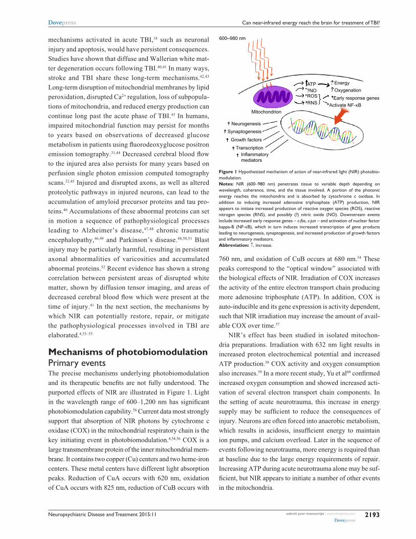
purported effects of NIR are illustrated in Figure 1. Light
in the wavelength range of 600–1,200 nm has significant
photobiomodulation capability.56 Current data most strongly
support that absorption of NIR photons by cytochrome c
oxidase (COX) in the mitochondrial respiratory chain is the
key initiating event in photobiomodulation.4,54,56 COX is a
large transmembrane protein of the inner mitochondrial mem-
brane. It contains two copper (Cu) centers and two heme-iron
centers. These metal centers have different light absorption
peaks. Reduction of CuA occurs with 620 nm, oxidation
of CuA occurs with 825 nm, reduction of CuB occurs with
760 nm, and oxidation of CuB occurs at 680 nm.54 These
peaks correspond to the “optical window” associated with
the biological effects of NIR. Irradiation of COX increases
the activity of the entire electron transport chain producing
more adenosine triphosphate (ATP). In addition, COX is
auto-inducible and its gene expression is activity dependent,
such that NIR irradiation may increase the amount of avail-
able COX over time.57
NIR’s effect has been studied in isolated mitochon-
dria preparations. Irradiation with 632 nm light results in
increased proton electrochemical potential and increased
ATP production.58 COX activity and oxygen consumption
also increases.59 In a more recent study, Yu et al60 confirmed
increased oxygen consumption and showed increased acti-
vation of several electron transport chain components. In
the setting of acute neurotrauma, this increase in energy
supply may be sufficient to reduce the consequences of
injury. Neurons are often forced into anaerobic metabolism,
which results in acidosis, insufficient energy to maintain
ion pumps, and calcium overload. Later in the sequence of
events following neurotrauma, more energy is required than
at baseline due to the large energy requirements of repair.
Increasing ATP during acute neurotrauma alone may be suf-
ficient, but NIR appears to initiate a number of other events
in the mitochondria.
Figure 1 Hypothesized mechanism of action of near-infrared light (NiR) photobio-modulation.Notes: NiR (600–980 nm) penetrates tissue to variable depth depending on wavelength, coherence, time, and the tissue involved. A portion of the photonic energy reaches the mitochondria and is absorbed by cytochrome c oxidase. in addition to inducing increased adenosine triphosphate (ATP) production, NiR appears to initiate increased production of reactive oxygen species (ROS), reactive nitrogen species (RNS), and possibly (?) nitric oxide (NO). Downstream events include increased early response genes – c-fos, c-jun – and activation of nuclear factor kappa-B (NF-κB), which in turn induces increased transcription of gene products leading to neurogenesis, synaptogenesis, and increased production of growth factors and inflammatory mediators.Abbreviation: ↑, increase.
κ
Neuropsychiatric Disease and Treatment 2015:11submit your manuscript | www.dovepress.com
Dovepress
Dovepress
2194
Henderson and Morries
Secondary eventsIn addition to the increase in ATP, the change in redox state
leads to greater oxidation and activation of the mitochondrial
permeability transition pore which alters intermembrane
potentials within the mitochondria.61 This may have a direct
or indirect effect on gene transcription and protein synthe-
sis.62 For example, transcription factors, such as redox factor-
1-dependent activator protein 1, activating transcription
factor/cyclic adenosine monophosphate response element
binding protein (ATF/CREB), and hypoxia-inducible factor
alpha, are upregulated in response to changes in redox states.
The change in redox state and NIR possibly directly63,64
result in the production of reactive oxygen species (ROS),
the most common of which is superoxide. Similarly, NIR
(670 nm) modulates the effects of reactive nitrogen species
in a model of multiple sclerosis.65 ROS are potent second
messenger molecules. ROS are involved in cell signaling,
enzyme activation, nucleic acid synthesis, protein synthesis,
and the activation of transcription factors.
The change in oxidation also likely contributes to the
displacement of nitric oxide (NO) from the COX molecule.
When bound, NO functions as an inhibitor by displacing
oxygen from the binding site on COX. When NO is dis-
placed, there is an increase in the activity of the electron
transport chain.66 In addition, NO serves as a vasodilator
and thus increases local blood flow.67 NO also functions
as a second messenger. Numerous tissue culture and ani-
mal studies have demonstrated the effect of NIR on NO
levels,4,68,69 including the upregulation of NO synthase
expression.70
Nuclear factor kappa-B (NF-κB) is a redox sensitive
transcription factor.71 This pro-survival transcription fac-
tor modulates the expression of numerous genes, including
ones involved in inflammation, early response (heat shock),
anti-apoptosis, cellular migration, and cell survival.71,72 NIR
(810 nm) activates NF-κB apparently via ROS in cultured
fibroblasts.72 NF-κB can be activated by ROS and indirectly
by the effects of ROS on inflammatory cytokines (eg, tumor
necrosis factor, interleukin-1).73
Tertiary eventsWith NIR exposure, induction of mitochondrial RNA
synthesis,74 as well as protein synthesis, occurs.75 More
recent work has shown NIR induces the upregulation and
downregulation of numerous genes both in the nucleus
and in the mitochondria of various cell types. Kushibiki
et al76 have reviewed the mitochondrial RNA expression
changes associated with photobiomodulation. Zhang et al77
demonstrated changes in mRNA expression of over 100
genes in cultured fibroblasts exposed to 653 nm NIR. Genes
involved in cell proliferation, such as mitogen-activated
protein kinase 11 (MAPk11), and cell-cycle progression
are increased.77 Apoptosis-inhibiting genes are upregulated
(eg, Janus kinase binding protein). Meanwhile, apoptosis-
promoting genes, such as heat shock 70k Da protein 1A
and caspase 6, are downregulated.77 Expression of genes
for antioxidants and inhibitors of the effects of ROS are
increased. Similar work in the retina has shown that genes
involved in cell survival, antioxidant production, transcrip-
tion, and growth factor production are also upregulated in
neural retina cells.78
Data from tissue culture and animal studies of NIR
reveal an increase in growth factor expression68,72,79,80 and
subsequent cell proliferation.4,12,68,69 Examples of these key
growth factors include nerve growth factor, brain-derived
neurotrophic factor, transforming growth factor-beta, and
vascular endothelial growth factor, which may contribute to
late brain remodeling after TBI.4,12,54,68,73,79–86 For example, a
fivefold increase in nerve growth factor mRNA transcription
occurred after irradiation of skeletal muscle cell culture with
633 nm NIR light.85
Recent data suggest that transcranial NIR phototherapy
can increase the process of neurogenesis in adult mice with
stroke or TBI. Increased numbers of neuroprogenitor cells
have been demonstrated in both the dentate gyrus of the
hippocampus and in the subventricular zone of the lateral
ventricle of mouse models.4,87,88 These cells also demonstrate
increased expression of a microtubule protein associated
with migrating neuroblasts.88 Some studies provide evi-
dence that NIR phototherapy may increase the process of
synaptogenesis.4,88 Together, these processes may aid in the
neuroplasticity responsible for neural repair and improved
function in cases of chronic TBI.
These cellular changes appear to persist for considerably
longer than the interval of light application89 (when delivered
at appropriate wavelengths and amplitudes).4 For example,
low level (red and) near-infrared light therapy (LLLT) of
a power density of 0.9–36 J/cm2 applied in a single treat-
ment at 24 hours post-stroke in animal models yielded a
reduction in neurological deficits, as well as histochemi-
cal evidence of neuron proliferation and migration.55,88,90,91
A single application of LLLT in rodent models of TBI had
similar benefits.4,87,92–94 Interestingly, these benefits were not
immediately apparent. Rather, a delay of 1–4 weeks was
noted, consistent with a progressive regeneration cascade
set in motion by the NIR exposure.
Neuropsychiatric Disease and Treatment 2015:11 submit your manuscript | www.dovepress.com
Dovepress
Dovepress
2195
Can near-infrared energy reach the brain for treatment of TBi?
SummaryDuring NIR phototherapy, absorption of red or NIR photons
by COX in the mitochondrial respiratory chain causes sec-
ondary molecular and cellular events, including activation
of second messenger pathways, changes in NO levels, and
growth factor production. NILT leads to the reduction of
excitotoxicity, the production of neurotrophic factors, the
modulation of ROS, the transcription of new gene products
with protective or pro-proliferative properties, and the
release of numerous growth factors for neurons and other
cells.4,64,68,73,82–84 NIR appears to initiate a cascade of subcellu-
lar events which can yield immediate, delayed, and persistent
beneficial changes in the injured neuron or other cell.
Properties of NIRLight has fundamental physical properties which are relevant
to its clinical use. Light is a form of electromagnetic radia-
tion which has properties of both waves and particles. Light
is characterized by its wavelength (distance between two
peaks), frequency, and amplitude. Light is also characterized
by its energy content. This energy is quantified as joules (J).
The amount of energy delivered per unit time constitutes the
power of light in watts (W = J/second). For medical applica-
tions, light is typically reported in terms of wavelength (nm),
energy (J), irradiance or power density (W/cm2), and radiant
exposure or fluence or dose (J/cm2).54,95
NIR has a number of biological effects, but it is critical
to understand the physical interactions between tissue and
light. When light impinges on the surface, a portion (∼10%)
is reflected.96 The energy that does penetrate the surface is
refracted or bent toward a line perpendicular to the surface.
This results from the particle property of light. These particles
are, of course, photons. The photons entering the tissue can be
transmitted through the tissue, scattered, or absorbed. Scatter
increases the volume of tissue impacted by the light. Photons
can change direction without loss of energy. Scattering is
particularly likely at interfaces between different tissues.
Reflection or refraction also occurs at such interfaces. These
effects contribute to shortening the distance to which light
will travel or penetrate into the tissue.96–98
Most tissues have the capacity to absorb light energy.
Usually this is mediated by a molecule absorbing a photon.
Molecules containing metal ions have a strong capacity for
absorbing photonic energy, but DNA and water also can. The
absorption of energy can induce a change in the confirmation
and/or function of the molecule.
Penetration of NIR through tissues is determined by
several factors: wavelength, energy, attenuation coefficient
(composed of scatter, refraction, and absorption), area of
irradiance, coherence, and pulsing. In general, longer wave-
lengths (up to 1,000 nm) will penetrate deeper; however, the
absorption of water begins to predominate above 1,000 nm.96
Increases in power density, in general, will lead to greater
penetration. More photons will traverse the tissue. The area
of surface irradiation also affects penetration due to scatter-
ing effects.
Coherence is a property of light waves in which waves
of monochromatic light are aligned such that any point
on the wave has the same amplitude and position as the
equivalent point in an adjacent wave. Temporal coherence
reflects the slight variations in the waveform over time. The
more consistent the waveform is, the higher the temporal
coherence. Monochromatic light typically has high temporal
coherence. Spatial coherence results from the divergence
of the light from the point of emission.66 Laser has virtually
no spatial divergence and creates a long narrow volume
of coherent light. LEDs are not monochromatic, but emit
light in a relatively narrow band over the peak wavelength.
LEDs also have significant spatial divergence and there-
fore a wide volume of space is radiated; however, within
that volume only a very small volume contains coherent
light. As Karu66 illustrates, the result is that noncoherent
LED sources likely only provide coherent light in a thin
volume, usually at surfaces. In contrast, laser generates a
long narrow volume of coherent light which can penetrate
deeper into tissues.
When coherent light enters a tissue, slight distortions
in the timing and the shape of the waves occur. As a result,
interference can occur between the waves. Polarization, the
angle at which a wave is vibrating, also contributes to inter-
ference. On a single wave basis, interference results when
the amplitude of the wave at a given point is different from
that of an adjacent wave and of the population of coher-
ent light waves. At the point of difference, the amplitudes
can either cancel each other out {[+x] + [-x]}, be additive
{[+x] + [+x]}, or any variation in between {[+x] + [-y]}.
The result of these interactions is a field of randomly dis-
tributed points of increased and decreased light intensity,
referred to as a speckle intensity pattern. Speckling can have
a significant impact on the effective penetration depth.99 As
such, areas of high intensity will penetrate further or will
have two to three orders of magnitude greater energy at a
given depth.99
Pulsing of NIR also increases the depth of penetration
and the amount of energy delivered to any given point at the
peak of a pulse. Yet, pulsing allows for troughs of energy

Neuropsychiatric Disease and Treatment 2015:11submit your manuscript | www.dovepress.com
Dovepress
Dovepress
2196
Henderson and Morries
output such that the overall energy delivered to the tissue
can be equivalent or even lower than that delivered by a
continuous emission. Pulsing is a property of lasers which
cannot be duplicated by LEDs.
Limitations of NILT protocolsPrior clinical applications of NIR photomodulation have
utilized LLLT emitters and prolonged courses of daily
treatments often extending over months.100,101 For example,
the first published study of NIR therapy for TBI in humans
described two cases of chronic mild TBI with significant
disability.100 Each patient had marked neuropsychological
improvement after a prolonged series of LLLT treatments
using 870 and 633 nm LED arrays over 4–72 months.
Yet, some clinical and laboratory studies of LLLT have
failed to consistently demonstrate benefit.15,16,102,103 For
example, Lavery et al15 demonstrated that LLLT (890 nm
LEDs delivering 1.3 J/cm2 for 40 minutes daily for
90 days) did not yield significant improvement in nerve
conduction velocity in patients with diabetic neuropathy.
Similarly, treatment of a rat model of contusive spinal
cord injury with LLLT (830 nm at 22.6 J/cm2 or 670 nm
at 28.4 J/cm2) for 30 minutes per day for 5 days resulted in
no significant functional improvement and no reduction in
lesion size.16 The identical treatment regimen was applied
to a rat model of TBI with no detectable improvement
in motor or sensory function or change in lesion size.16
In this animal model, Giacci et al calculated 2.6 J/cm2
reached the spinal cord with each treatment. This is within
the range of reported beneficial doses; yet, it was not
effective. Note that several studies have shown that LLLT
radiant energy is almost completely absorbed in the first
1 mm of skin.104,105
A clinical example of this discrepancy has unfolded in
the clinical trials for the treatment of stroke utilized in the
NEST-1 and NEST-2 trials.106,107 Lapchak12 reported that
the physical parameters of NILT in these studies may have
delivered insufficient energy to cortical tissues to be effec-
tive. Therein, 808 nm NIR with energy densities of 0.9 J/cm2
was applied to the human scalp at multiple sites for a total
of 40 minutes.106,107 Note that animal models of both stroke
and TBI indicate NIR energy densities in the range of
0.9–36 J/cm2 yields significant biochemical and behavioral
changes.3,4,81,87–90,93,94 The concern raised from the NEST
studies12 is that current clinical trials using LLLT to treat
TBI may yield negative or inaccurate efficacy data, not
because of the incapacity of NIR to invoke a change, but
due to a dose error. Doses that are effective when directly
applied to a monolayer of cells75,82 or when penetrating
0.2 mm through the skull of a rodent108 and the 5 mm
through the full thickness of the mouse brain,4,81,87,88,92–94,109
may be insufficient to penetrate to 20–30 mm into the
human brain.
We have been utilizing relatively high power (10–15 W)
lasers at the wavelengths of 810 and 980 nm in clinic to treat
TBI with positive results.3 The use of NIR in the treatment
of stroke and of TBI are reviewed in a companion paper.3
Skin is the first tissue encountered in the clinical application
of NIR phototherapy and represents a barrier to effective
penetration due to several factors. Human skin has multiple
layers and, therefore, multiple interfaces. Each interface is
a surface for scatter. In addition, each layer has different
inherent optical properties.97,110 The epidermis, comprised
in part of keratin, collagen, lipids, and melanin, has high
absorption in the ultraviolet range, but also absorbs light in
the infrared range of 600–1,100 nm.110 The dermis, comprised
in part of collagen, elastin, and proteoglycans, is of variable
thickness and penetration varies as a result. Scattering is a
predominate property of the dermis.110 The dermis is also
dense with blood vessels and the hemoglobin-rich blood
therein. While hemoglobin has absorption peaks at 450,
550, and 600 nm,97 it also absorbs photonic energy in the
clinical NIR range of 800–1,100 nm.110,111 The NIR absorp-
tion of hemoglobin depends upon its oxygenation status,
with carboxyhemoglobin having greater NIR absorption.111
Altogether, NIR photonic energy must first overcome the
hurdle of penetrating the skin to have an impact on deeper
structures.
We have shown clinical improvement in patients with
TBI utilizing high power NIR sources, but does high power
NIR penetrate deeper and/or with greater fluence compared
to LLLT? We have explored NIR penetration of tissues of
clinical relevance or previously modeled to quantify the
effective penetration at power densities ranging from the
50–200 mW levels used in LLLT87,88,90–94 to high power
levels used in clinical studies of TBI treatment.3 Specifically,
NIR in the wavelengths often used in clinical studies of red
and NIR photobiomodulation were utilized (650–670;16,100
810;87,93,94 880;100 980 nm112,113). Power levels ranged from
50 to 200 mW to model LLLT and from 6 to 15 W to model
high power NIR phototherapy. Specific tissues included
sheep skin and human skin to model skin penetration based
on issues raised in studies by Kolari and Airaksinen,114
Bjordal et al,8 and Esnouf et al.104 Sheep head, with skin,
skull, and brain intact, served as a model of human brain
penetration.3

Neuropsychiatric Disease and Treatment 2015:11 submit your manuscript | www.dovepress.com
Dovepress
Dovepress
2197
Can near-infrared energy reach the brain for treatment of TBi?
Recent findings concerning NIR attenuationethical considerationsAnimal tissue was obtained from local slaughter facilities and
no animals were sacrificed exclusively for these experiments.
Human tissue was obtained from ScienceCare (Denver, CO,
USA), a commercial agency which adheres to all federal and
state legal requirements through the Uniform Anatomical
Gift Act, and is accredited by the American Association of
Tissue Banks. All guidelines for the ethical handling and
disposal of human tissue were adhered to strictly. Ethics
approval was not sought for the in vivo human tissue studies,
because the authors themselves served as the subjects of the
tissue experiments. Verbal informed consent was exchanged
between the authors in the planning and preparation of these
in vivo human tissue studies.
ex vivo tissue studiesSheep skinLamb heads were obtained within 12 hours of slaughter.
Areas of skin were shaved and sections approximately
3×3 cm were excised. The thickness of the skin was measured
with precision calipers. A recently calibrated light meter
(Ophir-Spiricon LLC, North Logan, UT, USA) was posi-
tioned in a custom-made holder with a calibrated carriage to
hold an NIR emitter at a fixed and measurable distance from
the surface of the light meter. Several different NIR emitters
were utilized in this phase of the study to explore the effects
of frequency, power density, pulsing, and LED versus laser
on the penetration of NIR. Each emitter was positioned in the
carriage of the meter holder and set a fixed distance from the
meter surface. The baseline light transmission through air at
that distance was determined by five separate measurements.
The skin sample was then interposed and NIR transmission
through the skin sample was measured over five separate
trials each lasting 10 seconds. Temperature readings were
obtained using a laser digital sensor (Cen-Tec, Kunshan
City, People’s Republic of China) from the surface facing
the NIR emitter and the surface facing the light meter after
each transmission trial. This procedure was repeated for all
NIR emitters studied.
A 50 mW LED emitter at 810±20 nm was constructed to
emulate commercially available NIR diode devices which are
arranged in arrays and utilized in other studies of LLLT for
the treatment of the human brain. This LED was constructed
by arranging commercially available diodes in a concave
metal faceplate to emit 50.4±5.0 mW. This custom device
is referred to herein as “Custom LED”. Five commercially
available NIR emitters were also evaluated. The In Light pad
(In Light Wellness System, Albuquerque, NM, USA) has an
array of LEDs emitting 650 and 880 nm light at 200 mW. The
Eltech K-Laser 6D is a 6 W emitter (Eltech Srl, Treviso, Italy)
with dual wavelengths of 670/970 nm. The LiteCure LT1000
(LiteCure LLC, Newark, DE, USA) is a 10 W adjustable laser
NIR emitter with a dual wavelength of 810/980 nm and can
be set to continuous or pulsed light emission. The Diowave
810 nm laser (Technological Medical Advancements, Inc,
West Palm Beach, FL, USA) is adjustable up to 15 W, has a
wavelength of 810 nm, and can deliver continuous or pulsed
NIR. The Diowave 980 nm laser (Technological Medical
Advancements, Inc) also is adjustable up to 15 W and can
deliver continuous or pulsed NIR.
Penetration of sheep skinEx vivo studies of NIR penetration through fresh lamb skin
revealed a marked decrease in power density through a skin
thickness of only 2 mm (Table 1). The Custom 50 mW
810 nm LED did not appear to penetrate 2 mm of skin.
Similarly, the commercial 0.2 W 650/800 nm LED system
(In Light) did not show any detectable energy penetrating
the 2 mm of sheep skin. When compared to the power den-
sity of penetration through 2 mm of air, the 6 W LED of
wavelength 670/970 nm had an approximately 12%–20%
penetration, predominately in the red light range of 670 nm.
The 10 W combined 810/980 nm infrared laser (LiteCure)
showed a power density drop of 91% across 2 mm of skin.
The 15 W 810 nm laser (Diowave) demonstrated a 67% drop
in power density, while the 15 W 980 nm laser (Diowave)
demonstrated an 86% drop in power density across the same
thickness of skin. All of these levels of penetration were
statistically significant compared to the LED systems.
Using pulsed infrared light yielded much greater penetra-
tion. For example, 13.8% of the power density of a combined
810/980 nm infrared laser with a pulse frequency of 10 Hz
penetrated 2 mm of skin compared to only 8.6% of the con-
tinuous wave light of similar parameters. The 15 W 810 nm
laser with a pulse frequency of 10 Hz had a power density
drop of 66% across 2 mm of skin. The 15 W 980 nm infrared
laser with a pulse frequency of 10 Hz had only a 39% drop in
power density across a similar skin thickness. This difference
was not statistically significant with multiple comparisons,
but a trend was evident.
One concern about higher-powered infrared light sources
is the risk of tissue heating. Here, we observed the low
power LEDs made no significant temperature change in
the skin samples. The 10 W combined 810/980 nm and the

Neuropsychiatric Disease and Treatment 2015:11submit your manuscript | www.dovepress.com
Dovepress
Dovepress
2198
Henderson and Morries
15 W 810 nm infrared lasers made no significant temperature
change when keeping the emitter head in motion. Tem-
perature increase with the 15 W 980 nm infrared laser was
1.33°C. Notably, temperature change using 10 Hz pulsing
was not significant.
Animal skin serves as a useful model for human skin but
differs from human skin in many ways. The density of hair
follicles and melanin in the epidermis represent important
differences which could be predicted to impact NIR penetra-
tion. We also studied NIR penetration in human skin samples
obtained from a tissue bank.
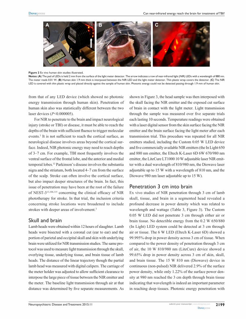
ex vivo human skin studiesThe same protocol was used to measure light transmission
through human skin. A full-thickness section of human skin
measuring 15×15 cm square was prepared by removing all
subcutaneous fat by dissection and isolating separate seg-
ments measuring 7×7 cm. The thickness of the human skin
was measured with digital calipers. Different NIR emitters
were again utilized. Each emitter was positioned in the car-
riage of the meter holder and set a fixed distance from the
meter surface. The baseline light transmission through air at
that distance was determined by five separate measurements.
The skin sample was then interposed and light transmission
through the skin sample was measured over five separate
trials each lasting 10 seconds. As shown in Figure 2, LEDs
were positioned against the skin sample with an interven-
ing sheet of clear plastic wrap. Temperature readings were
obtained using the laser digital sensor from the surface facing
the NIR emitter after each transmission trial. This procedure
was repeated for all NIR emitters studied.
Penetration of human skinEx vivo human skin was utilized to study transmission
of NIR photonic energy (Table 2, Figure 2). The Custom
0.05 W 810 nm LED did not appear to penetrate 1.9 mm of
human skin. The commercial 0.2 W 650/880 nm In Light
LED system delivered 0.01±0.002 W across 2 mm of air.
No energy could be detected penetrating the 1.9 mm thick-
ness of human skin with this device. Energy from the 10 W
combined 810/980 nm infrared laser could be detected pen-
etrating 1.9 mm of human skin and a power density drop of
89% across 1.9 mm of human skin was noted with 0.994 W
penetrating the tissue. The 15 W 810 nm laser demonstrated
an 83% drop in power density across a similar thickness of
human skin with 2.008 W penetrating the tissue. The 15 W
980 nm laser was not tested on human skin. Photonic energy
penetration of the two laser devices was statistically different Tab
le 1
Dat
a on
infr
ared
ligh
t pen
etra
tion
of e
x vi
vo s
kin
sam
ples
Inst
rum
ent
Wat
tsW
atts
at
0
mm
Wat
ts a
t 5
mm
Skin
thi
ckne
ss
(mm
)W
atts
pe
netr
atin
gT
emp
chan
ge (
°C)
Pul
se
(Hz)
Wat
ts a
t 5
mm
Skin
thi
ckne
ss
(mm
)W
atts
pe
netr
atin
gT
emp
chan
ge (
°C)
Cus
tom
LeD
810
nm0.
05
0.05
0±0.
001
0.02
0±0.
000
1.9
0.00
00±0
.000
00.
00%
-0.0
8±0.
00N
AN
AN
AN
AN
A
in L
ight
LeD
650/
880
nm0.
20.
020±
0.00
20.
010±
0.00
01.
90.
0000
±0.0
000
0.00
%-0
.05±
0.01
NA
NA
NA
NA
NA
K-L
aser
/elte
ch 6
D L
eD67
0/97
0 nm
63.
890±
0.00
40.
012±
0.00
31.
90.
0027
±0.0
010
21.5
0%a,
b
-0.1
9±0.
79N
AN
AN
AN
AN
A
Lite
Cur
e LT
1000
81
0/98
0 nm
10
5.16
0±0.
153
4.79
0±0.
058
1.9
0.41
20±0
.036
08.
60%
a–c
0.22
8±0.
306
102.
20±0
.050
2.0
0.30
4±0.
126
13.8
0%0.
928±
1.60
0
Dio
wav
e81
0 nm
159.
210±
0.04
08.
460±
0.00
62.
02.
8720
±0.6
670
33.9
0%a–
e
0.38
9±0.
322
104.
00±0
.020
2.0
1.37
2±0.
241
34.3
0%-0
.055
±0.4
57
Dio
wav
e98
0 nm
159.
590±
0.08
59.
080±
0.09
32.
01.
2620
±0.3
080
13.9
0%a–
c,f
1.33
±0.3
6210
4.47
±0.0
402.
02.
734±
0.27
961
.10%
0.88
9±0.
794
Not
es: T
he m
anuf
actu
rer’s
spec
ified
wat
t out
put,
the
actu
al o
utpu
t reg
ister
ed w
hen
the
infr
ared
ligh
t-em
ittin
g de
vice
was
dire
ctly
in c
onta
ct w
ith th
e m
eter
det
ecto
r, an
d th
e w
atts
whi
ch tr
aver
sed
5 m
m o
f air
are
repo
rted
. The
wat
ts r
ecor
ded
afte
r in
frar
ed li
ght f
rom
the
devi
ce p
enet
rate
d th
e st
ated
thic
knes
s of
ski
n al
ong
with
tem
pera
ture
(tem
p) c
hang
e at
the
surf
ace
of th
e sk
in c
lose
st to
the
infr
ared
em
itter
also
are
pro
vide
d. D
ata
on th
e ef
fect
s of
pul
sing
at 1
0 H
z ar
e pr
ovid
ed
whe
re r
elev
ant.
Num
bers
in b
old
are
perc
enta
ge c
hang
e in
pho
toni
c en
ergy
with
pen
etra
tion
of in
terp
osed
tiss
ue v
ersu
s an
equ
al d
istan
ce o
f air.
Sig
nific
ance
is in
dica
ted
with
sup
ersc
ripte
d le
tter
s –
signi
fican
ce le
vel a
t P
0.00
01 a
s de
term
ined
by
one
-tai
led
t-tes
t with
cor
rect
ions
for
mul
tiple
com
paris
ons.
a Cus
tom
ver
sus
othe
r; b in
Lig
ht v
ersu
s ot
her;
c K-L
aser
ver
sus
othe
r; d L
iteC
ure
vers
us o
ther
; e Dio
wav
e 81
0 nm
ver
sus
othe
r; f D
iow
ave
980
nm v
ersu
s ot
her
(P
0.01
).A
bbre
viat
ions
: NA
, not
app
licab
le; L
eD, l
ight
-em
ittin
g di
ode.
Neuropsychiatric Disease and Treatment 2015:11 submit your manuscript | www.dovepress.com
Dovepress
Dovepress
2199
Can near-infrared energy reach the brain for treatment of TBi?
from that of any LED device (which showed no photonic
energy transmission through human skin). Penetration of
human skin also was statistically different between the two
laser devices (P<0.000005).
For NIR to penetrate to the brain and impact neurological
injury (stroke or TBI) or disease, it must be able to reach the
depths of the brain with sufficient fluence to trigger molecular
events.3 It is not sufficient to reach the cortical surface, as
neurological disease involves areas beyond the cortical sur-
face. Indeed, NIR photonic energy may need to reach depths
of 3–7 cm. For example, TBI most frequently involves the
ventral surface of the frontal lobe, and the anterior and medial
temporal lobes.32 Parkinson’s disease involves the substantia
nigra and the striatum, both located 4–7 cm from the surface
of the scalp. Stroke can often involve the cortical surface,
but also impact deeper structures of the brain. In fact, this
issue of penetration may have been at the root of the failure
of NEST-313,106,115 concerning the clinical efficacy of NIR
phototherapy for stroke. In that trial, the inclusion criteria
concerning stroke locations were broadened to include
strokes with deeper areas of involvement.3
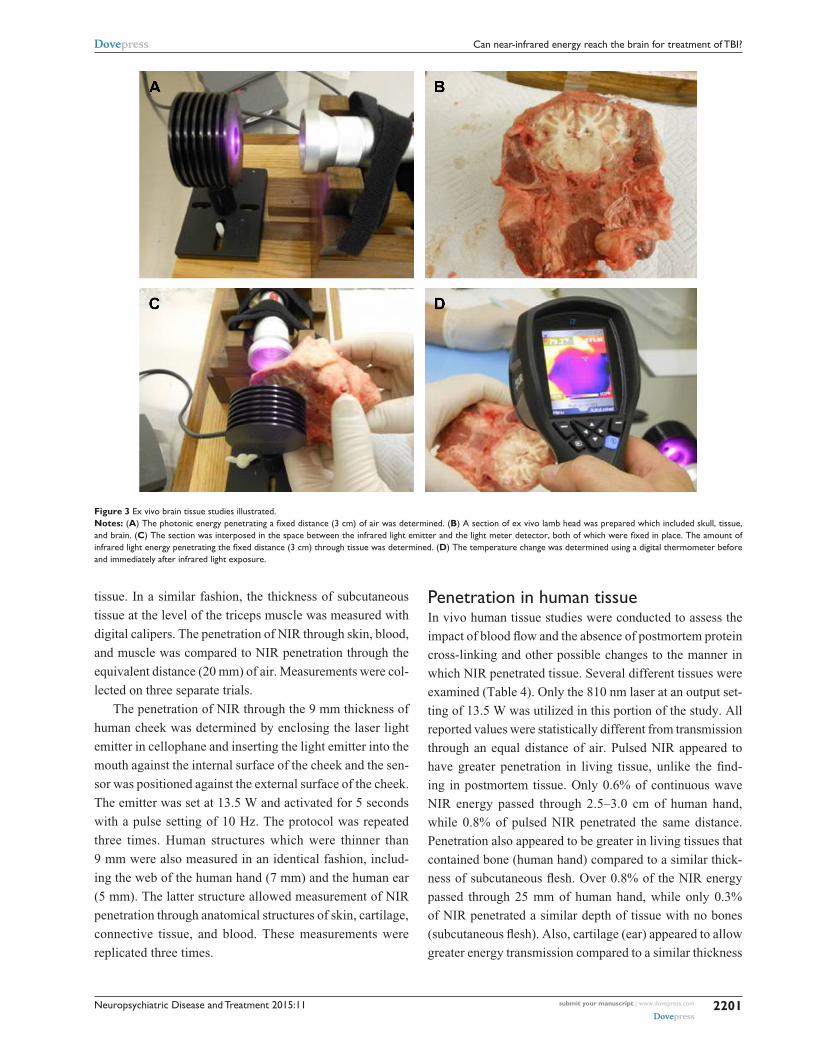
Skull and brainLamb heads were obtained within 12 hours of slaughter. Lamb
heads were bisected with a coronal cut (ear to ear) and the
portion of parietal and occipital skull and skin with underlying
brain were utilized for NIR transmission studies. The same pro-
tocol was used to measure light transmission through the skull,
overlying tissue, underlying tissue, and brain tissue of lamb
heads. The distance of the linear trajectory though the partial
lamb head was measured with digital calipers. The carriage of
the meter holder was adjusted to allow sufficient clearance to
interpose the large piece of tissue between the NIR emitter and
the meter. The baseline light transmission through air at that
distance was determined by five separate measurements. As
shown in Figure 3, the head sample was then interposed with
the skull facing the NIR emitter and the exposed cut surface
of brain in contact with the light meter. Light transmission
through the sample was measured over five separate trials
each lasting 10 seconds. Temperature readings were obtained
with a laser digital sensor from the skin surface facing the NIR
emitter and the brain surface facing the light meter after each
transmission trial. This procedure was repeated for all NIR
emitters studied, including the Custom 0.05 W LED device
and five commercially available NIR emitters (the In Light 650
and 880 nm emitter, the Eltech K-Laser 6D 6W 670/980 nm
emitter, the LiteCure LT1000 10 W adjustable laser NIR emit-
ter with a dual wavelength of 810/980 nm, the Diowave laser
adjustable up to 15 W with a wavelength of 810 nm, and the
Diowave 980 nm laser adjustable up to 15 W).
Penetration 3 cm into brainEx vivo studies of NIR penetration through 3 cm of lamb
skull, tissue, and brain in a segmented head revealed a
profound decrease in power density which was related to
wavelength and wattage (Table 3, Figure 3). The Custom
0.05 W LED did not penetrate 3 cm through either air or
brain tissue. No detectible energy from the 0.2 W 650/880
(In Light) LED system could be detected at 3 cm through
air or tissue. The 6 W LED (Eltech K-Laser 6D) showed a
99.995% drop in power density across 3 cm of tissue. When
compared to the power density of penetration through 3 cm
of air, the 10 W 810/980 nm (LiteCure) device showed a
99.65% drop in power density across 3 cm of skin, skull,
and brain tissue. The 15 W 810 nm (Diowave) device in
continuous (non-pulsed) NIR delivered 2.9% of the surface
power density, while only 1.22% of the surface power den-
sity at 980 nm reached the 3 cm depth through brain tissue
indicating that wavelength is indeed an important parameter
in reaching deep tissues. Photonic energy penetration with
Figure 2 ex vivo human skin studies illustrated. Notes: (A) The pad of LeDs is held 2 mm from the surface of the light meter detector. The arrow indicates a row of near-infrared light (NiR) LeDs with a wavelength of 880 nm. The meter reads 0.01 w. (B) Human skin 1.9 mm thick is interposed between the NiR LeD and the light meter detector. Thin plastic wrap covers the detector. (C) The NiR LeD is covered with thin plastic wrap and placed directly against the sample of human skin. Photonic energy could not be detected passing through 1.9 mm of human skin.

Neuropsychiatric Disease and Treatment 2015:11submit your manuscript | www.dovepress.com
Dovepress
Dovepress
2200
Henderson and Morries
the laser devices was statistically different from the LED
devices; however at this depth, the laser devices did not differ
significantly from each other using continuous NIR delivery.
Using pulsed NIR emission settings, lower total fluency and
heat is delivered to the skin surface, while energy delivered
to deeper structures at a pulse peak is greater. Pulsed energy
penetration to 3 cm was significantly different between the
three laser devices (P0.01).
Temperature changes at the skull surface ranged from
0.2°C to 3°C. Temperature change in the brain was less
than 1°C except when using the 980 nm 15 W laser in con-
tinuous emission setting. In the latter case, the temperature
variation was quite large. These data show less temperature
change using the pulsed setting regardless of wavelength
and power.
NIR transmission through living tissue is likely different
than through postmortem tissue for a number of reasons.
First, cross-linking of proteins is an early and progressive
event in death. Second, changes in interstitial fluids occur
within hours of death. Third, the perfusion of dermis and
deeper tissues in vivo creates scatter and refraction of NIR.
Fourth, the flow of blood also disperses heat from the site of
NIR application. Lastly, some authors have suggested that
NO created at the site of irradiation and carried throughout
the body in the blood is responsible for the beneficial effects
of NIR phototherapy.116 This effect could account for the
clinical benefits seen in TBI if NIR does not penetrate to
the depths of 3 cm or greater in living tissue. Jagdeo et al
modeled NIR penetration in living human tissue by serving
as their own author/volunteers.117 We have replicated
some of their work using a higher-powered NIR source.
Tissues included hand, cheek, ear, and triceps to model
the complex mixture of skin, bone, connective tissues, and
other tissues found in the pathway of NIR en route to the
brain.
in vivo human tissue studiesThe in vivo penetration of NIR through human tissue was
measured compared to NIR penetration of air. The Diowave
810 nm high power laser was the only NIR emitter utilized
in this portion of the study. The power output was set at 13.5
W for a duration of 5 seconds. First, the thickness of two
different human hands was measured with digital calipers.
The passage of NIR through 25 or 30 mm of air was repeat-
edly measured (Table 3). Then, the passage of NIR across
the equivalent distance of the palm of a human hand was
repeatedly measured. Intervening anatomical structures
included skin, tendon, bone, muscle, blood, and connective Tab
le 2
Dat
a on
infr
ared
ligh
t pe
netr
atio
n of
ex
vivo
hum
an s
kin
sam
ples
Inst
rum
ent
Wat
tsW
atts
at
0
mm
Wat
ts a
t 2
mm
Skin
thi
ckne
ss
(mm
)W
atts
pe
netr
atin
gT
emp
chan
ge (
°C)
Pul
se
(Hz)
Wat
ts a
t 2
mm
Skin
thi
ckne
ss
(mm
)W
atts
pe
netr
atin
gT
emp
chan
ge (
°C)
Cus
tom
LeD
810
nm0.
05
0.05
0±0.
001
0.02
0±0.
000
1.9
0.00
0±0.
000
0.00
%c,
d
-0.0
80±0
.000
NA
NA
NA
NA
NA
in L
ight
65
0/88
0 nm
0.20
0.02
0±0.
004
0.01
0±0.
002
1.9
0.00
0±0.
000
0.00
%c,
d
0.00
0±0.
000
NA
NA
NA
NA
NA
Lite
Cur
e LT
1000
81
0/98
0 nm
10.0
09.
140±
0.04
28.
680±
0.04
91.
90.
994±
0.06
711
.5%
a,b,
d
0.87
0±0.
412
104.
590±
0.02
31.
90.
372±
0.01
38.
1%a,
b,d
0.55
0±0.
170
Dio
wav
e81
0 nm
15.0
013
.050
±0.4
4012
.030
±0.0
111.
92.
008±
0.09
216
.7%
a–c
1.81
0±0.
447
106.
198±
0.01
91.
90.
790±
0.02
712
.7%
a–c
1.00
±0.5
0
Not
es: T
he m
anuf
actu
rer’
s sp
ecifi
ed w
att
outp
ut, t
he a
ctua
l out
put
regi
ster
ed w
hen
the
infr
ared
ligh
t-em
ittin
g de
vice
was
dir
ectly
in c
onta
ct w
ith t
he m
eter
det
ecto
r, a
nd t
he w
atts
whi
ch t
rave
rsed
2 m
m o
f air
are
rep
orte
d. T
he w
atts
re
cord
ed a
fter
infr
ared
ligh
t fr
om t
he d
evic
e pe
netr
ated
the
sta
ted
thic
knes
s of
ski
n al
ong
with
tem
pera
ture
(te
mp)
cha
nge
at t
he s
urfa
ce o
f the
ski
n cl
oses
t to
the
infr
ared
em
itter
als
o ar
e pr
ovid
ed. D
ata
on t
he e
ffect
s of
pul
sing
at
10 H
z ar
e pr
ovid
ed w
here
rel
evan
t. N
umbe
rs in
bol
d ar
e pe
rcen
tage
cha
nge
in p
hoto
nic
ener
gy w
ith p
enet
ratio
n of
inte
rpos
ed t
issu
e ve
rsus
an
equa
l dis
tanc
e of
air
. Sig
nific
ance
is in
dica
ted
with
sup
ersc
ript
ed le
tter
s –
sign
ifica
nce
leve
l at
P
0.00
01 a
s de
term
ined
by
one-
taile
d t-
test
with
cor
rect
ions
for
mul
tiple
com
pari
sons
. a Cus
tom
ver
sus
othe
r; b in
Lig
ht v
ersu
s ot
her;
c Lite
Cur
e ve
rsus
oth
er; d D
iow
ave
810
nm v
ersu
s ot
her
(P
0.01
).A
bbre
viat
ions
: NA
, not
app
licab
le; L
eD, l
ight
-em
ittin
g di
ode.
Neuropsychiatric Disease and Treatment 2015:11 submit your manuscript | www.dovepress.com
Dovepress
Dovepress
2201
Can near-infrared energy reach the brain for treatment of TBi?
tissue. In a similar fashion, the thickness of subcutaneous
tissue at the level of the triceps muscle was measured with
digital calipers. The penetration of NIR through skin, blood,
and muscle was compared to NIR penetration through the
equivalent distance (20 mm) of air. Measurements were col-
lected on three separate trials.
The penetration of NIR through the 9 mm thickness of
human cheek was determined by enclosing the laser light
emitter in cellophane and inserting the light emitter into the
mouth against the internal surface of the cheek and the sen-
sor was positioned against the external surface of the cheek.
The emitter was set at 13.5 W and activated for 5 seconds
with a pulse setting of 10 Hz. The protocol was repeated
three times. Human structures which were thinner than
9 mm were also measured in an identical fashion, includ-
ing the web of the human hand (7 mm) and the human ear
(5 mm). The latter structure allowed measurement of NIR
penetration through anatomical structures of skin, cartilage,
connective tissue, and blood. These measurements were
replicated three times.
Penetration in human tissueIn vivo human tissue studies were conducted to assess the
impact of blood flow and the absence of postmortem protein
cross-linking and other possible changes to the manner in
which NIR penetrated tissue. Several different tissues were
examined (Table 4). Only the 810 nm laser at an output set-
ting of 13.5 W was utilized in this portion of the study. All
reported values were statistically different from transmission
through an equal distance of air. Pulsed NIR appeared to
have greater penetration in living tissue, unlike the find-
ing in postmortem tissue. Only 0.6% of continuous wave
NIR energy passed through 2.5–3.0 cm of human hand,
while 0.8% of pulsed NIR penetrated the same distance.
Penetration also appeared to be greater in living tissues that
contained bone (human hand) compared to a similar thick-
ness of subcutaneous flesh. Over 0.8% of the NIR energy
passed through 25 mm of human hand, while only 0.3%
of NIR penetrated a similar depth of tissue with no bones
(subcutaneous flesh). Also, cartilage (ear) appeared to allow
greater energy transmission compared to a similar thickness
Figure 3 ex vivo brain tissue studies illustrated. Notes: (A) The photonic energy penetrating a fixed distance (3 cm) of air was determined. (B) A section of ex vivo lamb head was prepared which included skull, tissue, and brain. (C) The section was interposed in the space between the infrared light emitter and the light meter detector, both of which were fixed in place. The amount of infrared light energy penetrating the fixed distance (3 cm) through tissue was determined. (D) The temperature change was determined using a digital thermometer before and immediately after infrared light exposure.

Neuropsychiatric Disease and Treatment 2015:11submit your manuscript | www.dovepress.com
Dovepress
Dovepress
2202
Henderson and Morries
Tab
le 3
Dat
a on
infr
ared
ligh
t pe
netr
atio
n of
ex
vivo
lam
b sk
ull,
tissu
e, a
nd b
rain
Inst
rum
ent
Wat
tsW
atts
at
0
mm
Wat
ts a
t
30 m
mW
atts
pen
etra
ting
br
ain
at 3
0 m
mT
emp
chan
ge
at s
kin
(°C
)T
emp
chan
ge
at b
rain
(°C
)P
ulse
(H
z)W
atts
at
30 m
mW
atts
pen
etra
ting
br
ain
at 3
0 m
mT
emp
chan
ge
at s
kin
(°C
)T
emp
chan
ge
at b
rain
(°C
)
Cus
tom
LeD
810
nm0.
05
0.05
0±0.
001
0.00
0±0.
000
0.00
00±0
.000
00.
00%
c–e
0.00
0±0.
000
-0.0
40±0
.000
NA
NA
NA
NA
NA
in L
ight
LeD
650/
880
nm0.
200.
020±
0.00
20.
000±
0.00
00.
0000
±0.0
000
0.00
%c–
e
0.01
0±0.
000
-0.0
50±0
.010
NA
NA
NA
NA
NA
K-L
aser
/elte
ch 6
D L
eD67
0/97
0 nm
6.00
3.89
0±0.
004
0.00
9±0.
001
0.00
20±0
.000
022
.2%
a,b,
e,f
0.29
5±0.
877
0.11
3±0.
670
NA
NA
NA
NA
NA
Lite
Cur
e LT
1000
81
0/98
0 nm
10.0
05.
160±
0.15
32.
000±
0.03
00.
0070
±0.0
050
0.35
%a,
b
3.33
1±1.
410
0.11
0±0.
106
101.
580±
0.01
00.
0230
±0.0
050
1.46
%a–
c,e,
f
2.84
±3.9
000.
340±
0.47
8
Dio
wav
e81
0 nm
15.0
09.
210±
0.04
04.
000±
0.05
50.
1160
±0.0
200
2.90
%a–
c
0.22
0±0.
304
0.94
4±0.
179
102.
017±
0.02
90.
0478
±0.0
043
2.37
%a–
d,f
1.53
3±1.
200
1.00
0±1.
360
Dio
wav
e98
0 nm
15.0
09.
590±
0.08
56.
220±
0.12
70.
0764
±0.0
130
1.23
%a–
c
1.33
0±1.
390
2.89
0±2.
770
103.
137±
0.01
10.
0492
±0.0
070
1.57
%a–
e
1.55
0±1.
140
1.00
0±0.
465
Not
es: T
he m
anuf
actu
rer’
s sp
ecifi
ed w
att o
utpu
t, th
e ac
tual
out
put r
egis
tere
d w
hen
the
infr
ared
ligh
t-em
ittin
g de
vice
was
dir
ectly
in c
onta
ct w
ith th
e m
eter
det
ecto
r, a
nd th
e w
atts
whi
ch tr
aver
sed
30 m
m o
f air
are
rep
orte
d. T
he w
atts
re
cord
ed a
fter
infr
ared
ligh
t fr
om t
he d
evic
e pe
netr
ated
30
mm
of s
kull,
tis
sue,
and
bra
in, a
s w
ell a
s te
mpe
ratu
re (t
emp)
cha
nge
at t
he e
xter
ior
of t
he s
kull
(ski
n su
rfac
e) a
nd a
t th
e in
teri
or s
urfa
ce o
f the
bra
in a
re p
rovi
ded.
Dat
a on
the
ef
fect
s of
pul
sing
the
infr
ared
ligh
t at 1
0 H
z al
so a
re p
rovi
ded
whe
re r
elev
ant.
Num
bers
in b
old
are
perc
enta
ge c
hang
e in
pho
toni
c en
ergy
with
pen
etra
tion
of in
terp
osed
tiss
ue v
ersu
s an
equ
al d
ista
nce
of a
ir. S
igni
fican
ce is
indi
cate
d w
ith
supe
rscr
ipte
d le
tter
s –
sign
ifica
nce
leve
l at
P0.
0001
as
dete
rmin
ed b
y on
e-ta
iled
t-te
st w
ith c
orre
ctio
ns fo
r m
ultip
le c
ompa
riso
ns. a C
usto
m v
ersu
s ot
her;
b in L
ight
ver
sus
othe
r; c K
-Las
er v
ersu
s ot
her;
d Lite
Cur
e ve
rsus
oth
er; e D
iow
ave
810
vers
us o
ther
; f Dio
wav
e 98
0 ve
rsus
oth
er (P
0.
01).
Abb
revi
atio
ns: N
A, n
ot a
pplic
able
; LeD
, lig
ht-e
mitt
ing
diod
e.
Tab
le 4
Dat
a on
infr
ared
ligh
t pe
netr
atio
n of
in v
ivo
hum
an t
issu
e
Tis
sue
Dis
tanc
e to
m
eter
(m
m)
Wat
tsT
emp
ch
ange
(°C
)P
ulse
(H
z)W
atts
Tem
p ch
ange
(°C
)A
ir p
enet
rati
onT
issu
e pe
netr
atio
nA
ir p
enet
rati
onT
issu
e pe
netr
atio
n
Palm
of h
and
125
3.8±
0.00
0.02
3±0.
009
0.60
%a
+210
2.02
0±0.
017
0.01
8±0.
002
0.89
%a
+1.0
Palm
of h
and
230
3.9±
0.01
0.02
3±0.
007
0.59
%a
+210
2.01
0±0.
006
0.01
7±0.
001
0.85
%a
+1.0
Sub-
cut
flesh
20N
AN
AN
A10
1.58
3±0.
015
0.00
5±0.
003
0.32
%a
+0.2
Che
ek9
NA
NA
NA
103.
800±
0.01
00.
013±
0.00
30.
47%
a
+1.0
Han
d w
eb7
NA
NA
NA
104.
100±
0.16
70.
018±
0.00
20.
44%
a
+0.2
ear
with
car
tilag
e5
NA
NA
NA
104.
400±
0.03
00.
129±
0.00
72.
93%
a
+0.5
Not
es: F
or e
ach
hum
an ti
ssue
, the
dis
tanc
e m
easu
red,
the
wat
ts tr
aver
sing
the
dist
ance
of a
ir, t
he p
enet
ratin
g th
at d
ista
nce
in h
uman
tiss
ue, a
nd th
e te
mpe
ratu
re (t
emp)
cha
nge
at th
e sk
in s
urfa
ce im
med
iate
ly a
fter
infr
ared
ligh
t exp
osur
e ar
e pr
ovid
ed. D
ata
on t
he e
ffect
s of
pul
sing
the
infr
ared
ligh
t at
10
Hz
also
are
pro
vide
d w
here
rel
evan
t. N
umbe
rs in
bol
d ar
e pe
rcen
tage
cha
nge
in p
hoto
nic
ener
gy w
ith p
enet
ratio
n of
inte
rpos
ed t
issu
e ve
rsus
an
equa
l dis
tanc
e of
air
. a S
igni
fican
ce le
vel a
t P
0.00
01 a
s de
term
ined
by
one-
taile
d t-
test
with
cor
rect
ions
for
mul
tiple
com
pari
sons
.A
bbre
viat
ions
: NA
, not
app
licab
le; L
eD, l
ight
-em
ittin
g di
ode.
Neuropsychiatric Disease and Treatment 2015:11 submit your manuscript | www.dovepress.com
Dovepress
Dovepress
2203
Can near-infrared energy reach the brain for treatment of TBi?
of skin (hand web). Temperature change after 5 seconds was
2°C or less at the skin surface and dropped quickly possibly
related to blood flow.
Attenuation of NIR energyGiven the much greater distance involved in delivering
NIR in effective doses to the human brain, we examined
NIR penetration to 3 cm depth through the skin, skull, and
brain of ex vivo sheep head. As could be anticipated from
the penetration studies of skin, the low power NIR emitters
showed no evidence of penetration to this depth. The 6 W NIR
emitter transmitted less than 1/10 of 1% of its surface energy
through 3 cm of skull and brain. It did demonstrate that red
light (670 nm) transmitted better – 22% of the red light energy
that traversed 3 cm of air was able to penetrate 3 cm of tissue.
High-powered NIR lasers delivered between 0.5% and 3.0%
of their energy to the depth of the brain. Notably, penetra-
tion to this depth was significantly better using 810 nm NIR
than it was when using 980 nm NIR. This is advantageous
given the stronger evidence of 810 nm light being effective
in photomodulation of neurological function.87,88,91–94
These tissue studies indicate NIR energy penetrates to
depths of 3 cm whether in ex vivo or living tissue; however,
sufficient energy must be delivered to the skin surface. The
0.2 and 0.5 W NIR diodes yielded no detectable energy at
3 cm depth. High-powered NIR lasers were able to deliver
between 0.5% and 3.0% of their energy to the depth of 3 cm in
living or postmortem tissue, with living tissue absorbing more
energy over the intervening distance. Based on the power
density of 55–81 J/cm2 delivered to the skin of patients in
our clinical cases3,118 (using a 10–15 W NIR, 810/980 nm, or
810 nm laser), then the expected power density in the human
brain at 3 cm depth would be in the range of 0.8–2.4 J/cm2 –
precisely in the range shown to induce neurological benefit
in animal models.4,81,87,88,92–94 The safety of NIR exposure at
high doses has been explored in animal models119–121 and in
humans.3,55,115
Putting penetration in perspectiveOne of the earliest studies of NIR penetration and human
skin examined transmittance of 633 nm light through pro-
gressively thicker sections of human skin.102 Penetration
through 0.4 mm of epidermis was 78% for 633 nm light and
at 2 mm, the energy had dropped to approximately 5% of
the incident 633 nm light. Later, the same group found no
penetration of infrared light beyond 3 mm using the same
light sources.114 Bjordal et al8 also concluded that 90% of the
energy from 632 nm laser is lost in the skin. NIR at 820 nm
has slightly greater skin penetration. Approximately 89% of
820 nm light will penetrate 0.4 mm of epidermis and about
13.5% traverses 2 mm of skin,102 but 0% reaches a depth of
3 mm.114 Others found 80% of the energy from an 820 nm
source is lost in the skin.8 Human skin also was examined
by Esnouf et al104 using an 850 nm continuous light source
at 100 mW. They reported 34% of the light from the source
could penetrate 0.784 mm.104 The 904 nm laser may have
greater skin penetration.8 Using rat skin of an unspeci-
fied thickness, Joensen et al113 found 20% of the 810 nm
light from a 200 mW source penetrated skin. Our results
show considerably less penetration by low level NIR. We
found energy from a 50 mW 810 nm LED did not penetrate
2 mm of human or sheep skin. No energy could be detected
penetrating either human skin or sheep skin from a commer-
cially available 0.2 W LED (650+880 nm). In contrast, 9% of
the energy from the 10 W combined 810/980 nm continuous
wave infrared laser passed through 2 mm of skin (human or
sheep). The 15 W 810 laser in continuous mode delivered
33% of its energy through 2 mm of skin. While longer
wavelength is typically associated with greater penetration,
we found that only 14% of the energy from a 15 W 980 nm
laser in continuous mode penetrated 2 mm of (sheep) skin.
These data are consistent with the transmission properties
reported for skin, which has relatively good transmission
in the range of 810 nm and somewhat less transmission at
980 nm.122 Using pulsed infrared light yielded much greater
penetration. For example, 41% of the power density of a
combined 810/980 nm infrared laser with a pulse frequency
of 10 Hz penetrated 1.9 mm of human skin compared to
only 11% of the continuous wave light of similar param-
eters. The 15 W 810 nm laser with a pulse frequency of
10 Hz showed 69% of the energy penetrated 1.9 mm of
human skin compared to only 17% of the continuous wave
light of similar parameters.
Thicker segments of tissue have been studied by several
groups. Penetration through mouse skull (0.2 mm)108 and
overlying skin with 800–810 nm light from an LLLT emitter
ranges from 6.3%95 to 46%.109 Penetration through the entire
depth of a mouse brain (3–5 mm) is reportedly 1%–4%.109
Byrnes et al122 examined penetration of 810 nm continuous
light from a 150 mW source through skin, muscle, bone,
and spinal cord of the rat (∼24 mm). They found 6% of the
energy penetrated these tissues. Similarly, Giacci et al16 mea-
sured photonic energy transmission through an unspecified
thickness of skin and muscle overlying the spinal column
of the rat and found 6.6% of 670 nm light from a 0.5 W
emitter penetrated this distance. Penetration of 830 nm light

Neuropsychiatric Disease and Treatment 2015:11submit your manuscript | www.dovepress.com
Dovepress
Dovepress
2204
Henderson and Morries
was slightly greater at 11.3%.16 Penetration of these same
wavelengths from the skin of the rat head to the optic nerve
was estimated at 0.1%.16 Hudson et al112 precisely measured
photonic energy at varying depths of bovine muscle using a 1
W 808/980 nm emitter. They found 808 nm light had greater
penetration, despite the premise that longer wavelengths
travel further. Indeed, the attenuation of 808 nm light at 3 cm
was 99%, quite similar to the findings we describe here in
both living human hand and the more heterogenous media
of the ovine head.
In living human hand (25 mm), Jagdeo et al117 found
only 0.01%–0.09% of 830 nm light from a 0.5 W emitter
penetrated this distance. We found that 0.6% of 810 nm light
from a higher-powered source (10 W) was able to penetrate
25 mm of skin, bone, and soft tissue. This represents at least
a sixfold increase in penetration compared to previous work
with low power emitters.
Fitzgerald et al111 modeled light penetration into the brain
for a 670 nm and a 1,064 nm LED light source emitting
28 mW/cm2. Using a diffusion simulation computer model
with assumptions of 10 mm of overlying scalp and skull,
they derived that 2.5 mW/cm2 for 670 nm light and 13 mW/
cm2 for 1,064 nm light would reach the surface of the cere-
bral cortex.111 They further derived that at the center of the
brain (10 mm skull +46 mm brain =56 mm), power density
would be approximately 1.2×10⁻¹¹ W/cm2 for 670 nm light
and 1.4×10⁻⁷ W/cm2 for 1,064 nm light.111
Jagdeo et al also examined LLLT penetration through
isolated human skull from cadaver and found it was lim-
ited with only 7.4% of the light from an 830 nm 0.5 W
continuous LED device (Omnilux) penetrating the bone.117
Moreover, they found that no more than 0.5% of NIR at
830 nm penetrated approximately 10 mm of frontal skull
and overlying tissue in a cadaver model. Also, no detectable
NIR penetrated the temporal bone and overlying tissue.117
These studies of LLLT and the computer model indicate
that photonic energy from LLLT emitters does not appear
to deliver significant fluence to the depths required to treat
the human brain.
Our studies of 3 cm of sheep skull, brain, and overlying
soft tissue are the closest model of the clinical practice of
NILT for TBI. Again, NIR from devices generating less
than 1 W could not be detected at this depth. The energy
from a 6 W LED system showed a 99.995% drop across
3 cm of tissue. In contrast, 0.14% of the energy from a 10 W
810/980 nm device penetrated 3 cm of tissue. At 15 W, an
810 nm emitter in continuous mode delivered 1.26% of
the surface power density and 0.80% of the 980 nm device
emissions reached the 3 cm depth through brain tissue. Using
pulsed NIR emission settings, a lower overall power density
was delivered to the surface; however, similar penetration
was achieved.
Some have suggested that NIR has a dosing window and
have reported a bimodal response curve, such that high-dose
NIR could be detrimental;4,5 however, others have shown a
dose-dependent response to NIR with no detrimental effects
at higher doses.80
The penetration data mentioned and the data presented
herein challenge the presumption that only LLLT protocols
spanning weeks or months would possibly be clinically
effective. For example, using a 0.5 W 830 nm LED, Jagdeo
et al117 estimated the power density reaching the cerebral
cortex based on their model was 3 mW/cm2, which equates to
a fluence of 0.0064 J/cm2 – 1/140th of the minimum thought
to be necessary for ideal photobiomodulation.5 Anders and
colleagues noted NIR penetrates 4 cm into human cadaver
skin, skull, and brain using a 5 W laser (JJ Anders, personal
communication, January 13, 2015). From our data, we
estimate that in our clinical applications of high-powered
NIR lasers,3 we are delivering 0.64–1.95 J/cm2 to a depth of
30 mm. This is 100-fold greater fluence than that delivered
by an LED system, but within the range of fluence shown to
have beneficial biological effects.
ConclusionExtensive research has shown the fluence within the range
of 0.9–15.0 J/cm2 is most effective in activating the bio-
logical processes involved in reversing or mitigating the
pathophysiological effects of TBI. The attenuation of NIR
energy as it passes through tissue has been examined in
computer simulations, animal tissue, and human tissue.
NIR penetration in the human brain is subject to attenuation
by multiple tissues (skin, skull, dura, blood, cerebrospinal
fluid) and multiple interfaces which scatter, absorb, and
reflect the NIR to varying degrees. We have shown through
the use of higher wattage NIR lasers that we can deliver
fluence at therapeutic levels to the depths of the brain
without tissue heating or damage. The protocols have been
applied in our clinic with excellent clinical results and no
side effects.
AcknowledgmentsThe authors would like to acknowledge the technical assis-
tance of Phillip Hardinger, DC, DABCO, and Mr Charles
Vorwaller (Diowave Corporation/LiteCure Corporation).
Artistic creation Figure 1: Ms Taylor Tuteur.

Neuropsychiatric Disease and Treatment 2015:11 submit your manuscript | www.dovepress.com
Dovepress
Dovepress
2205
Can near-infrared energy reach the brain for treatment of TBi?
DisclosureDr Larry Morries is CEO of the Neuro-Laser Foundation,
a nonprofit foundation. He has a private practice in Lake-
wood, CO, USA.
Theodore A Henderson is president of The Synaptic
Space, a medical consulting firm and president of Dr Theodore
Henderson, Inc, a clinical service firm. He is also co-owner of
Neuro-Luminance, a clinical service organization, president
of the International Society of Applied Neuroimaging, and
CFO of the Neuro-Laser Foundation.
References 1. Hyder AA, Wunderlich CA, Puvanachandra P, Gururaj G, Kobusingye OC.
The impact of traumatic brain injuries: a global perspective. Neuro Rehabilitation. 2007;22(5):341–353.
2. Feigin VL, Forouzanfar MH, Krishnamurthi R, et al; Global Burden of Diseases, Injuries, and Risk Factors Study 2010 (GBD 2010), GBD Stroke Experts Group. Global and regional burden of stroke during 1990–2010: findings from the Global Burden of Disease Study 2010. Lancet. 2014;383(9913):245–254.
3. Morries LD, Cassano P, Henderson TA. Treatments for traumatic brain injury with emphasis on transcranial near infrared laser phototherapy. Neuropsychiatr Dis Treat. In press 2015.
4. Chung H, Dai T, Sharma SK, Huang YY, Carroll JD, Hamblin MR. The nuts and bolts of low-level laser (light) therapy. Ann Biomed Eng. 2012;40(2):516–533.
5. Huang YY, Chen AC, Carroll JD, Hamblin MR. Biphasic dose response in low level light therapy. Dose Response. 2009;7(4):358–383.
6. Enwemeka CS. Intricacies of dose in laser phototherapy for tissue repair and pain relief. Photomed Laser Surg. 2009;27(3):387–393.
7. Mester E, Mester AF, Mester A. The biomedical effects of laser applica-tion. Lasers Surg Med. 1985;5(1):31–39.
8. Bjordal JM, Couppé C, Chow RT, Tunér J, Ljunggren EA. A sys-tematic review of low level laser therapy with location-specific doses for pain from chronic joint disorders. Aust J Physiother. 2003;49(2): 107–116.
9. Basford JR, Sheffield CG, Harmsen WS. Laser therapy: a randomized, controlled trial of the effects of low-intensity Nd:YAG laser irra-diation on musculoskeletal back pain. Arch Phys Med Rehabil. 1999; 80(6):647–652.
10. Yang Z, Wu Y, Zhang H, et al. Low-level laser irradiation alters cardiac cytokine expression following acute myocardial infarction: a potential mechanism for laser therapy. Photomed Laser Surg. 2011; 29(6):391–398.
11. Tuby H, Maltz L, Oron U. Induction of autologous mesenchymal stem cells in the bone marrow by low-level laser therapy has profound beneficial effects on the infarcted rat heart. Lasers Surg Med. 2011; 43(5):401–409.
12. Lapchak PA. Taking a light approach to treating acute ischemic stroke patients: transcranial near-infrared laser therapy translational science. Ann Med. 2010;42(8):576–586.
13. Lampl Y, Zivin JA, Fisher M, et al. Infrared laser therapy for ischemic stroke: a new treatment strategy: results of the NeuroThera Effectiveness and Safety Trial-1 (NEST-1). Stroke. 2007;38(6):1843–1849.
14. Hacke W, Schellinger PD, Albers GW, et al; NEST 3 Committees and Investigators. Transcranial laser therapy in acute stroke treatment: results of neurothera effectiveness and safety trial 3, a phase III clinical end point device trial. Stroke. 2014;45(11):3187–3193.
15. Lavery LA, Murdoch DP, Williams J, Lavery DC. Does anodyne light therapy improve peripheral neuropathy in diabetes? A double-blind, sham-controlled, randomized trial to evaluate monochromatic infrared photoenergy. Diabetes Care. 2008;31(2):316–321.
16. Giacci MK, Wheeler L, Lovett S, et al. Differential effects of 670 and 830 nm red near infrared irradiation therapy: a comparative study of optic nerve injury, retinal degeneration, traumatic brain and spinal cord injury. PLoS One. 2014;9(8):e104565.
17. Veech RL, Valeri CR, VanItallie TB. The mitochondrial permeability transition pore provides a key to the diagnosis and treatment of traumatic brain injury. IUBMB Life. 2012;64(2):203–207.
18. Barkhoudarian G, Hovda DA, Giza CC. The molecular pathophysiology of concussive brain injury. Clin Sports Med. 2011;30(1):33–48.
19. Prins M, Greco T, Alexander D, Giza CC. The pathophysiology of traumatic brain injury at a glance. Dis Model Mech. 2013;6(6): 1307–1315.
20. Cheng G, Kong RH, Zhang LM, Zhang JN. Mitochondria in traumatic brain injury and mitochondrial-targeted multipotential therapeutic strategies. Br J Pharmacol. 2012;167(4):699–719.
21. Xiong Y, Gu Q, Peterson PL, Muizelaar JP, Lee CP. Mitochondrial dysfunction and calcium perturbation induced by traumatic brain injury. J Neurotrauma. 1997;14(1):23–34.
22. Lifshitz J, Friberg H, Neumar RW, et al. Structural and functional damage sustained by mitochondria after traumatic brain injury in the rat: evidence for differentially sensitive populations in the cortex and hippocampus. J Cereb Blood Flow Metab. 2003;23(2):219–231.
23. Lifshitz J, Sullivan PG, Hovda DA, Wieloch T, McIntosh TK. Mitochon-drial damage and dysfunction in traumatic brain injury. Mitochondrion. 2004;4(5–6):705–713.
24. Pandya JD, Nukala VN, Sullivan PG. Concentration dependent effect of calcium on brain mitochondrial bioenergetics and oxidative stress parameters. Front Neuroenergetics. 2013;18(5):10.
25. Violi F, Marino R, Milite MT, Loffredo L. Nitric oxide and its role in lipid peroxidation. Diabetes Metab Res Rev. 1999;15(4):283–288.
26. Singh IN, Sullivan PG, Hall ED. Peroxynitrite-mediated oxidative damage to brain mitochondria: Protective effects of peroxynitrite scavengers. J Neurosci Res. 2007;85(10):2216–2223.
27. Mustafa AG, Singh IN, Wang J, Carrico KM, Hall ED. Mitochondrial protection after traumatic brain injury by scavenging lipid peroxyl radicals. J Neurochem. 2010;114(1):271–280.
28. Sharma P, Su YA, Barry ES, Grunberg NE, Lei Z. Mitochondrial targeted neuron focused genes in hippocampus of rats with traumatic brain injury. Int J Crit Illn Inj Sci. 2012;2(3):172–179.
29. Ip EY, Zanier ER, Moore AH, Lee SM, Hovda DA. Metabolic, neu-rochemical, and histologic responses to vibrissa motor cortex stimula-tion after traumatic brain injury. J Cereb Blood Flow Metab. 2003; 23(8):900–910.
30. Hattori N, Huang SC, Wu HM, et al. Acute changes in regional cerebral (18)F-FDG kinetics in patients with traumatic brain injury. J Nucl Med. 2004;45(5):775–783.
31. Byrnes KR, Wilson CM, Brabazon F, et al. FDG-PET imaging in mild traumatic brain injury: a critical review. Front Neuroenergetics. 2014;5:13.
32. Raji CA, Tarzwell R, Pavel D, et al. Clinical utility of SPECT neu-roimaging in the diagnosis and treatment of traumatic brain injury: a systematic review. PLoS One. 2014;9(3):e91088.
33. Liu YR, Cardamone L, Hogan RE, et al. Progressive metabolic and structural cerebral perturbations after traumatic brain injury: an in vivo imaging study in the rat. J Nucl Med. 2010;51(11):1788–1795.
34. Toklu HZ, Hakan T, Biber N, Solakoğlu S, Oğünç AV, Sener G. The protective effect of alpha lipoic acid against traumatic brain injury in rats. Free Radic Res. 2009;43(7):658–667.
35. Cornelius C, Crupi R, Calabrese V, et al. Traumatic brain injury: oxi-dative stress and neuroprotection. Antioxid Redox Signal. 2013;19(8): 836–853.
36. Ziebell JM, Morganti-Kossmann MC. Involvement of pro- and anti-inflammatory cytokines and chemokines in the pathophysiology of traumatic brain injury. Neurotherapeutics. 2010;7(1):22–30.
37. Kumar A, Loane DJ. Neuroinflammation after traumatic brain injury: opportunities for therapeutic intervention. Brain Behav Immun. 2012; 26(8):1191–1201.
Neuropsychiatric Disease and Treatment 2015:11submit your manuscript | www.dovepress.com
Dovepress
Dovepress
2206
Henderson and Morries
38. Finnie JW. Neuroinflammation: beneficial and detrimental effects after traumatic brain injury. Inflammopharmacology. 2013;21(4):309–320.
39. Ramlackhansingh AF, Brooks DJ, Greenwood RJ, et al. Inflammation after trauma: microglial activation and traumatic brain injury. Ann Neurol. 2011;70(3):374–383.
40. Hall ED, Bryant YD, Cho W, Sullivan PG. Evolution of post-traumatic neurodegeneration after controlled cortical impact traumatic brain injury in mice and rats as assessed by the de Olmos silver and fluorojade staining methods. J Neurotrauma. 2008;25(3):235–247.
41. Metting Z, Cerliani L, Rödiger LA, van der Naalt J. Pathophysiological concepts in mild traumatic brain injury: diffusion tensor imaging related to acute perfusion CT imaging. PLoS One. 2013;8(5):e64461.
42. Leker RR, Shohami E. Cerebral ischemia and trauma – different etiolo-gies yet similar mechanisms: neuroprotective opportunities. Brain Res Brain Res Rev. 2002;39(1):55–73.
43. Naviaux RK. Metabolic features of the cell danger response. Mitochon-drion. 2014;16:7–17.
44. Lin AP, Liao HJ, Merugumala SK, Prabhu SP, Meehan WP 3rd, Ross BD. Metabolic imaging of mild traumatic brain injury. Brain Imaging Behav. 2012;6(2):208–223.
45. Boussi-Gross R, Golan H, Fishlev G, et al. Hyperbaric oxygen therapy can improve post concussion syndrome years after mild traumatic brain injury – randomized prospective trial. PLoS One. 2013;8(11):e79995.
46. DeKosky ST, Blennow K, Ikonomovic MD, Gandy S. Acute and chronic traumatic encephalopathies: pathogenesis and biomarkers. Nat Rev Neurol. 2013;9(4):192–200.
47. Lye TC, Shores EA. Traumatic brain injury as a risk factor for Alzheimer’s disease: a review. Neuropsychol Rev. 2000;10(2):115–129.
48. Sundman MH, Hall EE, Chen NK. Examining the relationship between head trauma and neurodegenerative disease: A review of epidemiology, pathology and neuroimaging techniques. J Alzheimers Dis Parkin-sonism. 2014;4. pii: 137.
49. McKee AC, Cantu RC, Nowinski CJ, et al. Chronic traumatic enceph-alopathy in athletes: progressive tauopathy after repetitive head injury. J Neuropathol Exp Neurol. 2009;68(7):709–735.
50. Hutson CB, Lazo CR, Mortazavi F, Giza CC, Hovda D, Chesselet MF. Traumatic brain injury in adult rats causes progressive nigrostriatal dopaminergic cell loss and enhanced vulnerability to the pesticide paraquat. J Neurotrauma. 2011;28(9):1783–1801.
51. Wong JC, Hazrati LN. Parkinson’s disease, parkinsonism, and traumatic brain injury. Crit Rev Clin Lab Sci. 2013;50(4–5):103–106.
52. Ryu J, Horkayne-Szakaly I, Xu L, et al. The problem of axonal injury in the brains of veterans with histories of blast exposure. Acta Neuropathol Commun. 2014;2(1):153.
53. Mochizuki-Oda N, Kataoka Y, Cui Y, Yamada H, Heya M, Awazu K. Effects of near-infra-red laser irradiation on adenosine triphosphate and adenosine diphosphate contents of rat brain tissue. Neurosci Lett. 2002;323(3):207–210.
54. Rojas JC, Gonzalez-Lima F. Low level light therapy of the eye and brain. Eye Brain. 2011;3:49–67.
55. Lapchak PA, Wei J, Zivin JA. Transcranial infrared laser therapy improves clinical rating scores after embolic strokes in rabbits. Stroke. 2004;35(8):1985–1988.
56. Karu TI, Kolyakov SF. Exact action spectra for cellular responses rel-evant to phototherapy. Photomed Laser Surg. 2005;23(4):355–361.
57. Wong-Riley MT, Liang HL, Eells JT, et al. Photobiomodulation directly benefits primary neurons functionally inactivated by toxins: role of cytochrome c oxidase. J Biol Chem. 2005;280(6):4761–4771.
58. Passarella S. He-Ne laser irradiation of isolated mitochondria. J Pho-tochem Photobiol B. 1989;3(4):642–643.
59. Pastore D, Greco M, Passarella S. Specific helium-neon laser sensitiv-ity of the purified cytochrome c oxidase. Int J Radiat Biol. 2000;76(6): 863–870.
60. Yu W, Naim JO, McGowan M, Ippolito K, Lanzafame RJ. Photomodu-lation of oxidative metabolism and electron chain enzymes in rat liver mitochondria. Photochem Photobiol. 1997;66(6):866–871.
61. Karu TI, Piatibrat LV, Tiflova OA, Nikogosian DN. [Specificity of the lethal and mutagenic actions of pico-second laser pulses of 532-nm wavelength.] Radiobiologiia. 1988;28(4):499–502. Russian.
62. Karu TI. Mitochondrial signaling in mammalian cells activated by red and near-IR radiation. Photochem Photobiol. 2008;84(5): 1091–1099.
63. Fujimaki Y, Shimoyama T, Liu Q, Umeda T, Nakaji S, Sugawara K. Low-level laser irradiation attenuates production of reactive oxygen species by human neutrophils. J Clin Laser Med Surg. 2003;21(3): 165–170.
64. Liang HL, Whelan HT, Eells JT, Wong-Riley MT. Near-infrared light via light-emitting diode treatment is therapeutic against rotenone- and 1-methyl-4-phenylpyridinium ion-induced neurotoxicity. Neuroscience. 2008;153(4):963–974.
65. Muili KA, Gopalakrishnan S, Eells JT, Lyons JA. Photobiomodulation induced by 670 nm light ameliorates MOG35–55 induced EAE in female C57BL/6 mice: a role for remediation of nitrosative stress. PLoS One. 2013;8(6):e67358.
66. Karu TI. Cellular mechanism of low power laser therapy: new ques-tions. In: Simunovic F, editor. Lasers in Medicine and Dentistry. Vol 3. Rijeka: Z Vitgraf; 2003:79–100.
67. Kobari M, Fukuuchi Y, Tomita M, Tanahashi N, Takeda H. Role of nitric oxide in regulation of cerebral microvascular tone and autoregula-tion of cerebral blood flow in cats. Brain Res. 1994;667(2):255–262.
68. Leung MC, Lo SC, Siu FK, So KF. Treatment of experimentally induced transient cerebral ischemia with low energy laser inhibits nitric oxide synthase activity and up-regulates the expression of transforming growth factor-beta 1. Lasers Surg Med. 2002;31(4):283–288.
69. Brondon P, Stadler I, Lanzafame RJ. A study of the effects of photo-therapy dose interval on photobiomodulation of cell cultures. Lasers Surg Med. 2005;36(5):409–413.
70. Moriyama Y, Moriyama EH, Blackmore K, Akens MK, Lilge L. In vivo study of the inflammatory modulating effects of low-level laser therapy on iNOS expression using bioluminescence imaging. Photochem Photobiol. 2005;81(6):1351–1355.
71. D’Angio CT, Finkelstein JN. Oxygen regulation of gene expression: a study in opposites. Mol Genet Metab. 2000;71(1–2):371–380.
72. Chen AC, Arany PR, Huang YY, et al. Low-level laser therapy activates NF-kB via generation of reactive oxygen species in mouse embryonic fibroblasts. PLoS One. 2011;6(7):e22453.
73. Häcker H, Karin M. Regulation and function of IKK and IKK-related kinases. Sci STKE. 2006;2006(357):re13.
74. Vacca RA, Marra E, Quagliariello E, Greco M. Activation of mitochon-drial DNA replication by He-Ne laser irradiation. Biochem Biophys Res Commun. 1993;195(2):704–709.
75. Greco M, Vacca RA, Moro L, et al. Helium-Neon laser irradiation of hepatocytes can trigger increase of the mitochondrial membrane poten-tial and can stimulate c-fos expression in a Ca2+-dependent manner. Lasers Surg Med. 2001;29(5):433–441.
76. Kushibiki T, Hirasawa T, Okawa S, Ishihara M. Regulation of miRNA Expression by Low-Level Laser Therapy (LLLT) and Photodynamic Therapy (PDT). Int J Mol Sci. 2013;14(7):13542–13558.
77. Zhang Y, Song S, Fong CC, Tsang CH, Yang Z, Yang M. cDNA microarray analysis of gene expression profiles in human fibroblast cells irradiated with red light. J Invest Dermatol. 2003;120(5):849–857.
78. Eells JT, Wong-Riley MT, VerHoeve J, et al. Mitochondrial signal transduction in accelerated wound and retinal healing by near-infrared light therapy. Mitochondrion. 2004;4(5–6):559–567.
79. Szymanska J, Goralczyk K, Klawe JJ, et al. Phototherapy with low-level laser influences the proliferation of endothelial cells and vascular endothelial growth factor and transforming growth factor-beta secretion. J Physiol Pharmacol. 2013;64(3):387–391.
80. von Leden RE, Cooney SJ, Ferrara TM, et al. 808 nm wavelength light induces a dose-dependent alteration in microglial polarization and resultant microglial induced neurite growth. Lasers Surg Med. 2013; 45(4):253–263.
Neuropsychiatric Disease and Treatment 2015:11 submit your manuscript | www.dovepress.com
Dovepress
Dovepress
2207
Can near-infrared energy reach the brain for treatment of TBi?
101. Nawashiro H, Wada K, Nakai K, Sato S. Focal increase in cerebral blood flow after treatment with near-infrared light to the forehead in a patient in a persistent vegetative state. Photomed Laser Surg. 2012;30(4):231–233.
102. Kolari PJ. Penetration of unfocused laser light into the skin. Arch Dermatol Res. 1985;277(4):342–344.
103. Franzen-Korzendorfer H, Blackinton M, Rone-Adams S, McCulloch J. The effect of monochromatic infrared energy on transcutaneous oxy-gen measurements and protective sensation: results of a controlled, double-blind, randomized clinical study. Ostomy Wound Manage. 2008;54(6):16–31.
104. Esnouf A, Wright PA, Moore JC, Ahmed S. Depth of penetration of an 850 nm wavelength low level laser in human skin. Acupunct Electrother Res. 2007;32(1–2):81–86.
105. Bashkatov AN, Genina EA, Kochubey VI, Tuchin VV. Optical proper-ties of human skin, subcutaneous and mucous tissues in the wavelength range from 400 to 2,000 nm. J Phys D Appl Phys. 2005;38(15): 2543–2555.
106. Zivin JA, Albers GW, Bornstein N, et al; NeuroThera Effective-ness and Safety Trial-2 Investigators. Effectiveness and safety of transcranial laser therapy for acute ischemic stroke. Stroke. 2009; 40(4):1359–1364.
107. Stemer AB, Huisa BN, Zivin JA. The evolution of transcranial laser therapy for acute ischemic stroke, including a pooled analysis of NEST-1 and NEST-2. Curr Cardiol Rep. 2010;12(1):29–33.
108. Choi JJ, Pernot M, Brown TR, Small SA, Konofagou EE. Spatio-temporal analysis of molecular delivery through the blood-brain barrier using focused ultrasound. Phys Med Biol. 2007;52(18): 5509–5530.
109. Khuman J, Zhang J, Park J, Carroll JD, Donahue C, Whalen MJ. Low-level laser light therapy improves cognitive deficits and inhibits microglial activation after controlled cortical impact in mice. J Neu-rotrauma. 2012;29(2):408–417.
110. Anderson RR, Parrish JA. The optics of human skin. J Invest Dermatol. 1981;77(1):13–19.
111. Fitzgerald M, Hodgetts S, Van Den Heuvel C, et al. Red/near-infrared irradiation therapy for treatment of central nervous system injuries and disorders. Rev Neurosci. 2013;24(2):205–226.
112. Hudson DE, Hudson DO, Wininger JM, Richardson BD. Penetration of Laser Light at 808 nm and 980 nm in Bovine Tissue Samples. Photomed Laser Surg. 2013;31(4):163–168.
113. Joensen J, Ovsthus K, Reed RK, et al. Skin penetration time-profiles for continuous 810 nm and Superpulsed 904 nm lasers in a rat model. Photomed Laser Surg. 2012;30(12):688–694.
114. Kolari PJ, Airaksinen O. Poor penetration of infra-red and helium neon low power laser light into the dermal tissue. Acupunct Electrother Res. 1993;18(1):17–21.
115. Huisa BN, Stemer AB, Walker MG, Rapp K, Meyer BC, Zivin JA; NEST-1 and -2 investigators. Transcranial laser therapy for acute ischemic stroke: a pooled analysis of NEST-1 and NEST-2. Int J Stroke. 2013;8(5):315–320.
116. Samoilova KA, Bogacheva ON, Obolenskaya KD, Blinova MI, Kalmykova NV, Kuzminikh EV. Enhancement of the blood growth promoting activity after exposure of volunteers to visible and infrared polarized light. Part I: stimulation of human keratinocyte proliferation in vitro. Photochem Photobiol Sci. 2004;3(1):96–101.
117. Jagdeo JR, Adams LE, Brody NI, Siegel DM. Transcranial red and near infrared light transmission in a cadaveric model. PLoS One. 2012; 7(10):e47460.
118. Henderson TA, Morries LD. SPECT perfusion imaging demonstrates improvement of TBI with transcranial near infrared laser phototherapy. Adv Mind Body Med. In press 2015.
119. Lapchak PA, Han MK, Salgado KF, Streeter J, Zivin JA. Safety profile of transcranial near-infrared laser therapy administered in combination with thrombolytic therapy to embolized rabbits. Stroke. 2008;39(11):3073–3078.
81. Xuan W, Agrawal T, Huang L, Gupta GK, Hamblin MR. Low-level laser therapy for traumatic brain injury in mice increases brain derived neurotrophic factor (BDNF) and synaptogenesis. J Biophotonics. 2014;8(6):502–511.
82. Frank S, Oliver L, Lebreton-De Coster C, et al. Infrared radiation affects the mitochondrial pathway of apoptosis in human fibroblasts. J Invest Dermatol. 2004;123(5):823–831.
83. Lubart R, Eichler M, Lavi R, Friedman H, Shainberg A. Low-energy laser irradiation promotes cellular redox activity. Photomed Laser Surg. 2005;23(1):3–9.
84. Mirsky N, Krispel Y, Shoshany Y, Maltz L, Oron U. Promotion of angiogenesis by low energy laser irradiation. Antioxid Redox Signal. 2002;4(5):785–790.
85. Schwartz F, Brodie C, Appel E, Kazimirsky G, Shainberg A. Effect of helium/neon laser irradiation on nerve growth factor synthesis and secretion in skeletal muscle cultures. J Photochem Photobiol B. 2002;66(3):195–200.
86. Meng C, He Z, Xing D. Low-level laser therapy rescues dendrite atro-phy via upregulating BDNF expression: implications for Alzheimer’s disease. J Neurosci. 2013;33(33):13505–13517.
87. Oron A, Oron U, Streeter J, et al. Low-level laser therapy applied transcranially to mice following traumatic brain injury significantly reduces long-term neurological deficits. J Neurotrauma. 2007;24(4): 651–656.
88. Oron A, Oron U, Chen J, et al. Low level laser therapy applied transcranially to rats after induction of stroke significantly reduces long-term neurological deficits. Stroke. 2006;37(10):2620–2624.
89. Lapchak PA. Transcranial near-infrared laser therapy applied to promote clinical recovery in acute and chronic neurodegenerative diseases. Expert Rev Med Devices. 2012;9(1):71–83.
90. Yip KK, Lo SC, Leung MC, So SK, Tang CY, Poon DM. The effect of low-energy laser irradiation on apoptotic factors following experimentally induced transient cerebral ischemia. Neuroscience. 2011;190:301–306.
91. Wu HM, Huang SC, Vespa P, Hovda DA, Bergsneider M. Redefin-ing the pericontusional penumbra following traumatic brain injury: evidence of deteriorating metabolic derangements based on positron emission tomography. J Neurotrauma. 2013;30(5):352–360.
92. Xuan W, Vatansever F, Huang L, et al. Transcranial low-level laser therapy improves neurological performance in traumatic brain injury in mice: effect of treatment repetition regimen. PLoS One. 2013;8(1):e53454.
93. Wu Q, Xuan W, Ando T, et al. Low-level laser therapy for closed-head traumatic brain injury in mice: effect of different wavelengths. Lasers Surg Med. 2012;44(3):218–226.
94. Ando T, Xuan W, Xu T, et al. Comparison of therapeutic effects between pulsed and continuous wave 810-nm wavelength laser irradiation for traumatic brain injury in mice. PLoS One. 2011;6(10): e26212.
95. Jenkins PA, Carroll JD. How to report low-level laser therapy (LLLT)/photomedicine dose and beam parameters in clinical and laboratory studies. Photomed Laser Surg. 2011;29(12):785–787.
96. Steiner R. Laser-tissue interactions. In: Raulin C, Karsai S, editors. Laser and IPL Technology in Dermatology and Aesthetic Medicine. Berlin and Heidelberg: Springer-Verlag; 2011:23–36.
97. Lister T, Wright PA, Chappell PH. Optical properties of human skin. J Biomed Opt. 2012;17(9):090901.
98. Wan S, Parrish JA, Anderson RR, Madden M. Transmittance of non-ionizing radiation in human tissues. Photochem Photobiol. 1981;34(6): 679–681.
99. Hode L. The importance of the coherency. Photomed Laser Surg. 2005;23(4):431–434.
100. Naeser MA, Saltmarche A, Krengel MA, Hamblin MR, Knight JA. Improved cognitive function after transcranial, light-emitting diode treatments in chronic, traumatic brain injury: two case reports. Photomed Laser Surg. 2011;29(5):351–358.
Neuropsychiatric Disease and Treatment
Publish your work in this journal
Submit your manuscript here: http://www.dovepress.com/neuropsychiatric-disease-and-treatment-journal
Neuropsychiatric Disease and Treatment is an international, peer-reviewed journal of clinical therapeutics and pharmacology focusing on concise rapid reporting of clinical or pre-clinical studies on a range of neuropsychiatric and neurological disorders. This journal is indexed on PubMed Central, the ‘PsycINFO’ database and CAS,
and is the official journal of The International Neuropsychiatric Association (INA). The manuscript management system is completely online and includes a very quick and fair peer-review system, which is all easy to use. Visit http://www.dovepress.com/testimonials.php to read real quotes from published authors.
Neuropsychiatric Disease and Treatment 2015:11submit your manuscript | www.dovepress.com
Dovepress
Dovepress
Dovepress
2208
Henderson and Morries
120. Ilic S, Leichliter S, Streeter J, Oron A, DeTaboada L, Oron U. Effects of power densities, continuous and pulse frequencies, and number of sessions of low-level laser therapy on intact rat brain. Photomed Laser Surg. 2006;24(4):458–466.
121. McCarthy TJ, De Taboada L, Hildebrandt PK, Ziemer EL, Richieri SP, Streeter J. Long-term safety of single and multiple infrared transcra-nial laser treatments in Sprague-Dawley rats. Photomed Laser Surg. 2010;28(5):663–667.
122. Byrnes KR, Waynant RW, Ilev IK, et al. Light promotes regeneration and functional recovery and alters the immune response after spinal cord injury. Lasers Surg Med. 2005;36(3):171–185.