neck mass differential diagnosis
TRANSCRIPT
Differential diagnosis of
neck swellings
DR SUMER YADAV
MCH – PLASTIC AND RECONSTRUCTIVE SURGEON

Tutorial outcomes
Understand the differential diagnosis
(DDx)
Know the aetiology of head and neck
swellings
Know the investigation used in the
management process

Approach of the neck mass
The general definition of a neck mass is
any abnormal enlargement, swelling, or
growth from the level of the base of skull to
the clavicles .
Definition

What Is DDx ?
the distinguishing of a disease or
condition from others presenting with
similar signs and symptoms
Emil Kraepelin father of DDx
Head and neck swellings
Numerous masses may develop in the head and
neck, and these may also be termed swellings,
growths, tumors, lumps, and bumps.
Although some swellings are cancerous , many are
not.
However, it is important to investigate if any
abnormal bump or swelling persists for more than
two weeks.

what type of structures found in the
head and neck region
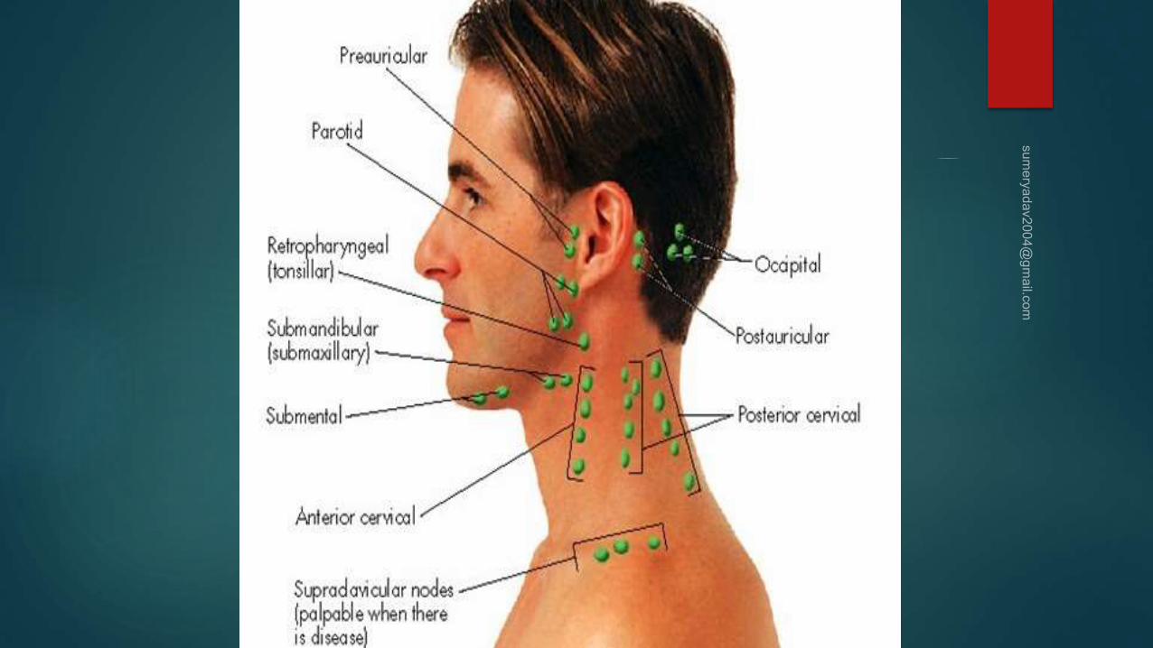
Lymph nodes
Salivary glands
Thyroid and parathyroid glands
Thymus
Potential spaces (fascia spaces)
Skin ,fat , fascia, muscles, nerves, vessels,
bones

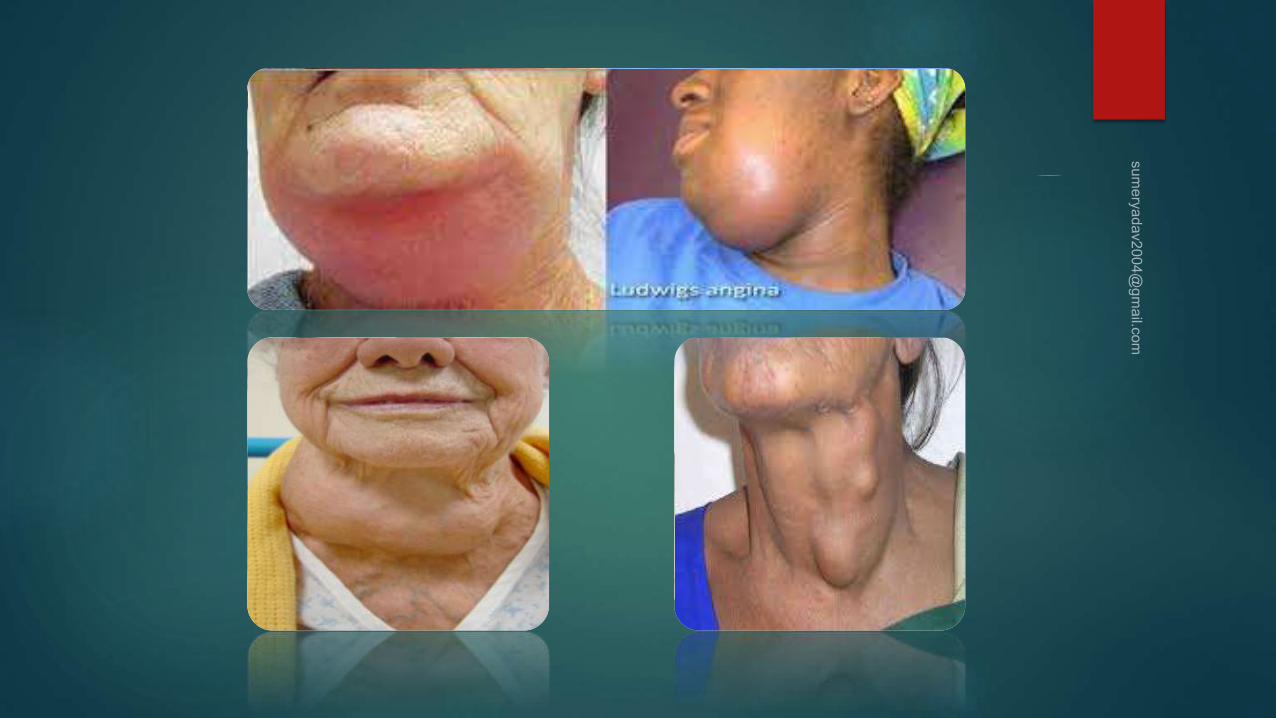
DDx of neck swellings

Approach of the neck mass
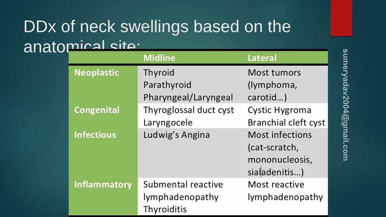
DDx of neck swellings based on the
anatomical site:


Symptoms Associated with Head &
Neck Lumps
Change in the voice including hoarseness that
persists for more than two weeks
Growth in the mouth
Swollen tongue
Blood in the saliva or phlegm
Swallowing problems
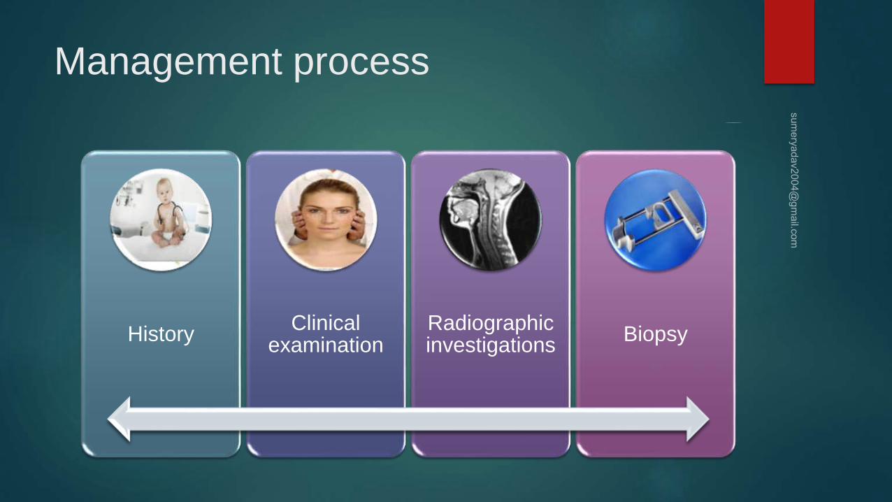
Management process
History Clinical
examination Radiographic investigations
Biopsy

Approach of the neck mass
The anterior triangle is
delineated by :
1. The anterior border of the SCM laterally,
2. The midline medially,
3. The lower border of the mandible
superiorly.
The SCM divides each side of the
neck into two major triangles,
anterior and posterior.
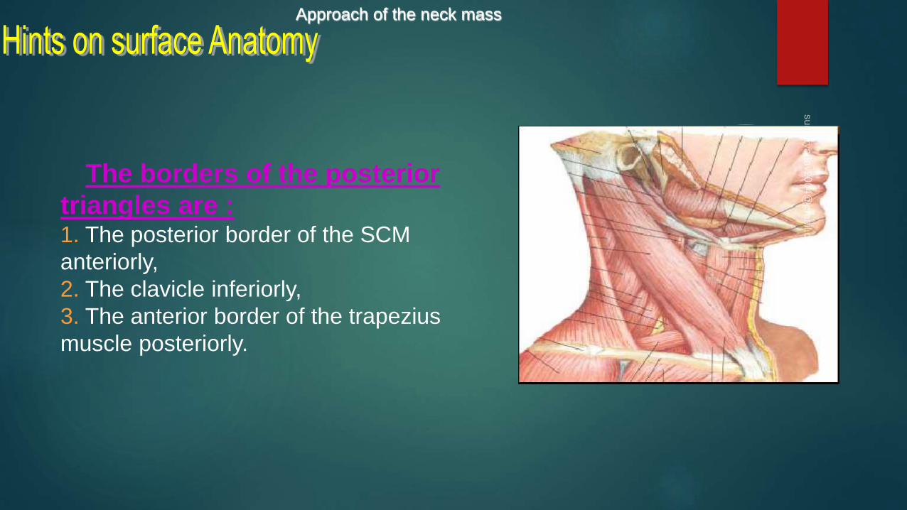
Approach of the neck mass
The borders of the posterior
triangles are :1. The posterior border of the SCM
anteriorly,
2. The clavicle inferiorly,
3. The anterior border of the trapezius
muscle posteriorly.
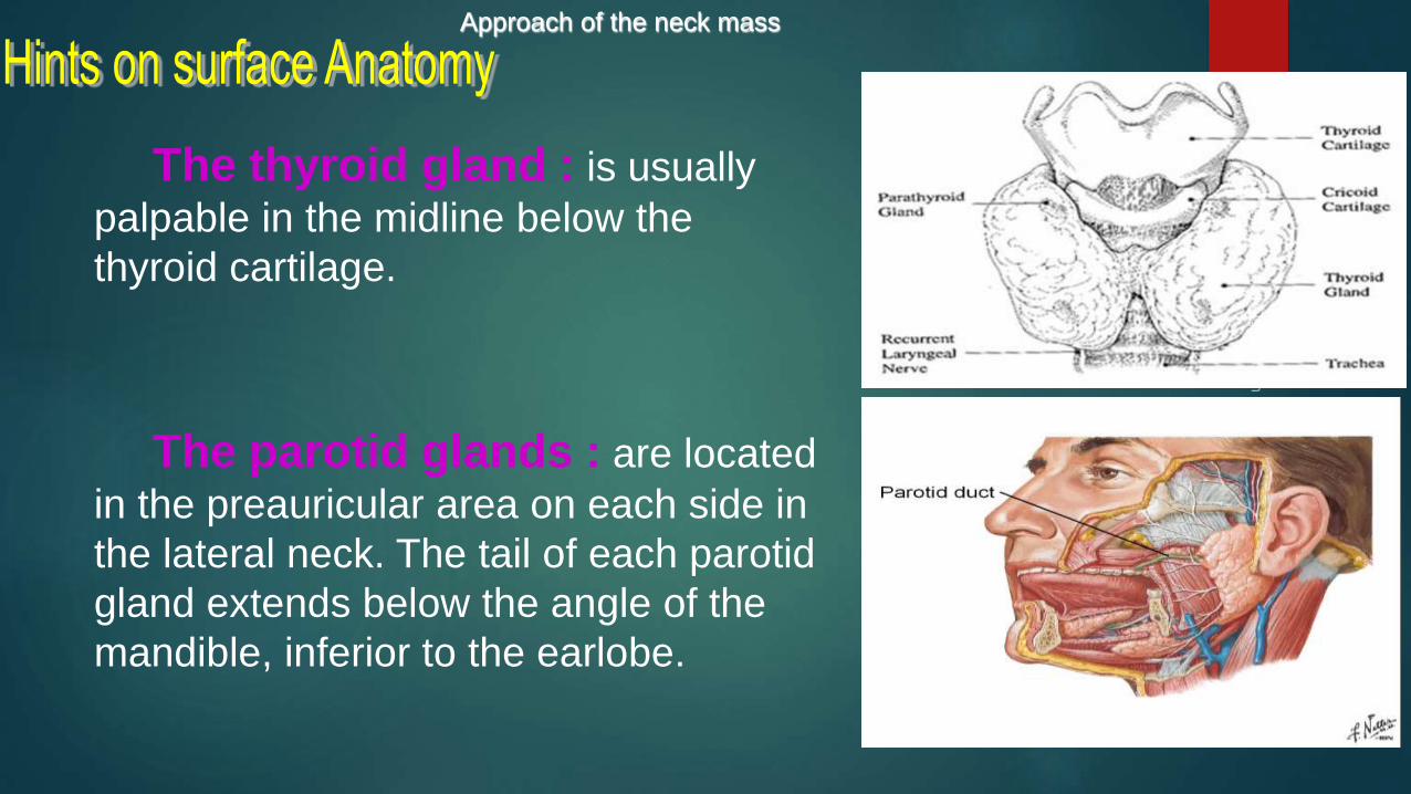
Approach of the neck mass
The thyroid gland : is usually
palpable in the midline below the
thyroid cartilage.
The parotid glands : are located
in the preauricular area on each side in
the lateral neck. The tail of each parotid
gland extends below the angle of the
mandible, inferior to the earlobe.


Approach of the neck mass
Submandibular glands : are
located within a triangle bounded by ….
the sternocleidomastoid muscle, the
posterior belly of the digastric muscle,
and the body of the mandible.
Lymph nodes : are
located throughout the head
and neck region .

Approach of the neck mass
The prominent landmarks of the
neck are :
1. hyoid bone,
2. Thyroid cartilage,
3. Cricoid cartilage,
4. Trachea,
5. Sternocleidomastoid muscles.

Approach of the neck mass
* Personal data :-
1. Age .
2. Sex .
3. Nationality .
* HPI :-
1. Duration . 2. Location .
3. Size . 4. How notice .
5. Painfull / painless . 6. Other masse .
7. Progression . 8. Trauma .
Approach of the neck mass
* Systemic Review :-
1. Symptoms of hypo. OR hyper. THYRODISM .
2. Symptoms which indicate malignancy .
3. Respiratory Symptoms .
4. GI Symptoms .
5. Symptoms which indicate infectious / inflammatory process . ( fever , wt loss , night sweat
6. Head & Neck Symptoms .
7. Compression Symptoms .

Approach of the neck mass
* General Examinations :-
1. Vital Signs .
2. General appearance of the pateint .
* Local Examination :-
1. Inspection :
a. site . b. shape .
c. color . d. relation to deglutition .
e. relation to tongue protrusion .

Approach of the neck mass
2. Palpation :
a. temperature . b. tenderness .
c. size . d. surface .
e. edge . f. consistence .
g. fluctuation . h. pulsatility .
i. relation to skin . j. mobility .
k. relation to underlying structures .

Approach of the neck mass
3. Percussion :on the sternum for retrosternal extension of the thyroid .
4. Auscultation :for bruits .

Approach of the neck mass
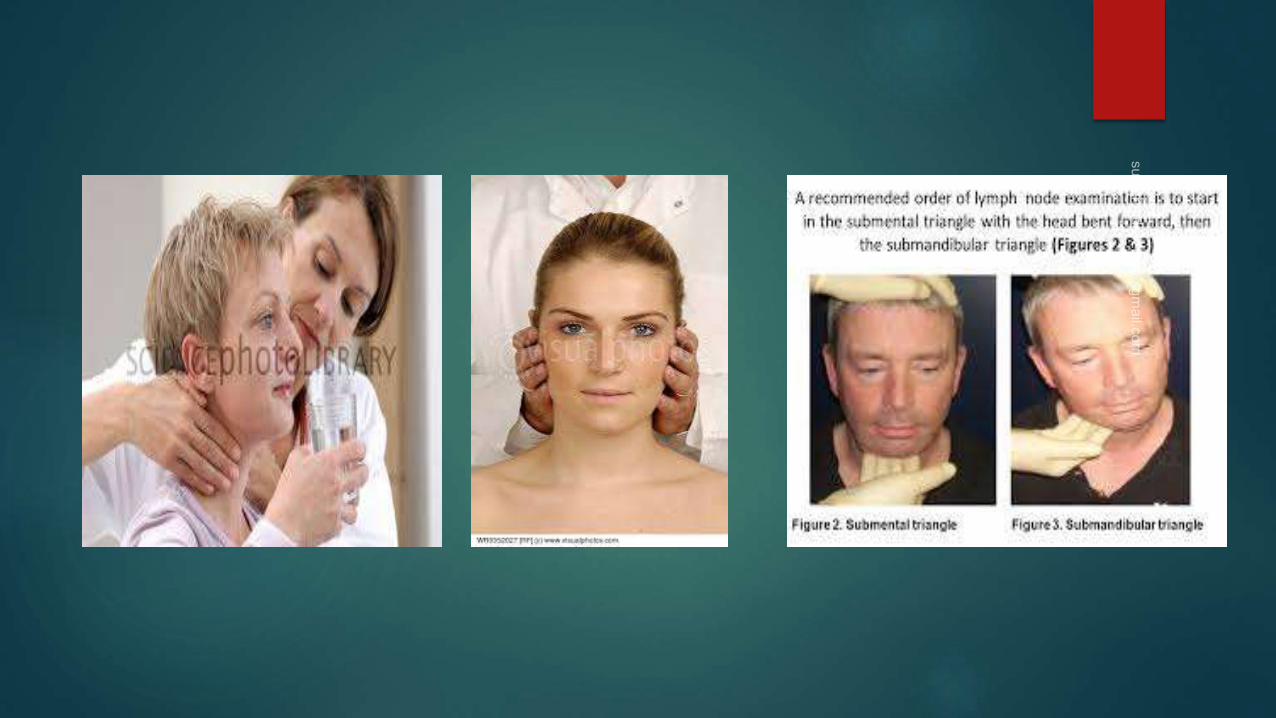
* Complete Head & Neck Examination :
1. look to the head for any mass or ulcer .
2. examine L.N.
Approach of the neck mass
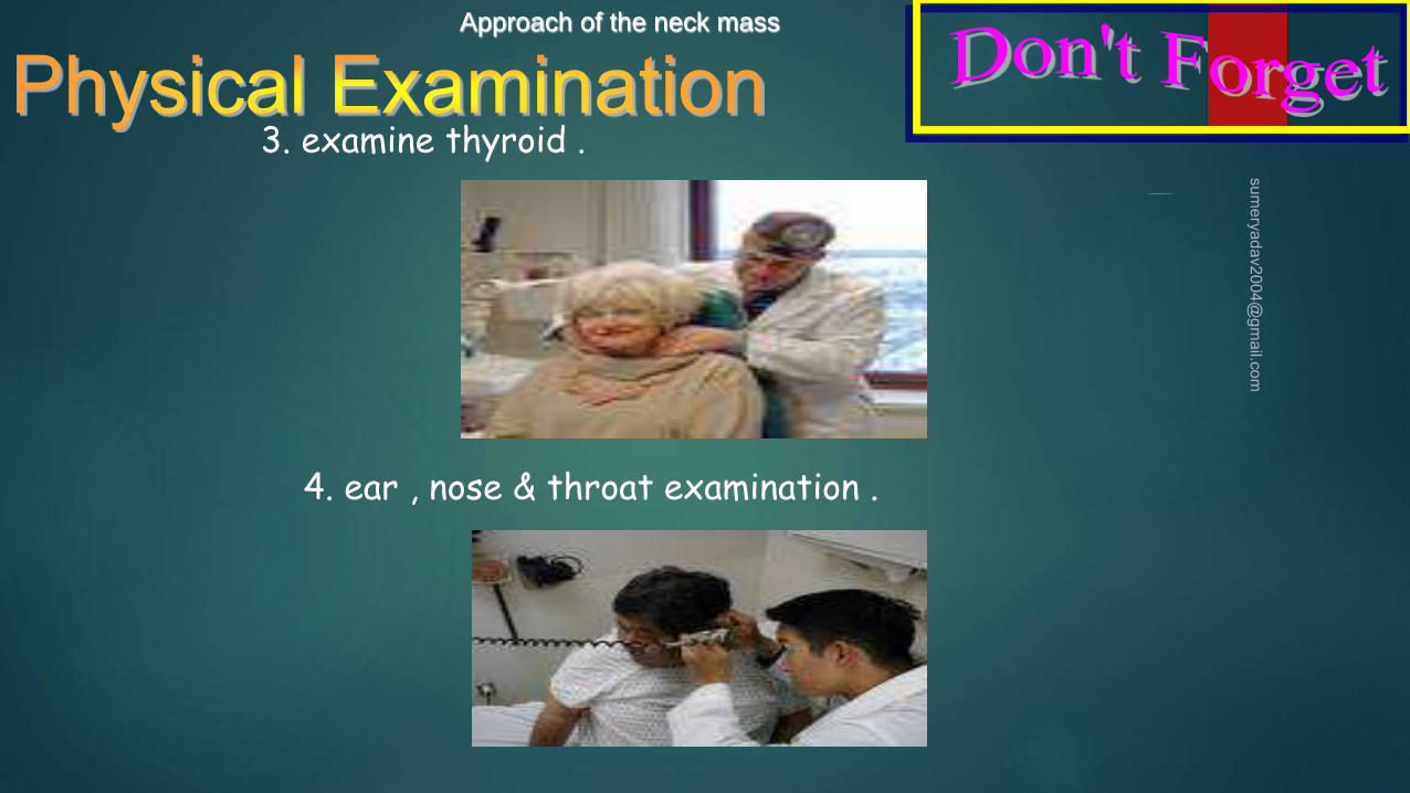
3. examine thyroid .
4. ear , nose & throat examination .

Approach of the neck mass
6. laryngoscope .
5. Mouth examination .
7. esophagi scope .


Summary of examination Examination of some masses / swelling may allow a physician to determine their
cause based on location, size, and consistency.
In other cases, however, additional tests may be required.
Changes in the skin – It is important to examine changes in the skin that could
indicate basal cell carcinoma, squamous cell cancer, and malignant melanoma.
Persistent Ear Pain or ear pain while swallowing may be a symptom of infection
or a growth in the throat.

Radiographic Investigation of the Head
and Neck Masses
MRI – Magnetic Resonance Imaging can clearly
highlight soft tissue pathologies better than the C.T. Scan.
It uses a magnetic field rather than x-rays (radiation).

Radiographic Investigation of the Head
and Neck Masses CT SCAN – Computed tomography is
less accurate than M.R.I for the soft
tissue examination
very useful to locate bony tumors and
their dimensions and extensions.
C.T with contrast is used to enhance
the visibility of abnormal tissue during
examination.

Radiographic Investigation of the Head
and Neck Masses PET (Positron Emission Tomography)
and SPECT (Single Photon Emission
Tomography) are useful after diagnosis
to help determine the grade of a tumor or
to distinguish between cancerous and
dead or scar tissue.
They involve injection with a
radioactive tracer.

Modality Basic Indications
Ultrasound Good for pediatric neck masses, thyroid masses. Differentiates cystic versus solid.
Computed tomographyWorkhorse imaging modality for adult neck masses. Provides three-dimensional
relationships, excellent detail of mucosal disease and involvement of adjacent bone.
Magnetic resonance imaging
Superior soft tissue delineation. Good for lesions of the salivary glands and tongue
(where dental amalgam may obscure the view on a CT). Modality of choice for
determining nerve enhancement. Consider for thyroid imaging in cases necessitating
radioiodine.
Radionuclide scanningUseful for midline lesions in children—differentiates functioning from nonfunctioning
tissue.
Positron emission tomographyUseful for staging of head and neck malignancies. Can be used in cases of unknown
primary malignant neck masses or treated neck disease.
Angiography/magnetic resonance
angiography/computed tomography angiography
Useful for lesions encasing the carotid and vascular lesions. Conventional angiography
should be considered for preoperative assessment in cases of potential carotid artery
sacrifice or where embolization is required.
Plain radiograph Generally should not be considered in the workup of a neck mass.

Biopsy
F.N.A.C – Fine Needle Aspiration Biopsy is
Safe
Rapid
Inexpensive
Presurgical planning
Avoids open biopsy


Treatment modalities
Medical
Surgical
Radiotherapy

Thank
s