neotropical diptera 15 - papavero & artigas 2008 - manual
TRANSCRIPT
1
Papavero & Artigas
Neotropical Diptera 15
Manual of Neotropical Diptera. Mydidae1
Neotropical DipteraDepto. de Biologia - FFCLRPUniversidade de São PauloRibeirão Preto, SP, Brazil
Neotropical Diptera 15: 1-58 (April 15, 2009)ISSN 1982-7121www.neotropicaldiptera.org
Nelson PapaveroMuseu de Zoologia, Universidade de São Paulo, São Paulo, SP, Brasil
Pesquisador Visitante do Departamento de Biologia, Faculdade de Filosofia, Ciências e Letras, Universidade de SãoPaulo, Ribeirão Preto, SP, Brasil
&
Jorge N. ArtigasDepartameno de Zoología
Universidad de ConcepciónConcepción, Chile
For general information and classification of the family, see Wilcox & Papavero (1971). Becher (1882) studied themouthparts, Zaitlan & Larson (1984) the head and Jahn (1930) the internal anatomy. Some species are mimics of Hymenoptera,especially Pompilidae (Rothschild, 1910; Zikan, 1942; Cooper, 1981; Meyer, McKenzie & Zalom, 1984; Nelson, 1986).
Adults (at least the males) feed upon flowers (Williams (1995) also found females of Mydas clavatus (Drury, 1773)feeding on flowers). Alcock (1989) described the mating system of Mydas ventralis Gertaecker, 1868 and Gibson (1965) theoviposition of Mydas clavatus (Drury, 1773).
As far as known, the larvae feed upon coleopterous larvae. The oldest record may be found in Westwood (1841:50, 51): he said that William Sharp MacLeay found Ceriomydas tricolor (Wiedemann, 1830) as “parasitic” on larvae of a“giant Prionidae” (Cerambycidae, Prioninae) [?maybe Stenodontes chevrolati Gahan, 1890], in Cuba. Walsh (1864), inIllinois, reared Mydas tibialis Wiedemann, 1828 from fibrous debris found in a hollow sycamore which containedcoleopterous larvae. Berg (1899) mentioned that Messiasia testaceiventris (Macquart, 1850) was always found associatedwith nests of Acromyrmex hystrix (Latreille, 1802) and Acromyrmex lundii (Guérin-Méneville, 1838), so probably thisspecies always preys upon coleopterous larvae found in the nests of Acromyrmex, as those of Gauromydas do in nestsof Atta ants. The most informative papers on mydid biology are those of Zikan (1942, 1944). He spent several yearsobserving the biology of a few species of mydids (especially Gauromydas heros (Perty, 1833), in the National Park ofItatiaia, State of Rio de Janeiro, Brazil. According to him, adult males commonly feed upon the nectar of flowers, especiallyAcacia paniculata Willdenow and Mimosa adherens Martius, and other Leguminosae and Compositae. Females apparentlyrely solely on the fatty substances accumulated in their abdomen for subistence. Zikan never observed females feeding onflowers. Male adults were found by him in the vicinity of the large nests of “saúva” ants (Hymenoptera, Formicidae, genusAtta), either flying around the nest or sitting on nearbly bushes and herbs. As a rule, only one male was to be seen in theneighbourhood of an ants’ nest. If another male approached, an aerial battle ensued, such as happens between malehummingbirds when one invades the territory of the other. The attacks between the two mydid males are followed by briefrespites, during which the two contenders keep flying, one facing the other. The attacks are then renewed, ending with the“defeat” of one of them, who leaves the field. Males seem to be attracted to areas with “saúva” nests by the sight of thelarge, bare, denuded earth mounds, in some cases several meters across, accumulated by the worker ants; this seems to be
1 This project was supported by FAPESP grants # 2003/10.274-9 and 2007/50878-1.
2
Manual of the Neotropical Diptera. Mydidae
Neotropical Diptera 15
corroborated by the fact that they are also attracted to similar areas resulting from other natural causes or even to man-made mounds. Near these sites mating takes place; sometimes the males make mistakes and try to copulate with females ofanother species, with other males, or with the large black pompilids, which they seem to mimic (cf. Fig. 2). If the matingcouple is disturbed, the female flies away very rapidly, carrying along the hanging male, until an eventual separation. Thefemales oviposit in the inerior of the ants’ nest. Zikán frequently found females with the body partially covered with earth,indicating that they were laying eggs in the loose soil of the nets’s entrance. The larvae live in the “garbage pans”[“panelas de lixo”] of the Atta nests, where the workers accumulate the garbage from the nest and the exhausted plantmedium on which the fungus Rhozites gongylophora Moeller is maintained by the ants. These residues of decayingplants attract several Coleoptera, especially some Melolonthidae (Dynastinae) of the genus Coelosis (Coelosis bicornis(Leske, 1779), Coelosis biloba (Linnaeus, 1767) and Coelosis inermis (Sternberg, 1908)), whose larvae feed upon thedebris, a fact discovered and published by Eidmann in 1937 (confirmed also by Pardo-Locarno, Morón & Gaigl, 2006, whoalso give a good figure of the larva of Coelosis biloba). Although he reported no actual observation, Zikan believed thatthe mydid larvae prey upon those dynastine larvae. The mature larva (Fig. 1A) abandons the “garbage pans” and digs upthe soil to a depth of 10-20 cm below the surface, where it constructs a pupation chamber. The pupal chamber or cell isalways situated above the “garbage pan”, sometimes quite far from it. There the pupa remains until the day of emergenceof the imago. Then, with the help of its strong spines (Figs. 1B-C), the pupa makes its way to the surface, where it remainshalf-buried until the hottest hours of the day, when the imago finally emerges. The adult walks for some distance andclimbs to a low bush or herb, where it dries its wings. The females can copulate a few moments later. Not all “ saúva” nestsharbour mydid larvae. The larvae are usually found in large nests with many “garbage pans”. Zikan found up to 16 exuviae(of two different species of mydids) in a next of Atta sexdens rubropilosa. Autuori (1952, 1971) also published on themydids associated with Atta. Genung (1959) found larvae of Mydas maculiventris Westwood, 1825 preying upon whitegrubs. Specimens of Messiasia pertenuis (Johnson, 1926), at the University of Arizona (Tucson), were reared from larvaeand pupae collected at the Santa Rita Range Reserve, Arizona, from nests of the banner-tailed kangaroo rat Dipodomysspectabilis Merriam (Rodentia, Heteromyidae) (cf. Wilcox & Papavero, 1975: 8), and were probably preying upon larvae ofscavenger beetles living in the rats’ den.
Rogers & Mattoni (1993) summarized what is known about the biology of Raphiomidas. Hogue (1967) describedthe pupa of Rhaphiomidas terminatus Cazier, 1941 and Steinberg, Dorsett, Shah, Jones & Burk, 1998 the pupa ofRhaphiomydas acton Coquillett, 1891.
Key to the genera
1. Scutum and scutellum with marginal bristles. Male terminalia (cf. Yeates & Irwin, 1996: 275, figs. 44-45) comparately large,epandrium divided medianly into two thick sclerites; hypandrium absent or entirely fused to bases of gonocoxites;gonocoxites fused in midline, with narrow, posteriorly directed extentions. Spiracle scar present on female sternite8. Proboscis elongate (length: 5.3-14.4 mm). Antennae short, flagellum of one segment of various shapes. Palpusone-segmented, palpal pit absent. Propresternum a single sclerite, pear-shaped; proprecoxal bridges present. Ccircumambient; r-m crossvein perpendicular to M, positioned closer to apex of discal cell than base; veins M1,M2 and R5 curving forward to join the wing margin before the wing apex; discal and m3 cells present; M3 andCuA1 united before wing margin, CuA1 + A1 fused just before margin. Abdomen elongate, cigar-shaped, taperingtowards apex. Ejaculatory apodeme small, cylindrical. Female terminalia with a row of stout spines on theacanthophorites (cf. Yeates & Irwin, 1996: 280, fig. 63); acanthophorites fused to tergite 9 dorsally; tergite 9 witha median, dorsal bridge. Three poorly sclerotized, relatively small spermathecae. Length, 19-35 mm. U. S. A.(California, Arizona, New Mexico, Nevada), Mexico (Baja California Norte, Baja California Sur, Sonora). SubfamilyRHAPHIOMYDINAE Williston, 1893 …………….…………………………….. Rhaphiomydas Osten Sacken, 1877
Scutum and scutellum devoid of bristles. Epandrium lobes never thick. Spiracle scar absent on female sternite 8. Othercombinations of characters ……..............................................................……………………………………………...… 2
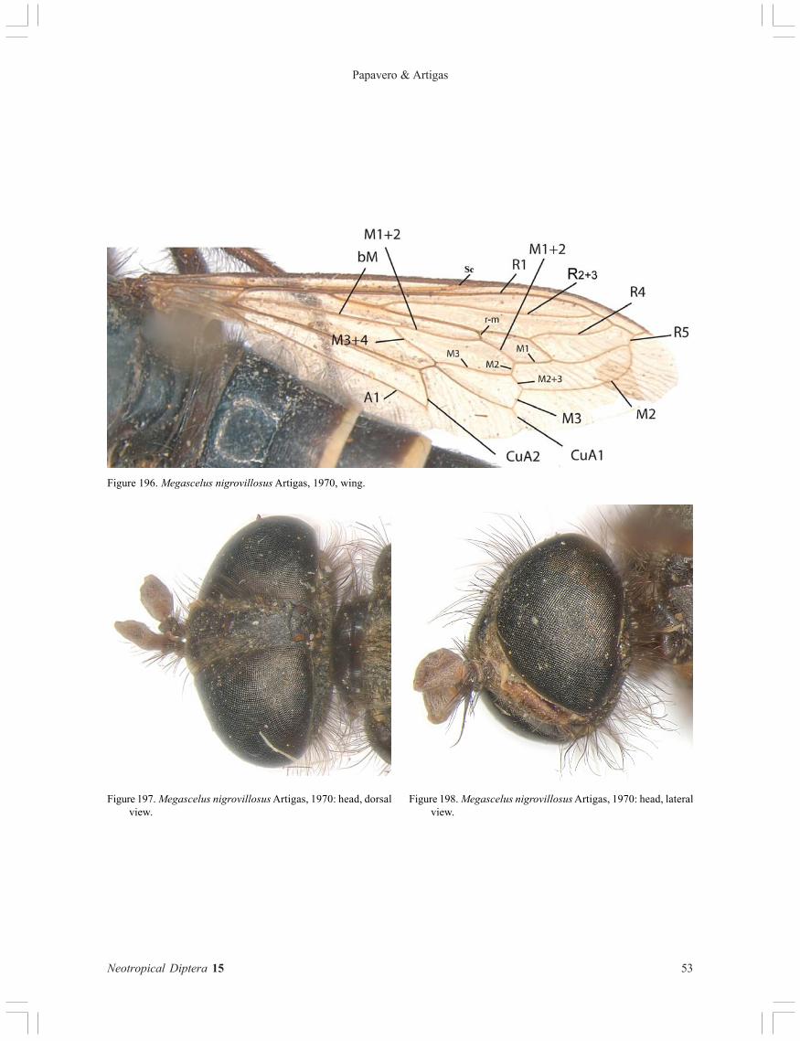
2 (1) C ends in R1, veins M1 and M2 present; M1 short, almost horizontal, ending in R5; M2 fused with M1 for a shortdistance and then curved towards anterior part of wing, ending in R5 before the wing apex; vein M3 + CuA1present; r-m crossvein occurring approximately midway along the discal cell at 90o to the longitudinal veins itjoins (Fig. 196). Second flagellomere of antennae absent (Figs. 197-198, 201-202); separate antennal insertion;lateral ocelli situated on either side of the ocellar tubercle (cf. fig. 5 of Yeates & Irwin, 1996; Fig. 197). A singleoccipital foramen. Mouthparts very small (Figs. 198, 201-202). Antepronotum short. Propresternum composed oftwo separate, rounded sclerites. Scutellum relatively large, extending posteriorly to the anterior margin of the

3
Papavero & Artigas
Neotropical Diptera 15
abdomen (Fig. 199). First tarsomere of hind leg about 5 times as long as wide (Fig. 200). First abdominal sternitesmall but present as a separate narrow band just behind the hind coxae. Bullae* absent on the posterolateralmargins of tergite 2. Hypandrium present and separated from gonocoxites, with a deep median notch (Fig. 204).Aedeagus as in Figures 205-206. Female genitalia with a row of stout spines on acanthophorites (cf. Yeates &Irwin, 1996: 280, figs. 61-62; Fig. 207), these pressed to tergite 9 dorsally; tergite 9 with a dorsal, median ridge.Furca narrow and elongate, with a posterior ventral scoop. Spermathecae as in Figure 208 (Chile). SubfamilyMEGASCELINAE Cazier, 1941 ................................................................................................ Megascelus Phlippi, 1865
C either circumambient or ending in R5 or ending in M1. M1 and M2 both present, but never as above, or completelyfused; M3 + CuA1 present or absent; r-m crossvein close to the apex of the discal cell and at an acute angle to theadjoining proximal region of M. Second flagellomere of antennae present. Antennal insertions very close together,so close that the borders of the articulations are confluent (cf. Fig. 3 of Yeates & Irwin, 1996). Ocellar tubercletakes the form of an elongate side. A dorsal and a ventral occipital foramen. Mouthparts large. Antepronotumelongate, contributing to the general lengthening of the cervical region. Propresternum pear-shaped. First tarsomereof hind legs about 5 times as long as wide, or shorter. Sternites of the first two abdominal tergites either fused orthe sternite of segment 1 has been lost. Bullae* present on the posterolateral margins of tergite 2 in both sexes(Figs. 47-60). Hypandrium either present, but fused with the anterior ventral margin of the gonocoxites, or absentor perhaps completely fused with the gonocoxites. Female genitalia variable. Furca not as above; if narrow andelongate (Apiophora), then without a posterior ventral scoop ……….....……………………….……………..……. 3
3 (2). First tarsomere of hind leg usually 5 times as long as wide …............................................................................................ 4First tarsomere of hind leg never 5 times as long as wide …........................................................................................……. 6
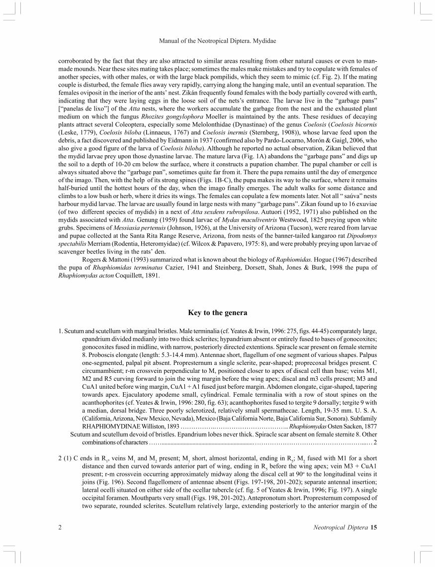
4 (3). C ends beyond apex of M2; veins M1 and M2 separate, M1 ends in R1 and M2 ends in C, above the wing apex; R4 fusedto R5 apically; M3 + CuA1 absent, i. e., cell m3 not petiolate (Fig. 3). Proboscis from very short (shorter than lengthof subcranial cavity) to very elongate (Fig. 4). Scape 3 times as long as pedicel (Figs. 5, 11, 25). Katatergite convex,bare of hairs. Hind femur not clavate (Fig. 6) and hind tibia cylindrical. Anatergite long and densely pilose. Abdominalbulla as in Fig. 52. Male terminalia as in Figs. 65-68 (see also Artigas & Palma, 1979: figs. 108-125, 138-139, 140-145).Female genitalia with spines on acanthophorites (Fig. 69). Female spermathecae (Figs. 69-70) reduced only to centralcapsule; genital furca as in Figs. 69-70 (see also Artigas & Palma, 1979: figs. 75-80). Length, 11-19 mm. (Chile,Argentina). Subfamily DIOCHLISTINAE Bequaert, 1963 ................................................. Mitrodetus Gerstaecker, 1868
Veins M1 and M2 completely fused, i. e., only one vein enters C between the apex of wing and apex of R1. Scape at most twotimes as long as pedicel. Other combinations of characters. Subfamily ECTYPHINAE Wilcox & Papavero, 1971 ......... 5
5 (4). Prementum subequal in length to subcranial cavity, labella short and slightly wider than mentum. Cell r4 usuallyclosed and short petiolate. Hind tibia with slender apical spur and several bristles; spur small in females of somespecies. Bulla as in Fig. 53. Male terminalia as in Figs. 71-73. Female genitalia with spines on acanthophorites (Fig.74). Female spermathecae and furca as in Figs. 74-75. Length, 18-25 mm. (U. S. A., Mexico) ................................................................................................…...................................................................................... Opomydas Curran, 1934
Prementum about one-half length of subcranial cavity, labella attached to prementum near its midpoint and subequal inlength to subcranial cavity (Fig. 10). Cell m4 usually broadly open. Hind tibia with apical spur and a bristle at base.Hypandrium fused only basally to gonocoxites, gonostyli absent (Figs. 76-78). Female genitalia with spines onacanthophorites (Fig. 79). Female spermathecae and furca as in Figs. 79-81. Length, 22-29 mm (U. S. A., Mexico) ........................................................................................................................................................... Heteromydas Hardy, 1944
6 (3). Cell r4 open, usually very widely open ...........................................................................................................................…... 7Cell r4 closed, or closed and petiolate …........................................................................................……................................. 11
7 (6). Hind tibia cylindrical (Fig. 211). (Northeastern Brazil, Chile). (Subfamily RHOPALIINAE Papavero & Wilcox, 1974) ... 8Hind tibia with ventral keel (carinate). (Southern Brazil, Chile, Argentina). (Subfamily APIOPHORINAE Papavero &
Wilcox, 1974) …........................................................................................…….…............................................................... 9
8 (7). Vein CuA1 + M3 present, i. e., cell m3 closed and petiolate. Labella almost half length of subcranial cavity. Bulla as inFig. 51. Male terminalia as in Artigas & Palma (1979: figs. 99-104, 146-149). Female spermathecae as in Figs. 82-84(see Artigas & Palma, 1979: fig. 82). Length, 11-17 mm. (Chile) ............................................. Midacritus Séguy, 1939
Vein CuA1 + M3 absent, i. e., cell m3 open (Fig. 212). Prementum short and attached to middle of labella, which isslightly shorter than length of subcranial cavity. Bulla as in Fig. 60. Male terminalia as in Figs. 85-87. Femalegenitalia with spines on acanthophorites (Fig. 88). Female spermathecae as in Figs. 88-89. Length, 10-14 mm.

4
Manual of the Neotropical Diptera. Mydidae
Neotropical Diptera 15
(Northeastern Brazil) ……………………………..………...... Pseudorhopalia Wilcox & Papavero, 1971 [Fig. 201-204]
9 (7). Katepimeron pilose (Fig. 214). Antennal flagellomere of a vivid reddish-orange color (Fig. 213). Abdomen beautifullyshining blue, deeply punctate (Fig. 215). Alula with fringe of squamose hairs (Fig. 216). Bulla as in Fig. 56. Femaletergite 10 with hairs. Female spermathecae as in Artigas & Palma (1979: figs. 81, 83). Male terminalia as in Figs. 90-92and Artigas & Palma (1979). Length, 18-23 mm. (Chile: Coquimbo to Talca) .... Paramydas Carrera & d’Andretta, 1948
Katepimeron bare. Female tergite 10 with spines on the acanthophorites. Other combinations of characters ............ 10
10 (9). Anal lobe of wing less than ½ as broad as long and alula with fringe of short, fine hairs (Fig. 217). Hind femur of male4-4.5, of female 4.5-5 times as long as broad. Bulla as in Fig. 47. Male terminalia as in Figs. 93-95 and as in Artigas &Palma (1979: figs. 93-98, 134-137). Female spermathecae as in Artigas & Palma (1979: figs. 84-86). Length, 14-19 mm.(Chile: between provinces of O’Higgins and Malleco) …............................... Apiophora Philippi, 1865 [Figs. 209-210]
Anal lobe of wing about as broad as long. Alula with dense fringe of squamose hairs. Hind femur of male 6, of female7 times as long as broad. Bulla as in Fig. 49. Male terminalia as in Figs. 96-98. Length, 21-28 mm. (Brazil: SantaCatarina) …............................................................................................…….…........ Eumydas Wilcox & Papavero, 1971
11 (6). Hind tibia cylindrical. (Americas). Subfamily MYDINAE Latreille, 1809 …................................................................... 12Hind tibia with ventral keel (carinate). Subfamily LEPTOMYDINAE Papavero & Wilcox, 1974) ................................... 23
12 (11). Prementum about one half length of subcranial cavity, labella attached to prementum near its midpoint andsubequal in length to subcranial cavity. Anterior margin of subcranial cavity situated at about two-fifths distancefrom lower eye margin to base of antennae …........................................................................................……............... 13
Prementum subequal in length to subcranial cavity, labella attached to prementum near its apical one-half, andextending out at about a 90o angle (Fig. 8). Anterior margin of subcranial cavity level with lower eye margin .... 14
13 (12). Antenna short, first flagellomere widened apically and subequal in length to scape and pedicel together; secondflagellomere as long as the three preceding segments (Fig. 18). Vein M3 + CuA1 absent. Bulla as in Fig. 48. Maleterminalia as in Figs. 99-101. Female genitalia only with hairs (Figs. 102-103). Female spermathecae and furca as inFigs. 102-103. Length, 20-27 mm. (Guiano-Brazilian subregion) Tribe DOLICHOGASTRINI Papavero & Wilcox,1974 ………………………................................................................................................ Dolichogaster Macquart, 1848
First flagellomere of antenna slender and at least twice as long as scape and pedicel together; second flagellomere shorterthan three preceding segments (Figs. 21-23). Vein M3 + CuA1 present. Bulla as in Fig. 50. Male terminalia as in Figs. 104-133. Female genitalia without spines. Female spermathecae as in Figs. 134-135. Length, 15-29 mm. (U. S. A.: Arizona, toArgentina: Buenos Aires). Tribe MESIIASIINI Papavero & Wilcox, 1974 ............................. Messiasia d’Andretta, 1951
14 (12). Facial gibbosity about as broad as high. Female tergite 9 narrower apically than basally. Male terminalia withsimple, or bifid and falciform, gonostyli. Length, 15-60 mm. (North and South Americas). Tribe MYDINI Latreille,1809 …........................................................................................…….…............................................................................ 15
Facial gibbosity about one and one-half times as broad as high (Fig. 7). Bulla as in Fig. 57. Female tergite 9 widerapically than basally, fluted (Figs. 64, 139). Male terminalia as in Figs. 136-138, with bifid gonostyli. Femalespermathecae and furca as in Figs. 139-141. Length, around 21 mm. (U. S. A., Mexico). Tribe PHYLLOMYDINIPapavero & Wilcox, 1974 ....…………………………………………………………………...... Phyllomydas Bigot, 1880
15 (14). Hind tibia with ventral keel underdeveloped, visible only on the basal half (or less) of the tibia; apical spur on hindtibia underdeveloped, always shorter than width of first tarsomere (still shorter, almost absent, in female). Epandriumtrapezoidal. Female spermathecae and furca as in Figs. 150-151. Length, 20-40 mm. (Neotropical, except Chile).Subtribe PROTOMYDINA Wilcox, Papavero & Pimentel, 1989 ..... Protomydas Wilcox, Papavero & Pimentel, 1989
Hind tibia with well-developed, very evident ventral keel, all along its length; apical spur of hind tibia well-developedin both sexes (longer in males), always longer than width of first tarsomere (up to two times as long as width offirst tarsomere). Epandium trapezoidal, subtrapezoidal, or triangular ....................................................................... 16
16 (15). Mesonotum without a definite pollinose pattern of stripes or spots (Americas). Subtribe MYDINA Latreille, 1809 ... 17Mesonotum with a very evident pattern of pollinose stripes or spots. (Neotropical, except Chile). Subtribe
STRATIOMYDINA Wilcox, Papavero & Pimentel, 1989 …......................................................................................... 21
17 (16). First tarsomere of hind leg long, at least subequal in length to tarsomeres 2-3 and always longer than tarsomere 5............................................................................................................................................................................................. 18

5
Papavero & Artigas
Neotropical Diptera 15
First tarsomere of hind leg not very long, subequal in length to tarsomere 2 and always shorter than tarsomere 5…........................................................................................…….…....................................................................................... 19
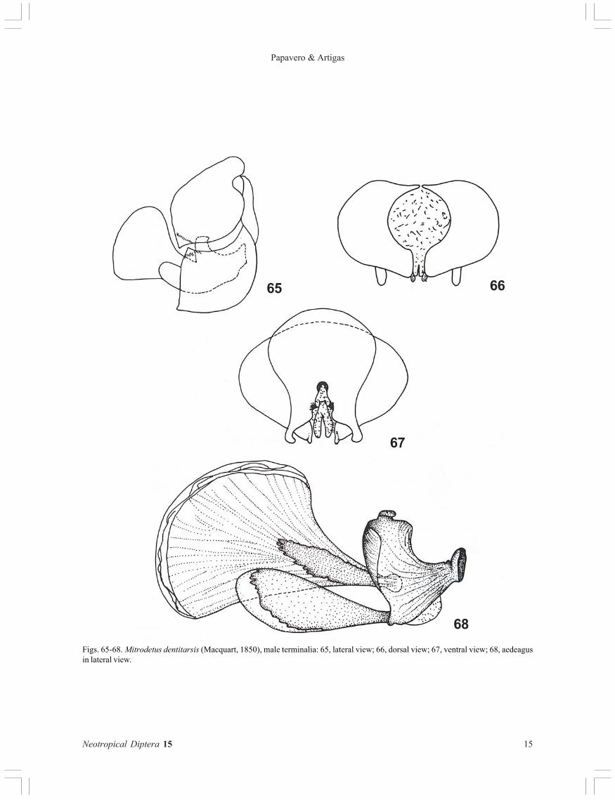
18 (17). Second flagellomere of antenna about six times as long as wide or more. Female spermathecae and furca as in Figs.152-154. (Neotropical, except Chile) …............................................. Gauromydas Wilcox, Papavero & Pimentel, 1989
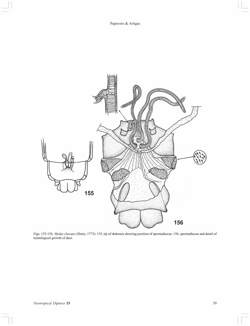
Second flagellomere about four times as long as wide or less (five times in Mydas boonei Curran) (Figs. 26, 27, 28).Female spermathecae as in Figs. 155-156. Mexico, extending into Nearctic region) .............. Mydas Fabricius, 1794
19 (17). No strong, differentiated bristles on legs. Fore tibia with a dense patch of black and orange red hairs. Alula broad,with a long, dense fringe of squamose hairs. Abdomen entirely cupreous-red, as wide as thorax. Male terminalliaas in Figs. 157-159. Female spermathecae as in Figs. 161-162. Length, 29-32 mm. (Surinam, Brazil: Pará) ...........................................................….......................................................................................... Mapinguari Papavero & Wilcox, 1974
Tibia with well-developed bristles. Other combinations of characters …......................................................................... 20
20 (19). Alula narrow, with a short, sparse fringe of hairs. Abdomen slender, narrower than thorax, mostly black, sometimesconstricted in the middle. Male terminalia as in Figs. 163-168. Female spermathecae as in Figs. 169-170. Length, 20-27 mm. (Neotropical, except Chile) ….................................................................................. Ceriomydas Williston, 1898
Alula broad, with dense fringe of squamose hairs (Fig. 178). Abdomen strongly petiolate, wasp-like, uniformly red(Fig. 175, 177). Bulla as in Fig. 176. Hind femur with 15 black tuberculate spines in two more or less irregular rowson venter (Fig. 174). Length, 29 mm. (Brazil: Pará) …................................. Utinga Wilcox, Papavero & Pimentel, 1989
21 (16). First tarsomere of hind leg long, subequal in length to tarsomeres 2-3 and longer than tarsomere 5....................... 22First tarsomere of hind leg very short, subequal in length to tarsomere 2 and shorter than tarsomere 5. Katepimeron
pilose (Baliomydas cubanus (Curran)) or bare. Male terminalia as in Figs. 179-181. (West Indies) .....................…........................................................................................…….….......... Baliomydas Wilcox, Papavero & Pimentel, 1989
22 (21). Targites 2-5 with the usual sparse, short, recumbent pilosity, directed backwards. Epandrium subtrapezoidal or triangular.Katepimeron bare (sparsely pilose in Stratiomydas colimas Wilcox, Papavero & Pimentel). Bulla as in Fig. 54. (Tropicallowland forests of Mexico and Central America, Peru) ......................... Stratiomydas Wilcox, Papavero & Pimentel, 1989
Tergites 2-5 with long, dense, recumbent hairs, directed outwards. Epandrial halves triangular. Katepimeron pilose.(Guiano-Brazilian subregion) …....................................................... Chrysomydas Wilcox, Papavero & Pimentel, 1989
23 (11). Katatergite bare. Vein CuA1 + M3 present (Fig. 182). Labella attached to prementum at its midpoint and slightlyshorter than subcranial cavity. Hind femur about 10 times as long as broad, venter with hairs and 2-3 subapicalspines. Bulla as in Fig. 58. Male terminalia as in Figs. 183-185. Female tergite 10 with apical hairs. Length, 12-18mm. (Peru) …........................................................................................……........... Plyomydas Wilcox & Papavero, 1971
Katatergite pilose, hairs short and inconspicuous in some females. Vein CuA1 + M3 absent. Proboscis obsolete tofunctional, varying from one half to three times length of subcranial cavity, labella attached to apex of prementum(Figs. 12, 13, 15, 16). Female tergite 10 with spines on acanthophorites (Figs. 61-62) ........................................... 24
24 (23). Second flagellomere of antenna longer than first flagellomere. Bulla as in Fig. 55. Male terminalia as in Figs. 186-188,gonostyle split apically, with two prongs, the inner one acute and not quite as long as the outer one. Femalespermathecae with only two capsules (Figs. 189-190). Length, 12-23 mm. (Canada: British Columbia, to Panama) ...…........................................................................................…….…................................................ Nemomydas Curran, 1934
Second flagellomere of antenna shorter than or subequal to length of first flagellomere. Bulla as in Fig. 59. Maleterminalia as in Figs. 191-193, gonostyle with only one apical prong. Female spermathecae with 3 capsules (Figs.194-195). Habitus as in Fitzgerald & Kondratieff, 1995: 33, fig. 24. Length, 9-20 mm. (U. S. A., Mexico) ...................................................…........................................................................................…….......... Pseudonomoneura Bequaert, 1961
* The bullae consist of ridges and grooves (figs. 12 and 13 of Yeates & Irwin, 1996), consistent with an evaporationsurface. The bullae probably function in the release of the pheromone product of an endocrine gland. The floor of eachgroove appears to be invaginated, and all secretions may communicate with the surface of the integument through thegrooves (Yeates & Irwin, 1996: 268).
The accompanying illustrations were extracted from Artigas (1973), Papavero & Wilcox (1974), Wilcox & Papavero(1971, 1975), Wilcox, Papavero & Pimentel (1989) and Zikan (1942, 1944).

6
Manual of the Neotropical Diptera. Mydidae
Neotropical Diptera 15
References
Alcock, J., 1989. The mating system of Mydas ventralis(Diptera: Mydidae). Psyche, Cambridge 96: 167-176,2 figs.
Artigas, J. C., 1973. Megascelus albovillosus, nueva especiede apioceratido de Chile y clave para la determinaciónde las especies del género (Diptera – Apioceratidae).Boln Soc. Biol. Concepción 42: 97-122, 38 figs.
Autuori, M., 1952. Fauna das “panelas de lixo” do sauveiro(Atta sp.). Mydaidae-Diptera. Ciência e Cultura, SãoPaulo 4 (3-4): 127.
Autuori, M., 1971. Ein Beitrag zur Fauna der Abraumkammernvon Atta rubropilosa Forel (Formicidae, Hymenoptera).Zeitschr. angew. Ent. 68: 76-78, 2 figs.
Becher, E., 1882. Zur Kenntniss der Mundtheile der Dipteren.Denkschr. Akad. Wien 45: 123-162, 4 pls.
Berg, C., 1899. Apuntes dipterológicos. Comunic. Mus. nac.Buenos Aires 1 (4): 124-130.
Claude-Joseph, Frère [Hno. Javier], 1928. Recherchesbiologiques sur les prédateurs du Chili. Ann. Sci. nat.,Zool. 11 (1): 67-207, 68 figs.
Cockerell, T. D. A., 1913. The first fossil mydaid fly. TheEntomologist 46: 207-208.
Cooper, W. E., 1981. Mimicry and spatial occupation of themydas fly, Mydas clavatus. J. Alabama Acad. Sci. 52(2): 58-65.
Eidmann, H., 1937. Die Gaste und Gastverhaltnisse derBlattschneiderameise Atta sexdens L. Ztschr. Morphol.Oekol. Tiere 32: 391-462.
Genung, W. G., 1959. Biological and ecological observationson Mydas maculiventris Westwood (Diptera:Mydaidae) as a predator of white grubs. FloridaEntomologist 42: 35-37.
Gibson, W. W., 1965. An observation on the ovipositionhabits of Mydas clavatus (Diptera: Mydaidae). J.Kansas ent. Soc. 38 (2): 196-197, fig. 1.
Greene, C. T., 1917. A contribution to the biology of NorthAmerican Diptera. Proc. ent. Soc. Washington 19: 147.
Hogue, C. L., 1967. The pupa of Rhaphiomidas terminatusCazier (Diptera: Apioceridae). Bull South CaliforniaAcad. Sci. 66: 49-70.
Jahn, L. A., 1930. The internal anatomy of the Mydas fly.Ohio J. Sci. 30: 85-94, 3 pls.
Meyer, R. P., T. L. McKenzie & F. G. Zalom, 1984. Associationof Mydas xanthopterus (Loew) (Diptera: Mydidae) andPepsis formosa Say (Hymenoptera: Pompilidae) in theChiricahua Mountains of southeastern Arizona. Pan-Pacific Entomologist 60 (4): 357.
Nelson, J. W., 1986. Ecological notes on male Mydas
xanthopterus (Loew) (Diptera: Mydidae) and theirinteractions with Hemipepsis ustulata Dahlbohm(Hymenoptrera: Pompilidae). Pan-Pacific Entomologist62 (4): 316-322.
Papayero, N. & J. Wilcox, 1974. Studies of Mydidaesystematics and evolution. II . Classification of theMydinae, with description of a new genus and a revisionof Ceriomydas Williston. Arqos Zool., São Paulo 25(1): 35-60, 10 figs.
Pardo-Locarno, L. C., M. Á. Morón & A. Gaigl, 2006. Losestados inmaduros de Coelosis biloba (Coleoptera:Melolonthidae: Dynastinae) y notas sobre su biología.Revta mexicana Biodiversidad 77: 215-224, 16 figs.
Rotschild, L.W. 1910. [Note to Austen’s Pl. XV]. Novitateszoologicae 28: 461.
Steinberg, M., D. Dorsett, C. Shah, C. E. Jones & J. Burk,1998. Pupal case of Rhaphiomidas acton Coquillett(Diptera: Mydidae) and behavior of newly-emergedadult. Pan-pacific Entomologist 74 (3): 178-180.
Walsh, B. D., 1864. On certain remarkable or exceptionallarvae, coleopterous, lepidopterous and dipterous, withdescriptions of several new genera and species, and ofseveral species injurious to vegetation, which havebeen already published in agricultural journals. Proc.Boston Soc. nat. Hist. (1862-1863) 9: 286-308.
Wilcox, J., N. Papavero & T. Pimentel, 1989. Studies ofMudidae (Diptera) systematics and evolution. IVb.Mydas and allies in the Americas (Mydini). MuseuParaense Emílio Goeldi, Belém.
Wilcox, J. & N. Papavero, 1971. The American genera ofMydidae (Diptera), with the description of three newgenera and two new species. Arquivos Zool., São Paulo21 (2): 41-119, 134 figs., 7 maps, 2 tables.
Wilcox, J. & N. Papavero, 1975. Studies of Mydidae (Diptera)systematics and evolution. III. The genus Messiasiad’Andretta in the Americas (Mydinae). Arquivos Zool.,São Paulo 26 (1): 1-47, 35 figs.
Williams, A. H., 1995. Adult female Mydas clavatus (Diptera:Mydidae) feeding on flowers in Wisconsin. The GreatLakes Entomologist 28 (3-4): 227-229.
Zaitlan, L. M. & J. R. Larsen, 1984. Morphology of the headof Mydas clavatus (Diptera: Mydidae). Intern. J. InsectMorphol. Embryol. 13 (2): 105-136.
Zikan, J. F., 1942. Also sobre a simbiose de Mydas com Atta.Rodriguésia, Rio de Janeiro 13 (2): 105-136.
Zikan, J. F., 1944. Novas observações sobre a biologia deMydas (Dipt.) e sua relação com os formigueiros desaúva. Bolm Minist. Agric., Rio de Janeiro 33: 43-55.

7
Papavero & Artigas
Neotropical Diptera 15
A
Fig. 1A-B. Gauromydas heros (Perty, 1833). A. Larva. B. Pupal skin, dorsal view. C. Pupa, lateral view.
1
B
C

8
Manual of the Neotropical Diptera. Mydidae
Neotropical Diptera 15
Figure 2. Pompilidae (Hymenoptera) (A) and its mimic Gauromydas heros (Perty, 1833) (B).
2
A
B

9
Papavero & Artigas
Neotropical Diptera 15
Figs. 3-6. Mitrodetus sp.: 3, wing; 4, head, lateral view; 5, antenna; 6. hind femur.
3
4
5
6

10
Manual of the Neotropical Diptera. Mydidae
Neotropical Diptera 15
Figs. 7-8. Phyllomydas bruesii Johnson, 1926, head in frontal view (7) and detail of proboscis in lateral view (8). Figs. 9-16, head,lateral view: 9, Apiophora rubrocincta (Blanchard, 1852); 10, Heteromydas bicolor Hardy, 1844; 11, Mitrodetus sp.; 12, Nemomydasmelanopogon Steyskal, 1956; 13, Nemomydas pantherinus (Gerstaecker, 1868); 14, Mydas clavatus (Drury, 1773); 15, Nemomydaslamia (Séguy, 1928); 16, Pseudonomoneura californica (Cole, 1970).
7
1011
13
89
12 14
1615

11
Papavero & Artigas
Neotropical Diptera 15
Figs. 17-33. Antennae: 17, Apiophora paulseni Philippi, 1863; 18, Dolichogaster brevicornis (Wiedemann, 1821); 19, Eumydascorupas Wilcox & Papavero, 1971; 20, Heteromydas bicolor Hardy, 1944; 21, Messiasia decor (Osten Sacken, 1886); 22, Messiasiamocoronga Wilcox & Papavero, 1975; 23, Messiasia pertenuis (Johnson, 1926); 24, Midacritus stuardoanus Séguy, 1929; 25,Mitrodetus dentitarsis (Macquart, 1850); 26, Mydas clavatus (Drury, 1773); 27, Mydas xanthopterus Loew, 1866; 28, Mydas luteipennisLoew, 1866; 29-30, Protomydas rubidapex (Wiedemann, 1830); 31, Nemomydas pantherinus (Gerstaecker, 1868). 32, Nemomydasmelanopogon Steyskal, 1956; 33, Opomydas athama (Séguy, 1928).
17
18
2120
2223
25
26
27
28
24
30
3132
33
29
19

12
Manual of the Neotropical Diptera. Mydidae
Neotropical Diptera 15
Figs. 34-46. Antennae: 34, Opomydas limbatus (Williston, 1886); 35, Opomydas townsendi (Williston, 1898); 36, Paramydas igniticornis(Bigot, 1857); 37, Plyomydas peruviensis Wilcox & Papavero, 1974; 38, Phyllomydas scitulus Williston, 1886); 39, Phyllomydasphyllocerus Bigot, 1880; 40, Phyllomydas bruesii Johnson, 1926; 41, Phyllomydas currani Hardy, 1943; 42, Pseudonomoneuracalifornica (Hardy, 1950); 43, Pseudonomoneura tinkhami (Hardy, 1950); 44, Pseudonomoneura micheneri (James, 1938); 45,Pseudonomoneura hirta (Coquillett, 1904); 46, Nemomydas lamia (Séguy, 1928).
34
39
37
36
38
41
42
43
4440 45
35
46

13
Papavero & Artigas
Neotropical Diptera 15
Figs. 47-60. Abdominal bullae: 47, Apiophora paulseni Philippi, 1865; 48, Dolichogaster brevicornis (Wiedemann, 1821), 49, Eumydascorupas Wilcox & Papavero, 1974; 50, Messiasia pertenuis (Johnson, 1933); 51, Midacritus stuardoanus Séguy, 1939; 52, Mitrodetusdentitarsis (Macquart, 1850); 53, Opomydas athama (Séguy, 1928); 54, Stratiomydas lividus (Curran, 1953); 55, Nemomydas venosus(Loew, 1866); 56, Paramydas igniticornis (Bigot, 1857); 57, Phyllomydas scitulus (Williston, 1886); 58, Plyomydas peruviensis Wilcox& Papavero, 1974; 59, Pseudonomoneura hirta (Coquillett, 1904); 60, Pseudorhopalia mirandai (d’Andretta & Carrera, 1951).
47
54
50
49
51
56
53
55
57
52
58
48
59
60

14
Manual of the Neotropical Diptera. Mydidae
Neotropical Diptera 15
Figs. 61-64. Female terminalia: 61, Pseudonomoneura californica (Hardy, 1950); 62, Nemomydas sp.; 63, Messiasia pertenuis (Johnson,1926); 64, Phyllomydas bruesii Johnson, 1926.
61
64
62
63
15
Papavero & Artigas
Neotropical Diptera 15
Figs. 65-68. Mitrodetus dentitarsis (Macquart, 1850), male terminalia: 65, lateral view; 66, dorsal view; 67, ventral view; 68, aedeagusin lateral view.
65
68
66
67

16
Manual of the Neotropical Diptera. Mydidae
Neotropical Diptera 15
70
69
Figs. 69-70. Mitrodetus dentitarsis (Macquart, 1850): 69, tip of abdomen, showing position of spermathecae and furca; 70, spermathecaand furca.

17
Papavero & Artigas
Neotropical Diptera 15
Figs. 71-73. Opomydas limbatus (Williston, 1886), male terminalia in ventral (71), dorsal (72) and lateral (73) views.
71 72
73

18
Manual of the Neotropical Diptera. Mydidae
Neotropical Diptera 15
Figs. 74-75. Opomydas limbatus (Williston, 1886): 74, tip of abdomen showing position of spermathecae and furca; 75, spermathecaeand furca.
74
75

19
Papavero & Artigas
Neotropical Diptera 15
Figs. 76-78. Heteromydas bicolor Hardy, 1944, male terminalia in ventral (76), dorsal (77) and lateral (78) views.
7677
78

20
Manual of the Neotropical Diptera. Mydidae
Neotropical Diptera 15
Figs. 79-81. Heteromydas bicolor Hardy, 1944: 79, tip of abdomen showing situation of spermathecae and furca; 80-81, spermathecaeand furca in different view.
79
8081

21
Papavero & Artigas
Neotropical Diptera 15
Figs. 82-84. Midacritus stuardoanus Séguy, 1939, male terminalia in ventral (82), dorsal (83) and lateral (84) views.
82 83
84

22
Manual of the Neotropical Diptera. Mydidae
Neotropical Diptera 15
Figs. 85-87. Pseudorhopalia mirandai (d’Andretta & Carrera, 1951), male terminalia in ventral (85), dorsal (86) and lateral (87) views.
85 86
87

23
Papavero & Artigas
Neotropical Diptera 15
Figs. 88, 88A, 89. Pseudorhopalia mirandai (d’Andretta & Carrera, 1951). 88. Situation of spermathecae and furca in the abdomen (tipof abdomen in lateral view in detail). 89. Spermathecae and furca.
88
89

24
Manual of the Neotropical Diptera. Mydidae
Neotropical Diptera 15
Figs. 90-92. Paramydas igniticornis (Bigot, 1857), male terminalia in ventral (90), dorsal (91) and lateral (92) views.
90 91
92

25
Papavero & Artigas
Neotropical Diptera 15
Figs. 93-95. Apiophora paulseni Philippi, 1865, male terminalia in ventral (93) and dorsal (94) views; 95, fused hypandrium andgonopods, lateral view.
9394
95

26
Manual of the Neotropical Diptera. Mydidae
Neotropical Diptera 15
Figs. 96-98. Eumydas corupas Wilcox & Papavero, 1974, male terminalia in ventral (96), dorsal (97) and lateral (98) views.
9697
98

27
Papavero & Artigas
Neotropical Diptera 15
Figs. 99-101. Dolichogaster brevicornis (Wiedemann, 1821), male terminalia in ventral (99), dorsal (100) and lateral (101) views.
99 100
101

28
Manual of the Neotropical Diptera. Mydidae
Neotropical Diptera 15
Figs. 102-103. Dolichogaster brevicornis (Wiedemann, 1821): 102, tip of abdomen showing situation of spermathecae and furca; 103,spermathecae and furca (tergite 9 flattened in the preparation).
103
102

29
Papavero & Artigas
Neotropical Diptera 15
Figs. 104-112. Mydidae, male terminalia. 104-106. Messiasia californica (Cole, 1970). 107-109. Messiasia décor (Oestren Sacken,1886). 110-112. Messiasia lanei d’Andretta, 1951. (dorsal view, 104, 107, 110; ventral view, 105, 108, 111; lateral view, 106, 109,112).
105104
108107
111110
106
109
112

30
Manual of the Neotropical Diptera. Mydidae
Neotropical Diptera 15
Figs. 113-121. Mydidae, male terminalia. 113-115. Messiasia painteri Wilcox & Papavero, 1975. 116-118. Messiasia perpolita(Johnson, 1933). 119-121. Messiasia pertenuis (Johnson, 1926). (dorsal view, 113, 116, 119; ventral view, 114, 117, 120; lateral view,115, 118, 121).
114113
117116
120119
115
118
121

31
Papavero & Artigas
Neotropical Diptera 15
Figs. 122-127. Mydidae, male terminalia. 122-124. Messiasia testaceiventris (Macquart, 1850). 125-127. Messiasia virgata (Wiedemann,1830). (dorsal view, 122, 125; ventral view, 123, 126; lateral view, 124, 127).
123122
126125
124
127

32
Manual of the Neotropical Diptera. Mydidae
Neotropical Diptera 15
Figs. 128-133. Mydidae, male terminalia. 128-130. Messiasia yacochuya Wilcox & Papavero, 1975. 131-133. Messiasia zikanid’Andretta, 1951. (dorsal view, 128, 131; ventral view, 129, 132; lateral view, 130, 133).
129128
132131
130
133

33
Papavero & Artigas
Neotropical Diptera 15
Figs. 134-135. Messiasia dalcyana d’Andretta, 1951: 134, tip of abdomen showing situation of spermathecae and furca; 135,spermathecae and furca.
134
135

34
Manual of the Neotropical Diptera. Mydidae
Neotropical Diptera 15
Figs. 136-138. Phyllomydas bruesii Johnson, 1926: male terminalia in ventral (136), dorsal (137) and lateral (138) views.
136 137
138

35
Papavero & Artigas
Neotropical Diptera 15
Figs. 139-141. Phyllomydas bruesii Johnson, 1926: 139, tip of abdomen showing situation of spermathecae; 140, spermathecae andfurca; 141, spermathecae.
139
141
140

36
Manual of the Neotropical Diptera. Mydidae
Neotropical Diptera 15
Figs. 142-149. Epandrial halves of: 142, Gauromydas apicalis (Wiedemann, 1830); 143, Protomydas caerulescens (Olivier, 1811); 144,Mydas clavatus (Drury, 1773); 145, Protomydas rubidapex (Wiedemann, 1830); 146, Gauromydas heros (Perty, 1833); 147, Baliomydasgracilis (Macquart, 1834); 148, Stratiomydas rufiventris (Macquart, 1850); 149, Gauromydas mystaceus (Wiedemann, 1830).
143142
145144
148147
144
146
149

37
Papavero & Artigas
Neotropical Diptera 15
Figs. 150-151. Protomydas coerulescens (Olivier, 1811): 150, tip of abdomen showing position of spermathecae; 151, spermathecae(with details and central capsule with teratological growth) and furca.
150
151
38
Manual of the Neotropical Diptera. Mydidae
Neotropical Diptera 15
Figs. 152-154. Gauromydas heros (Perty, 1833): 152, tip of abdomen showing position of spermathecae; 153, spermathecae; 154,detail of confluence of the three ducts of the spermathecae.
152
153154
39
Papavero & Artigas
Neotropical Diptera 15
Figs. 155-156. Mydas clavatus (Drury, 1773): 155, tip of abdomen showing position of spermathecae; 156, spermathecae and detail ofteratological growth of duct.
155
156

40
Manual of the Neotropical Diptera. Mydidae
Neotropical Diptera 15
Figs. 157-159. Mapinguari politus (Wiedemann, 1828), male terminalia in lateral (157), ventral (158) and dorsal (159) views.
157
158 159

41
Papavero & Artigas
Neotropical Diptera 15
Figs. 160-162. Mapinguari politus (Wiedemann, 1828): 160, dorsal view of abdomen’s apex; 161, ventral view of abdomen’s apex,showing situation of spermathecae; 162, spermathecae and detail of confluence of the three ducts of the spermathecae.
160
161 162

42
Manual of the Neotropical Diptera. Mydidae
Neotropical Diptera 15
Figs. 128-133. Mydidae, male terminalia. 163-165. Ceriomydas crassipes (Westwood, 1841). 166-168. Ceriomydas vespoides Papavero& Wilcox, 1974. (dorsal view, 163, 166; ventral view, 164, 167; lateral view, 165, 168).
163 164 165
166 167 168

43
Papavero & Artigas
Neotropical Diptera 15
Figs. 169-170. Ceriomydas crassipes (Westwood, 1841): 169, tip of abdomen showing situation of spermathecae; 170, spermathecae[notice the extreme development of accessory glands].
169
170

44
Manual of the Neotropical Diptera. Mydidae
Neotropical Diptera 15
Figs. 171-176. Utinga francai Wilcox, Papavero & Pimentel, 1989: 171-172, head in frontal (171) and lateral (172) views; 173, antenna;174, hind leg; 175, abdomen; 176, abdominal bulla.
171
174
172
173176
175

45
Papavero & Artigas
Neotropical Diptera 15
Figs. 177-178. Utinga francai Wilcox, Papavero & Pimentel, 1989: 174, habitus; 175, squama.
177
178

46
Manual of the Neotropical Diptera. Mydidae
Neotropical Diptera 15
Figs. 179-181. Baliomydas gracilis (Macquart, 1834), male terminalia in ventral (176), dorsal (177) and lateral (178) views.
179 180
181

47
Papavero & Artigas
Neotropical Diptera 15
Fig. 182. Plyomydas peruviensis Wilcox & Papavero, 1971, habitus.
182

48
Manual of the Neotropical Diptera. Mydidae
Neotropical Diptera 15
Figs. 183-185. Plyomydas peruviensis Wilcox & Papavero. 1971, male terminalia in ventral (183), dorsal (184) and lateral (185) views.
183
185
184

49
Papavero & Artigas
Neotropical Diptera 15
Figs. 186-188. Nemomydas pantherinus (Gerstaecker, 1868), male terminalia in ventral (186), dorsal (187) and lateral (188) views.
186 187
188

50
Manual of the Neotropical Diptera. Mydidae
Neotropical Diptera 15
Figs. 189-190. Nemomydas pantherinus (Gerstaecker, 1868): 189, tip of abdomen showing position of spermathecae; 190, spermathecaeand furca.
189
190

51
Papavero & Artigas
Neotropical Diptera 15
Figs. 191-193. Pseudonomoneura hirta (Coquillett, 1904), male terminalia in ventral (191), dorsal (192) and lateral (193) views.
191 192
193

52
Manual of the Neotropical Diptera. Mydidae
Neotropical Diptera 15
Figs. 194-195. Pseudonomoneura hirta (Coquillett, 1904): 194, tip of abdomen showing situation of spermathecae; 195, spermathecae.
195194
53
Papavero & Artigas
Neotropical Diptera 15
Figure 197. Megascelus nigrovillosus Artigas, 1970: head, dorsalview.
Figure 196. Megascelus nigrovillosus Artigas, 1970, wing.
Figure 198. Megascelus nigrovillosus Artigas, 1970: head, lateralview.

54
Manual of the Neotropical Diptera. Mydidae
Neotropical Diptera 15
Figure 199. Megascelus nigrovillosus Artigas, 1970: head and thorax, dorsal view.
Figure 200. Megascelus nigrovillosus Artigas, 1970: hind tarsus.

55
Papavero & Artigas
Neotropical Diptera 15
Figure1 201-208. Megascelus albovillosus Artigas, 1973. 201-202. Head, lateral and frontal views. 203-204. Male terminalia, lateraland ventral views. 205-206. Aedeagus, lateral and dorsal views. 207. Apex of female abdomen. 208. Spermathecae.
201 202
203
205
204
206207
208

56
Manual of the Neotropical Diptera. Mydidae
Neotropical Diptera 15
Figure 209. Pseudorhopalia mirandai (d’Andretta & Carrera, 1951), habitus.
Figure 212. Pseudorhopalia mirandai (d’Andretta & Carrera, 1951), wing.
Figure 211. Pseudorhopalia mirandai (d’Andretta & Carrera,1951), hind tibia and tarsus.
Figure 210. Pseudorhopalia mirandai (d’Andretta & Carrera,1951), head in ventral view.

57
Papavero & Artigas
Neotropical Diptera 15
Figure 214. Paramydas igniticornis (Bigot, 1857), pleura, showingpilose katepimeron.
Figure 213. Paramydas igniticornis (Bigot, 1857), head, lateralview.
Figure 214. Paramydas igniticornis (Bigot, 1857), abdomen, dorsal view.
Figure 215. Paramydas igniticornis (Bigot, 1857), alula.

58
Manual of the Neotropical Diptera. Mydidae
Neotropical Diptera 15
Figure 216. Apiophora quadricincta Artigas & Palma, 1979, base of wing.
Figure 217. Apiophora quadricincta Artigas & Palma, 1979, habitus.