nervous system part 1 - sinoe medical association
TRANSCRIPT

Nervous system part 1
Danil Hammoudi.MD


The central nervous system (CNS) is formed by :•the brain • spinal cord.
•These elements are enclosed within the skull and spinal vertebral canal.
•They are covered by the meninges,• the dura, •arachnoid •pia.
•Cerebrospinal fluid flows over the surface and fills the chambers (ventricles, central canal of the spinal cord).• Two primary cell types make up the CNS - the neurons, and the glia [NEUROGLIA].

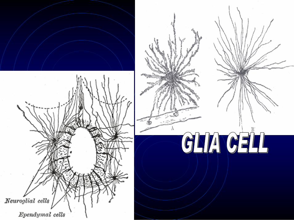
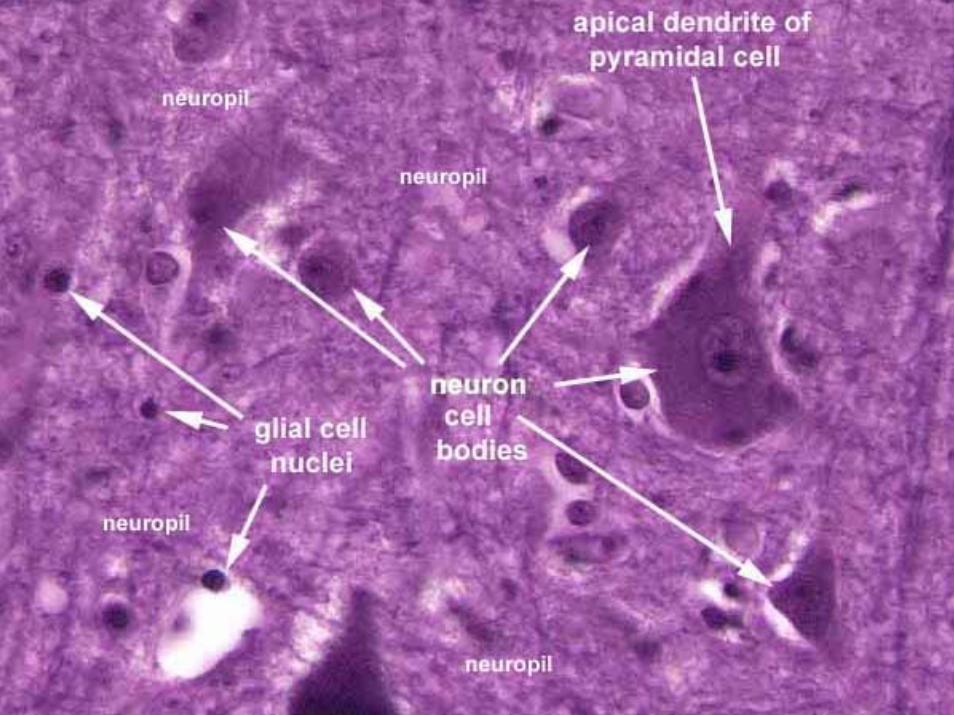
NEUROGLIA OR GLIA
• Glial cells, commonly called neuroglia or simply glia, are non-neuronal cells that provide
– support and nutrition,– maintain homeostasis, – form myelin, – and participate in signal transmission in
the nervous system.
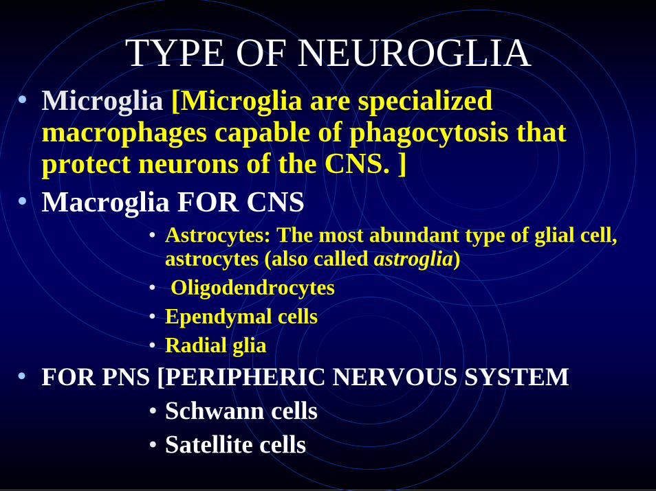
TYPE OF NEUROGLIA• Microglia [Microglia are specialized
macrophages capable of phagocytosis that protect neurons of the CNS. ]
• Macroglia FOR CNS• Astrocytes: The most abundant type of glial cell,
astrocytes (also called astroglia)• Oligodendrocytes• Ependymal cells• Radial glia
• FOR PNS [PERIPHERIC NERVOUS SYSTEM• Schwann cells• Satellite cells


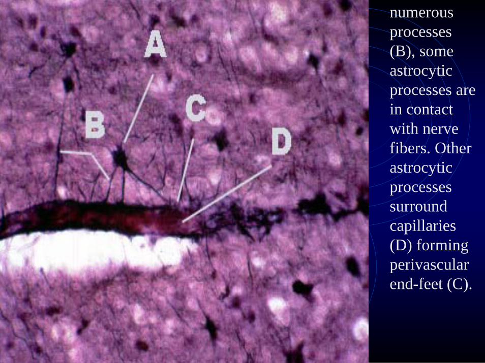
numerous processes (B), someastrocyticprocesses are in contact with nerve fibers. Otherastrocyticprocesses surround capillaries (D) formingperivascularend-feet (C).
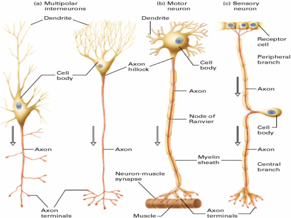
Neurons• There are many types of neuron based
on the size and shape of the cell body and the arrangement of the processes.
• Based on their staining neurons could be seen to be unipolar, bipolar ormultipolar.
• Most of the neurons within the CNS aremultipolar.
• The processes extending from the cell body are either axons or dendrites.
• Neurons usually have only one axon but many dendrites.

NEURONS
• Main role is to process and transmit information.
• neurons are found in the brain, the spinal cord and in the nerves and ganglia of the peripheral nervous system.
• Neurons are typically composed of a cell body, a dendritic tree and an axon.


Axons Dendrites
•Take information away from the cell body•Smooth Surface•Generally only 1 axon per cell•No ribosomes•Can have myelin •Branch further from the cell body
•Bring information to the cell body•Rough Surface (dendriticspines)•Usually many dendrites per cell•Have ribosomes•No myelin insulation •Branch near the cell body


Neurons are similar to other cells in the body because:1.1.Neurons are surrounded by a cell membrane.Neurons are surrounded by a cell membrane.2.2.Neurons have a nucleus that contains genes.Neurons have a nucleus that contains genes.3.3.Neurons contain cytoplasm, mitochondria and other Neurons contain cytoplasm, mitochondria and other organelles.organelles.4.4.Neurons carry out basic cellular processes such as Neurons carry out basic cellular processes such as protein synthesis and energy production.protein synthesis and energy production.
Neurons differ from other cells in the body because:1.1.Neurons haveNeurons have specialisedspecialised extensions called dendrites extensions called dendrites and axons. Dendrites bring information to the cell body and axons. Dendrites bring information to the cell body and axons take information away from the cell body.and axons take information away from the cell body.2.2.Neurons communicate with each other through an Neurons communicate with each other through an electrochemical process.electrochemical process.3.3.Neurons contain some specialized structures (for Neurons contain some specialized structures (for example, synapses) and chemicals (for example, example, synapses) and chemicals (for example, neurotransmitters).neurotransmitters).
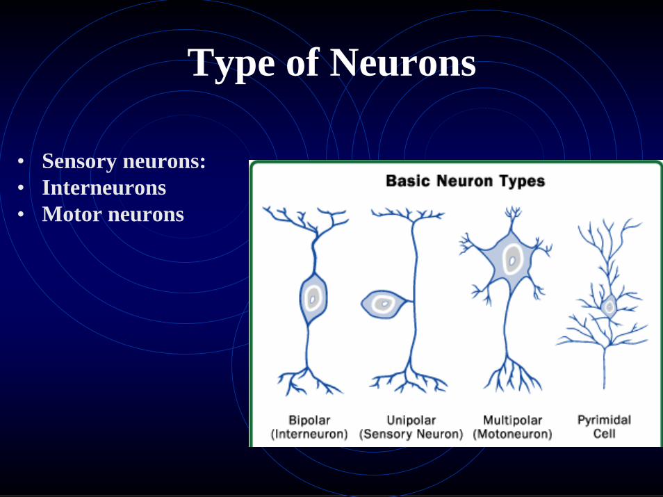
Type of Neurons
• Sensory neurons:• Interneurons• Motor neurons

Sensory neuronsThese run from the various types of stimulus receptors, e.g.,
••touch touch ••odor odor ••taste taste ••sound sound ••visionvision
to the central nervous system (CNS), the brain and spinal cord. The cell bodies of the sensory neurons leading to the spinal cord are located in clusters, called ganglia, next to the spinal cord. The axons usually terminate at interneurons.


Sensory Neurone: •Afferent Neuron – Moving away from a central organ or point •Relays messages from receptors to the brain or spinal cord
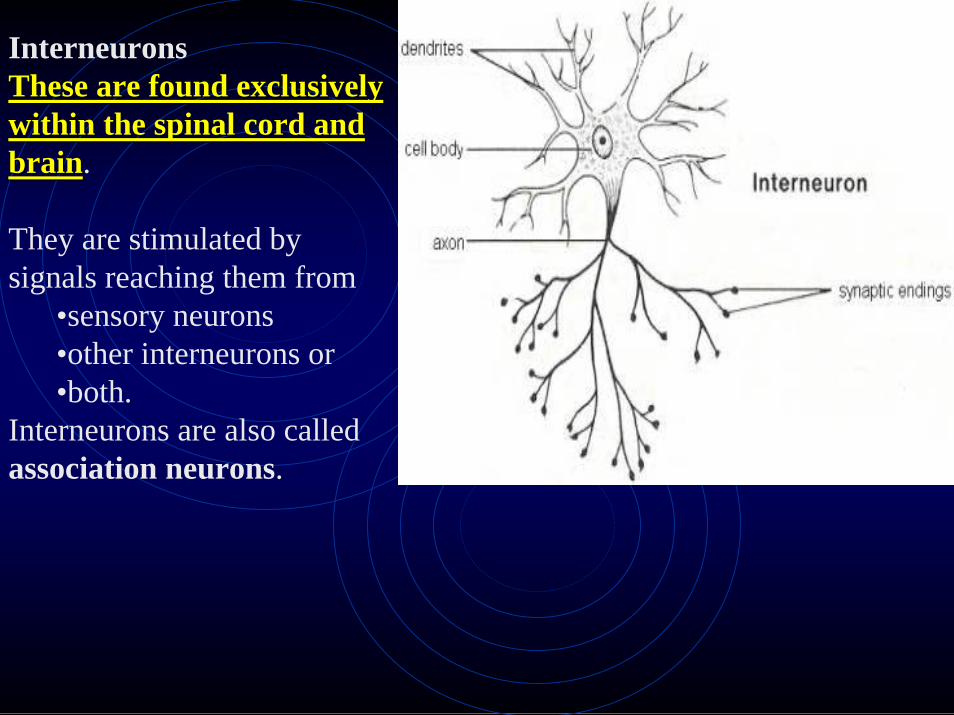
InterneuronsThese are found exclusively These are found exclusively within the spinal cord and within the spinal cord and brainbrain.
They are stimulated by signals reaching them from
•sensory neurons •other interneurons or •both.
Interneurons are also called association neurons.
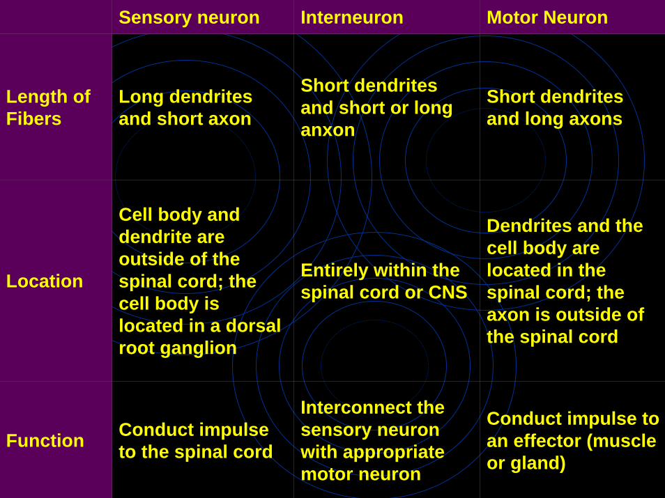
Sensory neuron Interneuron Motor Neuron
Length of Fibers
Long dendrites and short axon
Short dendrites and short or longanxon
Short dendrites and long axons
Location
Cell body and dendrite are outside of the spinal cord; the cell body is located in a dorsal root ganglion
Entirely within the spinal cord or CNS
Dendrites and the cell body are located in the spinal cord; the axon is outside of the spinal cord
Function Conduct impulse to the spinal cord
Interconnect the sensory neuron with appropriate motor neuron
Conduct impulse to an effector (muscle or gland)

Motor neuronsThese transmit impulses from the central nervous system to the
•muscles and •glands
that carry out the response.
Most motor neurons are stimulated by interneurons, although some are stimulated directly by sensory neurons.

Structural classificationMost neurons can be anatomically characterized as:
•Unipolar or Pseudounipolar- dendrite and axon emerging from same process.•Bipolar - single axon and single dendrite on opposite ends of the soma.•Multipolar - more than two dendrites
•Golgi I- neurons with long-projecting axonal processes.•Golgi II- neurons whose axonal process projects locally.
Some unique neuronal types can be identified according to their location in the nervous system and distinct shape.
Some examples are basket, Betz, medium spiny, Purkinje, pyramidal and Renshaw cells.
Functional classification
•Afferent neurons convey information from tissues and organs into the central nervous system.•Efferent neurons transmit signals from the central nervous system to the effector cells and are sometimes called motor neurons.•Interneurons connect neurons within specific regions of the central nervous system.
Afferent and efferent can also refer to neurons which convey information from one region of the brain to another.
Classification by action on other neurons
••Excitatory neuronsExcitatory neurons evoke excitation of their target neurons. Excitatory neurons in the brain are often glutamatergic. Spinal motoneurons use acetylcholine as their neurotransmitter.
••Inhibitory neuronsInhibitory neurons evoke inhibition of their target neurons. Inhibitory neurons are often interneurons. The output of some brain structures (neostriatum, globus pallidus, cerebellum) are inhibitory. The primary inhibitory neurotransmitters are GABA and glycine.
••ModulatoryModulatory neuronsneurons evoke more complex effects termed neuromodulation. These neurons use such neurotransmitters as dopamine, acetylcholine, serotonin and others.

Classification by discharge patternsClassification by discharge patterns
Neurons can be classified according to their electrophysiologicalcharacteristics:
•Tonic or regular spiking. Some neurons are typically constantly (or tonically) active. Example: interneurons in neurostriatum.•Phasic or bursting. Neurons that fire in bursts are called phasic.•Fast spiking. Some neurons are notable for their fast firing rates, for example some types of cortical inhibitory interneurons, cells inglobus pallidus.•Thin-spike. Action potentials of some neurons are more narrow compared to the others. For example, interneurons in prefrontal cortex are thin-spike neurons.
Classification by neurotransmitter releasedClassification by neurotransmitter releasedSome examples are cholinergic, GABA-ergic, glutamatergic anddopaminergic neurons.

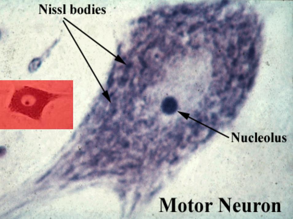
A Nissl body (or Nissl granuleor tigroid body) is a large
granular body found in nerve cells.
These granules are rough endoplasmic reticulum (with ribosomes) and are the site of protein synthesis.Nissl bodies show changes under various physiological conditions and in pathological conditions they may dissolve and disappear (karyolysis).
The axolemma is the membrane of a neuron's axon.It is responsible for maintaining the cell's membrane potential, and it contains channels through which ions can flow.
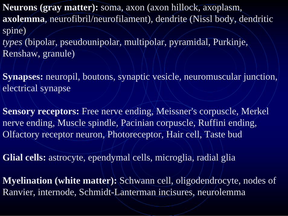
Neurons (gray matter): soma, axon (axon hillock, axoplasm,axolemma, neurofibril/neurofilament), dendrite (Nissl body, dendriticspine)types (bipolar, pseudounipolar, multipolar, pyramidal, Purkinje, Renshaw, granule)
Synapses: neuropil, boutons, synaptic vesicle, neuromuscular junction, electrical synapse
Sensory receptors: Free nerve ending, Meissner's corpuscle, Merkel nerve ending, Muscle spindle, Pacinian corpuscle, Ruffini ending, Olfactory receptor neuron, Photoreceptor, Hair cell, Taste bud
Glial cells: astrocyte, ependymal cells, microglia, radial glia
Myelination (white matter): Schwann cell, oligodendrocyte, nodes ofRanvier, internode, Schmidt-Lanterman incisures, neurolemma

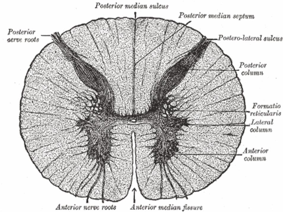
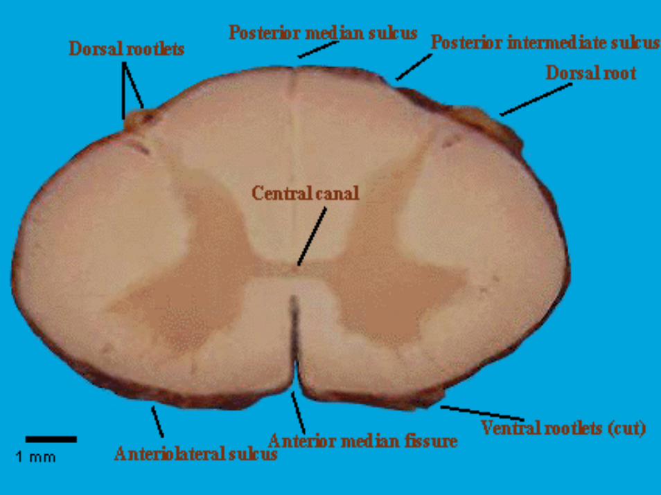
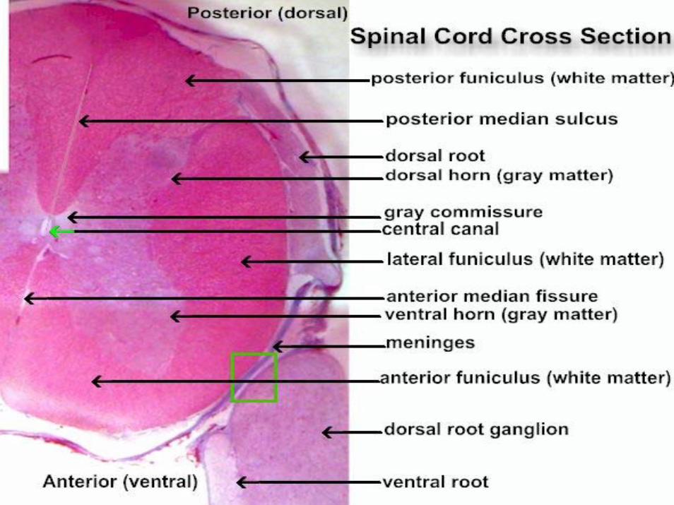
SPINAL CORD ANATOMY
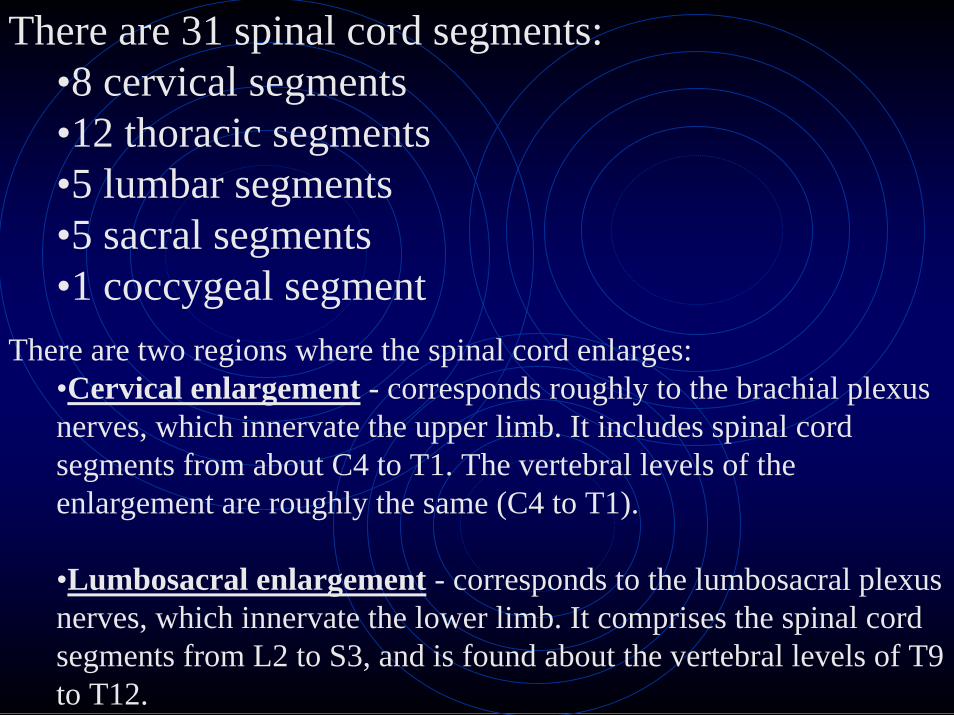
There are 31 spinal cord segments:•8 cervical segments•12 thoracic segments•5 lumbar segments•5 sacral segments•1 coccygeal segment
There are two regions where the spinal cord enlarges:•Cervical enlargement - corresponds roughly to the brachial plexus nerves, which innervate the upper limb. It includes spinal cord segments from about C4 to T1. The vertebral levels of the enlargement are roughly the same (C4 to T1).
•Lumbosacral enlargement - corresponds to the lumbosacral plexus nerves, which innervate the lower limb. It comprises the spinal cord segments from L2 to S3, and is found about the vertebral levels of T9 to T12.
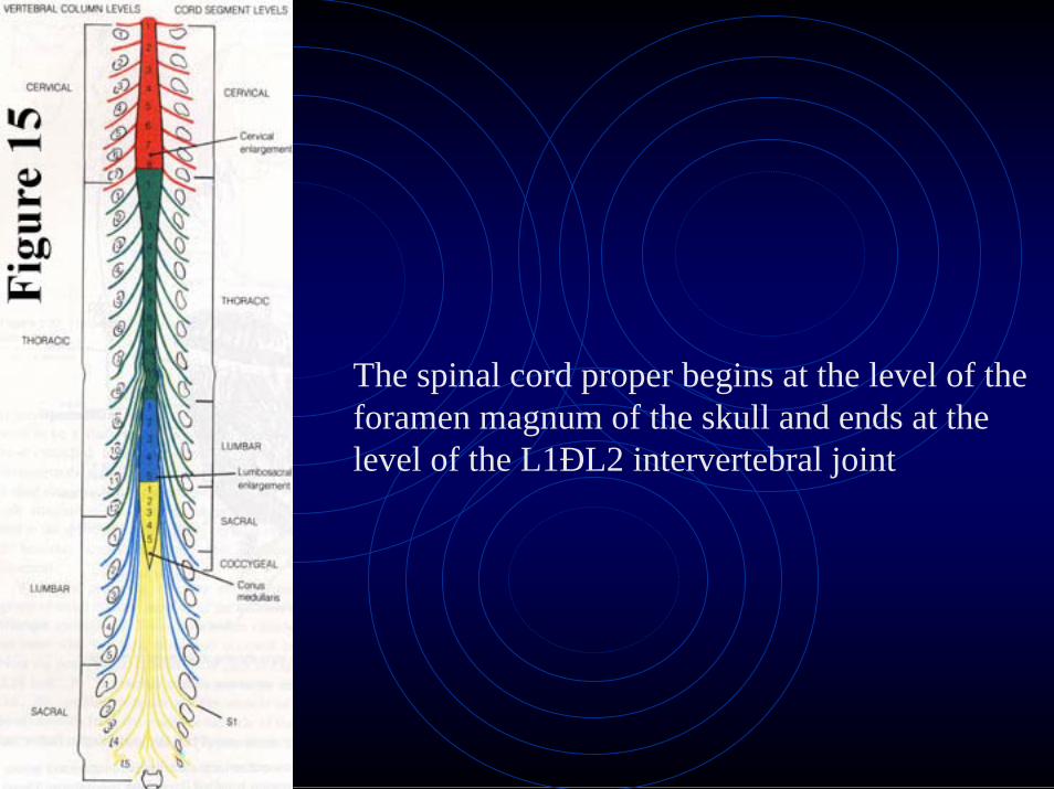
The spinal cord proper begins at the level of the foramen magnum of the skull and ends at the level of the L1ÐL2 intervertebral joint


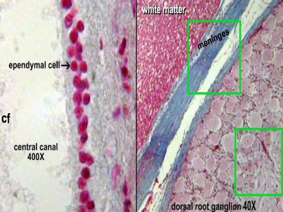
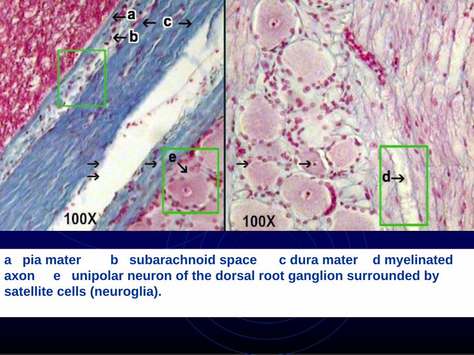
a pia mater b subarachnoid space c dura mater d myelinatedaxon e unipolar neuron of the dorsal root ganglion surrounded by satellite cells (neuroglia).
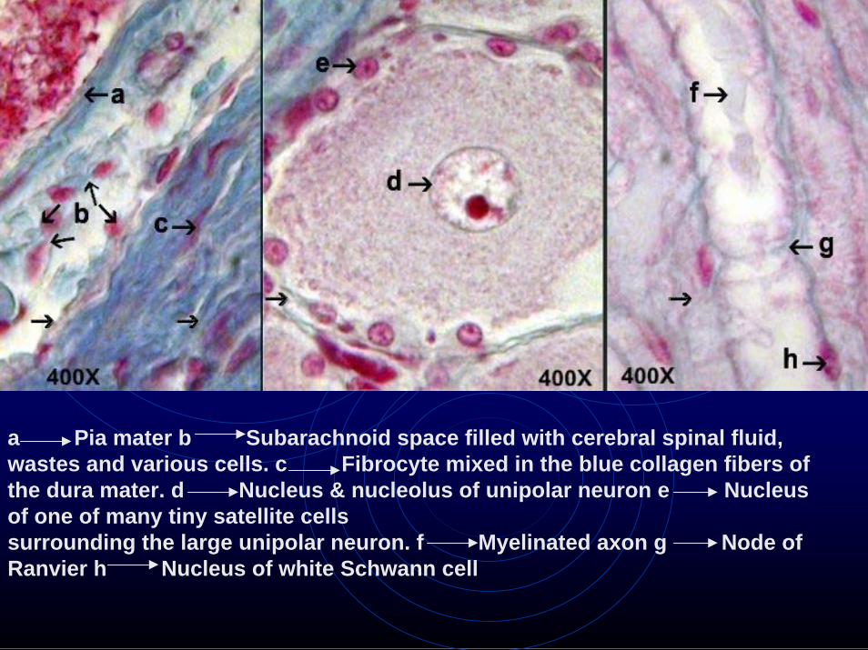
a Pia mater b Subarachnoid space filled with cerebral spinal fluid, wastes and various cells. c Fibrocyte mixed in the blue collagen fibers of the dura mater. d Nucleus & nucleolus of unipolar neuron e Nucleus of one of many tiny satellite cells surrounding the large unipolar neuron. f Myelinated axon g Node ofRanvier h Nucleus of white Schwann cell

a Synaptic bulbs over the motor end plate -neuromuscular junction b Neuron axon terminal - black fibers
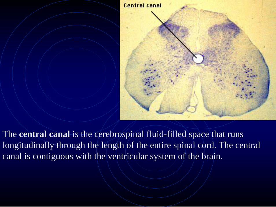
The central canal is the cerebrospinal fluid-filled space that runs longitudinally through the length of the entire spinal cord. The central canal is contiguous with the ventricular system of the brain.