neural development, cell-cell signaling, and the "two-hit
TRANSCRIPT

Neural Development, Cell-Cell Signaling, andthe "Two-Hit" Hypothesis of Schizophrenia
by Thomas M. Maynard, Linmarie Sikich, Jeffrey A. Lieberman,and Anthony'Samuel LaMantia
Abstact
To account for the complex genetics, the developmen-tal biology, and the late adolescent/early adulthoodonset of schizophrenia, the "two-hit" hypothesis hasgained increasing attention. In this model, genetic orenvironmental factors disrupt early central nervoussystem (CNS) development. These early disruptionsproduce long-term vulnerability to a "second hit" thatthen leads to the onset of schizophrenia symptoms.The cell-cell signaling pathways involved in nonaxialinduction, morphogenesis, and differentiation in thebrain, as well as in the limbs and face, could be targetsfor a "first hit" during early development. These samepathways, redeployed for neuronal maintenancerather than morphogenesis, may be targets for a "sec-ond hit" in the adolescent or adult brain.Furthermore, dysregulation of cell-cell signaling by a"first hit" may prime the CNS for a pathologicresponse to a "second hit" via the same signaling path-way. Thus, parallel disruption of cell-cell signaling inboth the developing and the mature CNS provides aplausible way of integrating genetic, developmental,and environmental factors that contribute to vulnera-bility and pathogenesis in schizophrenia.
Keywords: Development, signaling, schizophrenia,genetics, teratogenesis, neuronal circuits.
Schizophrenia Bulletin, 27(3):457-476,2001.
Schizophrenia is a common but complex mental disorderthat causes disruptions in thought processes, perceptions,and emotions. Although the effects of this disorder areprofound, there does not appear to be a single neurobio-logical cause, such as the widespread neural degenerationobserved in Alzheimer's disease, or a specific lesion in asingle brain region like the degeneration of the substantianigra observed in Parkinson's disease. In the absence of adiscrete cellular or anatomical pathology, the attention ofschizophrenia research has turned to more subtle anom-alies. Histopathological, pharmacological, and molecular
expression studies suggest that the integrity of neuronalcircuitry in the brain may be altered in schizophrenia.However, the pathological mechanisms that mediate thesechanges remain unclear.
Although several hypotheses suggest that aberrantembryonic development can contribute to schizophrenia,the onset of symptoms generally occurs many years afterthis process is completed. Thus, any disruptions to devel-opmental processes must result in long-term consequencesthat leave mature neurons or circuits vulnerable to subse-quent pathology. In addition, because the illness is oftenassociated with a progressive decline in functioning, it isimportant to consider what kinds of pathogenic processesmight continue to compromise abnormal neural circuits.Accordingly, it may be useful to consider how normalmechanisms of circuit construction and maintenance in thebrain might be disrupted, and how this might lead topathological changes thought to occur in schizophrenia.We will discuss one set of candidate mechanisms, the cell-cell signaling mechanisms that regulate the initial pattern-ing and formation of neuronal circuits as well as the sur-vival, growth, and plasticity of these circuits in the maturebrain. We will then consider how these mechanisms mightbe compromised in ways consistent with their roles in neu-ronal development and maintenance as well as the naturalhistory and clinical progression of schizophrenia.
The Elusive Pathogenesis ofSchizophrenia: Its Causes and Effects
The Causes of Schizophrenia. Decades of research intothe pathogenesis of schizophrenia have examined dozensof distinct genetic and environmental risk factors that canbe correlated with the natural history of the disorder;however, no single candidate mechanism has emerged.
Send reprint requests to Dr. A.-S. LaMantia, 260 MSRB, CB#7545,Department of Cell and Molecular Physiology, University of NorthCarolina, Chapel Hill, Chapel Hill, NC 27599; e-mail: [email protected].
457
Dow
nloaded from https://academ
ic.oup.com/schizophreniabulletin/article/27/3/457/1835125 by guest on 06 January 2022

Schizophrenia Bulletin, Vol. 27, No. 3, 2001 T.M. Maynard et al.
Instead, each factor may contribute to an increased vul-nerability to schizophrenia. Depending on the combina-tion of these risk factors, a pathologic threshold may bemet, thus leading to the emergence of disease symptoms.This conclusion is supported by several observations.
Schizophrenia is common. Schizophrenia is a com-mon psychiatric disorder. Estimates of lifetime prevalenceapproach 1 in 100. This high frequency suggests eitherthat the cause of the disorder is common or that there aremultiple causes that disrupt essential and vulnerable bio-logical functions and lead to symptom onset.
Schizophrenia is linked to many genetic risk fac-tors. Adoption, family, and twin studies provide strongevidence of the heritability of schizophrenia (Gottesmanand Wolfgram 1991). In addition, at least 15 genetic locihave been demonstrated to have weak to moderate link-ages to schizophrenia (Riley and McGuffin 2000). Anymodel for the disorder, therefore, must account for linkageto multiple genes.
Schizophrenia is not strictly genetic. Geneticsalone does not predict development of the disease. Theconcordance rate of schizophrenia is only about 50 per-cent in identical twins (Cardno and Gottesman 2000).Furthermore, these concordance rates may be reduced inmonozygotic twins who do not share a common amnioticenvironment as compared to those who do (Davis et al.1995). Thus, although genetic factors clearly can increasethe risk for schizophrenia, they cannot alone account forthe disease.
Schizophrenia is linked to environmental and non-genetic risk factors. Numerous environmental factors havebeen linked to schizophrenia. Births in the winter-springmonths (Torrey et al. 1997) increase schizophrenia risk pos-sibly because of maternal nutritional deficiencies (Hoek etal. 1996) or the prevalence of winter viral infections (Yolkenand Torrey 1995). In addition, the risk of developing schizo-phrenia is correlated with demographic factors, such as fam-ily size and locality, and the availability of an adequatematernal diet (Hulshoff Pol et al. 2000). Nevertheless, thesefactors alone are not sufficient to predict the development ofschizophrenia in any particular individual.
The Consequences of Schizophrenia. Although the spe-cific cause of schizophrenia is not yet clear, its conse-quences are well documented. These consequences do notappear to include widespread neurodegeneration or celldeath (Sapolsky 1993; Falkai et al. 1999); rather, a set ofsubtle changes to the neuronal circuitry of the brainappears to underlie the psychiatric symptoms that areobserved during the progression of the disease. This con-clusion is supported by the following observations.
Schizophrenia is not accompanied by consistentneuroanatomical anomalies. General markers of brainpathology, such as changes in ventricular size (Jones et al.
1994; Buchsbaum et al. 1997; Wright et al. 2000), havebeen associated with schizophrenia. Such changes, how-ever, are also associated with a number of neurologicaland psychiatric diseases and thus are not reliable predic-tors of schizophrenia. Alterations in specific cortical cellpopulations have been reported, such as a loss of neuropilor changes to "y-aminobutyric acid-ergic cells (GABA-ergic cells). There is, however, significant variability ineach of these findings within the schizophrenia population(Benes et al. 1991; Selemon et al. 1995; Beasley andReynolds 1997; Selemon et al. 1998). Thus, neuropatho-logical markers associated with the schizophrenia popula-tion are not absolute predictors of the disease in any par-ticular individual.
Molecular changes have been observed in thebrains of schizophrenia patients. There is an extensivecatalog of molecules whose expression levels change inthe brains of schizophrenia patients. These include reelin(Impagnatiello et al. 1998), SNAP25 (Thompson et al.1998), and neurotransmitter receptors (Joyce et al.1993a; Akbarian et al. 1995a; Eastwood et al. 1995;Goldsmith et al. 1997). In addition, changes in adhesionmolecules, cytoskeletal proteins, neurotrophins, and othercell-cell signaling molecules have been correlated withschizophrenia (Arnold et al. 1991; Vawter et al. 1998;Miyaoka et al. 1999; Takahashi et al. 2000). Diversity,rather than obvious functional relationships between spe-cific molecular families, is the primary characteristic ofthis list. Thus, the molecular changes seen in the brainsof schizophrenia patients may represent multiple finaltargets for pathology instead of a record of a singlepathological mechanism.
Schizophrenia affects the function of neuronalcircuits. In addition to these neuroanatomical changes,pharmacological manipulations suggest that synaptictransmission in some brain circuits may be compromised,particularly those involving dopaminergic, GABA-ergic,and glutaminergic neurotransmitters. Agents that increasedopaminergic tone (e.g., L-dopa, cocaine, or ampheta-mines), when chronically administered, can elicit halluci-nations and delusions that resemble the positive symp-toms of schizophrenia (Bell 1965; Snyder 1972; Post andKopanda 1976; Lieberman et al. 1987; Brady et al. 1991;Friedman and Sienkiewicz 1991; Young et al. 1997).Similarly, agents that act as partial antagonists at gluta-mate receptors (e.g., phencyclidine, ketamine) may alsoevoke psychotic symptoms (Javitt and Zukin 1991;Krystal et al. 1994; Rosse et al. 1994). In contrast, posi-tive symptoms in schizophrenia patients have beentreated for many years with pharmacological agents thatantagonize dopamine activity in the CNS (Creese et al.1976; Meltzer and Stahl 1976; Haracz 1982).
Schizophrenia affects synaptic transmission. Theeffects of schizophrenia on neuronal circuits are further
458
Dow
nloaded from https://academ
ic.oup.com/schizophreniabulletin/article/27/3/457/1835125 by guest on 06 January 2022

'Two-Hit" Hypothesis of Schizophrenia Schizophrenia Bulletin, Vol. 27, No. 3, 2001
supported by evidence of changes in the localization andfunction of neurotransmitters, including reduced expres-sion of dopamine receptor subtypes or dopaminergicsynaptic function in brain regions affected by schizophre-nia (Goldsmith et al. 1997; Laruelle 1998). In addition,reduced GAB A synthesis (Akbarian et al. 1995fc) andglutamate receptor hypofunction have been associatedwith the disease (Olney and Farber 1995; Mohn et al.1999).
Schizophrenia symptoms generally appear duringlate adolescence. It is widely accepted that schizophreniahas its origin during the early stages of neurodevelopment(Murray and Lewis 1987; Weinberger 1987; Weinberger1995). Subtle attentional, motor, and social problems arereported during childhood in people who are later diag-nosed with schizophrenia, although these problems arefrequent in the general population and do not reliably pre-dict the onset of the disease (Walker et al. 1996;Erlenmeyer-Kimling et al. 2000). Overt symptoms ofschizophrenia generally manifest themselves shortly afterthe onset of puberty. The onset of symptoms generallyoccurs at later ages for women than for men, suggestingthat gonadal steroid hormones may play at least some rolein the disease.
Schizophrenia is a progressive disorder. Often, thefirst overt manifestations of the illness are attenuated hal-lucinations and delusions that become more severe andpersistent over the course of weeks or months (Haas andSweeney 1992; Beiser et al. 1993). Early treatment withantipsychotic medications is often effective; however, alonger duration of untreated psychotic symptoms is asso-ciated with more severe symptoms during psychoticepisodes. These subsequent symptoms are resistant totreatment and more likely to recur (Loebel et al. 1992;Lieberman et al. 1996; Sheitman and Lieberman 1998;Robinson et al. 1999). This suggests that psychotic symp-toms themselves "sensitize" the brain and increase thepathology associated with the illness.
Built To Fail: The "Two-Hit"Hypothesis of Schizophrenia
In the absence of a singular genetic or environmentalpathogenic agent for schizophrenia, attention has turnedto disease models involving multiple factors. One suchmodel is the "two-hit" hypothesis (as described in Bayeret al. 1999). Two-hit models for schizophrenia are similarto those proposed for other complex diseases, such as can-cer (figure 1, top). Like schizophrenia, cancer is linked tonumerous genetic and environmental factors, although nosingle mechanism can account for all cases. In addition,although genetic risk factors are present from birth, can-cer often does not become apparent for many years. The
two-hit model for cancer provides a valuable frameworkfor assessing relationships between cell division, growth,survival, and death mechanisms that contribute to oncogen-esis. Although the loss of a single regulatory mechanismcould theoretically lead to a loss of cellular control, thereis a relatively high level of redundancy and resiliency inthe regulation of cellular functions. Thus, a completebreakdown in cellular control often occurs only after thedisruption of multiple regulatory mechanisms. A geneticmutation might make a particular cellular pathway vulner-able for transforming a specific cell type. Subsequently,an environmental carcinogen or a somatic mutation eventmight disrupt redundant or compensatory mechanisms,resulting in transformation and tumorigenesis.
The two-hit hypothesis for schizophrenia suggeststhat a prenatal genetic or environmental "first hit" disruptssome aspect of brain development, and establishesincreased vulnerability to a second hit that may occurlater in life (figure 1, bottom). Neither insult by itself issufficient to induce schizophrenia. Instead, the first hit"primes" the nervous system for the second, which thenprecipitates disease symptoms. Because the first hit isthought to occur during embryonic development, it seemscandidates for this hit should involve disruption of amechanism that is (1) susceptible to numerous genetic andenvironmental perturbations, and (2) capable of producinglong-term significant changes. In contrast, a second hitmight go unnoticed in a brain that had not been previously"primed" to respond in an aberrant way. One generalmechanism stands out as being both susceptible to manyperturbations and capable of producing long-termchanges: cell-cell signaling pathways initially involved ininduction and morphogenesis of the CNS (and other struc-tures), and later in ongoing maintenance of CNS neuronsand circuits, are ideal targets for two hits that might resultin schizophrenia vulnerability and pathogenesis.
Lessons From the Minor PhysicalAnomalies: Inductive Signaling andSchizophreniaMany severe neurological deficits, such as thoseobserved in Down's syndrome patients, are accompa-nied by physical malformations. These anomaliesinclude abnormal proportions or defects in the struc-tures of the head and face, limbs and digits, and internalorgans. Quantitative assessments of these anomalies(termed the "minor physical anomalies") (Waldrop et al.1968; Waldrop et al. 1978) show that they are correlatedwith psychological and behavioral problems. Minorphysical anomalies are observed more often amongadult schizophrenia patients than within the generalpopulation (Gualtieri et al. 1982; Guy et al. 1983;
459
Dow
nloaded from https://academ
ic.oup.com/schizophreniabulletin/article/27/3/457/1835125 by guest on 06 January 2022

Schizophrenia Bulletin, Vol. 27, No. 3, 2001 T.M. Maynard et al.
Figure 1. Schematic representations of two-hit models for cancer and schizophrenia
"Two hit" model of cancer
Genetic Environmental' jtvulnerability factore -4
Two hit" model of schizophrenia
Developmentaldisruptions
\Developing ^ ^ ^ w
CNS ^ ^ ^ "
Environmentalfactors
\Vulnerable »
CNS ^ ^
Target: Cell cycle pathways
Geneticpredisposition
Environmentalfactors
\
Controlledcelicyde
Genetic orenvironmental
disruption
Target: Cell signaling pathways
Environmental^factors •/
/Vote.—The two-hit model of cancer (top left) suggests that a first hit, such as a genetic predisposition to the disease, produces a vulnera-bility within specific cell populations, whereas a second hit, probably from environmental factors, induces the onset of the disease. Thismodel is complementary to the growing understanding of the molecular mechanisms that underlie the disorder (top right). At the molecu-lar level, the first hit likely corresponds to the loss of one gene within a set of redundant regulatory genes. The failure of one regulatorygene is likely compensated for by other parallel mechanisms, so a loss of cell cycle control likely occurs only after the subsequent loss ofadditional mechanisms. In a similar fashion, a two-hit model for schizophrenia (bottom left) would suggest that disruptions in the develop-ment of the central nervous system (CNS) produce a vulnerability to the disorder but that the onset of symptoms in a vulnerable brainwould be triggered by environmental factors. At the molecular and cellular levels, this model may represent how disruptions in early cellu-lar signaling events may leave some neuronal circuits vulnerable to disruptions in the signaling events that occur in the mature CNS (bot-tom right).
Ismail et al. 1998). In particular, schizophrenia patientsare more likely to have deviations in craniofacial pro-portions, including distance between the eyes, place-ment and symmetry of the ear pinnae, and cranial cir-cumference. Similarly, malformations of the digits ofthe hands and feet (including anomalous lengths orshapes of the digits and partial syndactyly of the toes)are more frequent in schizophrenia patients.
Malformations of limbs or facial structures are pre-sumably not the direct cause of schizophrenia. Theremay be, however, a similar set of "minor brain anom-alies" that develop in parallel to the visible defects inthe limbs and face. For instance, the olfactory bulbsappear reduced in size in some schizophrenia patients,and olfactory impairments are often seen (Moberg et al.1999; Turetsky et al. 2000). It is likely that both theminor physical anomalies and the minor brain anom-alies are the result of the same aberrant developmental
processes, which may be involved in the pathogenesisof schizophrenia. Indeed, a single developmental mech-anism, nonaxial mesenchymal/epithelial induction(LaMantia 1999; LaMantia et al. 2000), is essential forinitial morphogenesis and differentiation at all of thesesites.
Mesenchymal/Epithelial Induction, theMinor Physical Anomalies, and theForebrain
During the first trimester of embryogenesis, morphogene-sis of the limbs, heart, face, and forebrain relies on a com-plex set of cell-cell signaling interactions known as mes-enchymal/epithelial induction. These interactions resultfrom the apposition of two cell groups—epithelia, sheetsof cells that constitute the inner and outer surfaces of the
460
Dow
nloaded from https://academ
ic.oup.com/schizophreniabulletin/article/27/3/457/1835125 by guest on 06 January 2022
'Two-Hit" Hypothesis of Schizophrenia Schizophrenia Bulletin, Vol. 27, No. 3, 2001
embryo, and mesenchyme, the loosely arrayed accumula-tions of cells in the interstices between epithelial layers.The apposition of these two tissue compartments facili-tates the exchange of short- and long-range signals that inturn regulate the differentiation of both epithelial andmesenchymal derivatives. Although the mechanismsinvolved in mesenchymal/epithelial induction are [quite]complex, experimental studies in the limb and forebrainprovide insight into these signaling processes, includingthe identity of many [of the] important molecular compo-nents. Parallels between the limb and the forebrain sug-gest that mesenchymal/epithelial induction may be apotential target for a first hit in the two-hit model of schiz-ophrenia.
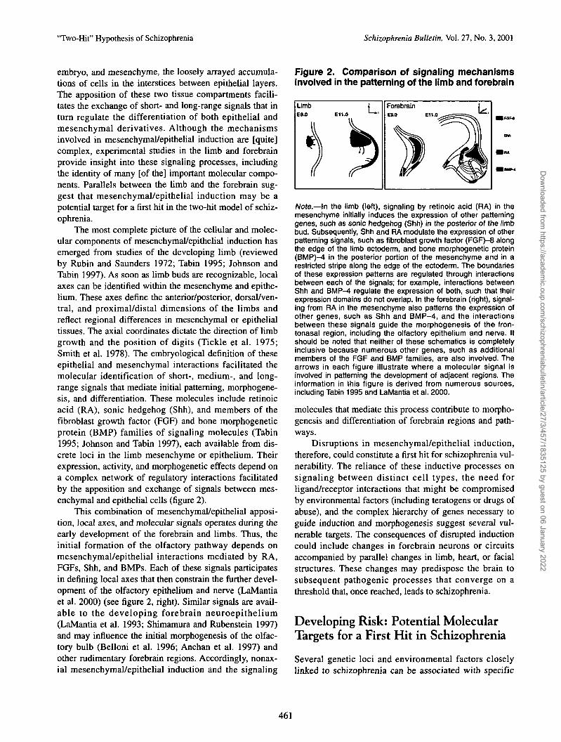
The most complete picture of the cellular and molec-ular components of mesenchymal/epithelial induction hasemerged from studies of the developing limb (reviewedby Rubin and Saunders 1972; Tabin 1995; Johnson andTabin 1997). As soon as limb buds are recognizable, localaxes can be identified within the mesenchyme and epithe-lium. These axes define the anterior/posterior, dorsal/ven-tral, and proximal/distal dimensions of the limbs andreflect regional differences in mesenchymal or epithelialtissues. The axial coordinates dictate the direction of limbgrowth and the position of digits (Tickle et al. 1975;Smith et al. 1978). The embryological definition of theseepithelial and mesenchymal interactions facilitated themolecular identification of short-, medium-, and long-range signals that mediate initial patterning, morphogene-sis, and differentiation. These molecules include retinoicacid (RA), sonic hedgehog (Shh), and members of thefibroblast growth factor (FGF) and bone morphogeneticprotein (BMP) families of signaling molecules (Tabin1995; Johnson and Tabin 1997), each available from dis-crete loci in the limb mesenchyme or epithelium. Theirexpression, activity, and morphogenetic effects depend ona complex network of regulatory interactions facilitatedby the apposition and exchange of signals between mes-enchymal and epithelial cells (figure 2).
This combination of mesenchymal/epithelial apposi-tion, local axes, and molecular signals operates during theearly development of the forebrain and limbs. Thus, theinitial formation of the olfactory pathway depends onmesenchymal/epithelial interactions mediated by RA,FGFs, Shh, and BMPs. Each of these signals participatesin denning local axes that then constrain the further devel-opment of the olfactory epithelium and nerve (LaMantiaet al. 2000) (see figure 2, right). Similar signals are avail-able to the developing forebrain neuroepithelium(LaMantia et al. 1993; Shimamura and Rubenstein 1997)and may influence the initial morphogenesis of the olfac-tory bulb (Belloni et al. 1996; Anchan et al. 1997) andother rudimentary forebrain regions. Accordingly, nonax-ial mesenchymal/epithelial induction and the signaling
Figure 2. Comparison of signaling mechanismsinvolved in the patterning of the limb and forebrain
Note.—In the limb (left), signaling by relinoic acid (RA) in themesenchyme initially induces the expression of other patterninggenes, such as sonic hedgehog (Shh) in the posterior of the limbbud. Subsequently, Shh and RA modulate the expression of otherpatterning signals, such as fibroblast growth factor (FGF)-8 alongthe edge of the limb ectoderm, and bone morphogenetic protein(BMP)-4 in the posterior portion of the mesenchyme and in arestricted stripe along the edge of the ectoderm. The boundariesof these expression patterns are regulated through interactionsbetween each of the signals; for example, interactions betweenShh and BMP-4 regulate the expression of both, such that theirexpression domains do not overlap. In the forebrain (right), signal-ing from RA in the mesenchyme also patterns the expression ofother genes, such as Shh and BMP-4, and the interactionsbetween these signals guide the morphogenesis of the fron-tonasal region, including the olfactory epithelium and nerve. Itshould be noted that neither of these schematics is completelyinclusive because numerous other genes, such as additionalmembers of the FGF and BMP families, are also involved. Thearrows in each figure illustrate where a molecular signal isinvolved in patterning the development of adjacent regions. Theinformation in this figure is derived from numerous sources,including Tabin 1995 and LaMantia et al. 2000.
molecules that mediate this process contribute to morpho-genesis and differentiation of forebrain regions and path-ways.
Disruptions in mesenchymal/epithelial induction,therefore, could constitute a first hit for schizophrenia vul-nerability. The reliance of these inductive processes onsignaling between distinct cell types, the need forligand/receptor interactions that might be compromisedby environmental factors (including teratogens or drugs ofabuse), and the complex hierarchy of genes necessary toguide induction and morphogenesis suggest several vul-nerable targets. The consequences of disrupted inductioncould include changes in forebrain neurons or circuitsaccompanied by parallel changes in limb, heart, or facialstructures. These changes may predispose the brain tosubsequent pathogenic processes that converge on athreshold that, once reached, leads to schizophrenia.
Developing Risk: Potential MolecularTargets for a First Hit in Schizophrenia
Several genetic loci and environmental factors closelylinked to schizophrenia can be associated with specific
461
Dow
nloaded from https://academ
ic.oup.com/schizophreniabulletin/article/27/3/457/1835125 by guest on 06 January 2022

Schizophrenia Bulletin, Vol. 27, No. 3, 2001 T.M. Maynard et al.
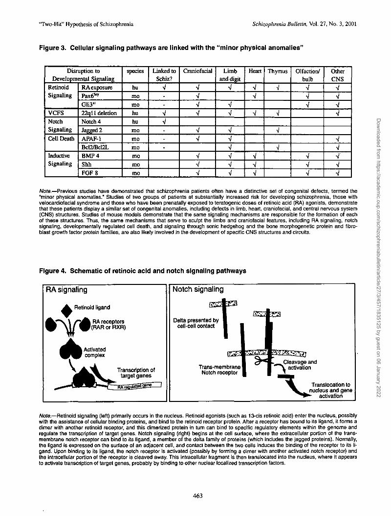
molecular and cellular mechanisms that contribute tomesenchymal/epithelial induction. Three of these—embryonic disruptions of retinoid signaling, haploinsuffi-ciency of chromosome 22qll, and genetic anomalies ofnotch signaling pathways—are not only linked to schizo-phrenia, but also associated with minor physical anom-alies (figure 3). Thus, a closer examination of these dis-ruptions in cell-cell signaling related to induction mayhelp to identify specific molecular mechanisms involvedin producing a first hit during neural development thatestablishes long-term vulnerability to schizophrenia.
Retinoid Signaling. RA is an important developmentalsignaling molecule critically involved in induction of thelimbs, heart, branchial arches, and forebrain (Linney andLaMantia 1994; LaMantia 1999). Embryonic RA expo-sure leads to a constellation of developmental deficits thatresemble the minor physical anomalies in schizophreniapatients. Thus, it has been suggested that embryonic sig-naling through this pathway may also be compromised inschizophrenia patients (Goodman 1998; LaMantia 1999).Moreover, many genetic loci linked to schizophrenia areclose to the sites of important genes in the retinoid signal-ing pathways (Goodman 1995). Apparently, RA signalingis an important factor for neural development, and earlydysregulation may contribute to CNS dysfunction.
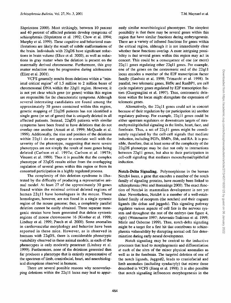
Retinoid signaling, like signaling of all members ofthe steroid-thyroid superfamily of hormones and recep-tors, involves a variety of transduction mechanisms (fig-ure 4, left). In addition to the ligand RA (the acidic formof vitamin A or retinol), there are two multigene familiesof receptor molecules: the retinoic acid receptors (RARs)and the retinoid-x-receptors (RXRs) (Yang et al. 1991;Mangelsdorf et al. 1992; Allenby et al. 1993). When a cellis exposed to RA (which is highly lipid soluble), the li-gand enters the cell and binds to nuclear RARs andRXRs. These receptors routinely heterodimerize uponbinding RA (thus, two separate receptor types, such as anRAR and an RXR, bind to one another). Subsequently, inconjunction with a number of corepressors and co-activa-tors (Minucci and Ozato 1996), RA ligand-receptor com-plexes transcriptionally activate specific genes (figure 4,left). This activation depends on direct binding of the acti-vated receptors to genomic regulatory sites known as RAresponse elements (RAREs).
The details of RA signaling indicate several stepswhere this process could be disrupted, leading to develop-mental anomalies that might confer schizophrenia vulner-ability. Teratogenic exposure to RA or RA deprivationresulting from a restricted maternal diet can alter develop-ment. For example, RA exposure during early fetal lifeleads to gross malformations of several structures includ-ing the limbs, face, heart, and CNS (Shenefelt 1972;
Lammer et al. 1985; Anchan et al. 1997). More subtledeficits in behavior have been reported after later fetalexposure to RA (reviewed by Adams and Lammer 1993).
Experimentally induced mutations in RA receptorsand cofactors have been more difficult to interpret. Singlenull mutations in several genes that contribute to RA sig-naling produce few detectable neural phenotypes, presum-ably because of functional redundancy and overlappingspatial distribution of various RA receptors and bindingproteins (Perez-Castro et al. 1989; Gustafson et al. 1993;Sucov and Evans 1995; Krezel et al. 1996). An exceptionto this trend is seen in the consequences of inactivating amajor retinoid synthetic enzyme, retinaldehyde dehydro-genase 2 (RALDH2), which disrupts embryogenesis at avery early stage (Niederreither et al. 1999). In addition,inactivation of the RAR(3 gene results in subtle butdetectable changes in long-term potentiation in the hip-pocampus (Chiang et al. 1998). Because of the potentialfor functional redundancy, other studies have examinedthe result of inactivating two receptors simultaneously.The absence of both RAR(3 and RXR7 leads to a dramaticbehavioral change, where both long-term potentiation andlong-term depression are diminished in the hippocampus(Chiang et al. 1998). In addition, the removal of bothgenes leads to significant deficits in locomotion, perhapsresulting from disrupted expression of dopamine receptors(Krezel et al. 1998). Together, these results demonstratethat retinoid signaling can influence some aspects ofhigher order cognitive functions. The current data, how-ever, do not address whether RA-sensitive behavioral andcognitive changes reflect altered RA-dependent neuraldevelopment, ongoing RA signaling required for maturebrain function, or both.
22qll Deletion Syndrome. Haploinsufficiency ofregions of chromosome 22qll leads to a constellation ofdevelopmental and behavioral anomalies (Goldberg et al.1993; Driscoll 1994; Ryan et al. 1997; Shprintzen 2000),collectively known as the 22q deletion syndrome (22qDS,also known as velocardiofacial syndrome [VCFS] orDiGeorge syndrome). This deletion syndrome is one ofthe best characterized genetic conditions linked to anincreased risk for schizophrenia (Cohen et al. 1999;Murphy et al. 1999). The phenotype of 22qDS is highlyvariable, even within families (Vincent et al. 1999), butlike RA-exposed individuals, patients with 22qll dele-tions have a characteristic set of heart, limb, and craniofa-cial anomalies that mirror the "minor physical anomalies"frequently observed in schizophrenia patients. Many peo-ple with 22qDS have cognitive problems (Golding-Kushner et al. 1985; Moss et al. 1999; Swillen et al.1999); behavioral or psychiatric difficulties are also com-mon (Papolos et al. 1996; Carlson et al. 1997a;
462
Dow
nloaded from https://academ
ic.oup.com/schizophreniabulletin/article/27/3/457/1835125 by guest on 06 January 2022
'Two-Hit" Hypothesis of Schizophrenia Schizophrenia Bulletin, Vol. 27, No. 3, 2001
Figure 3. Cellular signaling pathways are linked with the "minor physical anomalies"
Disruption toDevelopmental Signaling
RetinoidSignaling
VCFS
NotchSignaling
Cell Death
InductiveSignaling
RA exposure
Pax6^
GH3"
22ql 1 deletionNotch 4
Jagged 2
APAF-1Bcl2/Bcl2LBMP 4
Shh
FGF8
species
hu
mo
mo
hu
hu
mo
mo
mo
mo
mo
mo
Linked toSchiz?
V--
V---
Craniofacial
V
VV
V
Limband digit
VV
V
V
V
Heart
V
Thymus
V
V
Olfaction/bulb
V
V
VVV
OtherCNS
VVV
VVVVV
/Vote.—Previous studies have demonstrated that schizophrenia patients often have a distinctive set of congenital defects, termed the"minor physical anomalies.'' Studies of two groups of patients at substantially increased risk for developing schizophrenia, those withvelocardiofacial syndrome and those who have been prenatally exposed to teratogenic doses of retinoic acid (RA) agonists, demonstratethat these patients display a similar set of congenital anomalies, including defects in limb, heart, craniofacial, and central nervous system(CNS) structures. Studies of mouse models demonstrate that the same signaling mechanisms are responsible for the formation of eachof these structures. Thus, the same mechanisms that serve to sculpt the limbs and craniofacial features, including RA signaling, notchsignaling, developmental^ regulated cell death, and signaling through sonic hedgehog and the bone morphogenetic protein and fibro-blast growth factor protein families, are also likely involved in the development of specific CNS structures and circuits.
Figure 4. Schematic of retinoic acid and notch signaling pathways
RA signaling
• Retinoid ligand
j RA receptorsf(RAR or RXR)
Transcription oftarget genes
Notch signaling
Delta presented bycell-cell contact
Trans-membraneNotch receptor
Cleavage andactivation
Translocation tonucleus and gene
activation
Note.—Retinoid signaling (left) primarily occurs in the nucleus. Retinoid agonists (such as 13-cis retinoic acid) enter the nucleus, possiblywith the assistance of cellular binding proteins, and bind to the retinoid receptor protein. After a receptor has bound to its ligand, it forms adimer with another retinoid receptor, and this dimerized protein in turn can bind to specific regulatory elements within the genome andregulate the transcription of target genes. Notch signaling (right) begins at the cell surface, where the extracellular portion of the trans-membrane notch receptor can bind to its ligand, a member of the delta family of proteins (which includes the jagged proteins). Normally,the ligand is expressed on the surface of an adjacent cell, and contact between the two cells induces the binding of the receptor to its li-gand. Upon binding to its ligand, the notch receptor is activated (possibly by forming a dimer with another activated notch receptor) andthe intracellular portion of the receptor is cleaved away. This intracellular fragment is then translocated into the nucleus, where it appearsto activate transcription of target genes, probably by binding to other nuclear localized transcription factors.
463
Dow
nloaded from https://academ
ic.oup.com/schizophreniabulletin/article/27/3/457/1835125 by guest on 06 January 2022
Schizophrenia Bulletin, Vol. 27, No. 3, 2001 T.M. Maynard et al.
Shprintzen 2000). Most strikingly, between 10 percentand 40 percent of affected patients develop symptoms ofschizophrenia (Shprintzen et al. 1992; Chow et al. 1994;Murphy et al. 1999). These cognitive and behavioral man-ifestations are likely the result of subtle malformations ofthe brain. Individuals with 22qDS have significant reduc-tions in brain volume (Eliez et al. 2000), as well as reduc-tions in gray matter when the deletion is present on thematernally derived chromosome. Furthermore, this graymatter reduction may be progressive during adolescence(Eliez et al. 2001).
VCFS generally results from deletions within a "min-imal critical region" of 1.5 million to 2 million bases ofchromosomal DNA within the 22qll region. However, itis not yet clear which gene (or genes) within this regionare responsible for the characteristic symptoms. Althoughseveral interesting candidates are found among theapproximately 30 genes contained within this region,genetic mapping of 22qDS patients has not identified asingle gene (or set of genes) that is uniquely deleted in allaffected patients. Instead, 22qDS patients with similarsymptoms have been found to have deletions that do notoverlap one another (Amati et al. 1999; McQuade et al.1999). Additionally, the size and position of the deletionswithin 22qll do not appear to correlate well with theseverity of the phenotype, suggesting that more severephenotypes are not simply the result of more genes beingdeleted (Carlson et al. 1997a; Carlson et al. \991b;Vincent et al. 1999). Thus it is possible that the complexphenotype of 22qDS results either from the overlappingregulation of several genes within this region or from itsconcerted participation in a highly regulated process.
The complexity of this deletion syndrome is illus-trated by the difficulty of producing a representative ani-mal model. At least 27 of the approximately 30 genesfound within the minimal critical deleted regions ofhuman 22ql l have homologues in the mouse. Thesehomologues, however, are not found in a single syntenicregion of the mouse genome; thus, a completely paralleldeletion cannot be easily obtained. Three separate trans-genic strains have been generated that delete syntenicregions of mouse chromosome 16 (Kimber et al. 1999;Lindsay et al. 1999; Puech et al. 2000). Some anomaliesin cardiovascular morphology and behavior have beenreported in these mice. However, as is observed inhumans with 22qDS, there is considerable phenotypicvariability observed in these animal models, as each of thephenotypes is only modestly penetrant (Lindsay et al.1999). Furthermore, none of the deletions generated thusfar produces a phenotype that is entirely representative ofthe spectrum of limb, craniofacial, heart, and neurobiolog-ical disruptions observed in humans.
There are several possible reasons why nonoverlap-ping deletions within the 22qll locus may lead to appar-
ently similar neurobiological phenotypes. The simplestpossibility is that there may be several genes within thisregion that have similar functions during embryogenesis.There are a variety of inferred functions for genes withinthe critical region, although it is not immediately clearwhether these functions overlap. A more intriguing possi-bility is that several genes within this region may act inconcert. This could be a consequence of one (or more)22qll genes regulating other 22qll genes. For example,one of the genes on the centromeric end of the 22qlllocus encodes a member of the E2F transcription factorfamily (Gaubatz et al. 1998; Trimarchi et al. 1998). Inparallel, two telomeric genes, Htf9c and RanBPl, are cellcycle regulatory genes regulated by E2F transcription fac-tors (Guarguaglini et al. 1997). Thus, centromeric dele-tions within the locus might disregulate the expression oftelomeric genes.
Alternatively, the 22qll genes could act in concertbecause of their regulation by (or participation in) anotherregulatory pathway. For example, 22qll genes could beeither upstream regulators or downstream targets of mes-enchymal/epithelial signaling in the limbs, heart, face, andforebrain. Thus, a set of 22qll genes might be coordi-nately regulated by the cell-cell signals that mediateinduction, including FGFs, BMPs, Shh, and RA. It is pos-sible, therefore, that at least some of the complexity of the22qDS phenotype may be due not only to interactionsbetween 22qll genes, but also to their participation incell-cell signaling that mediates mesenchymal/epithelialinduction.
Notch-Delta Signaling. Polymorphisms in the humanNotch4 locus, a gene that encodes a member of the notchfamily of signaling proteins, have recently been linked toschizophrenia (Wei and Hemmings 2000). The exact func-tion of Notch4 in mammalian development is not yetclear. Nevertheless, Notch4 is a member of a well-estab-lished family of receptors (the notches) and their cognateligands (the deltas and jaggeds). This signaling pathwayregulates various aspects of cell fate in the nervous sys-tem and throughout the rest of the embryo (see figure 4,right) (Weinmaster 1997; Artavanis-Tsakonas et al. 1999;Miele and Osborne 1999). Thus, notch-delta signalingmight be a target for a first hit that contributes to schizo-phrenia vulnerability by disrupting normal cell fate deter-mination during early neural development.
Notch signaling may be central to the inductiveprocesses that lead to morphogenesis and differentiationat each of the sites of the minor physical anomalies aswell as in the forebrain. The targeted deletion of one ofthe notch ligands, Jagged2, leads to craniofacial andlimb anomalies (including syndactyly) that mirror thosedescribed in VCFS (Jiang et al. 1998). It is also possiblethat notch signaling influences morphogenesis in the
464
Dow
nloaded from https://academ
ic.oup.com/schizophreniabulletin/article/27/3/457/1835125 by guest on 06 January 2022

'Two-Hit" Hypothesis of Schizophrenia Schizophrenia Bulletin, Vol. 27, No. 3, 2001
limbs, heart, face, and forebrain by regulating the sur-vival of neural crest-associated mesenchymal cells thatare essential for normal mesenchymal/epithelial induc-tion. In avian embryos, the early survival, as well as theeventual differentiation, of some neural crest popula-tions appears to be regulated, at least in part, by notch-delta signaling (Maynard et al. 2000). In addition, notchsignaling may be directly involved in cell type specifica-tion within the developing forebrain. When notch sig-naling is disrupted in individual cells in the developingmouse or chicken forebrain, these cells adopt differentcell fates than they would normally (Wakamatsu et al.1999; Gaiano et al. 2000). Thus, disruptions of notchsignaling can directly or indirectly alter development atseveral sites associated with schizophrenia vulnerability.
It is currently difficult to assess the role that Notch4might play in developmental disruptions that contributeto the disease. Notch4 is primarily found in endothelialtissues and is expressed at only low levels in the devel-oping and mature CNS (Uyttendaele et al. 1996). Thus,expression patterns give little insight into Notch4's con-tribution to schizophrenia pathology. The Notch4 poly-morphisms linked to schizophrenia suggest that changesin the availability or localization of the gene, rather thana simple loss of function, may contribute to theincreased risk of schizophrenia. The two candidateNotch4 polymorphisms seen in schizophrenia pedigreesare near the start of the gene: one lies within the firstexon of the 30-exon gene, whereas the second lieswithin the upstream regulatory region (Wei andHemmings 2000). The first exon does not encode adomain involved in the function of the processed pro-tein. Instead, it may act as a "signaling peptide" thatbiases how the protein is processed and inserted into thecell membrane (Li et al. 1998). Thus, mutations in thisregion may alter the compartmentalization of the proteinwithin individual cells. Similarly, mutations upstream ofthe Notch4 gene are unlikely to alter protein function.They may, however, produce significant changes inexpression patterns or levels. Accordingly, in affectedpatients, alterations to the Notch4 gene may not lead toloss of function of the gene product, but to changes inthe expression pattern or localization of Notch4.
The Two-Hit Nature of SchizophreniaMay Reflect Neurodevelopmental"Priming"Cell-cell signaling—via retinoid dysregulation, haploin-sufficiency of 22qll genes, or disruptions in the notch-delta pathway, among other mechanisms—provides aplausible target for a first hit for schizophrenia vulnerabil-
ity. Two questions arise from these observations: first,what sort of disruptions might constitute a second hit; andsecond, is there a significant relationship between first andsecond hits?
A second hit must compromise the functionalintegrity of CNS neurons or circuits. Accordingly, muchof the apparent synaptic dysfunction that emerges withschizophrenia—for example, altered dopaminergic mod-ulation—may reflect the consequences of a second hit.In addition, some of the neuropathological changes asso-ciated with the disease—including ventricular enlarge-ment or reduction in neuropil—may be precipitated orexacerbated by a second hit. There is no clear consensuson what sorts of mechanisms might cause such func-tional or structural changes immediately before the onsetof disease symptoms. Based on the potential conse-quences of disrupted cell-cell signaling during develop-ment, it is plausible that the identities of second hitsmight be clarified by considering the nature of the hitsthat they follow.
Developmental cell-cell signaling mechanisms havetwo fairly consistent features that are potentially relevantto second hits in schizophrenia pathogenesis. First, thesesignals are used both in the embryo and in the adult toinfluence cell identity, maintenance, and function.Second, they can regulate their own function. This self-regulation often attenuates the effectiveness of the lig-and by downregulating receptors or upregulating cata-bolic cofactors. For both the RA and the notch signalingpathways, however, ligand-mediated receptor stimula-tion enhances signaling via upregulation of receptorsand related cofactors (Sucov and Evans 1995; Artavanis-Tsakonas et al. 1999). If these responses are sustained, amajor consequence of disrupted embryonic cell-cell sig-naling might be a silent and subtle—but nonetheless sig-nificant—change in the responses of mature cells to sim-ilar signals. Thus, a first hit may directly prime cell-cellsignaling pathways to respond aberrantly to a second hitto the CNS that exceeds a pathological threshold, lead-ing to schizophrenia.
"Developmental" Signaling Continuesin the Adult CNS
The signaling mechanisms described previously were ini-tially characterized by their roles in embryogenesis. Thus,they are often considered "developmental" signals.Nevertheless, members of each of these developmentalsignaling pathways—including retinoids and their recep-tors, 22qll genes, and notch receptors and ligands—areexpressed in adult tissues, including the mature CNS(Zetterstrom et al. 1994; Higuchi et al. 1995; Berezovska
465
Dow
nloaded from https://academ
ic.oup.com/schizophreniabulletin/article/27/3/457/1835125 by guest on 06 January 2022

Schizophrenia Bulletin, Vol. 27, No. 3, 2001 T.M. Maynard et al.
et al. 1998; Krezel et al. 1999). It is still far from clearwhat roles each of these signals may play in the adult, andspecifically in the adult brain. At least two possibilitiesexist: these pathways may be redeployed for unique pur-poses that do not reflect their developmental function, orthey may serve similar or identical functions to those theyserve in the embryo.
Retinoid Signaling. In addition to its roles during devel-opment, RA is required for proper functioning of themature CNS. Retinoid metabolites are necessary to main-tain normal vision by their requirement for phototrans-duction (Dowling and Wald 1960; Wald 1968). In addi-tion, dietary absence or excess of vitamin A, theprecursor to retinoic acid, leads to a diverse set of neuro-logical disturbances (Tansley 1933; Sorsby et al. 1966;Di Benedetto 1967; Feldman and Schlezinger 1970;Harris et al. 1998). As an extreme example, rats reared ona diet deficient in vitamin A develop severe neurologicaldeficits, culminating in paralysis and death (Aberle 1933;Zimmerman 1933). These disturbances are accompaniedby neuroanatomical disruptions as well, including severelesions of the spinal cord. In accordance with this maturefunction, RA receptors and related signaling moleculescontinue to be expressed in the adult CNS (Dev et al.1993; Zetterstrom et al. 1994; Krezel et al. 1999),although the mature pattern of receptor expression is sig-nificantly different from the patterns observed duringembryogenesis.
Some combination of retinoid receptors appears to bepresent in virtually every region of the adult brain.However, the expression patterns of RA receptors alonedo not indicate whether cell-cell signaling via RA actuallyoccurs. Fortunately, the consequences of RA signaling fortranscriptional regulation can be assessed with RA-responsive enhancer/promoter transgenes (Balkan et al.1992; Colbert et al. 1993). Using genomic sequences ofRAREs found in regulatory regions of endogenous genes,specific RA-sensitive enhancer/promoter constructs canbe made. These regulatory elements can drive reportergenes, such as p-galactosidase, in cells that express appro-priate RA receptors and have access to RA ligands. Thus,in mice carrying a RARE transgene, p-galactosidaseexpression indicates that individual cells respond toendogenous RA. Furthermore, RA is likely to activateendogenous genes in these cells.
Our recent observations of DR5 RARE-dependent,RA-mediated gene expression in adult RA-indicatortransgenic mice show that cells within the amygdala,habenula, cerebral cortex, olfactory bulb, and spinal cordare RA responsive in the mature CNS (figure 5). Thesecells have morphological and biochemical characteristicsof neurons. Thus, they have axonal and dendritic
Figure 5. Retinoid activated cells are presentwithin the mature central nervous system (CNS)
Amygdala
Note.—Studies of transgenic indicator mouse strains (Balkan etal. 1992) demonstrate that specific cells within the CNS continueto respond to retinoid signaling into maturity. Cryostat sectionsfrom several CNS regions were immunostained to detect cellsthat express p-galactosidase under the control of the retinoid-sen-sitive promoter. Sections from the amygdala, habenula, cerebralcortex, and olfactory bulb are shown. Although the transgenicallyexpressed marker protein is primarily found within the cell body,some faint staining can still be observed in the cellular processes.As such, it appears that the activated cells in the habenula areprojection neurons because faintly labeled processes can beobserved descending from the intensely labeled cell bodies. Incontrast, the cells in the other regions appear to be small, locallyprojecting neurons, as demonstrated by the high magnificationview of a labeled cortical cell.
processes; they are labeled with neuron-selective mark-ers and they are not recognized with glial markers.
Although the exact nature of each of these RA-activated cell populations has not yet been established,two are found at sites known for high levels of cellularplasticity in the mature nervous system. In the olfac-tory pathway as well as in the dorsal horn of the spinalcord, neuronal populations are capable of rapidchanges in gene expression, proliferation, and differen-tiation, and can form new circuits throughout life(Weiss et al. 1996; Bonfanti et al. 1997; Doetsch et al.1999). In addition, the coincidence of adult RA activa-tion with that in the embryo in both of these locations(Colbert et al. 1993; LaMantia et al. 1993) suggeststhat there may be shared vulnerability in some CNSneuronal populations to a first and second hit throughthe same molecular mechanism. Preliminary observa-tions suggest that the RA-activated neurons in theolfactory bulb and spinal cord as well as in the cortexand amygdala are small GABA-ergic interneurons.Anatomical studies of the brains of schizophreniapatients have demonstrated that similar cell populationswithin the cortex may be altered (Selemon andGoldman-Rakic 1999; Benes 2000). Thus, there may besome overlap between RA-sensitive cells and GABA-ergic neurons believed to be targets of schizophrenianeuropathology.
Furthermore, RA-activated cells within the amygdalaand habenula may participate in circuits that mediatealtered behavioral responses in schizophrenia patients.The amygdala can modulate a variety of emotional andbehavioral responses, such as conditioned fear responses
466
Dow
nloaded from https://academ
ic.oup.com/schizophreniabulletin/article/27/3/457/1835125 by guest on 06 January 2022

"Two-Hit" Hypothesis of Schizophrenia Schizophrenia Bulletin, Vol. 27, No. 3, 2001
and autonomic stress responses (Roozendaal et al. 1991;Oakes and Coover 1997). Similarly, the habenula relaysinformation from forebrain regions to limbic structures ofthe midbrain (Wang and Aghajanian 1977) and may func-tion in a regulatory feedback loop with dopaminergic cellsof the substantia nigra (Vincent et al. 1980; Sasaki et al.1988; Sasaki et al. 1990). In rodents, lesions to the habe-nula cause deficits in the ability to produce appropriatelymeasured responses to stimuli, especially under stress(Thornton and Bradbury 1989; Thornton et al. 1990; Vale-Martinez et al. 1997). Forebrain circuits that include habe-nular connections are sensitive to dopamine agonists andantagonists that enhance or inhibit these behaviors associ-ated with schizophrenia (Ellison 1994; Ellison et al.1996). Apparently, habenular circuits are sensitive todopaminergic manipulation that modulates behaviorsaltered in schizophrenia patients. Thus, RA-sensitive neu-rons in the amygdala and habenula may be targets for anRA-mediated second hit that compromises forebrain cir-cuits implicated in the behavioral pathology of schizo-phrenia.
These observations raise two final questions: If RAis the agent of a second hit, where is RA normally madeand what agents might alter its availability in the maturebrain? The choroid plexus, the secretory epitheliumassociated with the cerebral ventricles, is a likely site ofRA synthesis in the adult CNS. At least two RA biosyn-thetic enzymes—aldehyde oxidase and the retinalde-hyde dehydrogenase RALDH-2—are expressed bychoroid plexus cells in the adult and developing brain(Huang and Ichikawa 1994; Yamamoto et al. 1996;Bendotti et al. 1997; Yamamoto et al. 1998). It is alsopossible that subsets of adult neurons produce RAlocally, as is the case in the developing spinal cord,where RALDH2 is expressed in subsets of motoneurons(Sockanathan and Jessell 1998). Fluctuations in dietaryvitamin A might alter RA production in the adult brain.This could occur either via changes in synthetic enzymeexpression in choroid plexus cells or individual neu-rons, or by changes in RAR/RXR expression in RA-responsive neurons. A more intriguing possibility israised by the sensitivity of RA synthetic enzymes togonadal steroids such as testosterone. Aldehyde oxidasecan be regulated by testosterone (Kurosaki et al. 1999).Accordingly, retinoid levels in the brain may vary inresponse to testosterone levels. Conversely, in vitroexperiments demonstrate that RA can directly modulatethe production of testosterone (Chaudhary et al. 1989).Given the association of schizophrenia onset withpuberty and late adolescence, there may be a relation-ship between steroid levels (like that of testosterone)and an RA-mediated second hit that contributes toschizophrenia pathogenesis.
22qll Deletion Syndrome. The 22qDS patients, liketheir non-22q 11-deleted counterparts, usually developschizophrenia symptoms in late adolescence or earlyadulthood. It is possible that this late onset represents avulnerability established by the genetic deletion duringembryogenesis. Alternatively, at least some of the22qll genes may have functions in the mature CNS.Our preliminary observations demonstrate that morethan half of the mouse homologues of the typicallydeleted 22qll genes are expressed in the adult brain,suggesting that they have as-yet-undefined functions.Thus, haploinsufficiency of the 22qll region may act asa second hit, either by exacerbating deleterious effectsof developmental haploinsufficiency or by compromis-ing the function of some mature neuronal populations.
One candidate gene in the 22qll minimal criticalregion that could be involved in both a first and second hitis ARVCF (Armadillo Repeats deleted in VCFS), which islikely involved in cell-cell signaling in both the develop-ing and the mature brain. ARVCF is a member of thecatenin gene family, a group of related signaling geneshomologous to the Drosophila Armadillo gene. Thecatenin genes mediate cell signaling thorough the Wntpathway, which is involved in cell fate decisions, celladhesion, and cell proliferation. Although ARVCF has notyet been studied in the human brain, it is expressed in themature mouse brain. In addition, at least two other cateningenes are expressed in the adult human brain.Furthermore, these related genes—beta and gammacatenin—are expressed at significantly lower levels in thehippocampus of schizophrenia patient brain specimensthan in normal control brains (Cotter et al. 1998). Thus, itis possible that haploinsufficiency for ARVCF could com-promise mechanisms that regulate both the differentiationand the maintenance of neurons and circuits in patients atrisk for schizophrenia.
Notch-Delta Signaling. Notch signaling appears to playa role in the regulation or maintenance of synaptic con-nections in mature neurons (Berezovska et al. 1999;Sestan et al. 1999). Activation of the Notchl and Notch2signaling pathways in cultured cortical neurons alters thesize and extent of neuronal arborizations, suggesting thatnotch signaling is at least partly responsible for regulat-ing neuronal connectivity. Although the extension andspecification of neural processes is most evident duringembryogenesis, similar processes likely regulate thesynaptic connections of mature neurons as well. At leasttwo of the notch receptors (Notchl and Notch2) as wellas two of their ligands (Deltal and Jagged2) areexpressed throughout the cerebral cortex of the adultbrain (Higuchi et al. 1995; Berezovska et al. 1998).Thus, dysregulation of Notchl and Notch2 signaling in
467
Dow
nloaded from https://academ
ic.oup.com/schizophreniabulletin/article/27/3/457/1835125 by guest on 06 January 2022
Schizophrenia Bulletin, Vol. 27, No. 3, 2001 T.M. Maynard et al.
the mature brain might be a plausible target for a secondhit that contributes to schizophrenia pathogenesis byimpairing the normal maintenance of normal corticalaxonal and dendritic arbors.
It is unclear how Notch4 may be involved either inneuronal development or in the mature functioning ofneurons. The targeted deletion of Notch4 in mice does notproduce a detectable phenotype (Krebs et al. 2000). Inaddition, in the mature brain, Notch4 is expressed at highlevels in endothelial cells, but it is not present atdetectable levels in mature neurons. However, it is possi-ble that Notch4 may interact with the Notch 1 and Notch2signaling pathways because the targeted deletion ofNotch4 appears to dramatically enhance the effects ofmutations in the Notch 1 gene. Thus, it is possible thatalterations in Notch4 may act indirectly, through theNotch 1 and Notch2 genes, to facilitate signaling in thedeveloping or mature brain.
It is important to note that each of the signalingmechanisms discussed previously likely interacts witheach other. Thus, disruption of any one of these mecha-nisms may in turn disrupt others as well. For instance, RAcan modulate the effects of notch signaling in avian neuralcrest cell populations (Maynard et al. 20006). In addition,other signaling mechanisms such as those occurringthrough classical neurotransmitters—including dopamine,serotonin, GABA, and glutamate—that act both in thedeveloping brain and in the adult (Lauder 1993; LaMantia1995) may modulate pathogenic processes. Although the"mature" roles for many of these signaling mechanisms isnot yet clear, they may work together to regulate theintegrity of neurons and circuits in the mature brain.Accordingly, disrupted signaling via these pathways couldrepresent a second hit whose impact can be augmented orprefigured by developmental disruptions in the same path-way.
Second Hits and SubsequentSchizophrenia Pathology
Disruption of cell-cell signaling by a second hit might helpto explain the progressive course frequently observed inuntreated schizophrenia. It is possible that, when untreated,the prolonged dysregulation of neuronal signaling involvedin a psychotic episode may injure or stress individual neu-rons, which may in turn lead to further pathologic changes.Although such cellular stresses can sometimes activateapoptotic pathways, they also can induce the activation ofadditional signaling pathways, such as the "stress path-ways" involving members of the mitogen activated proteinkinase (MAPK) family. One potentially interesting set ofdownstream targets of these stress pathways is the RXR
retinoid receptors (Lee et al. 2000). Thus, prompt andaggressive treatment may not only attenuate immediateschizophrenia symptoms, but spare additional neurons andcircuits from further changes in cell signaling that reinforceor accelerate schizophrenia pathology.
Cell-Cell Signaling and SchizophreniaPathogenesis: A Vulnerable Mechanismfor Multiple Hits
Schizophrenia is unlikely to ever be linked to a singlegenetic or cell biological mechanism. The two-hitmodel, however, provides a framework for interpretingthe significance of many of the genetic and environ-mental factors correlated with this disorder. The inabil-ity to find a single genetic or environmental cause forcancer reflects the complexity and redundancy of regu-latory mechanisms involved in controlling cell prolifer-ation. In a similar manner, the multiple causal correla-tions seen in schizophrenia populations may reflect thecomplexity of cell-cell signaling processes that underliethe development and later maintenance of neuronal cir-cuits in the forebrain. Because cell-cell signaling is acomplex process that involves many genes and can beinterrupted by numerous environmental factors, thisprocess seems particularly vulnerable. Accordingly, thetwo-hit model of schizophrenia, when considered inlight of the cellular mechanisms that are the target ofeach hit, may have the same utility that the related two-hit model of cancer has had for understanding the rela-tionship between various causal agents and oncogene-sis. The processes that underlie these signalingmechanisms are complex, and current data do not per-mit easy conclusions to be drawn about the role of eachof these mechanisms in schizophrenia pathogenesis.Nevertheless, further studies of the developmental andmature roles of cell-cell signaling in the CNS may leadto new insights into the causes of several psychiatricdisorders, including schizophrenia.
References
Aberle, S.B.D. Neurological disturbances in rats reared ondiets deficient in vitamin A. Journal of Nutrition,7:445-161, 1933.
Adams, J., and Lammer, E.J. Neurobehavioral teratologyof isotretinoin. Reproductive Toxicology, 7(2): 175-177,1993.
Akbarian, S.; Huntsman, M.M.; Kim, J.J.; Tafazzoli, A.;Potkin, S.G.; Bunney, W.E., Jr.; and Jones, E.G. GABAAreceptor subunit gene expression in human prefrontal cor-
468
Dow
nloaded from https://academ
ic.oup.com/schizophreniabulletin/article/27/3/457/1835125 by guest on 06 January 2022

"Two-Hit" Hypothesis of Schizophrenia Schizophrenia Bulletin, Vol. 27, No. 3, 2001
tex: Comparison of schizophrenics and controls. CerebralCortex, 5(6):550-560, 1995a.
Akbarian, S.; Kim, J.J.; Potkin, S.G.; Hagman, J.O.;Tafazzoli, A.; Bunney, W.E., Jr.; and Jones, E.G. Geneexpression for glutamic acid decarboxylase is reducedwithout loss of neurons in prefrontal cortex of schizo-phrenics. Archives of General Psychiatry, 52(4):258-266;discussion 267-278, 19956.
Allenby, G.; Bocquel, M.T.; Saunders, M.; Kazmer, S.;Speck, J.; Rosenberger, M.; Lovey, A.; Kastner, P.; Grippo,J.F.; and Chambon, P. Retinoic acid receptors and retinoidX receptors: Interactions with endogenous retinoic acids.Proceedings of the National Academy of Sciences of theUnited States of America, 90(l):30-34,1993.
Amati, E; Conti, E.; Novelli, A.; Bengala, M.; Diglio,M.C.; Marino, B.; Giannotti, A.; Gabrielli, O.; Novelli,G.; and Dallapiccola, B. Atypical deletions suggest five22qll.2 critical regions related to the DiGeorge/velo-car-dio-facial syndrome. European Journal of HumanGenetics, 7(8):903-909,1999.
Anchan, R.M.; Drake, D.P.; Haines, C.F.; Gerwe, E.A.;and LaMantia, A.S. Disruption of local retinoid-mediatedgene expression accompanies abnormal development inthe mammalian olfactory pathway. Journal ofComparative Neurology, 379(2): 171-184, 1997.
Arnold, S.E.; Lee, V.M.; Gur, R.E.; and Trojanowski, J.Q.Abnormal expression of two microtubule-associated pro-teins (MAP2 and MAP5) in specific subfields of the hip-pocampal formation in schizophrenia. Proceedings of theNational Academy of Sciences of the United States ofAmerica, 88(23): 10850-10854,1991.
Artavanis-Tsakonas, S.; Rand, M.D.; and Lake, R.J.Notch signaling: Cell fate control and signal integration indevelopment. Science, 284(5415):770-776, 1999.
Balkan, W.; Colbert, M.; Bock, C ; and Linney, E.Transgenic indicator mice for studying activated retinoicacid receptors during development. Proceedings of theNational Academy of Sciences of the United States ofAmerica, 89(8):3347-3351, 1992.
Bayer, T.A.; Falkai, P.; and Maier, W. Genetic and non-genetic vulnerability factors in schizophrenia: The basisof the "two hit hypothesis." Journal of PsychiatricResearch, 33(6):543-548, 1999.
Beasley, C.L., and Reynolds, G.P. Parvalbumin-immunoreactive neurons are reduced in the prefrontal cor-tex of schizophrenics. Schizophrenia Research,24(3):349-355, 1997.
Beiser, M.; Erickson, D.; Fleming, J.A.; and Iacono, W.G.Establishing the onset of psychotic illness. AmericanJournal of Psychiatry, 150(9):1349-1354, 1993.
Bell, D.S. Comparison of amphetamine psychosis andschizophrenia. British Journal of Psychiatry,111:701-707, 1965.
Belloni, E.; Muenke, M.; Roessler, E.; Traverse G.; Siegel-Bartelt, J.; Frumkin, A.; Mitchell, H.F.; Donis-Keller, H.;Helms, C; Hing, A.V.; Heng, H.H.; Koop, B.; Martindale, D.;Rommens, J.M.; Tsui, L.C.; and Scherer, S.W. Identificationof sonic hedgehog as a candidate gene responsible for holo-prosencephaly. Nature Genetics, 14(3):353-356,1996.
Bendotti, C ; Prosperini, E.; Kurosaki, M.; Garattini, E.;and Terao, M. Selective localization of mouse aldehydeoxidase mRNA in the choroid plexus and motor neurons.Neuroreport, 8(9-10):2343-2349, 1997.
Benes, F.M. Emerging principles of altered neural cir-cuitry in schizophrenia. Brain Research. Brain ResearchReviews, 31(2-3):251-269,2000.
Benes, F.M.; McSparren, J.; Bird, E.D.; SanGiovanni,J.P.; and Vincent, S.L. Deficits in small interneurons inprefrontal and cingulate cortices of schizophrenic andschizoaffective patients. Archives of General Psychiatry,48(ll):996-1001, 1991.
Berezovska, O.; McLean, P.; Knowles, R.; Frosh, M.; Lu,F.M.; Lux, S.E.; and Hyman, B.T. Notchl inhibits neuriteoutgrowth in postmitotic primary neurons. Neuroscience,93(2):433^39, 1999.
Berezovska, O.; Xia, M.Q.; and Hyman, B.T. Notch isexpressed in adult brain, is coexpressed with presenilin-1,and is altered in Alzheimer disease. Journal ofNeuropathology and Experimental Neurology,57(8):738-745, 1998.
Bonfanti, L.; Peretto, P.; Merighi, A.; and Fasolo, A.Newly-generated cells from the rostral migratory streamin the accessory olfactory bulb of the adult rat.Neuroscience, 81(2):489-502, 1997.
Brady, K.T.; Lydiard, R.B.; Malcolm, R.; and Ballenger,J.C. Cocaine-induced psychosis. Journal of ClinicalPsychiatry, 52(12):509-512, 1991.
Buchsbaum, M.S.; Yang, S.; Hazlett, E.; Siegel, B.V., Jr.;Germans, M.; Haznedar, M.; O'Flaithbheartaigh, S.; Wei,T.; Silverman, J.; and Siever, L.J. Ventricular volume andasymmetry in schizotypal personality disorder and schizo-phrenia assessed with magnetic resonance imaging.Schizophrenia Research, 27(l):45-53, 1997.
Cardno, A.G., and Gottesman, I.I. Twin studies of schizo-phrenia: From bow-and-arrow concordances to Star WarsMx and functional genomics. American Journal ofMedical Genetics, 97(1):12-17, 2000.
Carlson, C ; Papolos, D.; Pandita, R.K.; Faedda, G.L.;Veit, S.; Goldberg, R.; Shprintzen, R.; Kucherlapati,
469
Dow
nloaded from https://academ
ic.oup.com/schizophreniabulletin/article/27/3/457/1835125 by guest on 06 January 2022

Schizophrenia Bulletin, Vol. 27, No. 3, 2001 T.M. Maynard et al.
R.; and Morrow, B. Molecular analysis of velo-cardio-facial syndrome patients with psychiatric disorders.American Journal of Human Genetics, 60(4):851-859,1997a.
Carlson, C ; Sirotkin, H.; Pandita, R.; Goldberg, R.;McKie, J.; Wadey, R.; Patanjali, S.R.; Weissman, S.M.;Anyane-Yeboa, K.; Warburton, D.; Scambler, P.;Shprintzen, R.; Kucherlapati, R.; and Morrow, B.E.Molecular definition of 22qll deletions in 151 velo-car-dio-facial syndrome patients. American Journal of HumanGenetics, 61(3): 620-629, 1997*.
Chaudhary, L.R.; Hutson, J.C.; and Stocco, D.M. Effect ofretinol and retinoic acid on testosterone production by ratLeydig cells in primary culture. Biochemical andBiophysical Research Communications, 158(2):400-406,1989.
Chiang, M.Y.; Misner, D.; Kempermann, G.; Schikorski,T.; Giguere, V.; Sucov, H.M.; Gage, F.H.; Stevens, C.F.;and Evans, R.M. An essential role for retinoid receptorsRARbeta and RXRgamma in long-term potentiation anddepression. Neuron, 21(6):1353-1361, 1998.
Chow, E.W.; Bassett, A.S.; and Weksberg, R. Velo-cardio-facial syndrome and psychotic disorders: Implications forpsychiatric genetics. American Journal of HumanGenetics, 54(2): 107-112, 1994.
Cohen, E.; Chow, E.W.; Weksberg, R.; and Bassett, A.S.Phenotype of adults with the 22qll deletion syndrome: Areview. American Journal of Human Genetics,86(4):359-365, 1999.
Colbert, M.C.; Linney, E.; and LaMantia, A.S. Localsources of retinoic acid coincide with retinoid-medi-ated transgene activity during embryonic develop-ment. Proceedings of the National Academy ofSciences of the United States of America,90(14):6572-6576, 1993.
Cotter, D.; Kerwin, R.; al-Sarraji, S.; Brion, J.P.;Chadwich, A.; Lovestone, S.; Anderton, B.; and Everall, I.Abnormalities of Wnt signalling in schizophrenia—evi-dence for neurodevelopmental abnormality. Neuroreport,9(7):1379-1383, 1998.
Creese, I.; Burt, D.R.; and Snyder, S.H. Dopamine recep-tor binding predicts clinical and pharmacologicalpotencies of antischizophrenic drugs. Science,192(4238):481^83, 1976.
Davis, J.O.; Phelps, J.A.; and Bracha, H.S. Prenatal devel-opment of monozygotic twins and concordance for schiz-ophrenia. Schizophrenia Bulletin, 21(3):357-366, 1995.
Dev, S.; Adler, A.J.; and Edwards, R.B. Adult rabbit brainsynthesizes retinoic acid. Brain Research,632(l-2):325-328, 1993.
Di Benedetto, R.J. Chronic hypervitaminosis A in anadult. JAMA, 201(9):700-702, 1967.
Doetsch, F ; Caille, I.; Lim, D.A.; Garcia-Verdugo, J.M.;and Alvarez-Buylla, A. Subventricular zone astrocytes areneural stem cells in the adult mammalian brain. Cell,97(6):703-716, 1999.
Dowling, J.E., and Wald, G. The biological function ofvitamin A acid. Proceedings of the National Academy ofSciences of the United States of America, 46:587-608,1960.
Driscoll, D.A. Genetic basis of DiGeorge and velocardio-facial syndromes. Current Opinions in Pediatrics,6(6):702-706, 1994.
Eastwood, S.L.; McDonald, B.; Burnet, P.W.; Beckwith,J.P.; Kerwin, R.W.; and Harrison, P.J. Decreased expres-sion of mRNAs encoding non-NMDA glutamate receptorsGluRl and GluR2 in medial temporal lobe neurons inschizophrenia. Molecular Brain Research, 29(2):211-223,1995.
Eliez, S.; Antonarakis, S.E.; Morris, M.A.; Dahoun, S.P.;and Reiss, A.L. Parental origin of the deletion 22qll.2and brain development in velocardiofacial syndrome: Apreliminary study. Archives of General Psychiatry,58(l):64-68, 2001.
Eliez, S.; Schmitt, J.E.; White, CD. ; and Reiss, A.L.Children and adolescents with velocardiofacial syndrome:A volumetric MRI study. American Journal of Psychiatry,157(3):409-415, 2000.
Ellison, G. Stimulant-induced psychosis, the dopaminetheory of schizophrenia, and the habenula. BrainResearch Reviews, 19(2):223-239, 1994.
Ellison, G.; Irwin, S.; Keys, A.; Noguchi, K.; and Sulur,G. The neurotoxic effects of continuous cocaine andamphetamine in habenula: Implications for the substratesof psychosis. NIDA Research Monograph, 163:117-145,1996.
Erlenmeyer-Kimling, L.; Rock, D.; Roberts, S.A.; Janal,M.; Kestenbaum, C ; Cornblatt, B.; Adamo, U.H.; andGottesman, I.I. Attention, memory, and motor skills aschildhood predictors of schizophrenia-related psychoses:The New York High-Risk Project. American Journal ofPsychiatry, 157(9): 1416-1422, 2000.
Falkai, P.; Honer, W.G.; David, S.; Bogerts, B.; Majtenyi,C ; and Bayer, T.A. No evidence for astrogliosis in brainsof schizophrenic patients: A post-mortem study.Neuropathology and Applied Neurobiology, 25(l):48-53,1999.
Feldman, M.H., and Schlezinger, N.S. Benign intracranialhypertension associated with hypervitaminosis A.Archives of Neurology, 22(1): 1-7, 1970.
470
Dow
nloaded from https://academ
ic.oup.com/schizophreniabulletin/article/27/3/457/1835125 by guest on 06 January 2022

'Two-Hit" Hypothesis of Schizophrenia Schizophrenia Bulletin, Vol. 27, No. 3, 2001
Friedman, A., and Sienkiewicz, J. Psychotic complica-tions of long-term levodopa treatment of Parkinson's dis-ease. Acta Neuroligica Scandinavica, 84(2): 111-113,1991.
Gaiano, N.; Nye, J.S.; and Fishell, G. Radial glial identityis promoted by Notch 1 signaling in the murine forebrain.Neuron, 26(2):395^04, 2000.
Gaubatz, S.; Wood, J.G.; and Livingston, D.M.Unusual proliferation arrest and transcriptional controlproperties of a newly discovered E2F family member,E2F-6. Proceedings of the National Academy ofSciences of the United States of America,95(16):9190-9195, 1998.
Goldberg, R.; Motzkin, B.; Marion, R.; Scambler, P.J.;and Shprintzen, R.J. Velo-cardio-facial syndrome: Areview of 120 patients. American Journal of MedicalGenetics, 45(3):313-319, 1993.
Golding-Kushner, K.J.; Weller, G.; and Shprintzen, R.J.Velo-cardio-facial syndrome: Language and psychologicalprofiles. Journal of Craniofacial Genetics andDevelopmental Biology, 5(3):259-266,1985.
Goldsmith, S.K.; Shapiro, R.M.; and Joyce, J.N.Disrupted pattern of D2 dopamine receptors in the tempo-ral lobe in schizophrenia: A postmortem study. Archives ofGeneral Psychiatry, 54(7):649-658, 1997.
Goodman, A.B. Chromosomal locations and modes ofaction of genes of the retinoid (vitamin A) system supporttheir involvement in the etiology of schizophrenia.American Journal of Medical Genetics, 60(4):335-348,1995.
Goodman, A.B. Three independent lines of evidence sug-gest retinoids as causal to schizophrenia. Proceedings ofthe National Academy of Sciences of the United States ofAmerica, 95(13):7240-7244, 1998.
Gottesman, 1.1., and Wolfgram, D.L. SchizophreniaGenesis: The Origins of Madness. New York, NY:Freeman, 1991.
Gualtieri, C.T.; Adams, A.; Shen, CD. ; and Loiselle,D. Minor physical anomalies in alcoholic and schizo-phrenic adults and hyperactive and autistic children.American Journal of Psychiatry, 139(5):640-643,1982.
Guarguaglini, G.; Battistoni, A.; Pittoggi, C ; DiMatteo, G.; Di Fiore, B.; and Lavia, P. Expression ofthe murine RanBPl and Htf9-c genes is regulated froma shared bidirectional promoter during cell cycle pro-gression. Biochemical Journal, 325(Pt l):277-286,1997.
Gustafson, A.L.; Dencker, L.; and Eriksson, U. Non-over-lapping expression of CRBPI and CRABPI during pattern
formation of limbs and craniofacial structures in the earlymouse embryo. Development, 117(2):451^60, 1993.
Guy, J.D.; Majorski, L.V.; Wallace, C.J.; and Guy, M.P.The incidence of minor physical anomalies in adult maleschizophrenics. Schizophrenia Bulletin, 9(4):571-582,1983.
Haas, G.L., and Sweeney, J.A. Premorbid and onset fea-tures of first-episode schizophrenia. SchizophreniaBulletin, 18(3):373-386, 1992.
Haracz, J.L. The dopamine hypothesis: An overview ofstudies with schizophrenic patients. SchizophreniaBulletin, 8(3):438^69, 1982.
Harris, E.W.; Loewenstein, J.I.; and Azar, D. Vitamin Adeficiency and its effects on the eye. InternationalOphthalmology Clinics, 38(1): 155-161, 1998.
Higuchi, M.; Kiyama, H.; Hayakawa, T; Hamada, Y.; andTsujimoto, Y. Differential expression of Notch 1 andNotch2 in developing and adult mouse brain. MolecularBrain Research, 29(2):263-272, 1995.
Hoek, H.W.; Susser, E.; Buck, K.A.; Lumey, L.H.; Lin,S.P.; and Gorman, J.M. Schizoid personality disorder afterprenatal exposure to famine. American Journal ofPsychiatry, 153(12): 1637-1639, 1996.
Huang, D.Y., and Ichikawa, Y. Two different enzymes areprimarily responsible for retinoic acid synthesis in rabbitliver cytosol. Biochemical and Biophysical ResearchCommunications, 205(2): 1278-1283, 1994.
Hulshoff Pol, H.E.; Hoek, H.W.; Susser, E.; Brown, A.S.;Dingemans, A.; Schnack, H.G.; van Haren, N.E.; PereiraRamos, L.M.; Gispen-de Wied, C.C.; and Kahn, R.S.Prenatal exposure to famine and brain morphology inschizophrenia. American Journal of Psychiatry,157(7): 1170-1172, 2000.
Impagnatiello, F.; Guidotti, A.R.; Pesold, C ; Dwivedi, Y;Caruncho, H.; Pisu, M.G.; Uzunov, D.P.; Smalheiser, N.R.;Davis, J.M.; Pandey, G.N.; Pappas, G.D.; Tueting, P.;Sharma, R.P.; and Costa, E. A decrease of reelin expressionas a putative vulnerability factor in schizophrenia.Proceedings of the National Academy of Sciences of theUnited States of America, 95(26): 15718-15723, 1998.
Ismail, B.; Cantor-Graae, E.; and McNeil, T.F. Minorphysical anomalies in schizophrenic patients and theirsiblings. American Journal of Psychiatry,155(12): 1695-1702, 1998.
Javitt, D.C., and Zukin, S.R. Recent advances in the phen-cyclidine model of schizophrenia. American Journal ofPsychiatry, 148(10): 1301-1308, 1991.
Jiang, R.; Lan, Y; Chapman, H.D.; Shawber, C ; Norton,C.R.; Serreze, D.V.; Weinmaster, G.; and Gridley, T.Defects in limb, craniofacial, and thymic development in
471
Dow
nloaded from https://academ
ic.oup.com/schizophreniabulletin/article/27/3/457/1835125 by guest on 06 January 2022

Schizophrenia Bulletin, Vol. 27, No. 3, 2001 T.M. Maynard et al.
Jagged2 mutant mice. Genes and Development,12(7): 1046-1057, 1998.
Johnson, R.L., and Tabin, C.J. Molecular models for ver-tebrate limb development. Cell, 90(6):979-990, 1997.
Jones, P.B.; Harvey, I.; Lewis, S.W.; Toone, B.K.; Van Os,J.; Williams, M.; and Murray, R.M. Cerebral ventricledimensions as risk factors for schizophrenia and affectivepsychosis: An epidemiological approach to analysis.Psychological Medicine, 24(4):995-1011, 1994.
Joyce, J.N.; Shane, A.; Lexow, N.; Winokur, A.;Casanova, M.F.; and Kleinman, J.E. Serotonin uptakesites and serotonin receptors are altered in the limbic sys-tem of schizophrenics. Neuropsychopharmacology,8(4):315-336, 1993.
Kimber, W.L.; Hsieh, P.; Hirotsune, S.; Yuva-Paylor, L.;Sutherland, H.F.; Chen, A.; Ruiz-Lozano, P.; Hoogstraten-Miller, S.L.; Chien, K.R.; Paylor, R.; Scambler, P.J.; andWynshaw-Boris, A. Deletion of 150 kb in the minimalDiGeorge/velocardiofacial syndrome critical region inmouse.. Human Molecular Genetics, 8(12):2229-2237,1999.
Krebs, L.T.; Xue, Y.; Norton, C.R.; Shutter, J.R.; Maguire,M.; Sundberg, J.P.; Gallahan, D.; Closson, V.; Kitajewski,J.; Callahan, R.; Smith, G.H.; Stark, K.L.; and Gridley, T.Notch signaling is essential for vascular morphogenesis inmice. Genes and Development, 14(11):1343-1352, 2000.
Krezel, W.; Dupe, V.; Mark, M.; Dierich, A.; Kastner, P.;and Chambon, P. RXR gamma null mice are apparentlynormal and compound RXR alpha +/-/RXR beta -/-/RXRgamma -/- mutant mice are viable. Proceedings of theNational Academy of Sciences of the United States ofAmerica, 93(17):9010-9014, 1996.
Krezel, W.; Ghyselinck, N.; Samad, T.A.; Dupe, V.;Kastner, P.; Borrelli, E.; and Chambon, P. Impaired loco-motion and dopamine signaling in retinoid receptormutant mice. Science, 279(5352):863-867, 1998.
Krezel, W.; Kastner, P.; and Chambon, P. Differentialexpression of retinoid receptors in the adult mouse centralnervous system. Neuroscience, 89(4): 1291-1300, 1999.
Krystal, J.H.; Karper, L.P.; Seibyl, J.P.; Freeman, G.K.;Delaney, R.; Bremner, J.D.; Heninger, G.R.; Bowers,M.B.; and Charney, D.S. Subanesthetic effects of the non-competitive NMDA antagonist, ketamine, in humans:Psychotomimetic, perceptual, cognitive, and neuroen-docrine responses. Archives of General Psychiatry,51(3):199-214, 1994.
Kurosaki, M.; Demontis, S.; Barzago, M.M.; Garattini, E.;and Terao, M. Molecular cloning of the cDNA coding formouse aldehyde oxidase: Tissue distribution and regula-tion in vivo by testosterone. Biochemical Journal, 341(Ptl):71-80, 1999.
LaMantia, A.; Bhasin, N.; Rhodes, K.; and Heemskerk, J.Mesenchymal/epithelial induction mediates olfactorypathway formation. Neuron, 28(2):411-425, 2000.
LaMantia, A.S. The usual suspects: GABA and glutamatemay regulate proliferation in the neocortex. Neuron,15(6): 1223-1225, 1995.
LaMantia, A.S. Forebrain induction, retinoic acid, andvulnerability to schizophrenia: Insights from molecularand genetic analysis in developing mice. BiologicalPsychiatry, 46(1): 19-30, 1999.
LaMantia, A.S.; Colbert, M.C.; and Linney, E. Retinoicacid induction and regional differentiation prefigure olfac-tory pathway formation in the mammalian forebrain.Neuron, 10(6): 1035-1048, 1993.
Lammer, E.J.; Chen, D.T.; Hoar, R.M.; Agnish, N.D.;Benke, P.J.; Braun, J.T.; Curry, C.J.; Fernhoff, P.M.; Grix,A.W., Jr.; and Lott, I.T. Retinoic acid embryopathy. NewEngland Journal of Medicine, 313(14):837-841, 1985.
Laruelle, M. Imaging dopamine transmission in schizo-phrenia: A review and meta-analysis. Quarterly Journal ofNuclear Medicine, 42(3):211-221, 1998.
Lauder, J.M. Neurotransmitters as growth regulatory sig-nals: Role of receptors and second messengers. Trends inNeuroscience, 16(6):233-240, 1993.Lee, H.Y.; Suh, Y.A.; Robinson, M.J.; Clifford, J.L.;Hong, W.K.; Woodgett, J.R.; Cobb, M.H.; Mangelsdorf,D.J.; and Kurie, J.M. Stress pathway activation inducesphosphorylation of retinoid X receptor. Journal ofBiological Chemistry, 275(41):32193-32199, 2000.
Li, L.; Huang, G.M.; Banta, A.B.; Deng, Y; Smith, T.;Dong, P.; Friedman, C; Chen, L.; Trask, B.J.; Spies, T.;Rowen, L.; and Hood, L. Cloning, characterization, andthe complete 56.8-kilobase DNA sequence of the humanNOTCH4gene. Genomics, 51(l):45-58, 1998.Lieberman, J.A.; Kane, J.M.; and Alvir, J. Provocativetests with psychostimulant drugs in schizophrenia.Psychopharmacology, 91(4):415^33, 1987.
Lieberman, J.A.; Koreen, A.R.; Chakos, M.; Sheitman,B.; Woerner, M.; Alvir, J.M.; and Bilder, R. Factors influ-encing treatment response and outcome of first-episodeschizophrenia: Implications for understanding the patho-physiology of schizophrenia. Journal of ClinicalPsychiatry, 57(Suppl 9):5-9, 1996.
Lindsay, E.A.; Botta, A.; Jurecic, V.; Carattini-Rivera, S.;Cheah, Y.C.; Rosenblatt, H.M.; Bradley, A.; and Baldini,A. Congenital heart disease in mice deficient for theDiGeorge syndrome region. Nature, 401(6751):379-383,1999.
Linney, E., and LaMantia, A.S. Retinoid signaling inmouse embryos. In: Wassarman, P.M., ed. Advances in
472
Dow
nloaded from https://academ
ic.oup.com/schizophreniabulletin/article/27/3/457/1835125 by guest on 06 January 2022

'Two-Hit" Hypothesis of Schizophrenia Schizophrenia Bulletin, Vol. 27, No. 3, 2001
Developmental Biology. Greenwich, CT: JAI Press, 1994.pp. 73-114.
Loebel, A.D.; Lieberman, J.A.; Alvir, J.M.; Mayerhoff,D.I.; Geisler, S.H.; and Szymanski, S.R. Duration of psy-chosis and outcome in first-episode schizophrenia.American Journal of Psychiatry, 149(9): 1183-1188, 1992.
Mangelsdorf, D.J.; Borgmeyer, U.; Heyman, R.A.; Zhou,J.Y.; Ong, E.S.; Oro, A.E.; Kakizuka, A.; and Evans, R.M.Characterization of three RXR genes that mediate theaction of 9-cis retinoic acid. Genes and Development,6(3):329-344, 1992.
Maynard, T.M.; Wakamatsu, Y; and Weston, J.A. Cellinteractions within nascent neural crest cell populationstransiently promote death of neurogenic precursors.Development, 127(21):4561-4572, 2000.
McQuade, L.; Christodoulou, J.; Budarf, M.; Sachdev, R.;Wilson, M.; Emanuel, B.; and Colley, A. Patient with a22q l l . 2 deletion with no overlap of the minimalDiGeorge syndrome critical region (MDGCR). AmericanJournal of Medical Genetics, 86(l):27-33, 1999.
Meltzer, H.Y., and Stahl, S.M. The dopamine hypothesisof schizophrenia: A review. Schizophrenia Bulletin,2(1): 19-76, 1976.
Miele, L., and Osborne, B. Arbiter of differentiation anddeath: Notch signaling meets apoptosis. Journal of CellPhysiology, 181(3):393^09, 1999.
Minucci, S., and Ozato, K. Retinoid receptors in tran-scriptional regulation. Current Opinions in GeneticDevelopment, 6(5):567-574, 1996.
Miyaoka, T.; Seno, H.; and Ishino, H. Increased expres-sion of Wnt-1 in schizophrenic brains. SchizophreniaResearch, 38(1): 1-6, 1999.
Moberg, P.J.; Agrin, R.; Gur, R.E.; Gur, R.C.; Turetsky,B.I.; and Doty, R.L. Olfactory dysfunction in schizophre-nia: A qualitative and quantitative review.Neuropsychopharmacology, 21(3):325-340, 1999.
Mohn, A.R.; Gainetdinov, R.R.; Caron, M.G.; and Koller,B.H. Mice with reduced NMD A receptor expression dis-play behaviors related to schizophrenia. Cell,98(4):427-436, 1999.
Moss, E.M.; Batshaw, M.L.; Solot, C.B.; Gerdes, M.;McDonald-McGinn, D.M.; Driscoll, D.A.; Emanuel, B.S.;Zackai, E.H.; and Wang, P.P. Psychoeducational profile ofthe 22qll.2 microdeletion: A complex pattern. Journal ofPediatrics, 134(2): 193-198, 1999.
Murphy, K.C.; Jones, L.A.; and Owen, M.J. High ratesof schizophrenia in adults with velo-cardio-facial syn-drome. Archives of General Psychiatry, 56(10):940-945,1999.
Murray, R.M., and Lewis, S.W. Is schizophrenia a neu-rodevelopmental disorder? British Medical Journal,295(6600):681-682, 1987.
Niederreither, K.; Subbarayan, V.; Dolle, P.; andChambon, P. Embryonic retinoic acid synthesis is essen-tial for early mouse post-implantation development.Nature Genetics, 21(4):444-448, 1999.
Oakes, M.E., and Coover, G.D. Effects of small amygdalalesions on fear, but not aggression, in the rat. Physiologyand Behavior, 61(l):45-55, 1997.
Olney, J.W., and Farber, N.B. Glutamate receptor dys-function and schizophrenia. Archives of GeneralPsychiatry, 52(12):998-1007, 1995.
Papolos, D.F.; Faedda, G.L.; Veit, S.; Goldberg, R.;Morrow, B.; Kucherlapati, R.; and Shprintzen, R.J.Bipolar spectrum disorders in patients diagnosed withvelo-cardio-facial syndrome: Does a hemizygous deletionof chromosome 22qll result in bipolar affective disorder?American Journal of Psychiatry, 153(12): 1541-1547,1996.
Perez-Castro, A.V.; Toth-Rogler, L.E.; Wei, L.N.; andNguyen-Huu, M.C. Spatial and temporal pattern ofexpression of the cellular retinoic acid-binding proteinand the cellular retinol-binding protein during mouseembryogenesis. Proceedings of the National Academy ofSciences of the United States of America,86(22):8813-8817, 1989.
Post, R.M., and Kopanda, R.T. Cocaine, kindling, andpsychosis. American Journal of Psychiatry,133(6):627-634, 1976.
Puech, A.; Saint-Jore, B.; Merscher, S.; Russell, R.G.;Cherif, D.; Sirotkin, H.; Xu, H.; Factor, S.; Kucherlapati,R.; and Skoultchi, A.I. Normal cardiovascular develop-ment in mice deficient for 16 genes in 550 kb of the velo-cardiofacial/DiGeorge syndrome region. Proceedings ofthe National Academy of Sciences of the United States ofAmerica, 97(18): 10090-10095, 2000.
Riley, B.P., and McGuffin, P. Linkage and associatedstudies of schizophrenia. American Journal of MedicalGenetics, 97(1):23^W, 2000.
Robinson, D.; Woerner, M.G.; Alvir, J.M.; Bilder, R.;Goldman, R.; Geisler, S.; Koreen, A.; Sheitman, B.;Chakos, M.; Mayerhoff, D.; and Lieberman, J.A.Predictors of relapse following response from a firstepisode of schizophrenia or schizoaffective disorder.Archives of General Psychiatry, 56(3):241-247, 1999.
Roozendaal, B.; Koolhaas, J.M.; and Bohus, B. Centralamygdala lesions affect behavioral and autonomic balanceduring stress in rats. Physiology and Behavior,50(4):777-781, 1991.
473
Dow
nloaded from https://academ
ic.oup.com/schizophreniabulletin/article/27/3/457/1835125 by guest on 06 January 2022
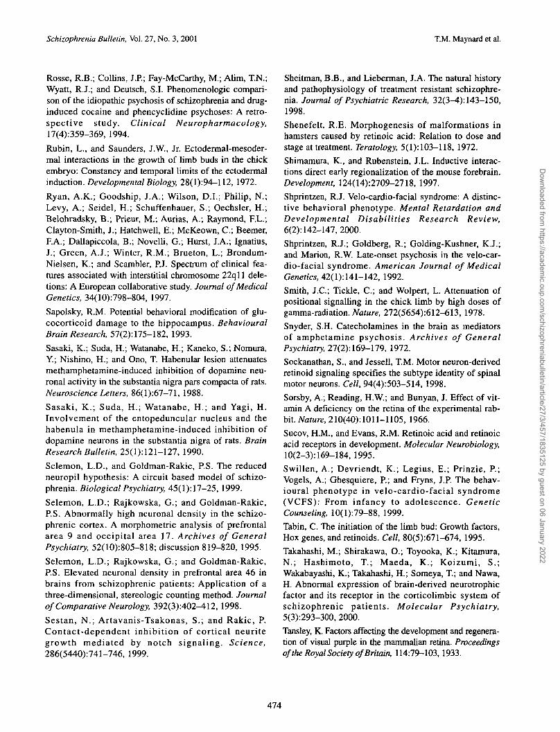
Schizophrenia Bulletin, Vol. 27, No. 3, 2001 T.M. Maynard et al.
Rosse, R.B.; Collins, J.P.; Fay-McCarthy, M.; Alim, T.N.;Wyatt, R.J.; and Deutsch, S.I. Phenomenologic compari-son of the idiopathic psychosis of schizophrenia and drug-induced cocaine and phencyclidine psychoses: A retro-spective study. Clinical Neuropharmacology,17(4):359-369, 1994.
Rubin, L., and Saunders, J.W., Jr. Ectodermal-mesoder-mal interactions in the growth of limb buds in the chickembryo: Constancy and temporal limits of the ectodermalinduction. Developmental Biology, 28(1):94—112, 1972.
Ryan, A.K.; Goodship, J.A.; Wilson, D.I.; Philip, N.;Levy, A.; Seidel, H.; Schuffenhauer, S.; Oechsler, H.;Belohradsky, B.; Prieur, M.; Aurias, A.; Raymond, F.L.;Clayton-Smith, J.; Hatchwell, E.; McKeown, C ; Beemer,F.A.; Dallapiccola, B.; Novelli, G.; Hurst, J.A.; Ignatius,J.; Green, A.J.; Winter, R.M.; Brueton, L.; Brondum-Nielsen, K.; and Scambler, P.J. Spectrum of clinical fea-tures associated with interstitial chromosome 22qll dele-tions: A European collaborative study. Journal of MedicalGenetics, 34(10):798-804, 1997.
Sapolsky, R.M. Potential behavioral modification of glu-cocorticoid damage to the hippocampus. BehaviouralBrain Research, 57(2):175-182, 1993.
Sasaki, K.; Suda, H.; Watanabe, H.; Kaneko, S.; Nomura,Y.; Nishino, H.; and Ono, T. Habenular lesion attenuatesmethamphetamine-induced inhibition of dopamine neu-ronal activity in the substantia nigra pars compacta of rats.Neuroscience Letters, 86(1):67-71, 1988.
Sasaki, K.; Suda, H.; Watanabe, H.; and Yagi, H.Involvement of the entopeduncular nucleus and thehabenula in methamphetamine-induced inhibition ofdopamine neurons in the substantia nigra of rats. BrainResearch Bulletin, 25(1):121-127, 1990.
Selemon, L.D., and Goldman-Rakic, P.S. The reducedneuropil hypothesis: A circuit based model of schizo-phrenia. Biological Psychiatry, 45(1): 17-25, 1999.
Selemon, L.D.; Rajkowska, G.; and Goldman-Rakic,P.S. Abnormally high neuronal density in the schizo-phrenic cortex. A morphometric analysis of prefrontalarea 9 and occipital area 17. Archives of GeneralPsychiatry, 52(1O):8O5-818; discussion 819-820, 1995.
Selemon, L.D.; Rajkowska, G.; and Goldman-Rakic,P.S. Elevated neuronal density in prefrontal area 46 inbrains from schizophrenic patients: Application of athree-dimensional, stereologic counting method. Journalof Comparative Neurology, 392(3):402-412, 1998.
Sestan, N.; Artavanis-Tsakonas, S.; and Rakic, P.Contact-dependent inhibition of cortical neuritegrowth mediated by notch signaling. Science,286(5440):741-746, 1999.
Sheitman, B.B., and Lieberman, J.A. The natural historyand pathophysiology of treatment resistant schizophre-nia. Journal of Psychiatric Research, 32(3-4): 143-150,1998.
Shenefelt, R.E. Morphogenesis of malformations inhamsters caused by retinoic acid: Relation to dose andstage at treatment. Teratology, 5(1): 103-118, 1972.
Shimamura, K., and Rubenstein, J.L. Inductive interac-tions direct early regionalization of the mouse forebrain.Development, 124(14):2709-2718, 1997.
Shprintzen, R.J. Velo-cardio-facial syndrome: A distinc-tive behavioral phenotype. Mental Retardation andDevelopmental Disabilities Research Review,6(2): 142-147, 2000.
Shprintzen, R.J.; Goldberg, R.; Golding-Kushner, K.J.;and Marion, R.W. Late-onset psychosis in the velo-car-dio-facial syndrome. American Journal of MedicalGenetics, 42(1):141-142, 1992.
Smith, i.C; Tickle, C ; and Wolpert, L. Attenuation ofpositional signalling in the chick limb by high doses ofgamma-radiation. Nature, 272(5654):612-613, 1978.
Snyder, S.H. Catecholamines in the brain as mediatorsof amphetamine psychosis. Archives of GeneralPsychiatry, 27(2): 169-179, 1972.
Sockanathan, S., and Jessell, T.M. Motor neuron-derivedretinoid signaling specifies the subtype identity of spinalmotor neurons. Cell, 94(4):503-514, 1998.
Sorsby, A.; Reading, H.W.; and Bunyan, J. Effect of vit-amin A deficiency on the retina of the experimental rab-bit. Nature, 210(40):1011-1105, 1966.
Sucov, H.M., and Evans, R.M. Retinoic acid and retinoicacid receptors in development. Molecular Neurobiology,10(2-3): 169-184, 1995.
Swillen, A.; Devriendt, K.; Legius, E.; Prinzie, P.;Vogels, A.; Ghesquiere, P.; and Fryns, J.P. The behav-ioural phenotype in velo-cardio-facial syndrome(VCFS): From infancy to adolescence. GeneticCounseling, 10(l):79-88, 1999.
Tabin, C. The initiation of the limb bud: Growth factors,Hox genes, and retinoids. Cell, 8O(5):671-674, 1995.
Takahashi, M.; Shirakawa, O.; Toyooka, K.; Kitamura,N.; Hashimoto, T.; Maeda, K.; Koizumi, S.;Wakabayashi, K.; Takahashi, H.; Someya, T.; and Nawa,H. Abnormal expression of brain-derived neurotrophicfactor and its receptor in the corticolimbic system ofschizophrenic patients. Molecular Psychiatry,5(3):293-300, 2000.
Tansley, K. Factors affecting the development and regenera-tion of visual purple in the mammalian retina. Proceedingsof the Royal Society ofBritain, 114:79-103,1933.
474
Dow
nloaded from https://academ
ic.oup.com/schizophreniabulletin/article/27/3/457/1835125 by guest on 06 January 2022

'Two-Hit" Hypothesis of Schizophrenia Schizophrenia Bulletin, Vol. 27, No. 3, 2001
Thompson, P.M.; Sower, A.C.; and Perrone-Bizzozero,N.I. Altered levels of the synaptosomal associated pro-tein SNAP-25 in schizophrenia. Biological Psychiatry,43(4):239-243, 1998.
Thornton, E.W., and Bradbury, G.E. Effort and stressinfluence the effect of lesion of the habenula complexin one-way active avoidance learning. PhysiologicalBehavior, 45(5):929-935, 1989.
Thornton, E.W.; Bradbury, G.E.; and Davies, C.Increased immobility in an automated forced swim-ming test following lesion of the habenula in rats:Absence of evidence for a contribution from motorimpairment. Behavioral Neuroscience, 104(l):37-43,1990.
Tickle, C; Summerbell, D.; and Wolpert, L. Positionalsignalling and specification of digits in chick limbmorphogenesis. Nature, 254(5497): 199-202, 1975.Torrey, E.F.; Miller, J.; Rawlings, R.; and Yolken, R.H.Seasonality of births in schizophrenia and bipolar dis-order: A review of the literature. SchizophreniaResearch, 28(1): 1-38, 1997.
Trimarchi, J.M.; Fairchild, B.; Verona, R.; Moberg, K.;Andon, N.; and Lees, J.A. E2F-6, a member of theE2F family that can behave as a transcriptional repres-sor. Proceedings of the National Academy of Sciencesof the United States of America, 95(6):2850-2855,1998.
Turetsky, B.I.; Moberg, P.J.; Yousem, D.M.; Doty,R.L.; Arnold, S.E.; and Gur, R.E. Reduced olfactorybulb volume in patients with schizophrenia. AmericanJournal of Psychiatry, 157(5):828-830, 2000.
Uyttendaele, H.; Marazzi, G.; Wu, G.; Yan, Q.;Sassoon, D.; and Kitajewski, J. Notch4/int-3, a mam-mary proto-oncogene, is an endothelial cell-specificmammalian Notch gene. Development,122(7):2251-2259, 1996.
Vale-Martinez, A.; Marti-Nicolovius, M.; Guillazo-Blanch, G.; Coll-Andreu, M.; and Morgado-Bernal, I.Effects of habenular lesions upon two-way activeavoidance conditioning in rats. Neurobiology ofLearning and Memory, 68(l):68-74, 1997.
Vawter, M.P.; Cannon-Spoor, H.E.; Hemperly, J.J.;Hyde, T.M.; VanderPutten, D.M.; Kleinman, J.E.; andFreed, W.J. Abnormal expression of cell recognitionmolecules in schizophrenia. Experimental Neurology,149(2):424-432, 1998.
Vincent, M.C.; Heitz, F.; Tricoire, J.; Bourrouillou, G.;Kuhlein, E.; Rolland, M.; and Calvas, P. 22qll dele-tion in DGS/VCFS monozygotic twins with discordantphenotypes. Genetic Counseling, 10(l):43^9, 1999.
Vincent, S.R.; Staines, W.A.; McGeer, E.G.; andFibiger, H.C. Transmitters contained in the efferents ofthe habenula. Brain Research, 195(2):479-484, 1980.Wakamatsu, Y.; Maynard, T.M.; Jones, S.U.; andWeston, J.A. NUMB localizes in the basal cortex ofmitotic avian neuroepithelial cells and modulates neu-ronal differentiation by binding to NOTCH-1. Neuron,23(1):71-81, 1999.Wald, G. The molecular basis of visual excitation.Nature, 219(156): 800-807, 1968.
Waldrop, M.F.; Bell, R.Q.; McLaughlin, B.; andHalverson, C.F., Jr. Newborn minor physical anom-alies predict short attention span, peer aggression, andimpulsivity at age 3. Science, 199(4328):563-565,1978.
Waldrop, M.F.; Pedersen, F.A.; and Bell, R.Q. Minorphysical anomalies and behavior in preschool children.Child Development, 39(2):391-400, 1968.Walker, E.F.; Lewine, R.R.; and Neumann, C.Childhood behavioral characteristics and adult brainmorphology in schizophrenia. Schizophrenia Research,22(2):93-101, 1996.Wang, R.Y., and Aghajanian, G.K. Physiological evi-dence for habenula as major link between forebrainand midbrain raphe. Science, 197(4298):89-91, 1977.Wei, J., and Hemmings, G.P. The NOTCH4 locus isassociated with susceptibility to schizophrenia. NatureGenetics, 25(4):376-377, 2000.Weinberger, D.R. Implications of normal brain devel-opment for the pathogenesis of schizophrenia.Archives of General Psychiatry, 44(7):660-669, 1987.Weinberger, D.R. From neuropathology to neurodevel-opment. Lancet, 346(8974):552-557, 1995.
Weinmaster, G. The ins and outs of notch signaling.Molecular and Cellular Neurosciences, 9(2):91-102,1997.Weiss, S.; Dunne, C; Hewson, J.; Wohl, C; Wheatley,M.; Peterson, A.C.; and Reynolds, B.A. MultipotentCNS stem cells are present in the adult mammalianspinal cord and ventricular neuroaxis. Journal ofNeuroscience, 16(23):7599-7609, 1996.Wright, I.C.; Rabe-Hesketh, S.; Woodruff, P.W.;David, A.S.; Murray, R.M.; and Bullmore, E.T. Meta-analysis of regional brain volumes in schizophrenia.American Journal of Psychiatry, 157(l):16-25, 2000.Yamamoto, M.; Drager, U.C.; Ong, D.E.; and McCaffery,P. Retinoid-binding proteins in the cerebellum andchoroid plexus and their relationship to regionalizedretinoic acid synthesis and degradation. European Journalof Biochemistry, 257(2):344-350, 1998.
475
Dow
nloaded from https://academ
ic.oup.com/schizophreniabulletin/article/27/3/457/1835125 by guest on 06 January 2022

Schizophrenia Bulletin, Vol. 27, No. 3, 2001 T.M. Maynard et al.
Yamamoto, M.; McCaffery, P.; and Drager, U.C.Influence of the choroid plexus on cerebellar develop-ment: Analysis of retinoic acid synthesis.Developmental Brain Research, 93(1-2): 182-190,1996.
Yang, N.; Schule, R.; Mangelsdorf, D.J.; and Evans,R.M. Characterization of DNA binding and retinoicacid binding properties of retinoic acid receptor.Proceedings of the National Academy of Sciences ofthe United States of America, 88(9):3559-3563,1991.
Yolken, R.H., and Torrey, E.F. Viruses, schizophrenia, andbipolar disorder. Clinical Microbiology Review,8(1):131-145, 1995.
Young, B.K.; Camicioli, R.; and Ganzini, L.Neuropsychiatric adverse effects of antiparkinsoniandrugs: Characteristics, evaluation and treatment. Drugsand Aging, 10(5): 367-383, 1997.
Zetterstrom, R.H.; Simon, A.; Giacobini, M.M.; Eriksson,U.; and Olson, L. Localization of cellular retinoid-bindingproteins suggests specific roles for retinoids in the adultcentral nervous system. Neuroscience, 62(3):899-918,1994.
Zimmerman, H.M. Lesions of the nervous system in vita-min deficiency: I. Rats on a diet low in vitaminA. Journal of Experimental Medicine, 57:215-228, 1933.
Acknowledgments
The authors wish to thank Ron Shatzmiller for his insight-ful comments and studies of the retiniod indicator trans-genes. In addition, we would like to thank AndrewGassman, Gloria Haskell, Naina Bhasin, and SamanthaGizerian for comments and suggestions about the manu-script. We would also like to thank Kenichi Tagoku forproviding preliminary expression data for the mouse22qll homologues. This work was supported by theNational Alliance for Research on Schizophrenia andDepression (NARSAD) and National Institutes of Healthgrants HD-29178 to A.S.L. and MH-33127 to J.A.L.
The Authors
Thomas M. Maynard, Ph.D., is a postdoctoral fellow inthe Department of Cell and Molecular Physiology in theSchool of Medicine of the University of North Carolina(UNC) in Chapel Hill, NC. Anthony-Samuel LaMantia,Ph.D., is an Associate Professor of Cell and MolecularPhysiology and a member of the UNC NeuroscienceCenter. Linmarie Sikich, M.D., is an Assistant Professor,and Jeffrey A. Lieberman, M.D., is a Professor and theVice Chairman for Research in the Department ofPsychiatry of the School of Medicine at the University ofNorth Carolina in Chapel Hill, NC.
476
Dow
nloaded from https://academ
ic.oup.com/schizophreniabulletin/article/27/3/457/1835125 by guest on 06 January 2022