neural oscillatory dynamics of inhibitory control in young ... · of their alcoholism-related...
TRANSCRIPT
Contents lists available at ScienceDirect
Biological Psychology
journal homepage: www.elsevier.com/locate/biopsycho
Neural oscillatory dynamics of inhibitory control in young adult bingedrinkersLee A. Holcomba, Siyuan Huanga, Stephen M. Cruzb, Ksenija Marinkovica,c,⁎
a Department of Psychology, San Diego State University, 5500 Campanile Dr, San Diego, CA, 92182, USAbDepartment of Biology, San Diego State University, 5500 Campanile Dr, San Diego, CA, 92182, USAc Department of Radiology, University of California, 9500 Gilman Dr, La Jolla, CA, 92093, USA
A R T I C L E I N F O
Keywords:Binge drinkingAlcoholEEGThetaBetaOscillationsGo/NoGoInhibitory controlResponse inhibition
A B S T R A C T
Alcohol consumption is often characterized by heavy episodic, or binge drinking, which has been on the rise. Theaim of this study was to examine the neural dynamics of inhibitory control in demographically matched groupsof young, healthy adults (N= 61) who reported engaging in binge (BD) or light drinking patterns (LD).Electroencephalography signal was recorded during a fast-paced visual Go/NoGo paradigm probing the abilityto inhibit prepotent responses. No group differences were found in task performance. BDs showed attenuatedevent-related theta (4–7 Hz) on inhibition trials compared to LDs, which correlated with binge episodes andalcohol consumption but not with measures of mood or disposition including impulsivity. A greater overalldecrease of early beta power (15–25 Hz) in BDs may indicate deficient preparatory “inhibitory brake” beforedeliberate responding. The results are consistent with deficits in the inhibitory control circuitry and are sug-gestive of allostatic neuroadaptive changes associated with binge drinking.
1. Introduction
Binge drinking is defined as alcohol consumption elevating theblood alcohol concentration (BAC) levels to at least 0.08 g/dL, whichusually occurs when four/five drinks are consumed by women or men,respectively, within two hours (National Institute on Alcohol Abuse &Alcoholism, 2017). However, many individuals exceed this level ofintake and consume alcohol at much higher levels (Naimi, Nelson, &Brewer, 2010; Terry-McElrath & Patrick, 2016). Binge pattern of ex-cessive drinking is associated with a range of negative consequencesand incurs high costs to society (Bouchery, Harwood, Sacks, Simon, &Brewer, 2011; Sacks, Gonzales, Bouchery, Tomedi, & Brewer, 2015). Itrepresents a major public health concern given rising prevalence rates(Hingson, Zha, & White, 2017), and the evidence that harmful con-sequences increase with hazardous drinking levels (Haber, Harris-Olenak, Burroughs, & Jacob, 2016).
It has been proposed that binge drinking may be a precursor to al-cohol use disorder (AUD) as alcohol consumption transitions from im-pulsivity to compulsivity (Kimbrough, Kim, Cole, Brennan, & George,2017; Koob & Le Moal, 2008a; Koob, 2013). Indeed, increased alcoholconsumption is associated with impaired self-control which couldcontribute to excessive drinking and may predict future heavy drinkingand alcohol dependence (Nigg et al., 2006; Paz, Rosselli, & Conniff,
2018). Behavioral disinhibition is considered to be an important di-mension in the development of AUD (Goldstein & Volkow, 2011; Koob& Volkow, 2010; Kwako, Momenan, Litten, Koob, & Goldman, 2016;Volkow, Fowler, Wang, & Goldstein, 2002) and prefrontally-mediateddeficits of inhibitory control and other executive functions have beenfound in individuals with AUD (Oscar-Berman and Marinkovic, 2007,Le Berre, Fama, & Sullivan, 2017; Oscar-Berman & Marinkovic, 2004;Sullivan & Pfefferbaum, 2005). Furthermore, neuroimaging evidenceindicates that acute alcohol intoxication primarily affects the prefrontalneurofunctional system subserving top-down cognitive control(Anderson et al., 2011; Kovacevic et al., 2012; Marinkovic,Rickenbacher, Azma, & Artsy, 2012; Marinkovic, Rickenbacher, Azma,Artsy, & Lee, 2013; Rosen, Padovan, & Marinkovic, 2016), includingimpairments of response inhibition (Gan et al., 2014; Kareken et al.,2013; Marinkovic, Halgren, Klopp, & Maltzman, 2000; Nikolaou,Critchley, & Duka, 2013; Schuckit et al., 2012).
Inhibitory control relies on the ability to suppress inappropriate orunwanted actions (Aron, Robbins, & Poldrack, 2014; Chikazoe, Konishi,Asari, Jimura, & Miyashita, 2007), but it also critically engages othercognitive functions including attentional control and working memory(Erika-Florence, Leech, & Hampshire, 2014; Hampshire, 2015). It hasbeen studied extensively with tasks that demand stopping or with-holding dominant responses, such as a Go/NoGo task (Aron et al., 2014;
https://doi.org/10.1016/j.biopsycho.2019.107732Received 25 January 2019; Received in revised form 17 July 2019; Accepted 18 July 2019
⁎ Corresponding author at: Department of Psychology, 5500 Campanile Dr., San Diego State University, San Diego, CA, 92182, USA.E-mail address: [email protected] (K. Marinkovic).
Biological Psychology 146 (2019) 107732
Available online 22 July 20190301-0511/ © 2019 Elsevier B.V. All rights reserved.
T
Simmonds, Pekar, & Mostofsky, 2008). This paradigm instructs parti-cipants to rapidly respond to target or “Go” stimuli (response activa-tion), and to withhold responding to occasional “NoGo” stimuli (re-sponse inhibition) (Garavan, Ross, & Stein, 1999). Functional magneticresonance imaging (fMRI) studies have indicated that successful per-formance on the Go/NoGo task primarily recruits prefrontal regions,including the ventral and lateral prefrontal cortices, the anterior cin-gulate cortex (ACC), the presupplementary motor area (preSMA), andthe basal ganglia among others (Aron et al., 2014; Criaud & Boulinguez,2013; Hampshire, Chamberlain, Monti, Duncan, & Owen, 2010; Levy &Wagner, 2011; Simmonds et al., 2008; Swick, Ashley, & Turken, 2011;Wiecki & Frank, 2013). Although fMRI is an excellent spatial mappingtool, its temporal resolution is low due to constraints imposed byneurovascular coupling (Buxton, 2002). In contrast, scalp electro-encephalography (EEG) measures neural activity directly and can pro-vide highly precise insight into the task-evoked neural activity in realtime but its spatial resolution is limited due to biophysical properties ofthe signal (Nunez & Srinivasan, 2006).
Because of its oscillatory nature, the EEG signal can be analyzedwithin the relevant frequency bands during task engagement (Amzica &Lopes da Silva, 2011; Basar, Basar-Eroglu, Karakas, & Schurmann,2001; Engel & Fries, 2010; Lundqvist, Herman, & Miller, 2018;Pfurtscheller & Lopes da Silva, 1999). Event-related theta oscillations(4–7 Hz) are sensitive to cognitive effort elicited by tasks probingcognitive control and performance monitoring (Brier et al., 2010;Cavanagh & Frank, 2014; Hanslmayr et al., 2008; Kovacevic et al.,2012; Rosen et al., 2016). Studies using source-localization of themagnetoencephalography (MEG) and EEG signal have shown that theACC and preSMA in the medial prefrontal cortex are major generatorsof event-related theta oscillations during such tasks (Hanslmayr et al.,2008; Kovacevic et al., 2012; Marinkovic, Rosen, Cox, & Kovacevic,2012; Marinkovic, Beaton, Rosen, Happer, & Wagner, 2019). Theseobservations have been confirmed with intracranial EEG recordingswhich have revealed that the ACC is a principal generator of the fronto-midline theta observed on the scalp (Cohen, Ridderinkhof, Haupt,Elger, & Fell, 2008; Wang, Ulbert, Schomer, Marinkovic, & Halgren,2005). Additional sources have been reported in the lateral prefrontalcortex (Beaton, Azma, & Marinkovic, 2018; Correas et al., 2018;Kovacevic et al., 2012; Marinkovic et al., 2019; Raghavachari et al.,2001; Rosen et al., 2016).
Studies manipulating acute alcohol intoxication have shown thatevent-related theta oscillations are attenuated by a moderate alcoholdose especially under high-conflict conditions during decision making(Beaton et al., 2018; Kovacevic et al., 2012; Marinkovic, Rosen et al.,2012, 2019; Rosen et al., 2016). Based on their association with AUD ingenetic linkage studies, theta oscillations have been suggested as anendophenotype indicating a predisposition to develop alcoholism orinhibitory-related disorders (Rangaswamy et al., 2007; Salvatore,Gottesman, & Dick, 2015). However, the supporting evidence of thetainvolvement in inhibitory control in binge drinkers or individuals withAUD is scarce. Most of the extant studies have used an equiprobable(50:50) Go/NoGo design which biases responding strategy towardstarget detection at the expense of inhibitory control (Wessel, 2018).Since the Go (target) stimuli are much more salient for task perfor-mance, they evoke greater prefrontal theta when measured with MEG,which is attenuated in BDs (Correas et al., 2018). EEG studies withequiprobable Go/NoGo tasks in young BDs have reported reduced thetapower in BDs to both target and nontarget stimuli (Lopez-Caneda et al.,2017). Similarly, in two EEG studies using this design, abstaining in-dividuals with chronic AUD, and offspring of individuals with AUD whowere deemed to be at high risk for developing alcoholism, showedlower event-related theta power than control participants (Kamarajanet al., 2004, 2006; Pandey et al., 2016). However, studies using theta toexamine inhibitory control in binge drinkers or in individuals with AUDwith an asymmetric Go/NoGo design are lacking. To address this gap inthe literature, the present study employed a fast-pace task variant with
a 80:20 Go/NoGo ratio which establishes Go response dominance andengages inhibitory control on NoGo trials (Wessel, 2018). This has al-lowed us to test the hypothesis that binge drinking is associated withimpaired inhibitory control and to examine whether this is reflected inattenuated task-dependent theta oscillations.
Furthermore, because this task requires countermanding of a pre-potent tendency to respond, we examined task-dependent beta oscil-lations (15–25 Hz) which provide temporally precise insight into an-ticipatory motor engagement, response preparation, inhibition, andexecution. Beta oscillations are considered to be the preferred fre-quency of the sensorimotor system and can serve as an index of thefunctional engagement of the underlying cortico-subcortical circuitry(Baker, 2007; Jenkinson & Brown, 2011; Khanna & Carmena, 2017;Kilavik, Zaepffel, Brovelli, MacKay, & Riehle, 2013). They are parti-cularly sensitive to the neural activity related to movement activationand inhibition (Engel & Fries, 2010; Jenkinson & Brown, 2011) and areaffected by alcohol intoxication (Marinkovic et al., 2000), but they havenot been examined in the context of binge drinking. Unlike event-re-lated theta power which increases in response to a salient stimulus, betapower is high at baseline and it Bdecreases during anticipatory, actual,or even imagined engagement of the motor system. Following a po-tential brief beta increase that may be inhibitory in nature (Pogosyan,Gaynor, Eusebio, & Brown, 2009; Swann et al., 2009), beta decrease(also termed “desynchronization”) is the principal characteristic ofevent-related beta power. It is easily observed during movement pre-paration as it presumably indicates readiness to execute a motor re-sponse (Baker, 2007; Engel & Fries, 2010; Jenkinson & Brown, 2011;Kilavik et al., 2013). The beta decrease is most dominant over thesensorimotor cortices which are the primary generators of the observedbeta changes (Beaton et al., 2018). After a command to execute or in-hibit a movement has been issued but before the actual response, betapower rebounds and increases above baseline levels (Cheyne,Bakhtazad, & Gaetz, 2006; Kilavik et al., 2013). The beta rebound hasshorter latency on NoGo trials on which there is no actual response,which can be interpreted as an active inhibition process (Khanna &Carmena, 2017; Solis-Escalante, Muller-Putz, Pfurtscheller, & Neuper,2012). These features make event-related beta oscillations well suitedfor tracking response preparation and execution stages, as well as post-movement adjustments of the motor system in real time (Beaton et al.,2018; Jenkinson & Brown, 2011). As the Go/NoGo task probes in-hibitory control with potential relevance to self-control dysregulationwhich is implicated in addiction (Baler & Volkow, 2006; Leeman,Patock-Peckham, & Potenza, 2012), investigating beta oscillatory ac-tivity in binge drinkers is of particular interest.
The aim of the current study was to examine the neural dynamics ofinhibitory control in young adults with and without histories of bingedrinking. Using a visual Go/NoGo task, the present study focused ontask-dependent event-related changes in theta (4–7 Hz) and beta(15–25 Hz) oscillations in order examine the neural indices of cognitiveand motor aspects of inhibitory control respectively in young adultsengaging in binge drinking. We hypothesized that individuals with ahistory of binge drinking would exhibit impaired inhibitory controlmanifested in suboptimal task performance, decreased event-relatedtheta power on NoGo trials, and alterations in the pattern of beta ac-tivity during response preparation.
2. Methods
2.1. Participants
Sixty-one healthy, non-smoking, right-handed individuals(M ± SD=23.41 ± 3.4 years of age, 31 females) participated in thisstudy. They were recruited from the local community through approvedads and postings and were queried about their alcohol and drug use andhealth history in a brief telephone screen interview. None of the par-ticipants reported drug or tobacco use at least one month prior to the
L.A. Holcomb, et al. Biological Psychology 146 (2019) 107732
2
study. They had no history of brain injury, or other neuropsychiatric ormedical problems, and none were taking medications at the time of thestudy. In the present study, a binge episode was defined as consumingsix/five or more drinks for men/women within a two hour time span.This criterion was adopted based on empirical evidence indicating thatthis level of drinking is more likely to result in BAC reaching or ex-ceeding 0.08% (Lange & Voas, 2001). Based on screening information,29 individuals were classified as binge drinkers (BD) if they reportedthree or more binge episodes in the past six months with at least oneepisode in the last month. On average, BDs reported 14.09 ± 13.6binge episodes in the past six months (Table 1), with median=10,mode= 20, range=3 to 72. The high end of the range was reported bya participant who weighed the most in the entire cohort. The nexthighest number of reported binge episodes in the past six months was30. The remaining 32 participants were Low Drinkers (LD), who re-ported no more than one binge episode in the past six months, .09 ± .3on average. The two groups were matched on age, sex, education,ethnicity/race, and family history of alcoholism. The study’s procedureswere approved by the Institutional Review Board and written informedconsent was obtained from each participant prior to the experiment.Participants were monetarily compensated for their participation. Thedata of four additional participants in the theta analysis and three fromthe beta analysis were discarded due to poor data quality.
2.2. Procedure
Participants completed a battery of questionnaires which includedhandedness (Oldfield, 1971) and medical history. They were askedabout their alcohol drinking habits, including the frequency, quantity,and the pattern of alcohol consumption (modified from Cahalan, Cisin,& Crossley, 1969), the magnitude of response to alcohol (Self-Rating ofthe Effects of Alcohol, SRE, Schuckit, Smith, & Tipp, 1997), the severityof their alcoholism-related symptoms (Short Michigan AlcoholismScreening Test, SMAST, Selzer, Vinokur, & Van Rooijen, 1975), theirmotives for engaging in alcohol use (Drinking Motive Questionnaire,DMQ, Kuntsche & Kuntsche, 2009), and the consequences of theirdrinking (Young Adult Alcohol Consequences Questionnaire, YAACQ,Read, Kahler, Strong, & Colder, 2006). They provided a detailed reporton their daily drinking during the past month (Timeline Followback,TLFB, Sobell & Sobell, 1996). Their disinhibition and impulsivity traitswere assessed by an abbreviated Impulsiveness Scale (AbbreviatedImpulsiveness Scale, ABIS, Coutlee, Politzer, Hoyle, & Huettel, 2014).Participants also completed questionnaires to measure their personality(Eysenck Personality Questionnaire, EPQ, Eysenck & Eysenck, 1975),depression (Patient Health Questionnaire-9, PHQ, Kroenke & Spitzer,2002), and anxiety (Generalized Anxiety Disorder, GAD, Spitzer,Kroenke, Williams, & Löwe, 2006). In addition, they completed the NIHToolbox Cognitive Battery (Gershon et al., 2013) which included testsprobing working memory, cognitive flexibility, processing speed, andepisodic memory (Table 1). Participants were screened for drug usewith a 12-panel urine multidrug test (Discover, American ScreeningCorporation) at the beginning of the recording session. They all testednegative and proceeded with the recording.
2.3. Experimental paradigm
Participants took part in a visual Go/NoGo task which probes theability to inhibit prepotent responses. They were presented with apseudorandomized series of ‘X’ and ‘Y’ letters and were instructed topress a button with their right index finger as quickly and as accuratelyas possible every time 'X' and 'Y' stimuli alternated (Go, 80% of trials)and to withhold responding when the stimuli repeated (NoGo, 20% oftrials) (Garavan et al., 1999). The task comprised a total of 685 stimulipresented for 230ms with a stimulus onset asynchrony (SOA) of1400 ± 200ms. A random jitter was added to each trial in 50ms in-crements to mitigate timing predictability. Stimuli were presented in-dividually in white font on a black background with the Presentationsoftware package (Version 18.1; www.neurobs.com) within a visualangle spanning .93° (horizontal) and 0.99° (vertical). At all other timesa fixation dot was presented in the middle of the screen.
2.4. Data acquisition and analysis
EEG signal was recorded with a 64-channel Brain Vision system(Brain Products GmbH, Germany) and was sampled continuously at500 Hz. The signal was referenced online to the nose, and a bipolarlyreferred vertical electro-oculogram (EOG) was recorded to monitoreyeblinks and eye movements. Electrode impedance was kept below 5kΩ.
2.4.1. Data preprocessingEEG data were analyzed using MATLAB (Mathworks, Natick, MA)
routines that incorporated publicly available algorithms includingFieldTrip (Oostenveld, Fries, Maris, & Schoffelen, 2011), and EEGLAB(Delorme & Makeig, 2004). Continuous data were band-pass filtered at0.1–100 Hz, and were segmented into epochs extending from -300 to800ms relative to each stimulus onset. A 300ms pad was added to thebeginning and end of the epoch to account for edge artifacts resultingfrom the Morlet wavelet convolution (Oostenveld et al., 2011). Noisychannels were removed by visual inspection and trials with large
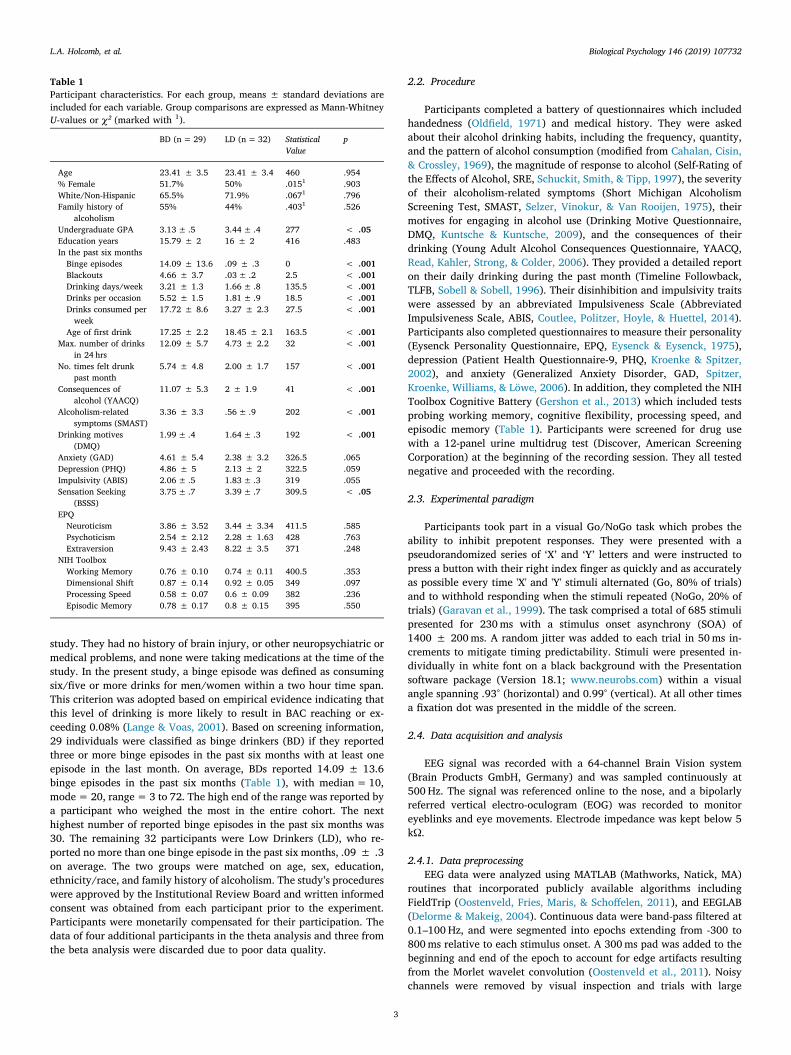
Table 1Participant characteristics. For each group, means ± standard deviations areincluded for each variable. Group comparisons are expressed as Mann-WhitneyU-values or χ² (marked with 1).
BD (n= 29) LD (n=32) StatisticalValue
p
Age 23.41 ± 3.5 23.41 ± 3.4 460 .954% Female 51.7% 50% .0151 .903White/Non-Hispanic 65.5% 71.9% .0671 .796Family history of
alcoholism55% 44% .4031 .526
Undergraduate GPA 3.13± .5 3.44± .4 277 < .05Education years 15.79 ± 2 16 ± 2 416 .483In the past six monthsBinge episodes 14.09 ± 13.6 .09 ± .3 0 < .001Blackouts 4.66 ± 3.7 .03± .2 2.5 < .001Drinking days/week 3.21 ± 1.3 1.66± .8 135.5 < .001Drinks per occasion 5.52 ± 1.5 1.81± .9 18.5 < .001Drinks consumed perweek
17.72 ± 8.6 3.27 ± 2.3 27.5 < .001
Age of first drink 17.25 ± 2.2 18.45 ± 2.1 163.5 < .001Max. number of drinks
in 24 hrs12.09 ± 5.7 4.73 ± 2.2 32 < .001
No. times felt drunkpast month
5.74 ± 4.8 2.00 ± 1.7 157 < .001
Consequences ofalcohol (YAACQ)
11.07 ± 5.3 2 ± 1.9 41 < .001
Alcoholism-relatedsymptoms (SMAST)
3.36 ± 3.3 .56± .9 202 < .001
Drinking motives(DMQ)
1.99± .4 1.64± .3 192 < .001
Anxiety (GAD) 4.61 ± 5.4 2.38 ± 3.2 326.5 .065Depression (PHQ) 4.86 ± 5 2.13 ± 2 322.5 .059Impulsivity (ABIS) 2.06± .5 1.83± .3 319 .055Sensation Seeking
(BSSS)3.75± .7 3.39± .7 309.5 < .05
EPQNeuroticism 3.86 ± 3.52 3.44 ± 3.34 411.5 .585Psychoticism 2.54 ± 2.12 2.28 ± 1.63 428 .763Extraversion 9.43 ± 2.43 8.22 ± 3.5 371 .248
NIH ToolboxWorking Memory 0.76 ± 0.10 0.74 ± 0.11 400.5 .353Dimensional Shift 0.87 ± 0.14 0.92 ± 0.05 349 .097Processing Speed 0.58 ± 0.07 0.6 ± 0.09 382 .236Episodic Memory 0.78 ± 0.17 0.8 ± 0.15 395 .550
L.A. Holcomb, et al. Biological Psychology 146 (2019) 107732
3

artifacts were removed with a threshold-based rejection. The defaultthreshold started at 100μV focusing on the posterior electrodes, but wasadjusted for each participant as needed with the goal of rejecting largeartifacts while keeping most trials with eyeblinks. This helped to opti-mize an independent component analysis (ICA) method (Delorme &Makeig, 2004) which was then used to detect and remove the eyeblinkand heartbeat artifacts. Data were analyzed in the time-frequency do-main by computing complex power spectrum of each trial with Morletwavelets within the theta (4–7 Hz) and beta (15–25Hz) bands (Beatonet al., 2018; Kovacevic et al., 2012). The wavelet results were ad-ditionally inspected for artifacts and the padding was removed. Theanalysis was conducted in a manner blind to group membership.Average event-related power is presented as percent signal change fromthe baseline (-300 to 0ms). Analysis of the raw power in the baselineindicated that the two groups did not differ in either theta or betabands, assuring that the observed group differences were indeed due toevent-related changes in power.
2.4.2. Data analysisData were analyzed for each channel which were then grouped into
frontal (Fz, F1, F2, F3, F4), central (Cz, C1, C2, C3, C4) and parietal (Pz,P1, P2, P3, P4) clusters and averaged within each cluster to analyzegroup and condition effects on theta power (see Fig. 2). For beta, onlythe central (Cz, C1, C2, C3, C4) electrode region was used (see Fig. 3) tocapture activity of the sensorimotor cortices which are the primarygenerators of event-related changes in beta oscillations (Baker, 2007;Beaton et al., 2018). Only trials on which responses were correctlyexecuted (Go) and withheld (NoGo) were included in the analysis. Byincorporating Go and NoGo trials in a 4:1 ratio, this task creates aprepotency to respond. As a consequence, effortful response inhibitionis needed to overcome it and withhold responses on NoGo trials. Thisresponse dominance also leads to occasional premature button pressing.All responses made between -250ms before and 200ms after the sti-mulus onset were counted as premature and were excluded from theanalysis.
2.4.3. Statistical analysisGroup differences in demographics were tested with χ², and those in
drinking habits, personality aspects, and cognitive functions wereanalyzed with a non-parametric Mann-Whitney U test to account forpossible violations of distribution normality (Table 1). Data were ana-lyzed with a mixed-design ANCOVA with Group as the between-sub-jects factor, Task Condition as the repeated measures factor, and im-pulsivity (Abbreviated Impulsiveness Scale, ABIS, Coutlee et al., 2014)as a covariate. Group differences between the frontal, central, andparietal clusters were additionally examined for theta. No effects of sexwere observed in the initial analyses for either the behavioral or elec-trophysiological data so this factor was subsequently removed from theanalysis. Associations between the principal EEG measures, re-presentative drinking variables, and several dispositional indices were
examined with a non-parametric Spearman’s Rho (rs) index that wascalculated across the whole sample. The following EEG measures wereincluded in the correlational analysis: theta NoGo, theta Go, and earlybeta averaged across both task conditions. Drinking variables com-prised the number of binge episodes, maximum number of drinks in24 h, average daily alcohol intake, and number of drinking days perweek, all assessed over the past 6 months. Mood and personality vari-ables included anxiety, depression, and impulsivity. A false discoveryrate approach (.20) (Hochberg & Benjamini, 1990) was used to correctfor multiple correlations.
3. Results
3.1. Behavioral measures
3.1.1. PerformanceAs shown in Fig. 1, participants responded more accurately to Go
trials (98.3%±3.6) than to NoGo trials (79.1%±12.2) resulting in amain effect of condition, F(1, 59)= 136.61, p < .001. No group dif-ferences were observed for response accuracy on either Go, F(1,59)= .39, p = .54, or NoGo trials, F(1, 59)= .06, p = .81. The LD(447.1ms ± 88.2) and BD (441.5ms ± 77.9) groups responded withcomparable speed, F(1, 59)= .07, p = .79.
3.1.2. Drinking habits, personality characteristics, and cognitive functionsTable 1. lists demographic characteristics and group differences in
drinking habits, experiences and motivational dimensions, personalitytraits, dispositional mood measures, and cognitive functions. BDs re-ported more binge episodes in the previous six months than LDs, higherlevels of alcohol consumption overall, they started drinking at an earlierage than LDs and experienced more negative consequences of drinkingincluding blackouts. They expressed higher levels of social, coping, andenhancement drinking motives. BDs reported higher sensation seeking,and marginally higher levels of impulsivity, anxiety, and depressionthan LDs. However, the two groups did not differ on personality traitsnor on cognitive tests.
3.2. Electrophysiological measures
3.2.1. Event-related theta powerEvent-related theta power peaked at ˜350 ms after stimulus
onset so the effects of Group and Condition were analyzed within atime interval of 300–400 ms (Fig. 2) to capture peak event-relatedchanges while controlling for impulsivity. Overall, there was a maineffect of Condition, as NoGo trials elicited greater event-relatedtheta power than Go trials, F(1, 56) = 7.9, p = .007. A Group xCondition interaction, F(1, 56) = 5.7, p = .02 was due to thetaattenuation for NoGo trials in the BD group, F(1, 56) = 8.27, p =.006, with group differences on Go trials not reaching significance,F(1, 56) = 1.70, p = .19. Region-specific analysis indicated that,compared to LDs, BDs had reduced theta power on NoGo trials atthe frontal, F(1, 56) = 5.2, p = .03, central, F(1, 56) = 9.75,p= .003, and parietal, F(1, 56) = 5.8, p= .02, electrode regions.In contrast, group differences on Go trials did not reach significancefor any electrode cluster including the frontal, F(1, 56) = .41, p =.52, central, F(1, 56) = 3.18, p= .08, and parietal, F(1, 56) = 1.87,p= .18 regions. Lower theta during response inhibition was asso-ciated with higher levels of drinking, as NoGo theta power corre-lated negatively with the number of reported binge episodes, rs =−.29, p= .03, daily alcohol intake, rs = −.26, p= .04, and theaverage number of weekly drinking days, rs = −.25, p = .05. Themaximum number of drinks consumed in 24 h in the previous sixmonths correlated with theta power on NoGo, rs = −.29, p = .04,and Go trials, rs = −.29, p = .04. None of the dispositional vari-ables were related to theta, all coefficients < .07, all p-values > .6.
Fig. 1. Accuracy and reaction times (means ± standard errors) are shown forthe low drinking (LD) and binge drinking (BD) groups and for the Go and NoGoconditions.
L.A. Holcomb, et al. Biological Psychology 146 (2019) 107732
4

3.2.2. Event-related beta powerEvent-related beta power is also expressed as percent signal change
from baseline (Fig. 3). It starts decreasing prior to stimulus onset inanticipation of making motor movement over the sensorimotor cortices.An early, transient increase in beta power during preparatory stage isvisible in LDs, followed by an overall beta decrease with a nadir at˜300ms and a rebound of beta power subsequent to issuing a motorcommand. A main effect of Group was observed within 50–125ms timewindow, as BDs had greater beta desynchronization than LDs, F(1,56)= 8.08, p= .006 (Fig. 3). Following the early transient increase inbeta power, the LD group maintained an overall higher level of betapower. This was reflected in a main effect of Group as measured at thebeta nadir (250–350ms), F(1, 56)= 5.06, p = .028 which, however,correlated with the early time interval, rs= .51, p< .001. As expected,beta power rebounded earlier on inhibitory NoGo trials, which wasconfirmed by a main effect of Condition (500–600ms), F(1,56)= 10.33, p = .002. No group differences were observed during thebeta rebound, F(1, 56)= 0.67, p = .42.
4. Discussion
The present study examined the neural dynamics of inhibitorycontrol in young adults as a function of their drinking patterns. In theabsence of differences in task performance, BD and LD groups differedon the neural indices of the engagement of cognitive control and thecircuitry subserving response preparation. Event-related theta oscilla-tions (4–7 Hz) were attenuated in BDs compared to LDs on trials re-quiring response inhibition as shown by the Group x Condition inter-action, which may indicate less efficient long-range top-downintegration engaged by the salient response suppression requirement.Decreased theta power on NoGo trials was associated with increasedlevels of binge and high-intensity drinking, and alcohol consumptionslevels but not dispositional or mood measures. An early, transient in-crease of event-related beta power (15–25 Hz) was observed in LDswhich is consistent with a brief “braking pause” during response pre-paration which may underlie deliberate decision to response or with-hold responding and which immediately precedes issuance of the motorexecution or inhibition commands. In contrast, BDs showed only a betadecrease which may be indicative of a deficient engagement of responseinhibition mechanisms. Even though the correlations between the earlybeta power and drinking variables did not survive correction for mul-tiple correlations, the lower levels of inhibition during the motor pre-paratory stage may be suggestive of allostatic neuroadaptive changes inneural transmission as a result of heavy episodic drinking patterns.Group differences in both theta and beta frequency bands were sig-nificant after controlling for self-reported impulsivity.
A Go/NoGo task with 80% Go trials, such as the one used in thecurrent study, probes inhibitory control by creating a prepotency torespond (Aron et al., 2014; Garavan et al., 1999; Wessel, 2018), asparticipants are required to withhold responding on a minority of trials.Because theta oscillations are associated with engagement of top-downcognitive control functions (Cavanagh & Frank, 2014; Kovacevic et al.,2012; Marinkovic et al., 2019; Rosen et al., 2016; Yamanaka &Yamamoto, 2010), they are well suited to examine the cognitive pro-cesses associated with behavioral control. In the current study, NoGotrials elicited much greater event-related theta power than Go trials, as
Fig. 2. Frontal, central, and parietal electrode clusters and group average time courses for event-related theta power averaged within each cluster. Overall, event-related theta power was greater on NoGo trials, it peaked at ˜350ms, and it was most prominent in the central region. BD participants had reduced NoGo theta powerin all three regions compared to LDs. *p < .05, **p< .01.
Fig. 3. Event-related beta power time course averaged over the central elec-trode cluster. LD participants show an early, transient increase in beta power(50–125ms). * p< .05, ** p < .01. Average Go reaction time is marked byarrow.
L.A. Holcomb, et al. Biological Psychology 146 (2019) 107732
5

would be expected based on their salience and inhibitory demands, inaddition to their low presentation frequency, and task relevance. Asignificant Group x Condition interaction indicated that BDs exhibitedattenuated theta activity selectively on NoGo trials (Fig. 2), suggestingthat binge drinking may be primarily associated with impaired pro-cesses that underlie inhibitory control. This novel finding is broadlyconsistent with previous reports of the selective vulnerability of the top-down circuitry underlying inhibitory control to alcohol intoxication(Anderson et al., 2011; Gan et al., 2014; Kareken et al., 2013; Kovacevicet al., 2012; Marinkovic, Rickenbacher et al., 2012, 2013; Marinkovicet al., 2019; Nikolaou et al., 2013; Rosen et al., 2016; Schuckit et al.,2012). Though less directly pertinent to inhibitory control per se, lowertheta has been reported in studies employing equiprobable Go/NoGotasks in large groups of individuals with AUD (Kamarajan et al., 2004;Pandey et al., 2016) and in young adult binge drinkers (Correas et al.,2018; Lopez-Caneda et al., 2017).
Functional imaging studies have reported decreased activity onNoGo or Stop-signal trials in BDs which correlated with measures ofalcohol intake (Ahmadi et al., 2013; Hu, Zhang, Chao, Krystal, & Li,2016) and impulsivity (Ahmadi et al., 2013). It has been proposed thatprotracted heavy alcohol intake is accompanied by incremental de-gradation of cognitive and motivational functions and that the resultingdisinhibition, as reflected in impaired self-control, plays a major role inaddiction Crews, Vetreno, Broadwater, & Robinson, 2016; Field,Schoenmakers, & Wiers, 2008; Goldstein & Volkow, 2002; Goldstein &Volkow, 2011; Koob & Volkow, 2010; Kwako et al., 2016; Volkow et al.,2002). Systematic reviews have confirmed deficient activity in the in-hibitory control network across a range of addictions (Luijten et al.,2014). This is broadly consistent with our findings of negative corre-lations between NoGo theta power and a range of drinking variablesincluding the number of self-reported binge episodes and weeklydrinking levels.
Previous studies have shown that impulsivity and other ex-ternalizing traits can predict future alcohol use (Finn, 2000; Littlefield,Stevens, & Sher, 2014; Regier et al., 1990; Verdejo-Garcia, Lawrence, &Clark, 2008). Indeed, dysregulation of impulse control concerns theinability to resist engaging in the activity that one declares to be un-wanted or even harmful. The inability to maintain inhibitory controlover drinking has been considered by some researchers to be funda-mental to drug abuse (Field, Wiers, Christiansen, Fillmore, & Verster,2010; Fillmore, 2003; Finn, 2000; Jentsch & Taylor, 1999; Sher & Trull,1994). Evidence suggests that the vulnerability to alcoholism shares acommon genetic component with externalizing traits which may pre-dispose individuals to a spectrum disorders including AUD (Begleiter &Porjesz, 1999; Dick et al., 2004; Heinz, Beck, Meyer-Lindenberg,Sterzer, & Heinz, 2011; Pihl, Peterson, & Lau, 1993; Schuckit, Smith, &Kalmijn, 2004). The current findings suggest that impulsivity as mea-sured with ABIS (Coutlee et al., 2014) did not drive the observed groupeffects on event-related theta power on inhibitory (NoGo) trials whichprevail when controlling for self-reported impulsivity, leading us tobelieve that these two systems have separate mediators. However,group differences indicating greater readiness to respond on the part ofBDs emerged from the analysis of beta oscillations.
In an effort to investigate the neural characteristics of the ability tosuppress a prepotent tendency to respond, we have analyzed event-re-lated beta oscillations which are sensitive to motor preparation. Betaoscillations are thought to reflect functional interactions between theneocortex and the basal ganglia as beta power typically decreases in alateralized and anticipatory manner during movement preparation andexecution (Baker, 2007; Jenkinson & Brown, 2011; Kilavik et al., 2013)with a maximal nadir over the sensorimotor cortex (Beaton et al., 2018;Litvak et al., 2011). In the current study, LDs had an early, transientincrease in the overall beta power at ˜100ms in contrast to BDs whoshowed only beta desynchronization. Beta increase is associated withmotor inhibition (Khanna & Carmena, 2017; Pogosyan et al., 2009;Swann et al., 2009) so this brief rise is suggestive of a momentary,
transient “inhibitory pause” prior to issuing the final motor commandto execute the response. It has been well established that motor in-hibition is subserved by the indirect pathway comprising cortical ex-citation of the striatum which inhibits the subthalamic-pallidal outputto the thalamus and the cortex resulting in motor hypoactivity (Haynes& Haber, 2013; Lanciego, Luquin, & Obeso, 2012; Zavala, Zaghloul, &Brown, 2015). Short latency of this transient beta increase is consistentwith engagement of the cortico-subthalamic hyperdirect pathwaywhich underlies rapid response suppression (Frank, 2006; Nambu,Tokuno, & Takada, 2002; Wessel & Aron, 2017). This finding suggeststhat in LDs, the motor response sequence incorporates a brief inhibitorystage that may facilitate a deliberate decision to respond or to withholdresponding possibly via lateral competition of alternative activations(Tunstall, Oorschot, Kean, & Wickens, 2002). In contrast, BDs did notexhibit this early beta increase which is consistent with their greaterreadiness to respond. Given that BDs regularly imbibe alcohol at higherlevels and have more high-intensity drinking episodes than BDs, it ispossible that the observed dysregulation of the early motor preparationphase reflects neural hyperexcitability. Indeed, we have reported find-ings on other neural indices indicating decreased inhibitory signalingduring wakeful rest in binge drinkers (Affan et al., 2018). These ob-servations are consistent with allostatic neuroadaptive changes (Kooband Le Moal, 2008b, Clapp, Bhave, & Hoffman, 2008; Koob & Le Moal,2005) whereby hazardous drinking results in downregulation of in-hibitory and upregulation of excitatory signaling (Finn & Crabbe, 1997;Most, Ferguson, & Harris, 2014; Roberto & Varodayan, 2017;Vengeliene, Bilbao, Molander, & Spanagel, 2008). With the majority ofintrinsic and efferent fibers being GABAergic (Lanciego et al., 2012),the basal ganglia are particularly vulnerable to the effects of binge-likedrinking which has been reported in animal models (Cuzon Carlsonet al., 2011; Wilcox et al., 2014) and human postmortem studies(Laukkanen et al., 2013).
In the current study the BD and LD groups did not differ in taskperformance despite clear group differences in both event-related thetaand beta bands. This finding is consistent with many other EEG studiesreporting group differences on neural measures in the absence of be-havioral deficits (Lopez-Caneda et al., 2012; Crego et al., 2009, 2010;Crego et al., 2012; Lopez-Caneda et al., 2013, 2017; Maurage, Pesenti,Philippot, Joassin, & Campanella, 2009; Petit et al., 2012). This di-vergence between the behavioral and direct measures of neural activityis indicative of greater EEG sensitivity to neural deficits associated withthe intermittent pattern of high-level drinking. Because binge drinkinghas been conceptualized as a transitional stage in a cyclic process po-tentially leading towards compulsive intake (Kimbrough et al., 2017;Koob & Le Moal, 2008a; Koob, 2013), EEG measures could potentiallyserve as biomarkers signifying transition to dependence.
Despite the notable novel findings of this study, there are alsolimitations that should be mentioned. The study employed a relativelysmall sample size which precluded a well-powered investigation ofpossible sex differences in inhibitory control. Though novel and unique,the findings of an early beta decrease in BDs that potentially signifydeficient response inhibition should be replicated in a larger cohort ofbinge drinkers, as well as individuals with AUD.
In conclusion, the present study used EEG and a visual Go/NoGotask to examine the neural dynamics of inhibitory control in BDs in aneffort to address existing gaps in the literature. Compared to LDs, BDsshowed reduced event-related theta power on NoGo trials, suggestingthat binge drinking is associated with deficits in the top-down circuitrysubserving inhibitory control. A unique and novel finding was an earlyreduction in event-related beta power in BDs, which may indicate adeficient preparatory “inhibitory brake” in these individuals which maybe suggestive of allostatic neuroadaptive changes associated with bingedrinking. The present study has contributed novel insights into the al-terations of cognitive and motor aspects of inhibitory control in bingedrinkers in the absence of performance deficits. Because binge drinkinghas been proposed as a transitional phase leading to chronic alcoholism,
L.A. Holcomb, et al. Biological Psychology 146 (2019) 107732
6
the present findings may inform future studies on heavy alcohol use.The alterations in brain signals could potentially serve as diagnosticindicators of a transition to dependence. When paired with alcohol-related cues, Go/NoGo paradigms can enhance neurofeedback-basedpreventive strategies focusing on inhibitory control for those at risk ofdeveloping alcoholism.
Declaration of Competing Interest
None.
Acknowledgements
This work was supported by start-up funds from the College ofSciences at San Diego State University and the National Institute onAlcohol Abuse and Alcoholism (R01-AA016624). The authors aregrateful to Rifqi Affan, Audrey Andrews, Nicole Fong, and MorganSlauter for assistance with data acquisition, and to Lauren Beaton,Laura Wagner, Joe Happer, and Martina Knezevic for assistance withdata analysis and manuscript preparation.
References
Affan, R. O., Huang, S., Cruz, S. M., Holcomb, L. A., Nguyen, E., & Marinkovic, K. (2018).High-intensity binge drinking is associated with alterations in spontaneous neuraloscillations in young adults. Alcohol, 70, 51–60.
Ahmadi, A., Pearlson, G. D., Meda, S. A., Dager, A., Potenza, M. N., Rosen, R., ... Tennen,H. (2013). Influence of alcohol use on neural response to go/no-go task in collegedrinkers. Neuropsychopharmacology, 38, 2197–2208.
Amzica, F., & Lopes da Silva, F. H. (2011). Cellular substrates of brain rhythms. In D.Schomer, & F. H. Lopes da Silva (Eds.). Niedermeyer’s electroencephalography: Basicprinciples, clinical applications, and related fields (pp. 33–63). Philadelphia: LippincottWilliams & Wilkins.
Anderson, B. M., Stevens, M. C., Meda, S. A., Jordan, K., Calhoun, V. D., & Pearlson, G. D.(2011). Functional imaging of cognitive control during acute alcohol intoxication.Alcoholism, Clinical and Experimental Research, 35, 156–165.
Aron, A. R., Robbins, T. W., & Poldrack, R. A. (2014). Inhibition and the right inferiorfrontal cortex: One decade on. Trends in Cognitive Sciences, 18, 177–185.
Baker, S. N. (2007). Oscillatory interactions between sensorimotor cortex and the per-iphery. Current Opinion in Neurobiology, 17, 649–655.
Baler, R. D., & Volkow, N. D. (2006). Drug addiction: The neurobiology of disrupted self-control. Trends in Molecular Medicine, 12, 559–566.
Basar, E., Basar-Eroglu, C., Karakas, S., & Schurmann, M. (2001). Gamma, alpha, delta,and theta oscillations govern cognitive processes. International Journal ofPsychophysiology: Official Journal of the International Organization of Psychophysiology,39, 241–248.
Beaton, L. E., Azma, S., & Marinkovic, K. (2018). When the brain changes its mind:Oscillatory dynamics of conflict processing and response switching in a flanker taskduring alcohol challenge. PloS One, 13, e0191200.
Begleiter, H., & Porjesz, B. (1999). What is inherited in the predisposition toward alco-holism? A proposed model. Alcoholism: Clinical and Experimental Research, 23,1125–1135.
Bouchery, E. E., Harwood, H. J., Sacks, J. J., Simon, C. J., & Brewer, R. D. (2011).Economic costs of excessive alcohol consumption in the U.S., 2006. American Journalof Preventive Medicine, 41, 516–524.
Brier, M. R., Ferree, T. C., Maguire, M. J., Moore, P., Spence, J., Tillman, G. D., ... Kraut,M. A. (2010). Frontal theta and alpha power and coherence changes are modulatedby semantic complexity in Go/NoGo tasks. International Journal of Psychophysiology:Official Journal of the International Organization of Psychophysiology, 78, 215–224.
Buxton, R. B. (2002). Introduction to functional magnetic resonance imaging. New York, NY:Cambridge University Press.
Cahalan, D., Cisin, I. H., & Crossley, H. M. (1969). American drinking practices: A nationalstudy of drinking behavior and attitudes. Monograph #6. New Brunswick, NJ: RutgersCenter of Alcohol Studies.
Cavanagh, J. F., & Frank, M. J. (2014). Frontal theta as a mechanism for cognitive control.Trends in Cognitive Sciences, 18, 414–421.
Cheyne, D., Bakhtazad, L., & Gaetz, W. (2006). Spatiotemporal mapping of cortical ac-tivity accompanying voluntary movements using an event-related beamforming ap-proach. Human Brain Mapping, 27, 213–229.
Chikazoe, J., Konishi, S., Asari, T., Jimura, K., & Miyashita, Y. (2007). Activation of rightinferior frontal gyrus during response inhibition across response modalities. Journalof Cognitive Neuroscience, 19, 69–80.
Clapp, P., Bhave, S. V., & Hoffman, P. L. (2008). How adaptation of the brain to alcoholleads to dependence: A pharmacological perspective. Alcohol Research & Health: theJournal of the National Institute on Alcohol Abuse and Alcoholism, 31, 310–339.
Cohen, M. X., Ridderinkhof, K. R., Haupt, S., Elger, C. E., & Fell, J. (2008). Medial frontalcortex and response conflict: Evidence from human intracranial EEG and medialfrontal cortex lesion. Brain Research, 1238, 127–142.
Correas, A., Lopez-Caneda, E., Beaton, L., Rodriguez Holguin, S., Garcia-Moreno, L. M.,
Anton-Toro, L. F., ... Marinkovic, K. (2018). Decreased event-related theta power andphase-synchrony in young binge drinkers during target detection: An anatomically-constrained MEG approach. Journal of Psychopharmacology (Oxford, England)269881118805498.
Coutlee, C. G., Politzer, C. S., Hoyle, R. H., & Huettel, S. A. (2014). An abbreviated im-pulsiveness scale constructed through confirmatory factor analysis of the BarrattImpulsiveness Scale version 11. Archives of Scientific Psychology, 2, 1–12.
Crego, A., Cadaveira, F., Parada, M., Corral, M., Caamano-Isorna, F., & RodriguezHolguin, S. (2012). Increased amplitude of P3 event-related potential in young bingedrinkers. Alcohol, 46, 415–425.
Crego, A., Holguin, S. R., Parada, M., Mota, N., Corral, M., & Cadaveira, F. (2009). Bingedrinking affects attentional and visual working memory processing in young uni-versity students. Alcoholism, Clinical and Experimental Research, 33, 1870–1879.
Crego, A., Rodriguez-Holguin, S., Parada, M., Mota, N., Corral, M., & Cadaveira, F.(2010). Reduced anterior prefrontal cortex activation in young binge drinkers duringa visual working memory task. Drug and Alcohol Dependence, 109, 45–56.
Crews, F. T., Vetreno, R. P., Broadwater, M. A., & Robinson, D. L. (2016). Adolescentalcohol exposure persistently impacts adult neurobiology and behavior.Pharmacological Reviews, 68, 1074–1109.
Criaud, M., & Boulinguez, P. (2013). Have we been asking the right questions when as-sessing response inhibition in go/no-go tasks with fMRI? A meta-analysis and criticalreview. Neuroscience and Biobehavioral Reviews, 37, 11–23.
Cuzon Carlson, V. C., Seabold, G. K., Helms, C. M., Garg, N., Odagiri, M., Rau, A. R., ...Grant, K. A. (2011). Synaptic and morphological neuroadaptations in the putamenassociated with long-term, relapsing alcohol drinking in primates.Neuropsychopharmacology, 36, 2513–2528.
Delorme, A., & Makeig, S. (2004). EEGLAB: An open source toolbox for analysis of single-trial EEG dynamics. Journal of Neuroscience Methods, 134, 9–21.
Dick, D. M., Li, T. K., Edenberg, H. J., Hesselbrock, V., Kramer, J., Kuperman, S., ...Foroud, T. (2004). A genome-wide screen for genes influencing conduct disorder.Molecular Psychiatry, 9, 81–86.
Engel, A. K., & Fries, P. (2010). Beta-band oscillations–signalling the status quo? CurrentOpinion in Neurobiology, 20, 156–165.
Erika-Florence, M., Leech, R., & Hampshire, A. (2014). A functional network perspectiveon response inhibition and attentional control. Nature Communications, 5, 4073.
Eysenck, H. J., & Eysenck, S. B. G. (1975). Manual of the eysenck personality questionnaire.London: Hodder & Staughton.
Field, M., Schoenmakers, T., & Wiers, R. W. (2008). Cognitive processes in alcohol binges:A review and research agenda. Current Drug Abuse Reviews, 1, 263–279.
Field, M., Wiers, R. W., Christiansen, P., Fillmore, M. T., & Verster, J. C. (2010). Acutealcohol effects on inhibitory control and implicit cognition: Implications for loss ofcontrol over drinking. Alcoholism, Clinical and Experimental Research, 34, 1346–1352.
Fillmore, M. T. (2003). Drug abuse as a problem of impaired control: Current approachesand findings. Behavioral and Cognitive Neuroscience Reviews, 2, 179–197.
Finn, D. A., & Crabbe, J. C. (1997). Exploring alcohol withdrawal syndrome. AlcoholHealth and Research World, 21, 149–156.
Finn, P. (2000). Acute effects of alcohol on cognition and impulsive-disinhibited beha-vior. In A. Noronha, M. Eckardt, & K. Warren (Vol. Eds.), Review of NIAAA’s neu-roscience and behavioral research portfolio: vol. 34, (pp. 337–356). Bethesda, MD: USDepartment of health and human services.
Frank, M. J. (2006). Hold your horses: A dynamic computational role for the subthalamicnucleus in decision making. Neural Networks: the Official Journal of the InternationalNeural Network Society, 19, 1120–1136.
Gan, G., Guevara, A., Marxen, M., Neumann, M., Junger, E., Kobiella, A., ... Smolka, M. N.(2014). Alcohol-induced impairment of inhibitory control is linked to attenuatedbrain responses in right fronto-temporal cortex. Biological Psychiatry, 76, 698–707.
Garavan, H., Ross, T. J., & Stein, E. A. (1999). Right hemispheric dominance of inhibitorycontrol: An event-related functional MRI study. Proceedings of the National Academy ofSciences of the United States of America, 96, 8301–8306.
Gershon, R. C., Wagster, M. V., Hendrie, H. C., Fox, N. A., Cook, K. F., & Nowinski, C. J.(2013). NIH toolbox for assessment of neurological and behavioral function.Neurology, 80, S2–6.
Goldstein, R. Z., & Volkow, N. D. (2002). Drug addiction and its underlying neurobio-logical basis: Neuroimaging evidence for the involvement of the frontal cortex. TheAmerican Journal of Psychiatry, 159, 1642–1652.
Goldstein, R. Z., & Volkow, N. D. (2011). Dysfunction of the prefrontal cortex in addic-tion: Neuroimaging findings and clinical implications. Nature Reviews Neuroscience,12, 652–669.
Haber, J. R., Harris-Olenak, B., Burroughs, T., & Jacob, T. (2016). Residual effects: Youngadult diagnostic drinking predicts late-life health outcomes. Journal of Studies onAlcohol and Drugs, 77, 859–867.
Hampshire, A. (2015). Putting the brakes on inhibitory models of frontal lobe function.NeuroImage, 113, 340–355.
Hampshire, A., Chamberlain, S. R., Monti, M. M., Duncan, J., & Owen, A. M. (2010). Therole of the right inferior frontal gyrus: Inhibition and attentional control. Neuroimage,50, 1313–1319.
Hanslmayr, S., Pastotter, B., Bauml, K. H., Gruber, S., Wimber, M., & Klimesch, W. (2008).The electrophysiological dynamics of interference during the Stroop task. Journal ofCognitive Neuroscience, 20, 215–225.
Haynes, W. I., & Haber, S. N. (2013). The organization of prefrontal-subthalamic inputs inprimates provides an anatomical substrate for both functional specificity and in-tegration: Implications for Basal Ganglia models and deep brain stimulation. TheJournal of Neuroscience: the Official Journal of the Society for Neuroscience, 33,4804–4814.
Heinz, A. J., Beck, A., Meyer-Lindenberg, A., Sterzer, P., & Heinz, A. (2011). Cognitiveand neurobiological mechanisms of alcohol-related aggression. Nature Reviews
L.A. Holcomb, et al. Biological Psychology 146 (2019) 107732
7

Neuroscience, 12, 400–413.Hingson, R. W., Zha, W., & White, A. M. (2017). Drinking Beyond the Binge Threshold:
Predictors, Consequences, and Changes in the U.S. American Journal of PreventiveMedicine, 52, 717–727.
Hochberg, Y., & Benjamini, Y. (1990). More powerful procedures for multiple significancetesting. Statistics in Medicine, 9, 811–818.
Hu, S., Zhang, S., Chao, H. H., Krystal, J. H., & Li, C. S. R. (2016). Association of drinkingproblems and duration of alcohol use to inhibitory control in nondependent youngadult social drinkers. Alcoholism, Clinical and Experimental Research, 40, 319–328.
Jenkinson, N., & Brown, P. (2011). New insights into the relationship between dopamine,beta oscillations and motor function. Trends in Neurosciences, 34, 611–618.
Jentsch, J. D., & Taylor, J. R. (1999). Impulsivity resulting from frontostriatal dysfunctionin drug abuse: Implications for the control of behavior by reward-related stimuli.Psychopharmacology (Berlin), 146, 373–390.
Kamarajan, C., Porjesz, B., Jones, K., Chorlian, D., Padmanabhapillai, A., Rangaswamy,M., ... Begleiter, H. (2006). Event-related oscillations in offspring of alcoholics:Neurocognitive disinhibition as a risk for alcoholism. Biological Psychiatry, 59,625–634.
Kamarajan, C., Porjesz, B., Jones, K. A., Choi, K., Chorlian, D. B., Padmanabhapillai, A., ...Begleiter, H. (2004). The role of brain oscillations as functional correlates of cogni-tive systems: A study of frontal inhibitory control in alcoholism. International Journalof Psychophysiology: Official Journal of the International Organization ofPsychophysiology, 51, 155–180.
Kareken, D. A., Dzemidzic, M., Wetherill, L., Eiler, W., 2nd, Oberlin, B. G., Harezlak, J., ...O’Connor, S. J. (2013). Family history of alcoholism interacts with alcohol to affectbrain regions involved in behavioral inhibition. Psychopharmacology (Berlin), 228,335–345.
Khanna, P., & Carmena, J. M. (2017). Beta band oscillations in motor cortex reflect neuralpopulation signals that delay movement onset. Elife, 6.
Kilavik, B. E., Zaepffel, M., Brovelli, A., MacKay, W. A., & Riehle, A. (2013). The ups anddowns of beta oscillations in sensorimotor cortex. Experimental Neurology, 245,15–26.
Kimbrough, A., Kim, S., Cole, M., Brennan, M., & George, O. (2017). Intermittent access toethanol drinking facilitates the transition to excessive drinking after chronic inter-mittent ethanol vapor exposure. Alcoholism, Clinical and Experimental Research, 41,1502–1509.
Koob, G. F. (2013). Theoretical frameworks and mechanistic aspects of alcohol addiction:Alcohol addiction as a reward deficit disorder. Current Topics in BehavioralNeurosciences, 13, 3–30.
Koob, G. F., & Le Moal, M. (2005). Plasticity of reward neurocircuitry and the’ dark side’of drug addiction. Nature Neuroscience, 8, 1442–1444.
Koob, G. F., & Le Moal, M. (2008a). Addiction and the brain antireward system. AnnualReview of Psychology, 59, 29–53.
Koob, G. F., & Le Moal, M. (2008b). Review. Neurobiological mechanisms for opponentmotivational processes in addiction. Philosophical Transactions of the Royal Society ofLondon Series B, Biological Sciences, 363, 3113–3123.
Koob, G. F., & Volkow, N. D. (2010). Neurocircuitry of addiction.Neuropsychopharmacology, 35, 217–238.
Kovacevic, S., Azma, S., Irimia, A., Sherfey, J., Halgren, E., & Marinkovic, K. (2012).Theta oscillations are sensitive to both early and late conflict processing stages:Effects of alcohol intoxication. PloS One, 7, e43957.
Kroenke, K., & Spitzer, R. L. (2002). The PHQ-9: A new depression diagnostic and severitymeasure. Psychiatric Annals, 32, 509–515.
Kuntsche, E., & Kuntsche, S. (2009). Development and validation of the drinking motivequestionnaire revised short form (DMQ-R SF). Journal of Clinical Child and AdolescentPsychology: the Official Journal for the Society of Clinical Child and AdolescentPsychology, American Psychological Association, Division 53, 38, 899–908.
Kwako, L. E., Momenan, R., Litten, R. Z., Koob, G. F., & Goldman, D. (2016). Addictionsneuroclinical assessment: A neuroscience-based framework for addictive disorders.Biological Psychiatry, 80, 179–189.
Lanciego, J. L., Luquin, N., & Obeso, J. A. (2012). Functional neuroanatomy of the basalganglia. Cold Spring Harbor Perspectives in Medicine, 2, a009621.
Lange, J. E., & Voas, R. B. (2001). Defining binge drinking quantities through resultingblood alcohol concentrations. Psychology of Addictive Behaviors: Journal of the Societyof Psychologists in Addictive Behaviors, 15, 310–316.
Laukkanen, V., Storvik, M., Hakkinen, M., Akamine, Y., Tupala, E., Virkkunen, M., ...Tiihonen, J. (2013). Decreased GABA(A) benzodiazepine binding site densities inpostmortem brains of Cloninger type 1 and 2 alcoholics. Alcohol, 47, 103–108.
Le Berre, A. P., Fama, R., & Sullivan, E. V. (2017). Executive functions, memory, andsocial cognitive deficits and recovery in chronic alcoholism: A critical review to in-form future research. Alcoholism, Clinical and Experimental Research, 41, 1432–1443.
Leeman, R. F., Patock-Peckham, J. A., & Potenza, M. N. (2012). Impaired control overalcohol use: An under-addressed risk factor for problem drinking in young adults?Experimental and Clinical Psychopharmacology, 20, 92–106.
Levy, B. J., & Wagner, A. D. (2011). Cognitive control and right ventrolateral prefrontalcortex: Reflexive reorienting, motor inhibition, and action updating. Annals of theNew York Academy of Sciences, 1224, 40–62.
Littlefield, A. K., Stevens, A. K., & Sher, K. J. (2014). Impulsivity and alcohol involvement:Multiple, distinct constructs and processes. Current Addiction Reports, 1, 33–40.
Litvak, V., Jha, A., Eusebio, A., Oostenveld, R., Foltynie, T., Limousin, P., ... Brown, P.(2011). Resting oscillatory cortico-subthalamic connectivity in patients withParkinson’s disease. Brain, 134, 359–374.
Lopez-Caneda, E., Cadaveira, F., Crego, A., Doallo, S., Corral, M., Gomez-Suarez, A., ...Rodriguez Holguin, S. (2013). Effects of a persistent binge drinking pattern of alcoholconsumption in young people: A follow-up study using event-related potentials.Alcohol and Alcoholism (Oxford, Oxfordshire), 48, 464–471.
Lopez-Caneda, E., Cadaveira, F., Crego, A., Gomez-Suarez, A., Corral, M., Parada, M., ...Rodriguez Holguin, S. (2012). Hyperactivation of right inferior frontal cortex inyoung binge drinkers during response inhibition: A follow-up study. Addiction, 107,1796–1808.
Lopez-Caneda, E., Rodriguez Holguin, S., Correas, A., Carbia, C., Gonzalez-Villar, A.,Maestu, F., ... Cadaveira, F. (2017). Binge drinking affects brain oscillations linked tomotor inhibition and execution. Journal of Psychopharmacology (Oxford, England), 31,873–882.
Luijten, M., Machielsen, M. W., Veltman, D. J., Hester, R., de Haan, L., & Franken, I. H.(2014). Systematic review of ERP and fMRI studies investigating inhibitory controland error processing in people with substance dependence and behavioural addic-tions. Journal of Psychiatry & Neuroscience: JPN, 39, 149–169.
Lundqvist, M., Herman, P., & Miller, E. K. (2018). Working memory: Delay activity, yes!Persistent activity? Maybe not. The Journal of Neuroscience: the Official Journal of theSociety for Neuroscience, 38, 7013–7019.
Marinkovic, K., Beaton, L. E., Rosen, B. Q., Happer, J. P., & Wagner, L. C. (2019).Disruption of frontal lobe neural synchrony during cognitive control by alcohol in-toxication. Journal of Visualized Experiments: JoVE.
Marinkovic, K., Halgren, E., Klopp, J., & Maltzman, I. (2000). Alcohol effects on move-ment-related potentials: a measure of impulsivity? Journal of Studies on Alcohol, 61,24–31.
Marinkovic, K., Rickenbacher, E., Azma, S., & Artsy, E. (2012). Acute alcohol intoxicationimpairs top-down regulation of Stroop incongruity as revealed by blood oxygen level-dependent functional magnetic resonance imaging. Human Brain Mapping, 33,319–333.
Marinkovic, K., Rickenbacher, E., Azma, S., Artsy, E., & Lee, A. K. (2013). Effects of acutealcohol intoxication on saccadic conflict and error processing. Psychopharmacology(Berlin), 230, 487–497.
Marinkovic, K., Rosen, B. Q., Cox, B., & Kovacevic, S. (2012). Event-related theta powerduring lexical-semantic retrieval and decision conflict is modulated by alcohol in-toxication: Anatomically-constrained MEG. Frontiers in Psychology, 3.
Maurage, P., Pesenti, M., Philippot, P., Joassin, F., & Campanella, S. (2009). Latent de-leterious effects of binge drinking over a short period of time revealed only byelectrophysiological measures. Journal of Psychiatry & Neuroscience: JPN, 34,111–118.
Most, D., Ferguson, L., & Harris, R. A. (2014). Molecular basis of alcoholism. Handbook ofClinical Neurology, 125, 89–111.
Naimi, T. S., Nelson, D. E., & Brewer, R. D. (2010). The intensity of binge alcohol con-sumption among U.S. Adults. American Journal of Preventive Medicine, 38, 201–207.
Nambu, A., Tokuno, H., & Takada, M. (2002). Functional significance of the cortico-subthalamo-pallidal’ hyperdirect’ pathway. Neurosciences Research, 43, 111–117.
National Institute on Alcohol Abuse and Alcoholism (2017). Drinking levels defined.Nigg, J. T., Wong, M. M., Martel, M. M., Jester, J. M., Puttler, L. I., Glass, J. M., ... Zucker,
R. A. (2006). Poor response inhibition as a predictor of problem drinking and illicitdrug use in adolescents at risk for alcoholism and other substance use disorders.Journal of the American Academy of Child and Adolescent Psychiatry, 45, 468–475.
Nikolaou, K., Critchley, H., & Duka, T. (2013). Alcohol affects neuronal substrates ofresponse inhibition but not of perceptual processing of stimuli signalling a stop re-sponse. PloS One, 8, e76649.
Nunez, P. L., & Srinivasan, R. (2006). Electric fields of the brain: The neurophysics of EEG.New York: Oxford University Press.
Oldfield, R. C. (1971). The assessment and analysis of handedness: The Edinburgh in-ventory. Neuropsychologia, 9, 97–113.
Oostenveld, R., Fries, P., Maris, E., & Schoffelen, J. M. (2011). FieldTrip: Open sourcesoftware for advanced analysis of MEG, EEG, and invasive electrophysiological data.Computational Intelligence and Neuroscience156869 2011.
Oscar-Berman, M., & Marinkovic, K. (2004). Alcoholism and the brain: An overview.Alcohol Research & Health: the Journal of the National Institute on Alcohol Abuse andAlcoholism, 27, 125–133.
Oscar-Berman, M., & Marinkovic, K. (2007). Alcohol: Effects on neurobehavioral func-tions and the brain. Neuropsychology Review, 17, 239–257.
Pandey, A. K., Kamarajan, C., Manz, N., Chorlian, D. B., Stimus, A., & Porjesz, B. (2016).Delta, theta, and alpha event-related oscillations in alcoholics during Go/NoGo task:Neurocognitive deficits in execution, inhibition, and attention processing. Progress inNeuro-psychopharmacology & Biological Psychiatry, 65, 158–171.
Paz, A. L., Rosselli, M., & Conniff, J. (2018). Identifying inhibitory subcomponents as-sociated with changes in binge drinking behavior: A 6-month longitudinal design.Alcoholism, Clinical and Experimental Research, 42, 1815–1822.
Petit, G., Kornreich, C., Maurage, P., Noel, X., Letesson, C., Verbanck, P., ... Campanella,S. (2012). Early attentional modulation by alcohol-related cues in young bingedrinkers: An event-related potentials study. Clinical Neurophysiology: Official Journalof the International Federation of Clinical Neurophysiology, 123, 925–936.
Pfurtscheller, G., & Lopes da Silva, F. H. (1999). Event-related EEG/MEG synchronizationand desynchronization: Basic principles. Clinical Neurophysiology: Official Journal ofthe International Federation of Clinical Neurophysiology, 110, 1842–1857.
Pihl, R. O., Peterson, J. B., & Lau, M. A. (1993). A biosocial model of the alcohol-ag-gression relationship. Journal of Studies on Alcohol Supplement, 11, 128–139.
Pogosyan, A., Gaynor, L. D., Eusebio, A., & Brown, P. (2009). Boosting cortical activity atBeta-band frequencies slows movement in humans. Current Biology: CB, 19,1637–1641.
Raghavachari, S., Kahana, M. J., Rizzuto, D. S., Caplan, J. B., Kirschen, M. P., Bourgeois,B., ... Lisman, J. E. (2001). Gating of human theta oscillations by a working memorytask. The Journal of Neuroscience: the Official Journal of the Society for Neuroscience, 21,3175–3183.
Rangaswamy, M., Jones, K. A., Porjesz, B., Chorlian, D. B., Padmanabhapillai, A.,Kamarajan, C., ... Begleiter, H. (2007). Delta and theta oscillations as risk markers in
L.A. Holcomb, et al. Biological Psychology 146 (2019) 107732
8

adolescent offspring of alcoholics. International Journal of Psychophysiology: OfficialJournal of the International Organization of Psychophysiology, 63, 3–15.
Read, J. P., Kahler, C. W., Strong, D. R., & Colder, C. R. (2006). Development and pre-liminary validation of the young adult alcohol consequences questionnaire. Journal ofStudies on Alcohol, 67, 169–177.
Regier, D. A., Farmer, M. E., Rae, D. S., Locke, B. Z., Keith, S. J., Judd, L. L., ... Goodwin, F.K. (1990). Comorbidity of mental disorders with alcohol and other drug abuse.Results from the Epidemiologic Catchment Area (ECA) Study [see comments]. Jama,264, 2511–2518.
Roberto, M., & Varodayan, F. P. (2017). Synaptic targets: Chronic alcohol actions.Neuropharmacology, 122, 85–99.
Rosen, B. Q., Padovan, N., & Marinkovic, K. (2016). Alcohol hits you when it is hard:Intoxication, task difficulty, and theta brain oscillations. Alcoholism, Clinical andExperimental Research, 40, 743–752.
Sacks, J. J., Gonzales, K. R., Bouchery, E. E., Tomedi, L. E., & Brewer, R. D. (2015). 2010national and state costs of excessive alcohol consumption. American Journal ofPreventive Medicine, 49, e73–e79.
Salvatore, J. E., Gottesman, I. I., & Dick, D. M. (2015). Endophenotypes for alcohol usedisorder: An update on the field. Current Addiction Reports, 2, 76–90.
Schuckit, M. A., Smith, T. L., & Kalmijn, J. (2004). The search for genes contributing tothe low level of response to alcohol: Patterns of findings across studies. Alcoholism,Clinical and Experimental Research, 28, 1449–1458.
Schuckit, M. A., Smith, T. L., & Tipp, J. E. (1997). The Self-Rating of the Effects of alcohol(SRE) form as a retrospective measure of the risk for alcoholism. Addiction, 92,979–988.
Schuckit, M. A., Tapert, S., Matthews, S. C., Paulus, M. P., Tolentino, N. J., Smith, T. L., ...Simmons, A. (2012). fMRI differences between subjects with low and high responsesto alcohol during a stop signal task. Alcoholism, Clinical and Experimental Research, 36,130–140.
Selzer, M. L., Vinokur, A., & Van Rooijen, L. (1975). A self-administered short michiganalcoholism screening test (SMAST). Journal of Studies on Alcohol, 36, 117–126.
Sher, K. J., & Trull, T. J. (1994). Personality and disinhibitory psychopathology:Alcoholism and antisocial personality disorder. Journal of Abnormal Psychology, 103,92–102.
Simmonds, D. J., Pekar, J. J., & Mostofsky, S. H. (2008). Meta-analysis of Go/No-go tasksdemonstrating that fMRI activation associated with response inhibition is task-de-pendent. Neuropsychologia, 46, 224–232.
Sobell, L., & Sobell, M. (1996). Timeline followback users’ manual for alcohol use. Toronto,Canada: Addiction Research Foundation.
Solis-Escalante, T., Muller-Putz, G. R., Pfurtscheller, G., & Neuper, C. (2012). Cue-inducedbeta rebound during withholding of overt and covert foot movement. ClinicalNeurophysiology: Official Journal of the International Federation of ClinicalNeurophysiology, 123, 1182–1190.
Spitzer, R. L., Kroenke, K., Williams, J. B., & Löwe, B. (2006). A brief measure for as-sessing generalized anxiety disorder: The GAD-7. Archives of Internal Medicine, 166,
1092–1097.Sullivan, E. V., & Pfefferbaum, A. (2005). Neurocircuitry in alcoholism: A substrate of
disruption and repair. Psychopharmacology (Berlin), 180, 583–594.Swann, N., Tandon, N., Canolty, R., Ellmore, T. M., McEvoy, L. K., Dreyer, S., ... Aron, A.
R. (2009). Intracranial EEG reveals a time- and frequency-specific role for the rightinferior frontal gyrus and primary motor cortex in stopping initiated responses. TheJournal of Neuroscience: the Official Journal of the Society for Neuroscience, 29,12675–12685.
Swick, D., Ashley, V., & Turken, U. (2011). Are the neural correlates of stopping and notgoing identical? Quantitative meta-analysis of two response inhibition tasks.Neuroimage, 56, 1655–1665.
Terry-McElrath, Y. M., & Patrick, M. E. (2016). Intoxication and binge and high-intensitydrinking among US young adults in their mid-20s. Substance Abuse, 37, 597–605.
Tunstall, M. J., Oorschot, D. E., Kean, A., & Wickens, J. R. (2002). Inhibitory interactionsbetween spiny projection neurons in the rat striatum. Journal of Neurophysiology, 88,1263–1269.
Vengeliene, V., Bilbao, A., Molander, A., & Spanagel, R. (2008). Neuropharmacology ofalcohol addiction. British Journal of Pharmacology, 154, 299–315.
Verdejo-Garcia, A., Lawrence, A. J., & Clark, L. (2008). Impulsivity as a vulnerabilitymarker for substance-use disorders: Review of findings from high-risk research,problem gamblers and genetic association studies. Neuroscience and BiobehavioralReviews, 32, 777–810.
Volkow, N. D., Fowler, J. S., Wang, G. J., & Goldstein, R. Z. (2002). Role of dopamine, thefrontal cortex and memory circuits in drug addiction: Insight from imaging studies.Neurobiology of Learning and Memory, 78, 610–624.
Wang, C., Ulbert, I., Schomer, D. L., Marinkovic, K., & Halgren, E. (2005). Responses ofhuman anterior cingulate cortex microdomains to error detection, conflict mon-itoring, stimulus-response mapping, familiarity, and orienting. The Journal ofNeuroscience: the Official Journal of the Society for Neuroscience, 25, 604–613.
Wessel, J. R. (2018). Prepotent motor activity and inhibitory control demands in differentvariants of the go/no-go paradigm. Psychophysiology, 55.
Wessel, J. R., & Aron, A. R. (2017). On the globality of motor suppression: Unexpectedevents and their influence on behavior and cognition. Neuron, 93, 259–280.
Wiecki, T. V., & Frank, M. J. (2013). A computational model of inhibitory control infrontal cortex and basal ganglia. Psychological Review, 120, 329–355.
Wilcox, M. V., Cuzon Carlson, V. C., Sherazee, N., Sprow, G. M., Bock, R., Thiele, T. E., ...Alvarez, V. A. (2014). Repeated binge-like ethanol drinking alters ethanol drinkingpatterns and depresses striatal GABAergic transmission. Neuropsychopharmacology,39, 579–594.
Yamanaka, K., & Yamamoto, Y. (2010). Single-trial EEG power and phase dynamics as-sociated with voluntary response inhibition. Journal of Cognitive Neuroscience, 22,714–727.
Zavala, B., Zaghloul, K., & Brown, P. (2015). The subthalamic nucleus, oscillations, andconflict. Movement Disorders: Official Journal of the Movement Disorder Society, 30,328–338.
L.A. Holcomb, et al. Biological Psychology 146 (2019) 107732
9