neurod1/b2 contributes to cell-speciï¬c transcription of the
TRANSCRIPT
MOLECULAR AND CELLULAR BIOLOGY,0270-7306/97/$04.0010
Nov. 1997, p. 6673–6682 Vol. 17, No. 11
Copyright © 1997, American Society for Microbiology
NeuroD1/b2 Contributes to Cell-Specific Transcriptionof the Proopiomelanocortin Gene
GINO POULIN, BENJAMIN TURGEON, AND JACQUES DROUIN*
Laboratoire de Genetique Moleculaire, Institut de Recherches Cliniquesde Montreal, Montreal, Quebec, Canada H2W 1R7
Received 24 March 1997/Returned for modification 27 May 1997/Accepted 8 August 1997
NeuroD1/b2 is a basic helix-loop-helix (bHLH) factor expressed in the endocrine cells of the pancreas andin a subset of neurons as they undergo terminal differentiation. We now show that NeuroD1 is expressed incorticotroph cells of the pituitary gland and that it is involved in cell-specific transcription of the proopio-melanocortin (POMC) gene. It was previously shown that corticotroph-specific POMC transcription dependsin part on the action of cell-restricted bHLH factors that were characterized as the CUTE (corticotrophupstream transcription element) (M. Therrien and J. Drouin, Mol. Cell. Biol. 13:2342–2353, 1993) complexes.We now demonstrate that these complexes contain NeuroD1 in association with various ubiquitous bHLHdimerization partners. The NeuroD1-containing heterodimers specifically recognize and activate transcriptionfrom the POMC promoter E box that confers transcriptional specificity. Interestingly, the NeuroD1 hetero-dimers activate transcription in synergy with Ptx1, a Bicoid-related homeodomain protein, which also con-tributes to corticotroph specificity of POMC transcription. In the adult pituitary gland, NeuroD1 transcriptsare detected in POMC-expressing corticotroph cells. Taken together with the restricted pattern of Ptx1 ex-pression, these results suggest that these two factors establish the basis of a combinatorial code for the pro-gram of corticotroph-specific gene expression.
Families of cell-restricted basic helix-loop-helix (bHLH)transcription factors control differentiation in many cell lin-eages. For example, the myogenic bHLH factors are involvedin various steps of myogenesis, and they do so as muscle-specific transcription factors (16, 40, 44, 46, 53, 55). Similarly,Tal-1/SCL is required for differentiation of the hematopoieticlineage (3, 48), and a growing family of neurogenic bHLHfactors has been identified in neural tissues. The Mash-1 andHES-1 bHLH factors that were originally cloned by homologyto the Drosophila genes achaete-scute, and Hairy and Enhancerof Split behave as positive and negative regulators, respectively,of neurogenesis (15, 18, 19). The functions of other neuraltissue-specific bHLH factors are not yet known (22, 32, 34).One member of this group, NeuroD1, was recently shown to beassociated with late neuronal differentiation in Xenopus laevis(30). The same factor was also isolated as b2, a cell-specifictranscription factor of the insulin gene (41). Thus, this so-called neural tissue-specific bHLH factor is also expressed in asmall subset of endocrine cells of the pancreas. We now reporton the expression and transcriptional role of NeuroD1/b2 inspecific endocrine cells of the anterior pituitary gland thatexpress the proopiomelanocortin (POMC) gene.
The development of the neuroendocrine system is highlyintegrated; indeed, pituitary development and hypothalamicdevelopment are intimately associated, as disruption of oneaffects the other (24–26). The pituitary gland develops from aplacodal invagination of the stomodeum starting at embryonicday 8.5 (E8.5) in mice (2). The structure formed by this invag-ination, known as Rathke’s pouch, develops in close contactwith the neuroepithelium of the ventral diencephalon fated tobecome the hypothalamus (47). A projection of the hypothal-
amus (the infudibulum) will develop into the posterior lobe ofthe pituitary, while Rathke’s pouch will become the anteriorand the intermediate lobes of the gland. Throughout morpho-genesis, specific hormone-producing cells differentiate accord-ing to a well-conserved sequence (52). The first cells to reachfinal differentiation in this sequence (at E12.5) are the POMC-expressing cells of the anterior lobe, the corticotrophs whichprocess POMC into ACTH (adrenocorticotropic hormone).Another POMC-expressing lineage appears later at E14.5: themelanotroph cells of the intermediate pituitary, which processPOMC into a-MSH (melanocyte-stimulating hormone) (20).
Previous work to identify mechanisms for corticotroph-spe-cific transcription of the POMC gene indicated that two regu-latory elements of the promoter are sufficient to recapitulatethis activity. Different transcription factors bind these two el-ements and exert synergistic effects on transcription (50, 51).Consistent with the apparent tissue specificity of this syner-gism, cognate DNA-binding proteins for these elements have arestricted distribution. One of these elements (CE3) is thebinding site of Ptx1, a bicoid-related homeogene expressed inthe pituitary (27). The activity of this element is synergisticallyand specifically enhanced in corticotroph cells by a target sitefor bHLH factors, an E-box element (Fig. 1A). This E box,DE2C, is bound by corticotroph-restricted bHLH proteins thatappear as two bands in gel retardation. These two complexeswere previously named CUTE (corticotroph upstream tran-scription element) (51). bHLH transcription factors must formdimers through their HLH domains in order to bind DNA;protein-DNA interactions depend on the basic region (31, 37).The class I (or ubiquitous) bHLH factors can form ho-modimers, and the class II (or tissue-restricted) bHLH factorsare active as heterodimers in association with class I factors(29). Since the CUTE complexes have a restricted distribution,they may very well be heterodimers of class I and II bHLHfactors. The DE2C E box of the POMC gene has the samesequence as the cell-specific E box of the insulin promoter thatwas used as probe to clone b2 (9, 14, 23, 41). Hence, the CUTE
* Corresponding author. Mailing address: Institut de RecherchesCliniques de Montreal, Laboratoire de Genetique Moleculaire, 110avenue des Pins Ouest, Montreal, QC, Canada H2W 1R7. Phone:(514) 987-5680. Fax: (514) 987-5575. E-mail: [email protected].
6673
Dow
nloa
ded
from
http
s://j
ourn
als.
asm
.org
/jour
nal/m
cb o
n 18
Oct
ober
202
1 by
79.
110.
54.1
18.
complexes may contain NeuroD1 or a related factor of similarDNA-binding specificity.
We now report that NeuroD1/b2-containing heterodimersspecifically bind and activate transcription from reporters con-taining the DE2C E box. Further, we show that these het-erodimers activate transcription synergistically with Ptx1 inheterologous cells. We also show that the CUTE complexescontain NeuroD1. In addition, NeuroD1 expression appears tobe restricted to a subset of pituitary cells that includes thecorticotrophs. These data support the model that NeuroD1 ispart of a transcriptional complex which triggers corticotroph-specific transcription and cell differentiation during pituitaryontogeny.
MATERIALS AND METHODS
Plasmids and oligonucleotides. The different reporter plasmids were con-structed in the vector pXP1-luciferase as described previously (43). The DE2C,CE1B, CE1A, DE2, and CE3 elements inserted into the reporter plasmids weremade from oligonucleotide sequences of the corresponding regions of the ratPOMC promoter as described previously (27, 51). The expression vectors of thebHLH factors used in this study were described in previous work: b2/NeuroD1(41), ME1a and ME1b (7), Pan1 and Pan2 (42), and Id (4).
Cell culture and nuclear extracts. L or CV-1 cells (21) were grown in Dul-becco’s modified Eagle’s medium (DMEM) with 10% newborn calf serum andmaintained at 37°C and 5% CO2. AtT-20 D16v cells (50) were grown under thesame conditions, but fetal bovine serum was used, also at a concentration of 10%.
The nuclear extracts were prepared as described previously (51). Briefly thecells were harvested using 0.5 mM EDTA in phosphate-buffered saline (PBS)and gently spun down. The supernatant was removed, and the cells were resus-pended in a buffer containing protease inhibitors, 10 mM Tris-HCl (pH 7.9),10 mM KCl, 0.1 mM EDTA, 0.1 mM EGTA, and 1 mM dithiothreitol. The cellswere left on ice for 15 min to swell, and then Nonidet P-40 was added to a finalconcentration of 0.66%. The cells were then vortexed vigorously for 10 s andcentrifuged at 12,000 rpm. The supernatant was subsequently removed, and thepellet of nuclei was resuspended in a buffer similar to that described above butcontaining 20 mM Tris-HCl (pH 7.9), 400 mM NaCl, and no KCl. The nucleiwere then shaken on a shaking platform for 30 min and spun down, and thesupernatant was kept at 270°C. The protein concentration was subsequentlyestimated by the Bradford assay.
Transfection assays. L and CV-1 cells were transfected by the calcium phos-phate coprecipitation method. The precipitate containing 3 mg of reporter plas-mid, 1 mg of expression vector, 1 mg of plasmid RSV-GH as an internal control,and carrier DNA up to a total of 10 mg was applied to 105 cells in a 35-mm petridish. Control samples contained equivalent amounts of empty expression vectoror pSP64. After 16 h, the medium was changed, and the cells were harvested 24 hlater, using 0.5 mM EDTA in PBS. In Fig. 8C, 1.5 mg of the reporter plasmid perdish was used; NeuroD1 expression vector was used at 1 mg per dish; Pan1, Ptx1,and Id effectors were used at 0.5 mg. Overexpression of different factors forpreparation of nuclear extracts and for use in electrophoretic mobility shiftassays (EMSAs) was accomplished by similar transfections using 5 mg of eachexpression plasmid. AtT-20 cells were transfected by using Lipofectamine (Phar-macia); 0.5 3 106 cells were plated into 35-mm petri dishes. Reporter plasmid(500 ng), Id expression vector (25 ng), and carrier DNA up to a total of 800 ngwere used together with 4.5 ml of Lipofectamine (Pharmacia) in a final volumeof 200 ml of DMEM without serum. The volume was made up to 1 ml withDMEM after 30 min and left for 4 h on the cells; 1 ml of DMEM containing 10%fetal bovine serum was then added. The cells were harvested 20 h later, using 0.5mM EDTA in PBS. Data were compared by Student’s t test.
Northern blot analysis. Total cellular RNA was isolated by the guanidiniumthiocyanate-phenol-chloroform method (8). RNA was analyzed by electrophore-sis on a 1.2% agarose gel by the RNA-glyoxal method (33). Transfer was per-formed on a Hybond-N (Amersham) membrane. Probe was made by randompriming on an ApaI-PstI fragment of unique (i.e., not conserved in NeuroD2 and-3) sequence in the 39 coding region of NeuroD1. Blots were washed for 30 minat 65°C in 0.13 SSC (13 SSC is 0.15 M NaCl plus 0.015 M sodium citrate)–0.1%sodium dodecyl sulfate (SDS). 18S rRNA was revealed by using an 18S RNA-specific oligonucleotide (ACG GTA TCT GAT CGT CTT CGA ACC) labeledwith [g-32P]ATP. Hybridization was performed in 63 SSC–5% Denhardt’s so-lution–1% SDS–0.05% sodium pyrophosphate–salmon sperm DNA (50 mg/ml)at 42°C overnight. Washes were performed in 63 SSC–0.05% sodium pyrophos-phate–1% SDS at 55°C for 45 min.
Histochemistry. Immunohistochemistry was performed with an anti-ACTHmonoclonal antibody (Cortex Biochemicals) produced against the amino-termi-nal part of ACTH and anti-thyroid-stimulating hormone beta subunit (TSHb)antibodies produced in rabbit (AFP4492192 provided by A. F. Parlow, PituitaryHormones and Antisera Center, Torrance, Calif.). Anti-mouse and anti-rabbitimmunoglobulin antibodies (Sigma) coupled to horseradish peroxidase wereused to reveal hormone-antibody complexes. Immunohistochemistry and in situhybridization procedures were as described previously (27, 28). Briefly, adultmale pituitaries were fixed in 4% paraformaldehyde, paraffin embedded, andsectioned in slices of 5-mm thickness. The hybridizations were carried out at52.5°C with a [35S]UTP-labeled probe. The washes were performed in 13 SSC–50% formamide at 62°C. The slides were then treated with RNase A (20 mg/ml)for 30 min at 37°C, followed by another 13 SSC–50% formamide wash at 62°C.Two different cRNA probes were generated for NeuroD1. One probe of about700 nucleotides contained 39 untranslated sequence and the C terminus ofNeuroD1 including the bHLH-coding domain, whereas another probe of 180nucleotides excluded the bHLH-coding region. These different probes gave sim-ilar results. Sense probes made from corresponding fragments did not give anysignificant signal.
EMSA. The gel shift assays were performed with 5% polyacrylamide gels (44:1acrylamide/bisacrylamide) in 40 mM Tris-HCl and 195 mM glycine (pH 8.5) at4°C. The probe sequences were 59-GAT CCG GAA GGC AGA TGG ACGCA-39 (DE2C) and 59-GAT CCG GGG CCA GGT GTG CGC TA-39 (CE1B).We used 10,000 cpm per reaction (;15 fmol) of double-stranded oligonucleotideprobes, end labeled by fill-in with Klenow enzyme in the presence of [a-32P]dCTP and purified on a Sephadex G-25 column. The reactions were per-formed in 20 ml containing 25 mM HEPES (pH 7.2), 150 mM KCl, 5 mMdithiothreitol, 10% glycerol, 1 to 2 mg of a 1:1 mixture of poly(dI-dC) andpoly(dA-dT), and 5 mg of nuclear extracts. The reaction mixtures were incubatedfor 30 min on ice prior to loading. The samples were separated by electrophoresisfor 3 to 3.5 h. For supershift experiments, the antibodies (from Ming Tsai [BaylorCollege of Medicine] or Santa Cruz Biotechnology, as indicated) were preincu-bated with the nuclear extracts for 15 min on ice prior to probe addition.
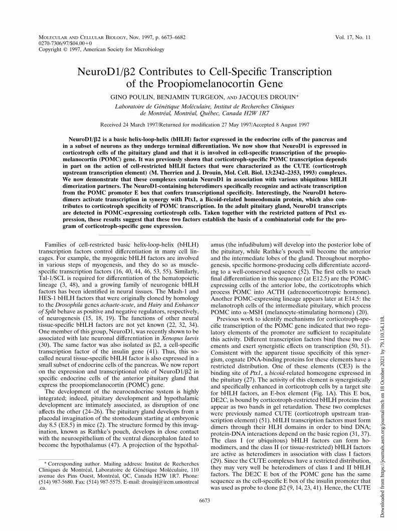
FIG. 1. Schematic representation of the rat POMC promoter (bp 2480 to163). (A) Relative positions of the DE2C, CE1B, and CE1A E boxes and of thePtx1 binding site (CE3) within the POMC promoter. (B) Nucleotide sequencesof the three E boxes of the promoter. Lowercase letters represent variant nu-cleotides by comparison to the consensus sequence NCANNTGN. (C) Effect ofbHLH factor expression on full-length POMC-luciferase reporter (50). Expres-sion vectors for NeuroD1 on/or Pan1 were transfected in CV-1 cells, and theluciferase (LUC.) activity (6 standard error of the mean) is shown relative tothat of the reporter alone. (D) Same experiment as in C performed with areporter plasmid containing a POMC promoter fragment deleted to bp 2323,which no longer contains the DE2C E box.
6674 POULIN ET AL. MOL. CELL. BIOL.
Dow
nloa
ded
from
http
s://j
ourn
als.
asm
.org
/jour
nal/m
cb o
n 18
Oct
ober
202
1 by
79.
110.
54.1
18.
RESULTS
Transcriptional specificity of NeuroD1. The CUTE com-plexes were previously characterized as bHLH factors thatspecifically bind the DE2C E box (51) and not other E boxes ofthe POMC promoter (35). The POMC promoter containsthree E boxes (Fig. 1A and B), but corticotroph-specific tran-scription can be conferred only by DE2C. Therefore, DE2Cplays a unique role by recruiting a corticotroph-restrictedbHLH factor(s) to the POMC promoter. Since b2/NeuroD1was shown to activate transcription from a similar E box in theinsulin promoter (41), we tested whether it acts on the POMCpromoter E boxes. Expression of NeuroD1 in CV-1 cells didnot affect the activity of a bp 2480 POMC-luciferase reporter,but cotransfection with the class I bHLH factor Pan1 resultedin significant activation (Fig. 1C). Interestingly, deletion of thedistal region of the promoter which contains the DE2C E boxprevented this activation (Fig. 1D), suggesting that the other Eboxes of the promoter are not targets for the NeuroD1 hetero-dimers. However, these E boxes are targets for class I bHLHfactors (Fig. 1C and D and reference 35).
To define the properties of each POMC promoter E box,three copies of each E box (DE2C, CE1B, and CE1A [Fig. 1Aand B]) were cloned upstream of a luciferase reporter contain-ing a minimal POMC promoter (bp 235 to 163 bp) fragment(50). Mammalian expression vectors for NeuroD1 and Pan1were cotransfected with these reporters into L cells. These cellsdo not express NeuroD1 (Northern blot analysis [data notshown]), nor do they express significant levels of class I bHLHfactors, as they are not detected in Western blots (data notshown) or in EMSA (see Fig. 3 and 4).
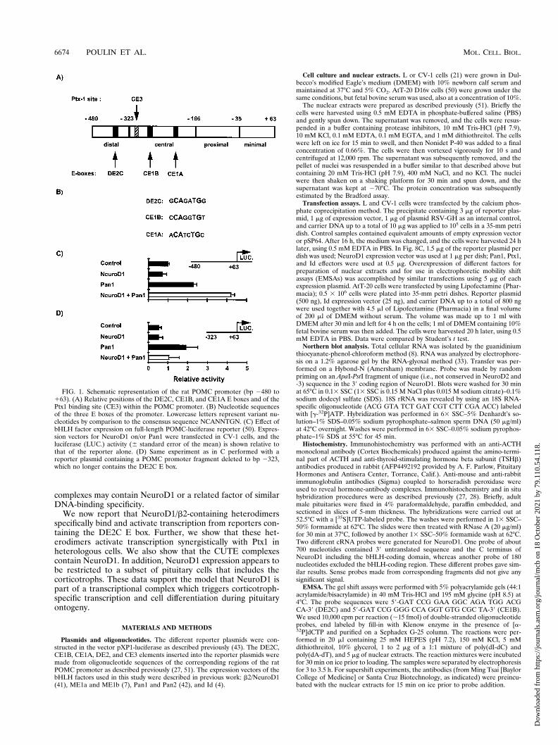
NeuroD1 overexpression in L cells did not increase the tran-scriptional activity of any of the three E-box reporter plasmids(Fig. 2, NeuroD1 compared to control). Overexpression ofPan1 alone did not affect the transcriptional activity of theDE2C reporter (Fig. 2A, Pan1) but enhanced the transcrip-tional activity of the CE1B and CE1A reporters (Fig. 2B and C,respectively). However, coexpression of NeuroD1 and Pan1
led to a significant increase of the DE2C reporter activity (Fig.2A, NeuroD1/Pan1 compared to NeuroD1 or Pan1) but didnot significantly enhance the activity of either the CE1B orCE1A reporter beyond the effect of Pan1 alone (Fig. 2B and C,Pan1 compared to NeuroD1/Pan1). Neither factor affected theactivity of reporters devoid of E boxes or containing a mutantE box (reference 51 and data not shown). Further, the fewnucleotide differences between the E boxes have marked ef-fects on their activation by NeuroD1 and/or Pan1. These re-sults strongly suggested that NeuroD1 transcriptional activity isdependent on specific recognition of the DE2C E box. Thus,the transcriptional properties of NeuroD1/Pan1 heterodimersare consistent with the properties of the CUTE complexes andof their target, DE2C (51). In contrast, the CE1B and CE1A Eboxes of the POMC promoter behave as targets of class Iubiquitous bHLH factors, albeit of different potencies.
FIG. 2. Specificity of NeuroD1 activation of POMC promoter E boxes. Re-porter plasmids containing three copies of E boxes DE2C (A), CE1B (B), andCE1A (C) were tested by cotransfection with expression vectors for NeuroD1and/or Pan1. Results (6 standard error of the mean) are the average of at leastthree separate experiments, each performed in duplicate. LUC., luciferase.
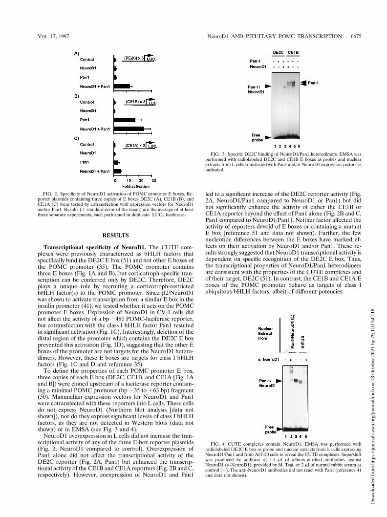
FIG. 3. Specific DE2C binding of NeuroD1/Pan1 heterodimers. EMSA wasperformed with radiolabeled DE2C and CE1B E boxes as probes and nuclearextracts from L cells transfected with Pan1 and/or NeuroD1 expression vectors asindicated.
FIG. 4. CUTE complexes contain NeuroD1. EMSA was performed withradiolabeled DE2C E box as probe and nuclear extracts from L cells expressingNeuroD1/Pan1 and from AtT-20 cells to reveal the CUTE complexes. Supershiftwas produced by addition of 1.5 ml of affinity-purified antibodies againstNeuroD1 (a-NeuroD1), provided by M. Tsai, or 2 ml of normal rabbit serum ascontrol (2). The anti-NeuroD1 antibodies did not react with Pan1 (reference 41and data not shown).
VOL. 17, 1997 NeuroD1 AND PITUITARY POMC TRANSCRIPTION 6675
Dow
nloa
ded
from
http
s://j
ourn
als.
asm
.org
/jour
nal/m
cb o
n 18
Oct
ober
202
1 by
79.
110.
54.1
18.
Specificity of NeuroD1/Pan1 heterodimer exerted at DNAbinding. The specificity of E-box recognition by NeuroD1/Pan1 heterodimers was tested by EMSA. The gel shift analyseswere performed with the DE2C and CE1B probes and nuclearextracts from L cells expressing Pan1, Pan1 and NeuroD1, orneither. Cells expressing only NeuroD1 did not exhibit bindingto either probe (data not shown), in agreement with experi-ments done with recombinant NeuroD1 (41). NeuroD1 andPan1 were both required to bind DE2C as a unique band inEMSA (Fig. 3, lane 3). In assays using the same nuclear ex-tracts with the CE1B probe, there was no significant binding ofsimilar migration (Fig. 3, lane 4 compared to lane 3). Instead,all bands observed with the CE1B probe were dependent onPan1 irrespective of the presence of NeuroD (Fig. 3, lane 5). Inagreement with the transfection data showing Pan1 transacti-vation of the CE1B but not the DE2C reporter (Fig. 2A andB), Pan1 dimer complexes did not bind DE2C (Fig. 3, lane 2).Taken together, these results demonstrate that the specificityof NeuroD1/Pan1 heterodimer action on transcription (Fig.2) is largely due to specific recognition of the DE2C E box(Fig. 3).
NeuroD1 is part of the CUTE complexes. To test whetherNeuroD1 is a component of the CUTE complexes of AtT-20
cells, we used anti-NeuroD1 antibodies (provided by M. Tsai)in supershift EMSAs. As control, we showed that NeuroD1/Pan1 heterodimers expressed in L cells (Fig. 4, lane 3) weresupershifted by the anti-NeuroD1 antibodies (Fig. 4, lane 2).The two CUTE complexes (51) of AtT-20 cells (Fig. 4, lane 4)were also supershifted in the presence of the anti-NeuroD1antibodies (Fig. 4, lane 5). Similar results (Fig. 6 and data notshown) were obtained with another antibody (Santa Cruz) madeagainst a C-terminal NeuroD1 peptide from a region unique toNeuroD1 by comparison to NeuroD2 and NeuroD3 (34).Thus, both CUTE bands of AtT-20 cells contain NeuroD1, andconversely, NeuroD1, rather than other related gene products,appears to be the major class II component of CUTE com-plexes.
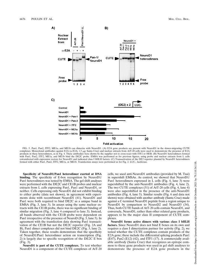
NeuroD1 forms active dimers with various class I bHLHfactors. Since NeuroD1 does not bind E boxes on its own andrequires a class I dimerization partner for activity (Fig. 2), wetested whether the CUTE complexes contain products of theE2A gene; these include the differential splicing products Pan1(E47), Pan2 (E12) (42), and ITF1 (17). A commercially avail-able antibody (Santa Cruz) that recognizes an epitope com-mon to these gene products was used in gel shift analyses todemonstrate the presence of E2A gene products in the
FIG. 5. Pan1, Pan2, ITF2, ME1a, and ME1b can dimerize with NeuroD1. (A) E2A gene products are present with NeuroD1 in the slower-migrating CUTEcomplexes. Monoclonal antibodies against E2A (a-E2A; 1.5 mg; Santa Cruz) and nuclear extracts from AtT-20 cells were used to demonstrate the presence of E2Aproducts in these heterodimers, as in Fig. 4. These antibodies were shown by the supplier not to cross-react with ITF2 or HEB. (B) NeuroD1 heterodimers formedwith Pan1, Pan2, ITF2, ME1a, and ME1b bind the DE2C probe. EMSA was performed as for previous figures, using probe and nuclear extracts from L cellscotransfected with expression vectors for NeuroD1 and indicated class I bHLH factors. (C) Transactivation of the DE2 reporter plasmid by NeuroD1 heterodimersformed with either Pan1, Pan2, ITF2, ME1a, or ME1b. Transfection assays were performed as for Fig. 2. LUC., luciferase.
6676 POULIN ET AL. MOL. CELL. BIOL.
Dow
nloa
ded
from
http
s://j
ourn
als.
asm
.org
/jour
nal/m
cb o
n 18
Oct
ober
202
1 by
79.
110.
54.1
18.
CUTE complexes of AtT-20 cells. As control, overexpressedNeuroD1/Pan1 heterodimers (Fig. 5A, lane 3) were super-shifted by the anti-E2A antibodies (Fig. 5A, lane 4). However,only the slower-migrating CUTE complex (Fig. 5A, lane 1) waspartially supershifted with saturating amounts of the anti-E2Aantibody (Fig. 5A, lane 2). This experiment suggested that E2Agene products are present in CUTE complexes. However, theymay account for only about 25% of the CUTE complexes.Specific antisera against the other mouse class I bHLH factorsare not available to test their presence in CUTE complexes. Inan attempt to determine whether other class I factors can actwith NeuroD, cotransfection assays and in vitro binding exper-iments by EMSA were performed. In transfection assays, allubiquitous bHLH factors tested (Pan1 and -2, ITF2, and HEB[ME1a and ME1b]) activated a DE2 reporter in the presenceof NeuroD1 (Fig. 5C) but not when transfected alone (data notshown). In EMSA, all class I bHLH factors formed DE2C-binding heterodimers with NeuroD1 (Fig. 5B). All of thesecomplexes migrated close to the position of the slower-migrat-ing CUTE complexes, suggesting that any of these could bepresent in CUTE complexes. Thus, ITF2 and/or ME1a/b (HEB)may constitute the other 50% of the slowly migrating CUTEband which is not accounted for by E2A products. Both ITF2and ME1a/b are expressed in AtT-20 cells (35). The identity ofthe NeuroD1 dimerization partner of the faster-migratingCUTE complex remains unknown.
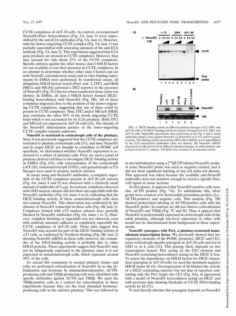
NeuroD1 is restricted to corticotroph cells of the pituitary.Since it was previously suggested that the CUTE complexes arerestricted to pituitary corticotroph cells (51), and since NeuroD1and its target DE2C are thought to contribute to POMC cellspecificity, we determined whether NeuroD1 expression is re-stricted to a subset of pituitary cells. First, we used a panel ofpituitary-derived cell lines to investigate DE2C-binding activityin EMSA (Fig. 6A); cells representative of the corticotroph(AtT-20), somatolactotroph (GH3), and gonadotroph (aT3)lineages were used to prepare nuclear extracts.
In assays using anti-NeuroD1 antibodies, a complete super-shift of the CUTE complexes present in AtT-20 cell extracts(Fig. 6A, lanes 8 and 9) was observed with a relatively smallamount of antibodies (0.5 mg). In contrast, complexes observedwith GH3 nuclear extracts did not show any supershift with theNeuroD1 antibodies (Fig. 6A, lanes 4 to 6), indicating that theDE2C-binding activity of these somatolactotroph cells doesnot contain NeuroD1. This observation was confirmed by theabsence of NeuroD1 transcripts in these cells (Fig. 6B, lane 3).Complexes formed with aT3 nuclear extracts were partiallyblocked by NeuroD1 antibodies (Fig. 6A, lanes 1 to 3). How-ever, complete blocking or supershift was not observed, evenwith antibody amounts sufficient to completely supershift theCUTE complexes of AtT-20 cells. These data suggest thatNeuroD1 may account for part of the DE2C-binding activity ofaT3 cells, as confirmed by Northern blotting (Fig. 6B, lane 2)showing NeuroD1 mRNA in these cells; however, the remain-der of the DNA-binding activity is probably due to otherbHLH proteins. These experiments suggest that NeuroD1 maynot be ubiquitously expressed in the pituitary since it is notexpressed in somatolactotroph cells, which represent around50% of the cells.
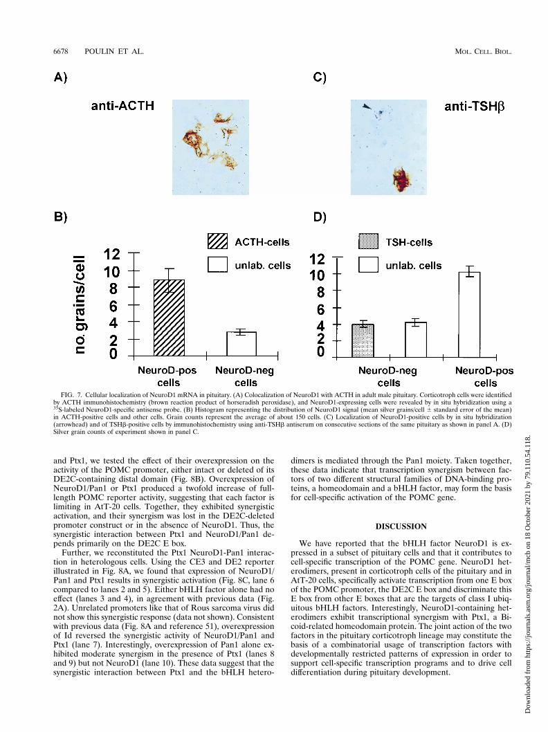
To extend this conclusion to normal pituitary tissues andcells, we performed colocalization of NeuroD1 by in situ hy-bridization and hormone by immunohistochemistry. ACTH-producing cells and TSHb-producing cells were identified withspecific antibodies against ACTH and TSHb. We used theTSHb-positive cells as a control for colocalization in theseexperiments because they are the least abundant hormone-producing cells in the anterior pituitary. This was followed by
in situ hybridization using a [35S]UTP-labeled NeuroD1 probe.A sense NeuroD1 probe was used as negative control, and itdid not show significant labeling of any cell (data not shown).This approach was taken because the available anti-NeuroDantibodies were not sensitive enough to reveal a specific Neu-roD signal in tissue sections.
At first glance, it appeared that NeuroD1-positive cells werealso ACTH positive (Fig. 7A). To substantiate this, silvergrains were counted over horseradish peroxidase-positive (i.e.,ACTH-positive) and negative cells. This analysis (Fig. 7B)showed preferential labeling of ACTH-positive cells with theNeuroD1 probe. In contrast, we did not observe colocalizationof NeuroD1 and TSHb (Fig. 7C and D). Thus, it appears thatNeuroD1 is preferentially expressed in corticotroph cells of theadult pituitary, although low-level expression in other cellswould not be discriminated from background in these experi-ments.
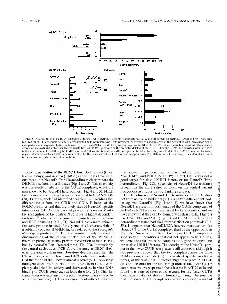
NeuroD1 synergizes with Ptx1, a pituitary-restricted home-odomain transcription factor. We previously showed that tworegulatory elements of the POMC promoter, DE2C and CE3,exert corticotroph-specific synergism in AtT-20 cells and not inGH3 or in L cells (51). This synergy likely depends on twotranscription factors: Ptx1 acting on the CE3 element andNeuroD1-containing heterodimers acting on the DE2C E box.To assess the dependence on bHLH factors for DE2C-depen-dent synergism in AtT-20 cells, we used the dominant negativebHLH factor Id (4). Overexpression of Id blunted the activityof a DE2C-containing reporter but not that of reporters con-taining only the Ptx1 target site CE3 (Fig. 8A), in agreementwith a model of NeuroD1 heterodimers acting on DE2C andwith previous data showing blockade of CUTE DNA-bindingactivity by Id (51).
To test directly whether this synergism depends on NeuroD1
FIG. 6. DE2C-binding activity in different pituitary lineages (aT3, GH3, andAtT-20 cells). (A) DE2C-binding activity in extracts (10 mg) from aT3, GH3, andAtT-20 cells. Supershift experiments were performed as for Fig. 4 and 5, usingantibodies (Santa Cruz) against NeuroD1 (a-NeuroD1) at 0, 0.5, and 0.8 mg perreaction. The binding activity detected in GH3 cells is bHLH, as it is supershiftedby the E2A monoclonal antibodies (data not shown). (B) NeuroD1 mRNAexpression in cells derived from different pituitary lineages, in adult pituitary andin head of an E11.5 embryo. 18S RNA was revealed as an internal control.
VOL. 17, 1997 NeuroD1 AND PITUITARY POMC TRANSCRIPTION 6677
Dow
nloa
ded
from
http
s://j
ourn
als.
asm
.org
/jour
nal/m
cb o
n 18
Oct
ober
202
1 by
79.
110.
54.1
18.
and Ptx1, we tested the effect of their overexpression on theactivity of the POMC promoter, either intact or deleted of itsDE2C-containing distal domain (Fig. 8B). Overexpression ofNeuroD1/Pan1 or Ptx1 produced a twofold increase of full-length POMC reporter activity, suggesting that each factor islimiting in AtT-20 cells. Together, they exhibited synergisticactivation, and their synergism was lost in the DE2C-deletedpromoter construct or in the absence of NeuroD1. Thus, thesynergistic interaction between Ptx1 and NeuroD1/Pan1 de-pends primarily on the DE2C E box.
Further, we reconstituted the Ptx1 NeuroD1-Pan1 interac-tion in heterologous cells. Using the CE3 and DE2 reporterillustrated in Fig. 8A, we found that expression of NeuroD1/Pan1 and Ptx1 results in synergistic activation (Fig. 8C, lane 6compared to lanes 2 and 5). Either bHLH factor alone had noeffect (lanes 3 and 4), in agreement with previous data (Fig.2A). Unrelated promoters like that of Rous sarcoma virus didnot show this synergistic response (data not shown). Consistentwith previous data (Fig. 8A and reference 51), overexpressionof Id reversed the synergistic activity of NeuroD1/Pan1 andPtx1 (lane 7). Interestingly, overexpression of Pan1 alone ex-hibited moderate synergism in the presence of Ptx1 (lanes 8and 9) but not NeuroD1 (lane 10). These data suggest that thesynergistic interaction between Ptx1 and the bHLH hetero-
dimers is mediated through the Pan1 moiety. Taken together,these data indicate that transcription synergism between fac-tors of two different structural families of DNA-binding pro-teins, a homeodomain and a bHLH factor, may form the basisfor cell-specific activation of the POMC gene.
DISCUSSION
We have reported that the bHLH factor NeuroD1 is ex-pressed in a subset of pituitary cells and that it contributes tocell-specific transcription of the POMC gene. NeuroD1 het-erodimers, present in corticotroph cells of the pituitary and inAtT-20 cells, specifically activate transcription from one E boxof the POMC promoter, the DE2C E box and discriminate thisE box from other E boxes that are the targets of class I ubiq-uitous bHLH factors. Interestingly, NeuroD1-containing het-erodimers exhibit transcriptional synergism with Ptx1, a Bi-coid-related homeodomain protein. The joint action of the twofactors in the pituitary corticotroph lineage may constitute thebasis of a combinatorial usage of transcription factors withdevelopmentally restricted patterns of expression in order tosupport cell-specific transcription programs and to drive celldifferentiation during pituitary development.
FIG. 7. Cellular localization of NeuroD1 mRNA in pituitary. (A) Colocalization of NeuroD1 with ACTH in adult male pituitary. Corticotroph cells were identifiedby ACTH immunohistochemistry (brown reaction product of horseradish peroxidase), and NeuroD1-expressing cells were revealed by in situ hybridization using a35S-labeled NeuroD1-specific antisense probe. (B) Histogram representing the distribution of NeuroD1 signal (mean silver grains/cell 6 standard error of the mean)in ACTH-positive cells and other cells. Grain counts represent the average of about 150 cells. (C) Localization of NeuroD1-positive cells by in situ hybridization(arrowhead) and of TSHb-positive cells by immunohistochemistry using anti-TSHb antiserum on consecutive sections of the same pituitary as shown in panel A. (D)Silver grain counts of experiment shown in panel C.
6678 POULIN ET AL. MOL. CELL. BIOL.
Dow
nloa
ded
from
http
s://j
ourn
als.
asm
.org
/jour
nal/m
cb o
n 18
Oct
ober
202
1 by
79.
110.
54.1
18.
Specific activation of the DE2C E box. Both in vivo (trans-fection assays) and in vitro (EMSA) experiments have dem-onstrated that NeuroD1/Pan1 heterodimers discriminate theDE2C E box from other E boxes (Fig. 2 and 3). This specificitywas previously attributed to the CUTE complexes, which arenow shown to be NeuroD1 heterodimers (Fig. 4 and 5). bHLHfactors interact with target sequences related to NCANNTGN(38). Previous work had identified specific DE2C residues thatdifferentiate it from the CE1B and CE1A E boxes of thePOMC promoter and that are likely sites of NeuroD1-specificinteractions (51). On the basis of previous studies on MyoD,the recognition of the central N residues is highly dependenton lysine122 situated in the junction region between the basicand HLH domains (10, 11, 31). NeuroD1 has a methionine atthe same position instead of the lysine; this is characteristic ofa subfamily of class II bHLH factors related to the Drosophilaatonal gene product (30). This methionine is likely involved indiscrimination of the central nucleotides of the POMC Eboxes. In particular, it may prevent recognition of the CE1B Ebox by NeuroD1/Pan1 heterodimers (Fig. 2B). Interestingly,the central nucleotides of CE1A are the same as DE2C. How-ever, previous work in our laboratory demonstrated that theCE1A E box, which differs from DE2C only by a T instead ofC at the 39 end of the E box, is almost inactive (51). Conversely,mutagenesis of this 39 nucleotide of DE2C from C to T com-pletely abolished its activity and decreased its in vitro DNAbinding to CUTE complexes at least threefold (51). This dis-crimination was explained by a putative steric clash caused bya T at this position (12). This is in agreement with other studies
that showed dependence on similar flanking residues forMyoD, Myc, and PHO4 (5, 13, 49). In fact, CE1A was not agood target for class I bHLH factors or for NeuroD1/Pan1heterodimers (Fig. 2C). Specificity of NeuroD1 heterodimerrecognition therefore relies as much on the central variantnucleotides as it does on the flanking residues.
CUTE is formed of NeuroD1 heterodimers. NeuroD1 doesnot form active homodimers (41). Using two different antibod-ies against NeuroD1 (Fig. 4 and 6), we have shown thatNeuroD1 is present in both bands of the CUTE complexes ofAtT-20 cells. These complexes must be heterodimers, and wehave shown that they can be formed with class I bHLH factorslike E2A, ITF2, and ME1 (Fig. 5B and C). All of the NeuroD1heterodimers tested had similar transactivation potentials (Fig.5B). It appears that NeuroD1/E2A heterodimers account forabout 25% of the CUTE complexes (half of the upper band inFig. 5A). Since only 50% of the upper CUTE complex issupershifted in conditions that did not appear to be limiting,we conclude that this band contains E2A gene products andother class I bHLH factors. The identity of the NeuroD1 part-ner in the lower CUTE complexes is still unknown, although itwas previously shown that the two complexes have the sameDNA-binding specificity (51). To verify if specific modifica-tion(s) of the class I bHLH factors might take place in AtT-20cells and account for the faster migration of the lower CUTEcomplexes, we overexpressed these factors in AtT-20 cells andfound that none of them could account for the faster CUTEcomplexes (data not shown). Formally, it might be possiblethat the lower CUTE complexes contain a splicing variant of
FIG. 8. Reconstitution of NeuroD1 synergism with Ptx1. (A) In NeuroD1- and Ptx1-expressing AtT-20 cells, both targets for NeuroD1 (DE2) and Ptx1 (CE3) arerequired for bHLH-dependent activity as demonstrated by Id overexpression. Data represent the average 6 standard error of the mean of at least three experiments,each performed in duplicate. LUC., luciferase. (B) The NeuroD1/Pan1 and Ptx1 synergism requires the DE2C E box. AtT-20 cells were lipofected with the indicatedexpression plasmids and with either the full-length bp 2480 POMC promoter or the promoter deleted of the DE2C E box (bp 2323). The activity shown is relativeto the basal activity of the full-length POMC reporter. (C) Reconstitution of NeuroD1 synergism with Ptx1 in heterologous cells (L). The DE2/CE3 reporter illustratedin panel A was cotransfected with expression vectors for the indicated factors. Ptx1 was described previously (27). Data represent the average 6 standard deviation oftwo experiments, each performed in duplicate.
VOL. 17, 1997 NeuroD1 AND PITUITARY POMC TRANSCRIPTION 6679
Dow
nloa
ded
from
http
s://j
ourn
als.
asm
.org
/jour
nal/m
cb o
n 18
Oct
ober
202
1 by
79.
110.
54.1
18.

NeuroD1 which is unique to AtT-20 cells and therefore notobserved in overexpression experiments in L cells (Fig. 4);however, this is not very likely, as in such a case, one mightexpect half of these faster-migrating complexes to also containE2A gene products. Interestingly, NeuroD1 is expressed inhamster insulinoma tumor cells, and its complexes with DE2Ccomigrated with the upper CUTE complexes and not with thelower (51). Taken together, these data suggest that a classI-type bHLH factor different from the ones tested in this workmay be expressed in AtT-20 cells. This unidentified factorwould produce the faster-migrating CUTE complexes as het-erodimers with NeuroD1.
NeuroD1 expression in pituitary. NeuroD1 is expressed inthe nervous system and in a- and b-cell-derived lines of thepancreas (41). It is also expressed in enteroendocrine secretincells (39). We now show that NeuroD1 is also expressed in asubset of pituitary cells (Fig. 7). These are predominantly thePOMC-expressing corticotroph cells. However, the sensitivityof the in situ hybridization is such that it may not reveallow-level expression in other cells. The analysis of pituitary-derived cell lines has confirmed NeuroD1 expression in amodel corticotroph line, AtT-20 cells (Fig. 4 and 6), but not incells of the somatolactotroph lineage, GH3 cells (6) (Fig. 6Aand B). The absence of NeuroD1 signal in TSHb-positivepituitary cells (Fig. 7C and D) was also confirmed by Northernblot analysis of RNA from the aTSH (1) cell line (data notshown). In contrast, NeuroD1-related DNA-binding activitywas detected in aT3 (54) cell extracts, a model of gonadotrophcells (Fig. 6A), and NeuroD1 expression was confirmed byNorthern blot analysis (Fig. 6B, lane 2). Thus, NeuroD1 mightbe expressed below in situ detection in adult pituitary gonado-trophs; alternatively, the aT3 might be representative of a fetaldifferentiation intermediate of the gonadotroph lineage, andNeuroD1 expression might be temporally limited in this lin-eage during pituitary development.
Two types of POMC-expressing cells, corticotrophs andmelanotrophs, are present in the pituitary. No expression ofNeuroD1 is seen in the intermediate lobe melanotrophs (datanot shown), indicating that NeuroD1 is not a marker of allPOMC-expressing cells. This observation is strongly supportiveof the conclusion that different transcriptional regulatorymechanisms are operative in the two POMC-expressing lin-eages. NeuroD1 is thus a marker of corticotrophs and could beuseful to differentiate melanotrophs from corticotrophs, eitherduring ontogeny or in human pituitary tumors.
NeuroD1 synergism with the homeobox factor Ptx1. Wehave previously documented the central role played by theNeuroD1 target sequence DE2C and the Ptx1 target sequenceCE3 for cell-specific transcription of POMC (27, 50, 51). For-mal promoter analysis had predicted that an interaction be-tween NeuroD1 and Ptx1 might form the scaffold upon whichother transcription factors become involved in POMC tran-scription. We now provide data showing transcriptional syner-gism between NeuroD1 heterodimers and Ptx1 (Fig. 8B andC). The NeuroD1-dependent synergism requires the DE2C Ebox, as it is not observed on a deleted POMC promoter thatstill contains the CE1A and CE1B E boxes (Fig. 8B). Thisinteraction was reconstituted in cells that express neither fac-tor, and its dependence on the bHLH dimers was supported byinterference with the bHLH dominant negative Id. It had pre-viously been shown that the CUTE complexes are disrupted byId (51), presumably by competing potential partners. Similardependence of the POMC promoter on bHLH activity wasshown in AtT-20 cells (Fig. 8A and reference 51). Synergismbetween bHLH and homeobox-containing factors was ob-served on the insulin promoter. Indeed, the lim homeodomain
factor lmx-1 was shown to synergize with dimers of Pan1, andtheir interaction was dependent on the lim domain (14). Pan1was also reported to activate transcription synergistically withanother homeobox factor, STF-1 (Pdx1, IPF-1) in the samesystem (45). These experiments were performed before thecloning of NeuroD1/b2: if Pan1/NeuroD1 heterodimers arethe active bHLH factors in b cells, we must conclude that Pan1is the most likely interaction partner of the homeobox fac-tor(s). Our data support the model that NeuroD1 hetero-dimers interact with Ptx1 through the class I bHLH moiety ofthe dimers, as suggested by the small synergism between Ptx1and Pan1 (without NeuroD1 [Fig. 8C, lane 8]). Since Pan1 didnot have any activity on its own (lane 3), it is possible that thePan1 effect is due to its recruitment by protein-protein inter-action with Ptx1; similar interactions were proposed betweenMyoD and MEF2A (36). Thus, it appears that NeuroD1 con-fers E-box selectivity to the heterodimers and that the ability tosynergize with Ptx1 or other homeobox factors (like lmx-1 orSTF-1) might be a property conferred by a class I bHLHpartner like Pan1.
Putative role of NeuroD1 in the pituitary. NeuroD1 has ahighly restricted pattern of expression: it is expressed only inthe nervous system and in a subset of pancreatic and pituitaryendocrine cells. Together with other neurogenic, bHLH fac-tors like MASH-1, neurogenin, and HES-1, NeuroD1 (and itsrelated factors NeuroD2 and NeuroD3) may constitute a de-velopmental code in which each factor plays a role at specifictimes or/and places during neural development (15, 18, 19, 32,34). These factors may have different functions, either in pat-tern formation or in cell commitment or differentiation. Thecurrent data on NeuroD1 function in Xenopus support a role incell differentiation during the later stages of neurogenesis (30).The expression of NeuroD1 in a limited subset of pituitary cellssuggests that NeuroD1 may play a similar role in pituitarylineage differentiation in addition to its role as transcriptionfactor. In particular, the high-level NeuroD1 expression incorticotrophs, both in the pituitary (Fig. 7A and B) and inAtT-20 cells (Fig. 3), is suggestive of a function in this lineage.The only other lineage for which some expression was detectedis the gonadotroph model aT3 cells (Fig. 6); however, aT3cells also express other factors (as yet unidentified) with re-lated DNA-binding specificity. Thus, the corticotrophs mightbe unique in the pituitary by their exclusive (of other bHLH)expression of NeuroD1. The synergistic action of NeuroD1with Ptx1 and their joint expression in corticotrophs may formthe basis of a combinatorial code for specification of this lin-eage.
In conclusion, we have shown that NeuroD1 is an essentialdeterminant for the transcriptional specificity of the DE2Celement of the POMC promoter. This specificity appears to beconferred by selective E-box recognition. Further, we haveshown that the transcriptional activity of NeuroD1/Pan1 het-erodimers is enhanced by interaction with a homeobox factor,Ptx1, that has a restricted pituitary pattern of expression.Taken together, these data support the model of a combina-torial action of transcription factors during pituitary develop-ment.
ACKNOWLEDGMENTS
We are grateful to Ming Tsai (Baylor College of Medicine, Houston,Tex.), Chris Nelson (Tularick), Toomas Neuman (Colorado State Uni-versity, Fort Collins), and Scott McDonald and Francois Guillemot(Strasbourg, France) for the gift of plasmids encoding hamsterNeuroD1 and Pan1/2, mouse ME1a/b, ITF-2, and NeuroD1, respec-tively. We are also grateful to Ming Tsai for the generous gift ofNeuroD antiserum and to A. F. Parlow, Pituitary Hormones and An-
6680 POULIN ET AL. MOL. CELL. BIOL.
Dow
nloa
ded
from
http
s://j
ourn
als.
asm
.org
/jour
nal/m
cb o
n 18
Oct
ober
202
1 by
79.
110.
54.1
18.
tisera Center, Torrance, Calif., for antisera against rat TSHb. Theexpert advice of Christian Lanctot was helpful in developing in situhybridization and immunohistochemistry, and we thank Michel Cham-berland for oligonucleotide synthesis. The expert secretarial assistanceof Lise Laroche was greatly appreciated.
This work was funded by a grant from the National Cancer Instituteof Canada with funds provided by the Canadian Cancer Society.
REFERENCES
1. Akerblom, I. E., E. C. Ridgway, and P. L. Mellon. 1990. An alpha-subunit-secreting cell line derived from a mouse thyrotrope tumor. Mol. Endocrinol.4:589–596.
2. Andersen, B., and M. G. Rosenfeld. 1994. Pit-1 determines cell types duringdevelopment of the anterior pituitary gland. A model for transcriptionalregulation of cell phenotypes in mammalian organogenesis. J. Biol. Chem.269:29335–29338.
3. Aplan, P. D., K. Nakahara, S. H. Orkin, and I. R. Kirsch. 1992. The SCLgene product: a positive regulator of erythroid differentiation. EMBO J.11:4073–4081.
4. Benezra, R., R. L. Davis, D. Lockshon, D. L. Turner, and H. Weintraub.1990. The protein Id: a negative regulator of helix-loop-helix DNA bindingproteins. Cell 61:49–59.
5. Blackwell, T. K., and H. Weintraub. 1990. Differences and similarities inDNA-binding preferences of MyoD and E2A protein complexes revealed bybinding site selection. Science 250:1104–1110.
6. Boockfor, F. R., and L. K. Schwarz. 1988. Cultures of GH3 cells contain bothsingle and dual hormone secretors. Endocrinology 122:762–764.
7. Chiaramello, A., K. Neuman, K. Palm, M. Metsis, and T. Neuman. 1995.Helix-loop-helix transcription factors mediate activation and repression ofthe p75LNGFR gene. Mol. Cell. Biol. 15:6036–6044.
8. Chomczynski, P., and N. Sacchi. 1987. Single-step method of RNA isolationby acid guanidium thyocyanate-phenol-chloroform-extraction. Anal. Bio-chem. 162:156–159.
9. Cordle, S. R., E. Henderson, H. Masuoka, P. A. Weil, and R. Stein. 1991.Pancreatic b-cell-type-specific transcription of the insulin gene is mediatedby basic helix-loop-helix DNA-binding proteins. Mol. Cell. Biol. 11:1734–1738.
10. Davis, R. L., P.-F. Cheng, A. B. Lassar, and H. Weintraub. 1990. The MyoDDNA binding domain contains a recognition code for muscle-specific geneactivation. Cell 60:733–746.
11. Davis, R. L., and H. Weintraub. 1992. Acquisition of myogenic specificity byreplacement of three amino acid residues from MyoD into E12. Science256:1027–1030.
12. Ellenberger, T., D. Fass, M. Arnaud, and S. C. Harrison. 1994. Crystalstructure of transcription factor E47: E-box recognition by a basic regionhelix-loop-helix dimer. Genes Dev. 8:970–980.
13. Fisher, F., and C. R. Goding. 1992. Single amino acid substitutions alterhelix-loop-helix protein specificity for bases flanking the core CANNTGmotif. EMBO J. 11:4103–4109.
14. German, M. S., J. Wang, R. B. Chadwick, and W. J. Rutter. 1992. Synergisticactivation of the insulin gene by a LIM-homeo domain protein and a basichelix-loop-helix protein: building a functional insulin minienhancer complex.Genes Dev. 6:2165–2176.
15. Guillemot, F., L. C. Lo, J. E. Johnson, A. Auerbach, D. J. Anderson, and A. L.Joyner. 1993. Mammalian achaete-scute homolog 1 is required for the earlydevelopment of olfactory and autonomic neurons. Cell 75:463–476.
16. Hasty, P., A. Bradley, J. Hsi Morris, D. G. Edmondson, J. M. Venuti, E. N.Olson, and W. H. Klein. 1993. Muscle deficiency and neonatal death in micewith a targeted mutation in the myogenin gene. Nature 364:501–506.
17. Henthorn, P., R. McCarrick-Walmsley, and T. Kadesch. 1990. Sequence ofthe cDNA encoding ITF-1, a positive-acting transcription factor. NucleicAcids Res. 18:677.
18. Ishibashi, M., S. L. Ang, K. Shiota, S. Nakanishi, R. Kageyama, and F.Guillemot. 1995. Targeted disruption of mammalian hairy and Enhancer ofsplit homolog-1 (HES-1) leads to up-regulation of neural helix-loop-helixfactors, premature neurogenesis, and severe neural tube defects. Genes Dev.9:3136–3148.
19. Ishibashi, M., K. Moriyoshi, Y. Sasai, K. Shiota, S. Nakanishi, and R.Kageyama. 1994. Persistent expression of helix-loop-helix factor HES-1 pre-vents mammalian neural differentiation in the central nervous system.EMBO J. 13:1799–1805.
20. Japon, M. A., M. Rubinstein, and M. J. Low. 1994. In situ hybridizationanalysis of anterior pituitary hormone gene expression during fetal mousedevelopment. J. Histochem. Cytochem. 42:1117–1125.
21. Jeannotte, L., M. A. Trifiro, R. K. Plante, M. Chamberland, and J. Drouin.1987. Tissue-specific activity of the pro-opioomelanocortin gene promoter.Mol. Cell. Biol. 7:4058–4064.
22. Kageyama, R., Y. Sasai, C. Akazawa, M. Ishibashi, K. Takebayashi, C.Shimizu, K. Tomita, and S. Nakanishi. 1995. Regulation of mammalianneural development by helix-loop-helix transcription factors. Crit. Rev. Neu-robiol. 9:177–188.
23. Karlsson, O., T. Edlund, J. B. Moss, W. J. Rutter, and M. D. Walker. 1987.A mutational analysis of the insulin gene transcription control region: ex-pression in beta cells is dependent on two related sequences within theenhancer. Proc. Natl. Acad. Sci. USA 84:8819–8823.
24. Kawamura, K., and S. Kikuyama. 1992. Evidence that hypophysis and hy-pothalamus constitute a single entity from the primary stage of histogenesis.Development 115:1–9.
25. Kawamura, K., and S. Kikuyama. 1995. Induction from posterior hypothal-amus is essential for the development of the pituitary proopiomelacortin(POMC) cells of the toad (Bufo japonicus). Cell Tissue Res. 279:233–239.
26. Kikuyama, S., H. Inaco, B. G. Jenks, and K. Kawamura. 1993. Developmentof the ectopically transplanted primordium of epithelial hypophysis (anteriorneural ridge) in Bufo japonicus embryos. J. Exp. Zool. 266:216–220.
27. Lamonerie, T., J. J. Tremblay, C. Lanctot, M. Therrien, Y. Gauthier, and J.Drouin. 1996. PTX1, a bicoid-related homeobox transcription factor involvedin transcription of pro-opiomelanocortin (POMC) gene. Genes Dev. 10:1284–1295.
28. Lanctot, C., B. Lamolet, and J. Drouin. 1997. The bicoid-related homeopro-tein Ptx1 defines the most anterior domain of the embryo and differentiatesposterior from anterior lateral mesoderm. Development 124:2817.
29. Lassar, A. B., R. L. Davis, W. E. Wright, T. Kadesch, C. Murre, A. Voronova,D. Baltimore, and H. Weintraub. 1991. Functional activity of myogenic HLHproteins requires hetero-oligomerization with E12/E47-like proteins in vivo.Cell 66:305–315.
30. Lee, J. E., S. M. Hollenberg, L. Snider, D. L. Turner, N. Lipnick, and H.Weintraub. 1995. Conversion of Xenopus ectoderm into neurons by Neu-roD, a basic helix-loop-helix protein. Science 268:836–844.
31. Ma, P. C., M. A. Rould, H. Weintraub, and C. O. Pabo. 1994. Crystalstructure of MyoD bHLH domain-DNA complex: perspectives on DNArecognition and implications for transcriptional activation. Cell 77:451–459.
32. Ma, Q. F., C. Kintner, and D. J. Anderson. 1996. Identification of neuroge-nin, a vertebrate neuronal determination gene. Cell 87:43–52.
33. Maniatis, T., E. F. Fritsch, and J. Sambrook. 1982. Molecular cloning: alaboratory manual. Cold Spring Harbor Laboratory, Cold Spring Harbor,N.Y.
34. McCormick, M. B., R. M. Tamimi, L. Snider, A. Asakura, D. Bergstrom, andS. J. Tapscott. 1996. NeuroD2 and NeuroD3: distinct expression patternsand transcriptional activation potentials within the NeuroD gene family.Mol. Cell. Biol. 16:5792–5800.
35. McDonald, S., G. Poulin, T. Lamonerie, M. Therrien, and J. Drouin. HLHtranscription factors act at multiple sites to control POMC gene expression.Submitted for publication.
36. Molkentin, J. D., B. L. Black, J. F. Martin, and E. N. Olson. 1995. Cooper-ative activation of muscle gene expression by mef2 and myogenic bhlh pro-teins. Cell 83:1125–1136.
37. Murre, C., P. S. McCaw, and D. Baltimore. 1989. A new DNA binding anddimerization motif in immunoglobulin enhancer binding, daughterless,MyoD, and myc proteins. Cell 56:777–783.
38. Murre, C., P. S. McCaw, H. Vaessin, M. Caudy, L. Y. Jan, Y. N. Jan, C. V.Cabrera, J. N. Buskin, S. D. Hauschka, A. B. Lassar, H. Weintraub, and D.Baltimore. 1989. Interactions between heterologous helix-loop-helix proteinsgenerate complexes that bind specifically to a common DNA sequence. Cell58:537–544.
39. Mutoh, H., B. P. Fung, F. J. Naya, M. J. Tsai, J. Nishitani, and A. B. Leiter.1997. The basic helix-loop-helix transcription factor beta2/neurod is ex-pressed in mammalian enteroendocrine cells and activates secretin geneexpression. Proc. Natl. Acad. Sci. USA 94:3560–3564.
40. Nabeshima, Y., K. Hanaoka, M. Hayasaka, E. Esumi, S. Li, and I. Nonaka.1993. Myogenin gene disruption results in perinatal lethality because ofsevere muscle defect. Nature 364:532–535.
41. Naya, F. J., C. M. M. Stellrecht, and M. J. Tsai. 1995. Tissue-specificregulation of the insulin gene by a novel basic helix-loop-helix transcriptionfactor. Genes Dev. 9:1009–1019.
42. Nelson, C., L. P. Shen, A. Meister, E. Fodor, and W. J. Rutter. 1990. Pan: atranscriptional regulator that binds chymotrypsin, insulin, and AP-4 en-hancer motifs. Genes Dev. 4:1035–1043.
43. Nordeen, S. K. 1988. Luciferase reporter gene vectors for analysis of pro-moters and enhancers. BioTechniques 6:454–456.
44. Olson, E. N., and W. H. Klein. 1994. bHLH factors in muscle development:dead lines and commitments, what to leave in and what to leave out. GenesDev. 8:1–8.
45. Peers, B., J. Leonard, S. Sharma, G. Teitelman, and M. R. Montminy. 1994.Insulin expression in pancreatic islet cells relies on cooperative interactionsbetween the helix loop helix factor E47 and the homeobox factor STF-1.Mol. Endocrinol. 8:1798–1806.
46. Rudnicki, M. A., P. N. Schnegelsberg, R. H. Stead, T. Braun, H. H. Arnold,and R. Jaenisch. 1993. MyoD or Myf-5 is required for the formation ofskeletal muscle. Cell 75:1351–1359.
47. Schwind, J. 1928. The development of the hypophysis cerebri of the albinorat. Am. J. Anat. 41:295–319.
VOL. 17, 1997 NeuroD1 AND PITUITARY POMC TRANSCRIPTION 6681
Dow
nloa
ded
from
http
s://j
ourn
als.
asm
.org
/jour
nal/m
cb o
n 18
Oct
ober
202
1 by
79.
110.
54.1
18.

48. Shivdasani, R. A., E. L. Mayer, and S. H. Orkin. 1995. Absence of bloodformation in mice lacking the T-cell leukaemia oncoprotein tal-1/SCL. Na-ture 373:432–434.
49. Sun, X.-H., and D. Baltimore. 1991. An inhibitory domain of E12 transcrip-tion factor prevents DNA binding in E12 homodimers but not in E12 het-erodimers. Cell 64:459–470.
50. Therrien, M., and J. Drouin. 1991. Pituitary pro-opiomelanocortin geneexpression requires synergistic interactions of several regulatory elements.Mol. Cell. Biol. 11:3492–3503.
51. Therrien, M., and J. Drouin. 1993. Cell-specific helix-loop-helix factorrequired for pituitary expression of the pro-opiomelanocortin gene. Mol.
Cell. Biol. 13:2342–2353.52. Voss, J. W., and M. G. Rosenfeld. 1992. Anterior pituitary development:
short tales from dwarf mice. Cell 70:527–530.53. Weintraub, H. 1993. The MyoD family and myogenesis: redundancy, net-
works, and thresholds. Cell 75:1241–1244.54. Windle, J. J., R. I. Weiner, and P. L. Mellon. 1990. Cell lines of the pituitary
gonadotrope lineage derived by targeted oncogenesis in transgenic mice.Mol. Endocrinol. 4:597–603.
55. Zhang, W., R. R. Behringer, and E. N. Olson. 1995. Inactivation of themyogenic bHLH gene MRF4 results in up-regulation of myogenin and ribanomalies. Genes Dev. 9:1388–1399.
6682 POULIN ET AL. MOL. CELL. BIOL.
Dow
nloa
ded
from
http
s://j
ourn
als.
asm
.org
/jour
nal/m
cb o
n 18
Oct
ober
202
1 by
79.
110.
54.1
18.