neurofibromatosis andsmall bowel adenocarcinoma: an...
TRANSCRIPT

Gut, 1987, 28, 1173-1176
Neurofibromatosis and small bowel adenocarcinoma:an unrecognised associationT J JONES AND T L MARSHALL
From the Department ofMorbid Anatomy, The London Hospital Medical College, London, and theDepartment ofHistopathology, Central Pathology Laboratory, Stoke-on-Trent, Staffordshire
SUMMARY We present a review of the reported cases and describe a fifth of neurofibromatosis withsmall bowel adenocarcinoma and concurrent leiomyomas, indicating a previously unrecognisedassociation.
Neurofibromatosis is known to be associated with anincreased incidence of neoplasms including phaeo-chromocytoma gliomata and neurofibrosarcoma.'Gastrointestinal involvement is well documented,predominantly consisting of neurofibromas, ganglio-neuromas and schwannomas." Epithelial tumoursincluding colonic,' pancreatic4 and small intestinaladenocarcinomas have all been reported.'
Case report
The patient, a 59 year old white man initiallypresented in July 1980 with a soft, well circumscribedmass 1-5 cm in diameter on the right thigh. This wasexcised and histology showed it to consist of spindlecells in a fibrillary stroma, appearances typical of aneurofibroma. On further clinical examination hewas found to have a cafe-au-lait area on the left thigh;his father had been diagnosed as having neurofibro-matosis in the past (no biopsy available). He wastherefore diagnosed as a case of neurofibromatosisand remained asymptomatic for the next five years.He next presented in February 1985 with inter-
mittent colicky abdominal pain associated withvomiting, diarrhoea and 10 kg weight loss. Onexamination his abdomen was distended, withincreased bowel sounds and tenderness over the rightiliac fossa. Initial laboratory investigations showed a
Address for correspondence: Dr T J Jones, Dept of Morbid Anatomy. TheLondon Hospital, London Et tBB.Received for publication 5 Februatry 1987.
neutrophil leucocytosis (WCC 19 6x10 with 80%neutrophils). A diagnosis of small bowel obstructionwas made and laparotomy revealed an area ofstenosis in the middle third of the ileum with multipleserosal nodules. Thirty centimetres of ileum wereresected and submitted for histological examinationOn opening the bowel an ulcerated stenosing lesionwas noted 15 cm from the distal plane of excisionalong with multiple nodules on the rosal surfacesituated principally on the mesenteric order.
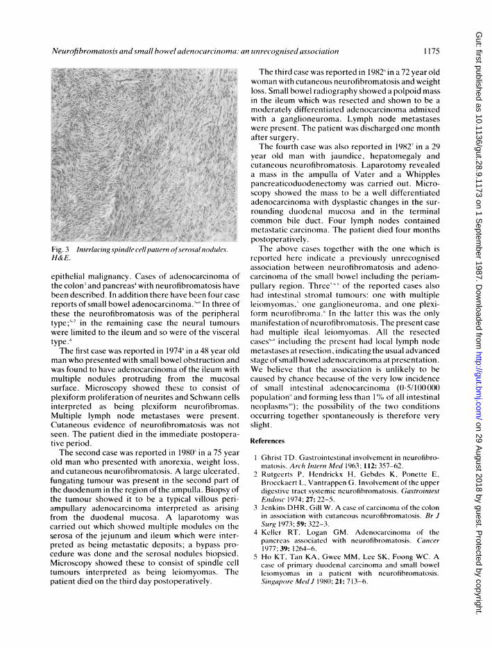
Microscopy of the stenosing lesion showed it to bea moderately differentiated adenocarcinoma (Fig. 1)which had penetrated through to the serosal surfacewith metastases in the mesenteric lymph nodes (Fig.2). The serosal nodules were shown to consist ofspindle cells with cigar shaped nuclei arranged ininterlacing bundles (Fig. 3); they arose from themuscularis propria and extended into the submucosaand onto the serosal surface. No neural elementswere seen. Tissue from the nodules was placed inglutaraldehyde, postfixed in osmium tetroxide andexamined using an AEI Corinth 500 Electron micro-scope. This showed intracellular filaments, adherentbasement membrane like material and pinocytoticvacuoles. The overall appearances were of tumoursof smooth muscle origin.
Discussion
Neurofibromatosis with involvement of the gastro-intestinal tract is well documented' 2 and usuallyconsists of neurofibromas, ganglioneuromas, andleiomyomas; much rarer is the development of
1173
on 29 August 2018 by guest. P
rotected by copyright.http://gut.bm
j.com/
Gut: first published as 10.1136/gut.28.9.1173 on 1 S
eptember 1987. D
ownloaded from

rig. I - -, 0!.Fig. 1 Section showing infiltrating adenocarcinoma. H& E. x50
j -.
Fig. 2 Metastatic adenocarcinomna in siabcapsular region ofmnesenteric Ivmph izode. H& E. x550
on 29 August 2018 by guest. P
rotected by copyright.http://gut.bm
j.com/
Gut: first published as 10.1136/gut.28.9.1173 on 1 S
eptember 1987. D
ownloaded from
Neurofibromnatosis and smrall bowel adenocarcinoma: an unrecognised association 1175
H&E.~~~~~~~.
Fig.39 intherlac maiindleclasethernofeuroalntumours
epThelfirsasemaigancyrepores of adenocarcinomar oldthe wholndpresnreaswithnmloeurofbsromatoionhavereorsofon smal boweladenocarcinomaofthe threewitmuthesple neuofibs romtrudisnga ofro the periphealsurfae;" Minheremaininshowed thesneuatouconsisto
wnerpelmted to theileu plexifor weureofthviscroas.
* . .j
tyvepe.'oc .
The secondcase was reported in1974' in a48 yearol
.manwho presented withaacgs cll bwerofseronoles
epithelialtmalinnyeCssoadenocarcinoma ofteiemwt
nth colonandodupasnreineurofibromatosishe hucsave
sufunating tmicourwscopresentwinthe setoconsisat of
blee duesrimbdinedrdtionofntherehave Beewou cafsmrporets as boelnadenocarcino maIerr ni treeasi
fthne the neurofuibromoswa thlaperiphwasltypred inutwihereminngeasmuthpe nedurle tuourshsere. lTe atthe dileum andheo wherie ote vistera-
tyveperiodThe firstdcase was reported in 1974Win a48 yearol
man who presented withsmal boweiaobstruct onsn
wasd foutndeouhav adenrfbocarcsinom of thre ileumawthd
multipe nsoules protru ing the muocdlesriofsa
eMicroscopyshowed these to consistofvilepellampulainterpreted as beingp ormreurofasibrosCutaeoudeidene ofl neurosa omapartoi y was
sareen oth patient dioedinltheipemmediatespostopera
Thetsecodcasbein weastaireportdins aO inpas75ryea
feungatin tuour wasnrsetitheseoa ondlebiparteofthectuourop showed iths tob cnityopicadvllou pen-
tuornepreted as beingmeatai leposismypmass pro-
patient died on the third day postoperatively.
The third case was reported in 1982 in a 72 year oldwoman with cutaneous neurofibromatosis and weightloss. Small bowel radiography showed a polpoid massin the ileum which was resected and shown to be amoderately differentiated adenocarcinoma admixedwith a ganglioneuroma. Lymph node metastaseswere present. The patient was discharged one monthafter surgery.The fourth case was also reported in 19827 in a 29
year old man with jaundice, hepatomegaly andcutaneous neurofibromatosis. Laparotomy revealeda mass in the ampulla of Vater and a Whipplespancreaticoduodenectomy was carried out. Micro-scopy showed the mass to be a well differentiatedadenocarcinoma with dysplastic changes in the sur-rounding duodenal mucosa and in the terminalcommon bile duct. Four lymph nodes containedmetastatic carcinoma. The patient died four monthspostoperatively.The aibove cases together with the one which is
reported here indicate a previously unrecognisedassociation between neurofibromatosis and adeno-carcinoma of the small bowel including the periam-pullary region. Three& "' of the reported cases alsohad intestinal stromal tumours: one with multipleleiomyomas,' one ganglioneuroma, and one plexi-form neurofibroma.i In the latter this was the onlymanifestation of neurofibromatosis. The present casehad multiple ileal leiomyomas. All the resectedcases' including the present had local lymph nodemetastases at resection, indicating the usual advancedstage of small bowel adenocarcinoma at presentation.We believe that the association is unlikely to becaused by chance because of the very low incidenceof small intestinal adenocarcinoma (0(5/100000population" and forming less than 1%Yo of all intestinalneoplasms''); the possibility of the two conditionsoccurring together spontaneously is therefore veryslight.
References
1 Ghrist TD. Gastrointestinal involvement in neurofibro-matosis. Arch Ititerni Med 1963; 112: 357-62.
2 Rutgcerts P, Hendrickx H, Gebdes K, Ponette E,Brocckaert L, Vantrappen G. Involvement of the upperdigestive tract systemic neurofibromatosis. GastrointestEndosc 1974; 27: 22-5.
3 Jenkins DHR, Gill W. A case of carcinoma of the colonin association with cutaneous neurofibromatosis. Br JSlurg 1973; 59: 322-3.
4 Keller RT, Logan GM. Adenocarcinoma of thepancreas associated with neurofibromatosis. Cancer1977; 39: 1264-6.
5 Ho KT, Tan KA, Gwee MM, Lee SK, Foong WC. Acase of primary duodenal carcinoma and small bowelIciomyomas in a patient with neurofibromatosis.Singapore Med J 1980; 21: 713-6.
on 29 August 2018 by guest. P
rotected by copyright.http://gut.bm
j.com/
Gut: first published as 10.1136/gut.28.9.1173 on 1 S
eptember 1987. D
ownloaded from

1176 Jones and Marshall
6 Nelson AM. Small bowel adenocarcinoma associatedwith neurofibromatosis. Am J Gastroenterol 1982; 77:149-51.
7 McGunchey JJ, Santer GJ, Haqqani MT. Primaryadenocarcinoma of the duodenum associated withcutaneous neurofibromatosis (von Recklinghausensdisease). Postgrad Med J 1982; 58: 115-6.
8 Albores-Saavedra J, Alcantra-Vazqueza A, Cruz-Ortiz
H, Olvera-Rabiela J, Rodriguez-Martinez A. Associa-tion of neurofibromatosis and adenocarcinoma of thesmall intestine. Patologia 1974; 12: 89-98.
9 Cancer Statistics Registrations. England and Wales;series MB1 No. 14. London: Office of populationcensuses and surveys, 1982.
10 Morson BC, Dawson IMP. Gastrointestinal pathology.Oxford: Blackwell, 1979: 408.
on 29 August 2018 by guest. P
rotected by copyright.http://gut.bm
j.com/
Gut: first published as 10.1136/gut.28.9.1173 on 1 S
eptember 1987. D
ownloaded from