neuropathogenesis of congenital cytomegalovirus infection
TRANSCRIPT

8/10/2019 Neuropathogenesis of Congenital Cytomegalovirus Infection
http://slidepdf.com/reader/full/neuropathogenesis-of-congenital-cytomegalovirus-infection 1/43
Neuropathogenesis of Congenital
Cytomegalovirus Infection: Disease
Mechanisms and Prospects for InterventionMaxim C.-J. Cheeran,
1 James R. Lokensgard,
1 and Mark R. Schleiss
2,*
Author information ► Copyright and License information ► This article has been cited by other articles in PMC.
Go to:
ABSTRACT
Congenital cytomegalovirus (CMV) infection is the leading infectious cause of mental
retardation and hearing loss in the developed world. In recent years, there has been an improved
understanding of the epidemiology, pathogenesis, and long-term disabilities associated withCMV infection. In this review, current concepts regarding the pathogenesis of neurological
injury caused by CMV infections acquired by the developing fetus are summarized. The
pathogenesis of CMV-induced disabilities is considered in the context of the epidemiology ofCMV infection in pregnant women and newborn infants, and the clinical manifestations of brain
injury are reviewed. The prospects for intervention, including antiviral therapies and vaccines,
are summarized. Priorities for future research are suggested to improve the understanding of thiscommon and disabling illness of infancy.
Go to:
INTRODUCTION
Congenital cytomegalovirus (CMV) infection is a major public health concern. CMV causes
serious neurodevelopmental sequelae, including mental retardation, cerebral palsy, and
sensorineural hearing loss (SNHL). Even with antiviral therapy, these injuries are often
irreversible. The pathogenesis of injury to the developing fetal central nervous system (CNS) isunknown. This review focuses on potential pathogenic mechanisms by which CMV injures the
CNS. This includes analysis of the cell types infected with CMV, the pattern of injury to the fetal
brain, and the long-term neurodevelopmental impact. Multiple mechanisms are proposed to play
a potential role(s) in CNS injury. These include the following: CMV acting as a “teratogen,”disrupting normal cellular differentiation and morphogenesis pathways; the impact on apoptosis
and antiapoptotic mechanisms; the role of neural stem cells; the critical developmental windows
of susceptibility; the role of the inflammatory processes in potentiating CNS injury; and the potential pathogenic impact of CMV on the endovascular system. Insights into the pathogenesis
of CNS injury caused by CMV have been obtained from studies in primate, mouse, and guinea
pig models. An improved understanding of the pathogenesis of CNS injury should help directtranslational vaccine and antiviral interventions that can be applied to the management of
infected infants.

8/10/2019 Neuropathogenesis of Congenital Cytomegalovirus Infection
http://slidepdf.com/reader/full/neuropathogenesis-of-congenital-cytomegalovirus-infection 2/43
Background and Epidemiology of Congenital CMV Infection
Magnitude of the problem.
Congenital CMV infection is the major cause of birth defects and childhood disorders in the
United States. It is estimated that about 40,000 children (0.2 to 2% of all deliveries) are bornwith CMV, resulting in about 400 fatal cases each year (44). Only 10 to 15% of children with
congenital CMV infection exhibit clinical signs at birth, although even children who appear
asymptomatic at birth are at risk for neurodevelopmental sequelae (34). Most children (60 to
90%) with symptomatic infection, and 10 to 15% of those with asymptomatic infection, developone or more long-term neurological sequelae, such as mental retardation, psychomotor
retardation, SNHL, and ophthalmologic abnormalities (12, 35, 91, 142). Current estimates
indicate that approximately 8,000 children are affected each year with some neurological
sequelae related to in utero CMV infection. This incidence is far greater than that of better-known childhood disorders, such as Down syndrome (4,000/year), fetal alcohol syndrome
(5,000/year), or spinal bifida (3,500/year), making congenital CMV infection the most common
cause of birth defects and childhood disabilities in the United States (44). Considering the publichealth significance of CMV-related long-term neurological disabilities, it is surprising that more
attention is not paid to understanding the neuropathogenesis of congenital CMV infection. This
review addresses current concepts regarding the epidemiology, pathogenic mechanisms, andintervention strategies being considered for this important clinical problem.
Prevalence and risk factors.
CMV infection is ubiquitous in the human population, and most individuals are eventually
infected. Human CMV is a large DNA virus belonging to the family Herpesviridae. Like allherpesviruses, CMV establishes a lifelong latency in the host, with periodic reactivations (180).
The overall age-adjusted prevalence of CMV in the United States is about 60%. Although only0.5 to 1% of children acquire CMV in utero, 40% acquire the infection within the first decade of
life. Seroprevalence increases to >80% by the age of 60 (132, 255). Seroprevalence varies amongdifferent socioeconomic and ethnic groups and increases among individuals with proximity to
infected children or working in childcare facilities (83, 193, 255). It is quite well documented
that the risk of congenital CMV is the greatest from a primary infection (i.e., infection in aseronegative individual) of the mother during pregnancy. Transplacental transmission of virus
occurs in about one-third of mothers with primary CMV infection (39, 85, 132), and
approximately one-half of these infections in utero result in a symptomatic clinical syndrome (6).Epidemiological data suggest that the timing of acquisition of primary infection relative to the
establishment of pregnancy is an important factor in establishing the risk to the fetus for in utero
transmission (222). Although women who are CMV seropositive preconception are less likely togive birth to an infant with congenital CMV than women who have a primary CMV infectionduring pregnancy, transplacental transmission with its attendant sequelae still occurs in this
setting (fetal infection rate of 1.4% [132]). Transmission in this setting appears to be related to
reinfection of seropositive women with new strains of CMV (34, 39). Maternal nonprimaryinfections account for the major disease burden associated with congenital CMV. It has recently
become appreciated that congenitally infected infants born to women with preconception
immunity are at substantial risk for long-term neurological sequelae (39, 81, 85, 225). In a study

8/10/2019 Neuropathogenesis of Congenital Cytomegalovirus Infection
http://slidepdf.com/reader/full/neuropathogenesis-of-congenital-cytomegalovirus-infection 3/43
of 300 children with confirmed congenital CMV, Ross et al. observed that in congenitally
infected babies born to seropositive women, the incidence of hearing loss and other congenital
damage was similar to that observed in congenital infection occurring in the setting of primarygestational infection (226). Better data are needed regarding the incidence of congenital CMV
infection, since congenital infection rates have been examined in relatively few populations. The
advent of routine newborn screening for congenital CMV could provide a clearer picture of theoverall disease burden (224).
Seronegative women of child-bearing age (15 to 44 years of age) undergoing primary infectionhave the highest risk of transplacental transmission of CMV. For a pregnant women, exposure to
CMV-infected children, often her own children who have acquired infection in group day care, is
a common primary source of infection (3). Young children shed virus at mucosal surfaces for
prolonged periods of time. It is well documented that both symptomatic and asymptomaticinfants excrete virus in the urine and saliva for many years after birth. Virus shedding in the
urine is often detectable until ∼10 years of age, with the mean shedding interval being ∼4 years(189). In addition, between 15 to 70% of children acquire CMV infection in group day care
settings and continue to shed the virus for 6 to 48 months (mean = 18 months) after primaryinfection (3). Because of chronic nature of CMV infection in young children, they serve as an
excellent reservoir for the virus. Pregnant women who have provided care to young children ayear before delivery have an increased risk for maternal CMV infection, and this situation
increases the risk of transmission of the virus to the fetus (83). CMV infection is readily
transmitted to the pregnant mother at mucosal surfaces via infected urine, saliva, or other bodilyfluids, but respiratory or aerosol transmission is not common (169). There is also potential for
fomite-mediated transmission of CMV (238). Simple hygienic practices such as hand washing
can dramatically reduce infection rates in pregnant mothers (44).
Sexual activity is an important mode of virus transmission in women of reproductive age (256).
CMV can readily be isolated from the genital tracts of both sexes (42). In young women, a riskfactor for congenital CMV infection is the recent onset of sexual activity (83). This study notedthat early sexual debut (<16 years), a history of multiple sexual partners, and a history of
sexually transmitted infections were not risk factors for transplacental transmission (83), possibly
because these activities are associated with seroconversion rates early after the onset of sexualactivity but prior to the onset of child-bearing. Since a longer interval between primary infection
and pregnancy allows women sufficient time to develop high-avidity antibody to CMV, this in
turn may result in a decreased risk of transplacental transmission (248, 255).
In utero infection is believed to be due to maternal viremia with attendant hematogenous spread
to the fetus. The rate of materno-fetal transmission is influenced by numerous factors, including
trimester of exposure, maternal age, CMV serostatus, character of maternal immunity, and viralloads. The risk of fetal transmission appears to increase with gestational age, but neurological
outcomes are more severe when infection occurs during the first trimester (6, 184, 196, 198).
However, viral transmission can occur during the entire gestation period, and neurologicaloutcomes may still be seen from infections acquired in late gestation (198, 254). Young maternal
age increases the risk for congenital CMV infection. Women who are 20 years of age or less at
delivery have a three-times-greater likelihood of delivering an infected infant than older women(42, 84). This increase in age-related risk may be due to a greater probability of primary

8/10/2019 Neuropathogenesis of Congenital Cytomegalovirus Infection
http://slidepdf.com/reader/full/neuropathogenesis-of-congenital-cytomegalovirus-infection 4/43
exposure to the virus in this age group or may be a combination of age-related biological effects
on CMV replication (83, 85).
Although is generally accepted that preconception immunity to CMV provides a substantial
protective effect against materno-fetal transmission, there have been reports suggesting that
maternal antibody titers alone may not be a good indicator of fetal protection (34, 39, 85, 224).The presence of maternal antibodies has been shown to be associated with a decreased incidence
of CMV and with improved neurological outcomes in the setting of congenital infection (71, 83).
Paradoxically, immunoglobulin G (IgG) antibodies to the viral glycoprotein B (gB) after primaryinfection are significantly increased in maternal and newborn delivery sera for infants who
develop hearing loss (33, 38), suggesting a possible increased exposure to viral antigen. It
appears that the qualitative aspects of the antibody response (i.e., presence of neutralizing, high-
avidity antibodies) are a critical indicator of fetal protection (33, 148). Antibody-mediated protection against fetal transmission is not absolute. As already noted, the failure of
preconception immunity to provide complete protection against congenital CMV infection may
be strongly related to reinfection with different strains of CMV with new antibody specificities
(39). This observation complicates the development of CMV vaccines based on single proteinssuch as envelope glycoproteins, since immunity to glycoproteins from one strain may not protect
against reinfection with another strain with a different protein-coding sequence in keyneutralizing epitopes.
Maternal antibodies are known to effectively cross the maternal-fetal interface, conferring
passive immunity to infections. However, antibody may actually facilitate transmission of CMVacross the placenta barrier. CMV has been shown to utilize maternal IgG to cross the placenta
via transcytosis as IgG-virion complexes utilizing the neonatal Fc receptor (FcRn) that is
expressed on the surface of syncytiotrophoblasts (16). It is postulated that IgG-virion complexesformed of high-avidity neutralizing antibodies may be quickly neutralized by villus core
macrophages on the fetal side, whereas low-avidity antibody complexes allow virus to escape the
macrophages and infect the fetus (161). Thus, in this model, the timing of infection relative to
the establishment of pregnancy and the antibody avidity to CMV are critical determinants of protection. Low-avidity antibodies persist for up to 20 weeks after a primary infection (148), and
this may be a window of high risk. Once infection of the fetus is established, it is not clear what
role antibody plays in ameliorating the risk of injury. In one study, passive administration ofCMV antibodies in the postnatal period did not alter the development of certain neurological
sequelae, including progressive hearing loss, in the setting of congenital CMV infection (37).
The chief benefit of antibody may be its effect on prevention of transplacental transmission.However, there is recent evidence that CMV immune globulin may be therapeutic for a fetus
already infected in utero. Therapeutic administration of high-titer CMV Ig during pregnancy in
women with evidence of primary infection has been shown in uncontrolled trials to decrease
transmission to the fetus, improve ultrasonic abnormalities in the developing fetus, and improveoverall placental health (5, 184). Controlled studies are required to confirm these observations
and to further examine the potential benefits of immune globulin both in utero and in the
newborn.
CMV infection acquired in utero has the potential to result in considerable neurodevelopmental
morbidity. Remarkably, hearing loss due to congenital CMV infection can progress through early
8/10/2019 Neuropathogenesis of Congenital Cytomegalovirus Infection
http://slidepdf.com/reader/full/neuropathogenesis-of-congenital-cytomegalovirus-infection 5/43
childhood even when it is clinically unapparent at birth. While it is difficult to accurately predict
the severity of congenital CMV infection, several predictive criteria have been suggested (149).
Serial ultrasonograms or cranial computed tomography scans are useful to detect overt pathological alterations in the fetal brains of symptomatic children and can accurately predict
development of cognitive and motor deficiencies (11, 36, 188). Recently, head ultrasound has
also been shown to be of value in predicting the magnitude of injury in the newborn CNS in thesetting of congenital infection (11). The lack of detectable lesions in asymptomatic newbornsmay not preclude them from developing hearing loss later in life (11). Lazzarotto et al. suggested
a three-pronged approach for the diagnosis of and prediction of outcomes in congenital CMV
infection (149): (i) screening for maternal antibodies to determine primary infection in themother, including assessment of an avidity index of CMV IgG; (ii) prenatal ultrasound to
examine for the presence of fetal abnormalities; and finally (iii) amniocentesis with quantitative
PCR analysis for CMV-specific DNA in the amniotic fluid. Thus far, quantitative analysis of
viral load has proved to be the best predictor for neurological damage in congenital CMVinfection (149). Levels of greater than 1,000 genome copy equivalents in the amniotic fluid were
100% predictive of fetal transmission, and higher loads (>5,000 copies) were predictive of
symptomatic infections (149). The increase in viral genomes in the amniotic fluid may reflect themagnitude of the viral load in the fetus, manifest as viral replication in the fetal kidney and
excretion via the fetal urine.
Go to:
PATHOLOGY
Among the primary clinical manifestations associated with congenital CMV infection, the most
devastating are those involving the developing CNS, since in contrast to other end-organ injury,
CNS injury is generally believed to be irreversible. The most commonly observed symptoms of
CMV infection at birth are intrauterine growth retardation, purpura, jaundice,hepatosplenomegaly, microencephaly, hearing impairment, and thrombocytopenia (11, 142).
While clinical signs due to abnormalities of the reticuloendothelial system (such as anemia,hepatosplenomegaly, and jaundice) are transient, neurological deficits either are evident at birth
and typically persist for life or tend to become evident (as SNHL) in early childhood.
Brain Structural Anomalies, Imaging Abnormalities, and Clinical Correlation
The earliest demonstrated structural brain abnormalities have been observed through fetal
imaging studies, as early as 28 weeks of gestation, using either magnetic resonance images orultrasonograms. T2- and T1-weighted magnetic resonance imaging scans of CMV-infected fetal
brains show white matter abnormalities reflective of acute responses, such as loss of intermediatezone layer, focal necrosis, and hemorrhaging. More commonly seen are chronic lesions due to
infection, which include ventricular dilatation, white matter gliosis, atrophy (volume loss), parenchymal cysts, ependymal cysts, calcifications, and cortical malformations (most notably
polymicrogyria [22]). Fetal sonographic analyses at between 22 and 37 weeks of gestation also
detect structural changes in the brain. Transvaginal ultrasonograms show different patterns ofabnormal periventricular hyper/hypoechogenicity, ventricular adhesions, cystic formation around
the ventricles, ependymal protrusions, abnormal sulcations, and hypoplastic corpus collosum

8/10/2019 Neuropathogenesis of Congenital Cytomegalovirus Infection
http://slidepdf.com/reader/full/neuropathogenesis-of-congenital-cytomegalovirus-infection 6/43
(162). Periventricular cysts develop during the second trimester, cerebellar lesions probably are
the result of fetal infection before 18 weeks of gestation, and abnormal sulcations probably are
due to injury between 18 and 24 weeks (162). Fetal imaging studies are useful in determining thetime and extent of fetal infection, and these findings may in turn help prognosticate neurological
outcomes (23).
Neonatal and postnatal imaging of children with symptomatic CMV is almost always associated
with structural brain abnormalities similar to those observed in the infected fetus (Fig. (Fig.1).1).
The most frequent of these is the presence of intracranial calcifications (50%). Computedtomography scans also show other abnormal changes, such as ventriculomegaly, white matter
changes, polymicrogyria, cysts, structural abnormalities, and extensive encephalopathy (36,
188). Other abnormalities observed in the spectrum of neuroimaging/pathological abnormalities
include lissencephaly, porencephaly, and schizencephaly (Fig. (Fig.1C).1C). Abnormal resultsfrom cranial ultrasonograms (showing periventricular or parenchymal calcifications, increased
ventricular size, and cerebellar lesions) performed during the first week of life are helpful for
symptomatic children in predicting development of some neurological deficit in life (11).
Typically, asymptomatic children do not show extensive calcifications or ventriculomegaly(283). The presence of distinct but subtle patterns of white matter lesions with or without
polymicrogyria and in combination with anterior temporal lobe cysts is suggestive of CMVinfection (274). At least one case of asymptomatic white matter magnetic resonance lesions has been reported in an infant who also developed SNHL (104). The extent to which these white
matter lesions seen in magnetic resonance imaging correlate with development of hearing loss or
other neurological syndromes is yet to be determined.
FIG. 1. Neurological outcomes of congenital CMV infection. Examples of computed tomography (A)
and magnetic resonance imaging (B and C) of three infants with severe symptomatic congenital
CMV infection with CNS involvement are shown. The classical pattern of ...
Auditory Abnormalities
CMV is currently the leading cause of nonhereditary SNHL in children (192). CMV infection is
associated with 10 to 60% of all SNHL in children. CMV-related SNHL either can be manifested
at birth or may be progressive in nature, with deterioration in hearing potentially occurring over
the first several years of life. Numerous epidemiological studies have linked congenital CMVinfection to the development of SNHL (196), but the viral and/or host inflammatory mechanisms
involved in the pathogenesis of auditory dysfunction remain unclear. In the era of routine
immunization for Haemophilus influenzae type B infection, CMV has now emerged as the mostcommon infectious disease associated with SNHL in children (196).
The frequency of hearing loss in children due to CMV infection is between 0.2 and 1.3/1,000live births (196). The risk of SNHL is higher among children with symptomatic CMV infections
(30 to 65%) than among those with asymptomatic congenital infection (7 to 15%). Hearing loss

8/10/2019 Neuropathogenesis of Congenital Cytomegalovirus Infection
http://slidepdf.com/reader/full/neuropathogenesis-of-congenital-cytomegalovirus-infection 7/43
is often the only sequela identified in the latter group (61, 115, 283). Various studies define
SNHL in children differently, complicating the estimation of the full magnitude of the effect of
CMV on the incidence of hearing disabilities. One useful definition, proposed by Fowler et al.,defined normal hearing in a child as perception of sounds that range between 0 and 20 dB at
frequencies of between 20 and 20,000 Hz. SNHL was defined as an air-bone gap of less than 10
dB and greater than 21-dB thresholds for the affected frequencies (81, 82). Another aspect ofCMV-related SNHL, which has only recently been appreciated, is that the rate of hearing lossincreases with age. Hearing loss of less than 20-dB threshold was seen in 5.3% of congenitally
infected children at birth (among 338), which increased to 6.5% at 3 months, 8.4% by 1 year, and
15% by 6 years of age. Although virtually all states in the United States mandate universalnewborn hearing screening, such screening will fail to identify the majority of cases of CMV-
associated SNHL, due to the large proportion of affected children who have hearing loss that has
its onset in later childhood or progressively increases over time (81).
The progressive nature of SNHL suggests that there may be a chronic infection in the CNS or
endolabryrinth that continues to be active throughout early childhood. Alternatively, it may
reflect an alteration in developmental gene expression resulting from in utero infection, althoughthe absence of structural anomalies in the endolabyrinth argues against this hypothesis. Viral
load appears to correlate with the risk of SNHL. Increased levels and longer duration of urinaryexcretion of CMV in both symptomatic and asymptomatic congenitally infected children, earlyin life, are associated with development of hearing loss. Infants with less than 5,000 PFU/ml
infectious virus in the urine or less than 10,000 genome copies/ml in peripheral blood may have
a lower likelihood of developing progressive hearing loss (13, 35). Furthermore, children withSNHL or progressive SNHL continue to excrete virus in the urine for >4 years, suggesting that
the risk of SNHL is related to ongoing active viral replication and high viral burden in
congenitally infected children (189).
Pathogenesis of CMV Labyrinthitis
CMV-induced hearing loss is believed to be caused by virus-induced labyrinthitis (259). Innerear histology from congenitally infected infants shows damage to structures including the
vestibular endolymphatic system and the vestibular organs (saccule and utricle) and collapse of
the saccular membrane (62). Damage is restricted to the endolymphatic structures, with minor
involvement of the cochlea, manifest mainly as hydrops at the basal turn (62, 217). Inclusion- bearing cells are seen in the epithelium of the endolymphatic sac and are positive for CMV
antigens (17, 62). It is postulated that CMV enters the endolymph via the stria vascularis (258),
and, compatible with this hypothesis, viral DNA can be detected in the perilymph by quantitative
PCR analysis (26, 261). To better understand the damage caused by CMV infection of the inner
ear, experimental animal models have been extensively used. Elsewhere in this review, the pathogenesis of CMV-induced labyrinthitis in animal models will be discussed.
Cell Types Infected
The neurotropism of CMV is evident from the predominance of CNS abnormalities observed inthe setting of symptomatic congenital infection. However, although the brain is a major target of
end-organ damage in this setting, the precise cellular targets of infection remain incompletely
8/10/2019 Neuropathogenesis of Congenital Cytomegalovirus Infection
http://slidepdf.com/reader/full/neuropathogenesis-of-congenital-cytomegalovirus-infection 8/43
characterized. Inclusion bodies are detected during postmortem histological analysis of the brain
(201, 236), but few or no histological data identifying the different cell types infected during
congenital CMV are available. Most of what we know today about susceptibility of brain cells toCMV infection has been afforded by experiments performed on cultured human brain cells and
from animal models of brain infection. In primary human cell culture systems or brain-derived
cell lines it has been shown that practically all cell types in the brain have some degree ofsusceptibility to CMV infection. Brain microvascular endothelial cells (EC) (77, 146, 208),astrocytes (157), neuronal cells (208), oligodendroglial cells (253), microglia/macrophages (213,
239), and neural progenitor/stem cells (51, 167) have a propensity for CMV infection. However,
these different cell types vary in their ability to support a complete viral replication cycle, whichin turn is largely controlled by the transcription factor milieu within the cell during infection.
Astrocytes, the major cell type, constituting about 70% the brain, support CMV replication.Primary human fetal astrocyte cultures show productive and cytopathic viral replication, with
immediate-early (IE) gene expression and early gene promoter upregulation. Titers of infectious
virus in cell supernatants show an increase of 2 to 3 log10 units over a course of 5 days (157,
168). Different astroglial cell lines support CMV replication to different levels. Some cell lines,such as BHRA, HS-63, and U373-MG, are permissive for complete viral replication, while in
some (glioblastoma and T98G cells) replication is aborted at the IE stage (208).
Astrocytes, in association with brain microvasculature EC (BMVEC), form the blood-brain
barrier, a structure that maintains the highly regulated solute and cellular microenvironment in
the CNS (1). EC derived from the microvasculature also support productive CMV replication(77, 208). Lytic viral replication is supported by BMVEC, whereas, in contrast to BMVEC, EC
from the aorta afford persistent viral replication for up to 30 days without cell lysis (77). Three
viral genes have been identified as being critical for CMV infection in EC: UL128, UL130, andUL131A. These genes are expressed in the infected cell postentry during the viral replication
cycle (2, 146, 245). These genes have a striking tendency to mutate during cell culture
propagation, and this may in turn be related to observed differences in tropism among clinical
isolates and laboratory strains (9, 106). Interestingly, CMV infection of microvascular EC promotes monocyte activation, migration, and infection, which may be a potential mechanism of
viral dissemination into the brain (29).
In contrast to astrocytes, primary differentiated human neurons are refractory to CMV
replication. Highly purified primary neuronal cultures (>90% neurons) contain a small
percentage of dividing astrocytes that support viral replication, but viral gene products cannot bedetected in neurons (157). The block in viral replication is effected at the level of the major IE
promoter (MIEP), not during viral entry (51). The human CMV MIEP, one of the first viral
transcriptional regulatory elements activated in a susceptible cell, has numerous transcription
modulator elements that may be regulated by the state of membrane polarization or celldifferentiation (51, 207, 281). The inhibition of MIEP-mediated transcription in resting neurons
is effectively reversed by membrane depolarization. The induction of MIEP activity by
potassium-mediated depolarization is dependent on activating the cyclic AMP response element binding protein (CREB) binding elements (281). This observation may have relevance for the
pathogenesis of CMV-associated labyrinthitis, since the endolymphatic compartment of the
cochlea is a high-potassium, low-sodium environment, whereas the perilymphatic compartment

8/10/2019 Neuropathogenesis of Congenital Cytomegalovirus Infection
http://slidepdf.com/reader/full/neuropathogenesis-of-congenital-cytomegalovirus-infection 9/43
consists of a low-potassium, high-sodium milieu (280). Furthermore, a recent study
demonstrated that the MIEP block in neurons can be synergistically reversed by activating the
cyclic AMP signaling pathway and inhibiting histone deacetylase-mediated viral gene silencing(130, 172). Similar experiments with undifferentiated human oligodendroglioma cells,
representative of immature oligodendrocytes, demonstrate that oligodendrocytes, like neurons,
may not be fully permissive for CMV infection. However, CMV IE, US11, and gB geneexpression is induced in human oligodendroglioma cells upon differentiation with phorbolmyristate acetate, without production of viral progeny (253). Taking these findings together, it
appears that the state of cell differentiation as well as functional status may modulate
permissiveness to CMV brain infection in utero.
Microglia, the end-differentiated resident brain macrophages, also do not support productive
CMV infection (157). CMV DNA has been demonstrated in infected microglial cells in theabsence of detectable viral IE proteins (212, 213). Although controversial, it is believed that
brain microglia may be replenished from bone marrow-derived precursors that migrate into the
brain (reviewed in reference 223). It has been shown in many studies that myeloid precursor cells
may be a site for CMV latency and a vehicle of viral dissemination in the host (105, 122, 136).Although myeloid precursors and monocytes are not productively infected by CMV (135, 145),
they support productive CMV infection at certain stages of differentiation (247). In addition, EC-adapted viral strains have been shown to infect both macrophages and dendritic cells (93, 244). Itis not known if brain macrophages are a potential source of viral infection during fetal
development, but the proximity of vascular macrophages to CMV-susceptible cells in the CNS
could play a major role in viral dissemination into the brain.
Go to:
PATHOGENESIS
Developmental Biology Paradigm: Is CMV a Teratogen?
Although it is often stated that CMV is a teratogen for the developing fetus, there is in fact little
evidence to support a direct teratogenic role in developing fetal tissue. However, several studies
suggest that CMV infection may lead to birth defects, either by direct chromosomal injury or bymodulation of developmental gene expression. In a study using human fibroblasts, infection with
CMV during the S phase of the cell cycle resulted in two specific chromosome 1 breaks at
positions 1q42 and 1q21. Purified virions, and not infected cell supernatants alone, were
responsible for the effect, which could be blocked by coincubation of virus with neutralizingantibody. UV-inactivated virus was as efficient as untreated virus in inducing specific damage to
chromosome 1, suggesting a requirement for viral adsorption/penetration but not de novo viralgene expression (80). Two loci present near this breakpoint may be of particular interest:
DFNA7 and USH2A. The DFNA7 gene has been linked to the inheritance of an autosomaldominant, nonsyndromic, progressive form of hearing loss (74). Perturbation of the DFNA7 gene
caused by CMV-induced breakage could conceivably be linked to the development of the
progressive SNHL. The USH2A gene, which is physically closest to the most prevalent CMV-induced break described by Fortunato et al. (80), encodes a protein important in the pathogenesis
of Usher's syndrome type II, an autosomal recessive disorder responsible for both SNHL and

8/10/2019 Neuropathogenesis of Congenital Cytomegalovirus Infection
http://slidepdf.com/reader/full/neuropathogenesis-of-congenital-cytomegalovirus-infection 10/43
blindness (73, 263). The possible relationship between these chromosomal breaks and the CMV-
induced sequelae of SNHL, as well as a possible link to the visual impairment caused by CMV,
requires further experimental evaluation.
Apoptosis and Cell Cycle Changes
Programmed cell death, or apoptosis, is a mechanism whereby damaged or infected cells are
eliminated from the tissue by an “autodestructive pathway” so as to maintain homeostasis. The
apoptotic process is essential for the elimination of damaged or poorly developing cells during
organogenesis and is also considered a critical defense mechanism to purge virus-infected cellsfrom the host. To sustain a relatively slow replication cycle and the propensity to establish a
lifelong infection in the host, the CMV genome has retained gene products that serve as
countermeasures against cellular antiviral processes, including apoptosis. Two distinct pathways
mediate apoptosis in the mammalian cell. One, the intrinsic pathway, triggers cellular sensor proteins such as p53 and initiates a cascade of biochemical signals leading to the mitochondrial
release of cytochrome c. The other is an extrinsic pathway activated by external signals,
primarily involving the immune system, and consequent phosphorylation of receptor deathdomains, such as those in the tumor necrosis factor (TNF) receptor family and FAS, by their
respective ligands. Apoptotic signals, both intrinsic and extrinsic, converge to induce the
activation of the caspase family of proteases. These eventually lead to proteolysis of the cellarchitecture, metabolic derangement, genomic fragmentation, and cell death. Viral replication
and biosynthesis constitute a cell stressor that results in the inevitable outcome, cell death
(reviewed in references 27 and 28).
CMVs have evolved mechanisms to delay the intrinsic apoptotic signaling pathway, presumably
to allow time for completion of their relatively slow replication cycles. The viral IE proteins, IE1
and IE2, which are the major transcriptional regulators of viral replication, are known for their
ability to inhibit apoptosis (290). When infected with CMV, human astrocytes turn over phosphatidylserine molecules to the extracellular surface, an early cellular alteration that marks
apoptotic cells for destruction by macrophages. However, the subsequent nuclear changes in theapoptotic cycle are delayed (i.e., DNA degradation) until later in the viral replication cycle (157).
Viral replication in astrocytes and other cells is known to induce the proapoptotic cell sensor p53
(157, 178, 252). Inhibition or delay of late apoptotic events in CMV-infected cells is associated
with sequestering of cytoplasmic p53 by viral IE2 (157, 268). However, CMV IE genes bythemselves are not sufficient to prevent cell death. Human CMV carries two antiapoptotic genes
that suppress virus-induced apoptosis in the late replication phase. The CMV UL36 gene encodes
the viral inhibitor of caspase 8 activation (vICA), and UL37 (exon 1) encodes viral
mitochondrion-localized inhibitor of apoptosis (vMIA) (97, 219, 246). vICA inhibits apoptosis
by binding to procaspase 8 and prevents its activation to an active form (246). Rodent andmacaque CMVs also carry a UL36 homolog, but M36 (the murine CMV [MCMV] homolog)
appears to be essential for viral replication in vivo (96, 170). On the other hand, vMIA inhibitsapoptosis by interacting with Bax (a proapoptotic molecule) and sequestering it within the
mitochondrial membrane as an inactive form (97). vMIA physically interacts with cytosolic Bax
to form high-molecular-weight oligomers and subsequently prevents the formation of themitochondrial permeabilization core complex and release of cytochrome c (14, 209). Viral
replication requires expression of vMIA, and deletion of the gene can be counteracted by

8/10/2019 Neuropathogenesis of Congenital Cytomegalovirus Infection
http://slidepdf.com/reader/full/neuropathogenesis-of-congenital-cytomegalovirus-infection 11/43
inhibiting cellular apoptotic responses using caspase inhibitors or the adenoviral E1B 19-kDa
antiapoptotic protein (219). In addition, vMIA disrupts the mitochondrial reticular network
formation (171) and is responsible for the cell architectural changes during infection (210).Additional viral genes, whose mechanisms of action have yet to be elucidated, are known to
inhibit apoptosis in infected cells, such as UL38 of CMV, which blocks caspase-dependent
apoptosis (267), and the MCMV gene M41, identified as a Golgi apparatus-resident apoptosisinhibitor (43). Given that homologs of some antiapoptotic viral genes have been identified in both rodent and macaque CMVs (170), it has now become possible to experimentally investigate
the in vivo relevance of viral functions that subvert cell death processes during congenital CMV
brain infection.
While inhibition of cell death may be essential for virus survival in the susceptible host,
apoptotic neuronal damage is observed in brains of patients with congenital CMV infection, particularly around the periventricular zone. However, postmortem examination of brain tissue
from patients who developed neurological sequelae due to a symptomatic congenital CMV
infection indicates that neuronal and glial apoptoses are absent or rare, with spatial and temporal
distance from the initial acute viral infection (64). Viral gene products are absent in areasassociated with neuronal apoptosis. Similarly, in the mouse model of CMV brain infection,
neuronal apoptosis in MCMV-infected brains is seen in areas both close and distal to theinfection site, but the dying cells rarely demonstrate the presence of viral antigen (241). This phenomenon, indicative of bystander apoptosis in uninfected cells, has also been observed in
CMV infection of the retina (32). The lack of association between viral products of infection and
apoptosis in vivo is further substantiated by the resistance of infected neurons to glutamate-induced apoptosis in vitro (141). This suggests an indirect role for virus-induced neuronal loss by
apoptosis. One of the mechanisms of indirect neuronal loss can be explained by virus-induced
neuroinflammatory responses, involving cytokines, chemokines, and metabolic intermediates
that result in neurotoxicity around and distal to the infection site.
The TNF family of ligands, which includes FasL/Apo1L/CD95L and Apo2L/TRAIL (TNF-
related apoptosis-inducing ligand), are extrinsic apoptosis signals expressed in response to tissueinjury or inflammation. The ligation of death receptors by FasL and TRAIL induces the direct
activation of upstream caspases in the apoptosis signaling cascade (15). FasL, but not TRAIL, is
expressed in the CNS along with all of its cognate receptors. The expression of FasL isupregulated during inflammation, presumably to “kill” infiltrating activated T cells and prevent
irreparable damage to the CNS. Very little is known about the extrinsic apoptotic pathway in the
brain, but there is increasing evidence of its role in regulating normal brain development and itscompromised state in neurological disorders (reviewed in references 57 and 183). The human
eye is yet another immune-privileged site, like the brain, where retinal cells express FasL and
TRAIL. CMV infection of retinal pigment epithelial (RPE) cells upregulates FasL expression,
via transactivation by the viral IE2 gene product, which in turn induces apoptosis of Tlymphocytes (55, 59). Interestingly, cells that are IE2 positive, but not the IE-negative bystander
RPE cells, are resistant to FasL-induced apoptosis. The IE2-mediated block in apoptosis in RPE
cells is associated with the induction of expression of cellular cFLIP (Fas-associated death
domain-like interleukin-1β-converting enzyme-inhibitory protein), an antiapoptotic molecule thatinhibits the activation of caspase 8 by FADD (Fas-associated protein with death domain). CMV
IE2 expression also inhibits TRAIL-induced apoptosis in retinal cells, indicating that the block is
8/10/2019 Neuropathogenesis of Congenital Cytomegalovirus Infection
http://slidepdf.com/reader/full/neuropathogenesis-of-congenital-cytomegalovirus-infection 12/43
indeed mediated at the level of a common signaling molecule (FADD) in the apoptosis pathway
(56). Similar upregulation of FasL and TRAIL is seen with CMV infection of dendritic cells,
indicating that the virus may have evolved a multilayered immune evasion strategy, which inturn may result in the apoptotic damage of bystander cells (56, 216).
In addition to inhibiting apoptosis, CMV IE and early gene products IE 72, IE86, pp71, and pUL69 alter cell cycle progression in human fibroblasts by interacting with cell cycle regulatory
proteins. In quiescent fibroblasts, CMV infection quickly (in 6 to 12 h) activates cell cycle
regulatory proteins and accelerates cell cycle progression from G0/G1 to early S phase. At laterstages of infection, when viral DNA replication is initiated, progression of cellular DNA
synthesis is inhibited, and the cell cycle is arrested at a pseudomitotic (G2/M) stage, where
chromosome segregation and cytokinesis are blocked (41, 114, 123, 159, 249). Inhibition of
cellular DNA synthesis is essential for viral replication (202). Modulation of cell cycle progression in infected cells enables the virus to maximize the availability of cellular DNA
replication machinery without competition from the cellular genome for the same resources (for
a review, see reference (46). In the developing brain, CMV productively infects astrocytes and
neural precursor cells, cell types known for their ability to undergo cell division in vivo.However, as noted, neurons, a terminally differentiated cell type, are not permissive to
productive viral infection (51, 157, 167). It is likely that cell cycle alterations in the neuralstem/precursor cell populations residing in the ventricular regions of the brain are criticallyimportant in the neuropathogenesis of congenital CMV infection. However, very little is known
about the effects that viral infection has on neural stem cells (see below), except that infection
inhibits cell proliferation and alters differentiation profiles (51, 190, 191).
Neural Stem Cells in CMV Infection
Neural stem/precursor cells, located predominantly in the subventricular zone and subgranular
zone of the hippocampus in the adult mammalian brain, have taken center stage in medicalresearch because of their ability to migrate, proliferate, and differentiate into neurons, astrocytes,
and oligodendrocytes. These cells potentially can repopulate damaged brain cells and aid in theestablishment of new neuronal circuits during memory formation (89, 182, 266). In brain
infections of both congenitally infected children and adults, CMV preferentially infects cells in
the ventricular or subventricular regions (100, 201, 236), indicating the possibility of CMV
replication in the neural stem/precursor cells residing in the region. With currently available protocols it is now possible to maintain human neural precursor cells in culture and differentiate
them into neurons, astrocytes, and oligodendrocytes (182). At least a few studies have
demonstrated that human CMV replicates efficiently in undifferentiated human neural precursor
cells in vitro (51, 167, 190). The extent to which these versatile cells are infected in utero may
determine the outcome of CNS sequelae associated with congenital CMV infection. Duringdifferentiation, susceptibility to CMV infection could then be retained in glial cells but not in
differentiated neurons. The apparent refractiveness of differentiated neurons to CMV infectionmay, at least in part, be explained by expression of the transcription factor C/EBP β, including a
dominant negative isoform that retains its DNA binding domain but has lost the transcriptional
activation domain (194). The CMV MIEP has C/EBP binding sites immediately downstreamfrom its proximal NF-κB binding sequence. The dominant negative isoform of C/EBP binds to
these enhancer regions and inhibits transcription from the CMV MIEP (51, 211). This is yet

8/10/2019 Neuropathogenesis of Congenital Cytomegalovirus Infection
http://slidepdf.com/reader/full/neuropathogenesis-of-congenital-cytomegalovirus-infection 13/43
another possible mechanism for repression of viral gene expression in neurons. CMV infection
of human neural precursor cells inhibits their differentiation into both neurons and astrocytes,
perhaps due to virus-induced apoptosis in cells undergoing differentiation (190, 191). CMVreplication also inhibits neural precursor cell proliferation, possibly by altering cell cycle
mechanisms (51, 191). It is possible that disruption of cellular processes in neural precursor cells
may indeed account for a large portion of the structural and migratory abnormalities seen duringcongenital human CMV infection.
The neural stem cell niche in the mouse has been well defined and extensively studied. Thesubependymal area around the ventricles in murine models of CMV brain infection is selectively
predisposed to CMV infection. Ex vivo cultures of thin brain slices, also called organotypic brain
cultures, are useful models to determine cell susceptibility to CMV infection while keeping the
three-dimensional architecture of the organ intact. MCMV infection of neural stem cells inhibitstheir growth and decreases their ability to differentiate into neuronal phenotypes. However, like
their human counterpart, stem cells differentiated into glia retain their susceptibility to CMV
infection in the mouse (139). Virus-infected cells in organotypic brain cultures immunostain for
GFAP (an astrocyte marker) and for nestin and Mushashi (stem cell markers). The susceptibilityof brain cells to CMV infection in an organ culture system does not differentiate between
C57BL/6 (a relatively resistant strain) and BALB/c (a susceptible strain) mouse brains (128),indicating that strain-dependent differences in resistance to CMV infection are not mediated byan altered susceptibility of the brain cells themselves. There is, however, a lack of infection in
mature neurons in the murine model. Although neural stem cells are present in relatively small
numbers in the adult mouse brain compared to the fetus or neonate, they are also susceptible toCMV infection (Fig. (Fig.2).2). Interestingly, the primordial embryonic stem (ES) cells are
refractory to MCMV infection, which is similar to the case for neurons (the prototypic end-
differentiated cell). As the ES cells differentiate toward a glial phenotype, they become
susceptible to CMV infection (166), indicating that the state of cellular differentiation may playan important role in determining susceptibility to CMV infection, most likely determined by the
cellular transcriptional factor milieu.
FIG. 2.
Cellular tropism of CMV brain infection in vivo. Coronal murine brain sections immunostainedwith anti-β-galactosidase antibody display staining (green), indicative of viral infection with the
lacZ -containing recombinant MCMV RM461. (A) Double ...
Infection of neural stem cells may affect not only prenatal development but also postnatalneuronal development. In mouse studies, fetal brain infection at embryonic day 14.5 (E14.5) to
E15.5 shows a dramatic decrease in the numbers of developing neurons (determined by
bromodeoxyuridine pulsing) migrating to distal cerebral cortical layers II and III, by half at postnatal day 7. The deeper layers of the cortex (III and IV) were inundated with both infected
and immature neuronal cells, indicative of impeded migration of developing neurons to the distal
cortical layers (241). CMV infection from the ventricular region is presumed to be carried into

8/10/2019 Neuropathogenesis of Congenital Cytomegalovirus Infection
http://slidepdf.com/reader/full/neuropathogenesis-of-congenital-cytomegalovirus-infection 14/43
the cortical structures by migration of developing neurons. Similar delayed migration of infected
neurons is also seen in the cerebellar cortices of CMV-infected neonatal mice. Although
postnatal development of the cerebellum is only delayed and not arrested, the delay indevelopment is associated with decreased proliferation and differentiation of granular neuron
precursors, potentially mediated by the loss of response to neurotrophins in infected precursors
and the induction of inflammatory responses in mononuclear cells (137).
It is interesting to note that infected neural cells retain their ability to migrate, albeit aberrantly
(137, 241). CMV-infected migrating neurons express only IE antigens, but glial cells, particularly those seen around the ventricles, express both the IE and late viral proteins (242).
The IE viral proteins, IE2 (M128) and IE3 (M122), are expressed in the immature neuronal cells
lining the ventricular zone, which decreases as the infection progresses. Additionally, CMV IE
expression diverges with differentiation of immature cells in the neonate; IE3 is expressed predominantly in glial cells and IE2 in neurons. Expression of IE2 is concentrated in the cortex
and is sustained in cortical neurons up to 2 weeks postinfection (120). It is not clear if MCMV
IE2 expression in neurons, which is generally considered to be nonessential for viral replication
(163), will alter neuronal physiology.
Developmental Susceptibility to CMV Infection
Neurological symptoms due to CMV infection are relatively unique to congenital infection. In
the CMV-infected adult, neurological complications are rare, except in cases of severe
immunodeficiency (such as in advanced AIDS and organ transplant patients). The mechanismsthat relate to differences in the manifestations and frequency of CMV-induced brain disorders
are poorly understood. One explanation for varying disease manifestations may be directly
related to the presence of a higher proportion of actively dividing immature neural stem cells in
the fetal brain at different fetal ages. The human brain begins development as a thin sheet of
neuroepithelial cells that proliferate rapidly to form the neural tube, as early as 4 weeks ofgestation. The center of the neural tube ultimately forms the ventricular system, while the
neuroepithelial cell divides asymmetrically, along the longitudinal and horizontal axis and inthickness, to form the different brain regions, while sustaining the astrocyte-like neural stem or
progenitor cells in the subventricular zone (reviewed in reference 174). The active proliferation
of neuroblasts occurs between 5 and 25 weeks of gestation, and the bulk of glial cell
development takes place between 20 and 40 weeks. Development of the cortical layer, however, begins as early as 7 weeks, and immature neurons migrate to new regions along a radial axis and
form neural networks (reviewed in reference 65).
Neuroepithelial cells (167) and neural progenitor cells (51) isolated from developing human
brain tissue are susceptible to CMV infection. In contrast, undifferentiated ES cells do notsustain viral replication (98, 143). The block in viral replication appears to be at the MIEP, withexpression being significantly suppressed in human ES cells (143, 286). Given these data, it can
be assumed that while the early embryo is not susceptible to CMV infection, the fetus may be
infected at as early as 4 weeks of gestation. CMV infections at different gestational ages mayhave distinct effects on the cellular and developmental patterning of the brain that may ultimately
determine neurological outcomes.

8/10/2019 Neuropathogenesis of Congenital Cytomegalovirus Infection
http://slidepdf.com/reader/full/neuropathogenesis-of-congenital-cytomegalovirus-infection 15/43
While the neuropathogenesis of human CMV infection is not clearly understood, mouse models
of congenital CMV infection demonstrate that susceptibility to CMV and outcomes of brain
abnormalities are directly related to the gestational age at infection (270). Similar to the case forhuman ES cells, mouse ES cells do not support productive CMV replication but become
susceptible upon differentiation to a glial phenotype (166). In concordance, early blastocysts and
postimplantation mouse embryos are not prone to CMV infection, presumably due to a lack ofsusceptible cells. The mouse embryo acquires permissiveness at E7.5 (125). Infection at themidgastrulation stage (E8.5) results in the development of micropthalmia and cerebral
hypoplasia due to infection in the mesodermal tissue (269). Infection of the placenta, at E12.5,
results in CMV brain infection, and 25% of the mice showed evidence of microencephaly andgrowth retardation associated with viral infection in the brain (271). The virus, in animals with
brain infection (27%), was located predominantly in the subventricular zone, a region where the
neural stem cells also reside (271). Adult mouse brains infused with epidermal growth factor to
stimulate neural stem cell proliferation showed higher levels of infected cells and viral titers inthe brain (108), suggesting that the susceptibility of adult brains depends on the quantity of
neural stem cells present. The numbers of susceptible cells, including neural stem cells and
immature glial/neuronal cells, decrease as the brain develops into adulthood and the spatialdistribution of susceptible cells becomes more localized to the ventricular and cortical marginal
areas (128). It is unclear how the infection of these neural stem cells may affect the neurological
outcomes of CMV brain infection.
Second, it is unwise to overlook the possibility that altered fetal immune responses to CMV may
explain the increased susceptibility to neuronal abnormalities due to congenital infection. Initialimmune responses to viral brain infection are mediated through the nonspecific cellular
responses of macrophages, microglia, and NK cells, as well as through the production of
cytokines and other soluble mediators by the resident glial cells (astrocytes and microglia). After
this initial innate response, adaptive immunity develops and mediates antigen-specific defenses.Both innate and adaptive responses are critical components for defense against viral brain
infection (49). In the mouse model, CMV infection in the neonate generates an attenuated
interferon (IFN) response compared to a similar infection in the adult (273). Whether the
attenuated response is due to a poor glial cell response or proportionally lower numbers of glialcells in the neonate needs to be defined. Neonatal mice infected with CMV in the CNS show
evidence of activation of NK cells and macrophages (139). While these local tissue responses are
induced by CMV, their involvement in the clearance of viral infection is currently speculative. Inaddition to the innate responses by resident glial cells, adaptive responses in the fetus may be
altered and thus may predispose to CMV infection (see below).
Neuroinflammatory Processes
The traditional view that the brain is immune privileged due to its immunological inert nature
and physical separation from the somatic immune system has been dramatically changed by anumber of studies in the last decade (90). It is now clear that local CNS cellular responses,
mediated largely by astrocytes and microglia, and the somatic immune system interact actively
during brain infections. Neuroinflammation has both protective and neurotoxic effects thatmediate the outcome of an insult. It is now known that a myriad of CNS-specific responses

8/10/2019 Neuropathogenesis of Congenital Cytomegalovirus Infection
http://slidepdf.com/reader/full/neuropathogenesis-of-congenital-cytomegalovirus-infection 16/43
modulate effector functions of both resident glial cells and infiltrating somatic immune cells,
resulting in a specialized response that mediates immune privilege (45).
Ontogeny of the immune response.
The increased susceptibility of the fetus and neonate to many viral infections, including humanimmunodeficiency virus (HIV), CMV, and herpes simplex virus, is not due to a lack of immune
effectors but is associated with their decreased reactivity to antigen compared to adult
immunocytes (reviewed in reference 10). Immune cells develop as early as 3 to 4 weeks ofgestation, at which time the human embryonic yolk sacs show evidence of granulocyte,
macrophage, and erythroid precursors. These primitive immune cells migrate from the yolk sac
to the liver by 6 weeks to form the first fetal hematopoeitic organ. The liver provides the nichefor further differentiation of primitive precursor cells to macrophages, pro-T and pro-B
lymphocytes, and granulocytes. The spleen is fully developed and functional by 18 weeks of
gestation, with adequate numbers of functional accessory cells available for antigen presentation.
Mature fetal T and B cells are first seen in the fetal circulation as early as 16 weeks of gestation
(the development of the immune system in mouse and humans has been extensively reviewed[24, 118, 177]).
Altered immune responses of the fetus may increase susceptibility.
Both clinical and experimental evidence demonstrates that the immune system during pregnancyis skewed to elicit predominantly Th2 responses, thus altering host susceptibility to various
pathogens (121). Cytokines, such as interleukin-10 (IL-10), IL-5, and IL-4, predominate at the
materno-fetal interface of the placenta, which is essential for maintenance of pregnancy (38, 47).It has been postulated that the microenvironment at the materno-fetal interface selectively
downregulates Th1 responses in the fetus, resulting in decreased IFN-γ production, while B cells
continue to respond to antigen stimulation to produce IgG and IgM antibodies (118).
Congenitally infected human fetuses can elicit a robust cell-mediated immune response
composed predominantly of CD8 lymphocyte effectors, with lower numbers of activated CD4 Tcells. Immunophenotypic analysis of the lymphocyte response indicates a switch in circulating T
cells toward higher proportions of CMV-specific activated and terminally differentiated effector
phenotype (HLA-DR +, CD95
+, and CD45RA
+ CD28
−) as early as 22 to 29 weeks of gestation
(72, 164). However these effector CD8+ cells were poor IFN-γ producers in response to CMV
antigens (72, 112) and had lower levels of perforin-positive activated cells, although they
produced granzyme A (164). The CD8 T-cell response is directed predominantly to two viral
proteins, IE1 and pp65. The IE1-specific T cells are detectable for up to 1 year after birth andform the bulk of the T-cell response to CMV later in life (95). The early responses to pp65 and
IE1 are elicited by peptides derived from multiple regions of the viral proteins, and the peptide
recognition repertoire broadens with age (94). While it is suggested that the CD8 T-cell
responses are protective against CMV, it is not clear if decreased cytokine responses ordifferences in peptide recognition patterns in the fetal response determine the neurological
outcome of congenital CMV. Gaining insights into the role of T-cell responses in fetal infection
will help in the design of better vaccines for CMV infection. CD8 T cells are critical in protection against MCMV brain infection (21, 49). Interestingly, the lymphocyte response to

8/10/2019 Neuropathogenesis of Congenital Cytomegalovirus Infection
http://slidepdf.com/reader/full/neuropathogenesis-of-congenital-cytomegalovirus-infection 17/43
neonatal brain infection shows a preponderance of CD8+ T cells and is focused against a single
immunodominant IE1 epitope (IE1 exon 4168-176) during the acute phase (21). The relevance of
immune responses to specific epitopes and their role in protecting against CMV brain infection isstill unclear. Nevertheless, investigations such as this in animal models of congenital infection
need to devote increased emphasis to the study of the fetal cellular response to CMV infection in
utero.
Cytokine-mediated damage.
It is well documented that immune responses in the CNS are mediated by both resident brain
cells and immune effectors that infiltrate brain tissue in response to infection or injury. Resident
glial cells are the intrinsic sensors in the brain and respond quickly and effectively toneurological insults through the production of soluble mediators, i.e., cytokines and chemokines
(reviewed in references 158 and 243). While some of these cytokines have neuroprotective
function, overexpression of cytokines may result in neurodegeneration and damage within the
CNS (181). Therefore, effective regulation of the innate and adaptive immune responses in the
CNS may play a critical role in maintaining the delicate balance between the control of viralinfection and immunopathology.
Cultured human glial cells, derived from 16- to 20-week-old fetal brain tissue, respond to CMV
infection by expressing a number of immune mediators, including chemokines and cytokines
(223). Astroglial cells, which constitute 70% of brain cells, produce chemokines in response toCMV infection. The chemokine response by astrocytes is predominated by the production of
CCL2 and less so by the production of CXCL8, CCL3, and CCL5 (53, 54). Interestingly, the
cytokine response to CMV infection in astrocytes is restricted to transforming growth factor β,
an anti-inflammatory cytokine, which may have an effect on viral replication (138), but none ofthe proinflammatory cytokines tested (TNF-α, IL-1β, IL-6, IFN-α, IFN-β, and IFN-γ) were found
to be induced. On the other hand, microglial cells (resident brain macrophages) respond to CMVinfection by producing TNF-α and IL-6 as well as CXCL10, CCL2, CCL3, and CCL5 (53, 54).The proinflammatory cytokines TNF-α and IFN-γ inhibit viral replication in astrocytes by
suppressing the CMV MIEP (50). TNF-α induced transcriptional inhibition of the MIEP was
mediated at a specific region of the enhancer situated between bp −583 and −242 (Fig. (Fig.3).3).This distal enhancer region is essential for viral replication, but the requirement can be overcome
by using a higher multiplicity of infection (173). These findings suggest that proinflammatory
cytokine production in the brain may have a protective role in controlling viral spread.
FIG. 3.
TNF-α inhibits CMV MIEP activity in human astrocytes. (A) Schematic description of various
truncated MIEP LacZ reporter constructs used to test the effect of cytokines on promoter activityin human astrocytes. M, modulator sequence; U, unique ...
Chemokines are responsible for recruiting peripheral immune cells into the CNS during infection(119). In a mouse model of CMV brain infection, we have demonstrated that active recruitment
8/10/2019 Neuropathogenesis of Congenital Cytomegalovirus Infection
http://slidepdf.com/reader/full/neuropathogenesis-of-congenital-cytomegalovirus-infection 18/43
of T cells into the CNS is essential for protection. While the initial infection established during
the first 3 days is controlled in the absence of peripheral lymphocytes, sustained protection and
control of viral spread within the CNS are mediated by a perforin-dependent cytotoxic (CD8+) T-
cell response. Viral infection also induces CXCL9 and CXCL10, which are known T-cell
chemoattractants that precede lymphocyte infiltration. In addition, the infiltrating lymphocytes,
which are a source for IFN-γ in the brain, transiently amplify the virus-initiated CXCL10response (48, 49). This acute cytokine response, although not critical for protection against CMV brain infection in this model, is regulated by the anti-inflammatory cytokine IL-10. Interestingly,
lack of IL-10 expression leads to a severely dysregulated IFN-γ response and renders a benign
CMV brain infection lethal. Although lack of IL-10 has little effect on viral clearance, the levelsof IL-6, IFN-γ, CXCL10, and CCL2 are dramatically increased. However, not all the cytokines
are dysregulated in the absence of IL-10; TNF-α, IL-1β, and CCL5 levels are relatively
unaffected (52). Interestingly, human CMV carries its own IL-10 homolog that inhibits CXCL10
production in human microglial cells, which consequently inhibits lymphocyte migration (53).An analogous IL-10 homolog has not been identified in MCMV. The IL-10-mediated control of
IFN-γ responses in the infected mouse brain is mediated predominantly by CD45hi
CD11b− cells,
a phenotype that characterizes infiltrating lymphocytes (52). The brain-infiltrating leukocyte profile in the absence of IL-10 and the mechanisms that regulate cytokine induction in brain cells
are currently under investigation. It appears that protection against CMV infection and the
regulation of cytokine responses are mediated by distinctly separate mechanisms, both of which
are essential for protecting the brain from deleterious consequences of viral infection.
It is known that infiltration of peripheral cells into the brain and the resultant production of the proinflammatory cytokine milieu are the first steps leading to many neurological disorders (195).
However, the mechanisms that cause neurotoxicity during CMV brain infection are not fully
understood. There is evidence for two possible paradigms for cytokine-induced brain damage: (i)
cytokines and their inducible cellular by-products are neurotoxic, or (ii) cytokines produced inresponse to viral infection alter neural stem cell migration and differentiation. Recent studies of
neonatal CMV infection in mice have suggested that the delay in cerebellar development due to
infection is associated with the inflammatory response which transiently perturbs the
developmental program (137). More studies are needed to determine the contributions of thesemechanisms to CMV neuropathogenesis.
Placental Insufficiency: Vascular Damage, Hypoxia, and Altered Permeability
The efficiency of viral transmission at the different stages of placental development may
influence fetal infection. The placenta, a six-layer barrier that separates the maternal and fetal
circulations, is progressively eroded by the invasion of trophoblasts into the maternal decidua,
ultimately fusing together to form syncytiotrophoblasts (107). It is not until the second trimesterthat the invasion of trophoblasts effectively fenestrates the maternal bloodstream enough to allow
exchange of oxygen and nutrients between the maternal and fetal blood to occur. Although thereis evidence that CMV-related pathology is mediated by direct infection of the fetus, recent
studies have shown that while the placenta serves as an amplifying reservoir and effective
conduit for viral transmission, CMV infection of placental cells may also contribute to the pathogenesis of congenital CMV infection by altering placental formation, ultimately resulting in
placental insufficiency (6, 200).
8/10/2019 Neuropathogenesis of Congenital Cytomegalovirus Infection
http://slidepdf.com/reader/full/neuropathogenesis-of-congenital-cytomegalovirus-infection 19/43
CMV infection of placental cytotrophoblasts perturbs their cellular gene expression profile. Of
note is the robust repression of genes associated with trophoblast differentiation and invasion and
with formation/stabilization of the extracellular matrix (230). CMV infection markedly decreasesthe expression of α1β1 integrin (laminin/collagen receptor) and other integrin molecules (α9 and
β6) on cytotrophoblasts. Consequently, CMV-infected trophoblasts demonstrate impaired cell
adhesion and invasion properties (78, 161). In addition, viral infection alters the activity ofmatrix metalloproteinases (MMP), in particular MMP9, in the placenta and inhibits expression ofHLA-G molecules on cytotrophoblasts (78, 287). Upregulation of MMP9, together with the
expression of integrin molecules and tissue inhibitor of metalloproteinases, is essential for
coordinating placental remodeling and modulating the depth of trophoblast invasion duringnormal development (25). Human CMV infection of trophoblasts results in the expression of the
viral IL-10 homolog and also induces cellular IL-10, both of which inhibit MMP9 expression in
placental cells (287). It is interesting to note that cellular proteins dysregulated during CMV
infection of placental trophoblasts are similar to those altered in preeclamsia, a conditioncharacterized by poor placentation and intrauterine growth reduction (6, 152).
The timing of trophoblast infection during gestation would determine pregnancy outcomesassociated with placental insufficiency. Trophoblast infection seen during the first trimester in
chorionic villi (199) could adversely affect placental development. Infection of the trophoblastearly in gestation can impair proper implantation and hence contribute to pregnancy loss. In thelater stages of pregnancy, improper development of the placenta may result in intrauterine
growth reduction and other fetal outcomes resulting from placental pathology. Ultrasound
examination of the placenta at between 16 and 36 weeks of gestation showed a significantthickening in pregnant women with primary CMV infection. Placental pathology is strongly
associated with fetal and neonatal disease (147). In addition, it is possible that CMV-mediated
inhibition of the immunoregulatory major histocompatibility complex molecule HLA-G
increases the susceptibility of invading trophoblasts to elimination by the maternal immuneresponse, further endangering placental formation. Hence, it is plausible that some of the clinical
features of cytomegalic inclusion disease could be explained by fetal hypoxia resulting from
placental insufficiency and hypoperfusion, which could in turn contribute to the pathogenesis of
brain abnormalities such as polymicrogryria. Many of these symptoms are resolved after birth, presumably with proper nutrition and adequate oxygenation. Furthermore, postnatal CMV
infections are not associated with the symptoms described for placental insufficiency, suggesting
that placental insufficiency may play a critical role in the pathogenesis of congenital CMV (6).
Go to:
ANIMAL MODELS
Although many elegant experiments investigating the pathogenic mechanisms of CMV braininfection have been performed in animal models, an obvious caveat with these animal systems is
the strict host specificity of CMVs, requiring the use of viruses that may have a different biology
than their human counterparts. However, these systems have been used to model many aspects ofcongenital infection, including neuropathogenesis and responses to vaccines which could not
otherwise be tested for efficacy. The development of CMV cross-species chimeras has also
helped bridge some genetic differences among this group of viruses.

8/10/2019 Neuropathogenesis of Congenital Cytomegalovirus Infection
http://slidepdf.com/reader/full/neuropathogenesis-of-congenital-cytomegalovirus-infection 20/43
Given this caveat, CMVs have comparable genetic makeups, with many genes that have both
sequence and functional homologs. They generally have similar pathogenic mechanisms in their
host species, which can be used to model human CMV infections. Among the animal models forCMVs (summarized in Table Table1),1), only guinea pig CMV (GPCMV), porcine CMV
(PCMV), and rhesus macaque CMV (RhCMV) are known for their ability to cross the placental
barrier during a natural infection, resulting in fetal infection (reviewed in reference 228).Anatomically, the guinea pig and the rhesus macaque placentations are classified ashemomonochorial, similar to the case for the human placenta, and are characterized by distinct
villous projections, cytotrophoblast invasion, and fenestration of maternal blood vessels that
extend into the maternal myometrium. However, anatomical differences in the placentalstructure, fetal fenestrations into the maternal blood vessels, and extent of fetal-maternal
interaction alone do not contribute to in utero transmission. Structurally, the pig placenta forms a
more robust placental barrier with a relatively unique epitheliochorial anatomy. During natural
infection, PCMV can be transmitted to the fetus, producing disease in multiple fetal organssystems (70). Interestingly, a recently isolated strain of rat CMV (RCMV) strain, ALL-03, has
been shown to cause congenital infection in the pregnant rat (156). In addition, the mouse
placenta can also be infected with CMV in severely immunodeficient mice, demonstrating pathological outcomes similar to those of human congenital CMV infection (285). Regardless of
the mechanisms of transmission into the fetus, the ability of the virus to infect the brain is the
single most important contribution to prognosis of CMV infection in the neonate (48, 221, 269)
and hence is a critically important experimental end point to keep in perspective in the study ofanimal models of CMV-associated diseases.
TABLE 1.
Comparison of animal models of CMV infection and pathogenesis
Murine Model
MCMV models have provided tremendous insights into CMV neuropathogenesis and the role of
immune responses in controlling infection (48, 221, 271). The advantages of using the murinemodel are as follows: (i) the fact that the characteristics of CMV infection in the mouse are
comparable to those of human CMV infection, in that CMV infection in the immunodeficienthost and the fetus renders pathology and symptoms similar to those seen during human infection;
(ii) the similarity of the MCMV genome to the human CMV genome at the genetic andnucleotide composition levels, making it a useful model to asses the role of viral genes in disease
pathogenesis (218); and (iii) a well-characterized immune system, a relative small animal size,
short gestational periods, and the availability of a large number of reagents, including transgenicand knockout animals. In mice, since they are altricial mammals, many developmental processes

8/10/2019 Neuropathogenesis of Congenital Cytomegalovirus Infection
http://slidepdf.com/reader/full/neuropathogenesis-of-congenital-cytomegalovirus-infection 21/43
continue to occur during the postnatal period, enabling the use of neonates to investigate effects
of infection on brain development.
With rare exceptions (285), the placental barrier is presumed to be refractory to CMV
transmission in the mouse, most likely due to effectiveness of the three-cell-thick trophoblast
layer that separates the maternal and fetal circulations. When this placental barrier iscircumvented, by direct infection of either the placenta or embryo, the fetal brain becomes
susceptible to CMV infection (151, 269). Susceptibility of the brain increases with the age of the
fetus. The earliest infection of the embryo can be demonstrated after E7.5, whereas early blastocyst and ES cells are refractory to MCMV infection (125, 166). MCMV-infected
blastocysts implanted into mice show neither signs of abnormal implantation nor developmental
abnormalities. The embryos tested at E11 are not positive for viral antigens (270). Virus-positive
cells are seen predominantly in the ventricular zone and subventricular zone and occasionally inthe pyramidal layer of the hippocampus and some cortical regions when infection is at later
stages of gestation (151). Embryos that survive late gestational infection often show signs of
growth retardation and microencephaly. During the course of maturation from neonate to adult,
the susceptibility of brain to CMV infection decreases. It is postulated that this decrease insusceptibility may be due to an age-dependent decrease in the number of susceptible cells in the
developing brain (128) or to an age-dependent increase in the ability of the immune response, both innate and adaptive, to protect against infection (139).
The physiological effects of CMV infection on neurons have not been well studied. It is possible
that the brain may serve as a site for viral latency, based on the long-lasting expression ofMCMV IE genes in the cortices of in utero-infected mice, presumably as a result of maturation
of infected neural stem cells into neurons (120). MCMV-infected neurons display altered
electrophysiological responses as early as 24 h postinfection (273). The resting potential ofinfected neurons becomes significantly depolarized (i.e., becomes more positive) and hence
attenuates amplitude, duration, and frequency of evoked action potentials. In addition, stepwise
enhancement of voltage-evoked electrophysiological responses is significantly attenuated in
MCMV-infected neurons (273). The N -methyl-d-aspartate (NMDA) receptor, an ionotropicglutamate receptor subtype seen on both neurons and glial cells, plays a central role in higher
brain functions, such as memory and learning, by mediating an influx of Ca2+
ions (276). CMV
infection of neonatal brain markedly decreases expression of NMDA receptor subunit 1 in theCA1 neurons of the hippocampus. This decrease in NMDA receptor subunit 1 attenuates
neuronal responses to glutamate, in that glutamate-induced excitotoxicity is dampened in
infected neurons (140). Whether electrophysiological changes in the infected neuron result froma decrease in ion channels or disruption of other intercellular processes is as yet unknown.
Furthermore, the implication of these findings for the pathophysiological outcome of congenital
CMV infection needs further investigation.
Guinea Pig Model
Among the small animal models of congenital CMV infection, GPCMV offers some uniqueadvantages compared to rodent models. Chief among these advantages is the fact that GPCMV
crosses the guinea pig placenta, causing infection in utero. Thus, the guinea pig model is
particularly well suited to the study of vaccines designed to interrupt transplacental transmission

8/10/2019 Neuropathogenesis of Congenital Cytomegalovirus Infection
http://slidepdf.com/reader/full/neuropathogenesis-of-congenital-cytomegalovirus-infection 22/43
of infection. The unique biology of the guinea pig and the use of this model to study congenital
CMV infection are briefly summarized below.
The biology of the guinea pig has been the subject of several reviews (69, 187, 214). The guinea
pig (Cavia porcellus) had at one time been included as a member of the order Rodentia, but
recent molecular phylogenetic analyses indicate that the guinea pig is actually a member of theorder Caviomorpha (67). There are three common breeds of guinea pigs: the English (short-
haired) breed, the Angora/Peruvian (long-haired) breed, and the Abyssinian breed, which is
characterized by its so-called rosette hair pattern. Of the types used in infectious diseaseresearch, derivatives of the Dunkin-Hartley line of short-haired guinea pigs are most commonly
utilized. Inbred guinea pig strains are also available, including strain 2 and strain 13 animals.
Guinea pigs have relatively lengthy gestational periods, ranging from 65 to 70 days. The average
litter size is three newborn pups per pregnant animal. An aspect of guinea pig reproductive
biology that makes this model particularly well suited to the study of congenital infection is the
structure and histology of the guinea pig placenta. As reviewed above, the guinea pig placenta is
hemomonochorial, containing a single trophoblast layer separating maternal and fetalcirculations, a feature that is histologically very similar to the human placenta (127, 150). This
feature of the guinea pig has enabled the exploration of the pathogenesis of a number ofclinically important transplacentally acquired infections, including syphilis (282) and listeriosis
(18, 19).
The guinea pig has been also been used for the evaluation of the pathogenesis and prevention of
congenital CMV infection. However, human CMV will not infect guinea pigs, so the species-
specific CMV indigenous to guinea pigs, GPCMV, is employed in these studies. The history and
biology of GPCMV have recently been reviewed (235). Virtually all studies of GPCMV pathogenesis appear to have been conducted with the strain originally isolated by Hartley in 1957
from infected guinea pig salivary glands, which was provided to the American Type CultureCollection (ATCC) as strain 22122 (111). Although it causes a latent, persistent infection in thesalivary gland, GPCMV does not commonly cause serious disease in animals in the vivarium.
However, there has been one report of two guinea pigs which were housed conventionally in
separate animal facilities and had not been experimentally manipulated but were found to haveevidence of disseminated CMV disease at necropsy (275).
A summary of features that make the GPCMV model valuable for the study of congenital CMV pathogenesis is included in Table Table1.1. Like congenitally infected infants, congenitally
GPCMV-infected guinea pigs exhibit a variety of forms of end-organ disease and injury,
including injury to the CNS (101, 129). Importantly, infection of the cochlea, achieved either via
direct inoculation or via transplacental transmission in utero, can produced SNHL, mimickingthe most important complication of congenital CMV infection in infants. In one study,
experimentally infected guinea pigs, inoculated with GPCMV through the round window of the
cochlea, developed profound SNHL. These experimentally inoculated cochleas contained
inflammatory and cytomegalic inclusion cells and showed various degrees of degenerativechanges by histopathological analyses. Of interest, the number of virally infected cells was
relatively small, relative to the histopathology observed. Based on this result, it was hypothesized
that the histopathology might be mediated by inflammation, in addition to the cytopathic effect

8/10/2019 Neuropathogenesis of Congenital Cytomegalovirus Infection
http://slidepdf.com/reader/full/neuropathogenesis-of-congenital-cytomegalovirus-infection 23/43
of the virus (129). This hypothesis was recently supported by a study of the impact of viral
immune modulation genes in which a GPCMV recombinant deleted of a CC chemokine gene
was found to have a reduced propensity for producing SNHL following intracochlear inoculation(237). SNHL has also been observed following congenital infection in guinea pigs. In a study of
congenital GPCMV labyrinthitis, primary maternal infection in guinea pigs during the first or
second trimester of pregnancy resulted in congenital infection in 64% of the offspring. Of thecongenitally infected neonates, it was found that 28% had significant auditory deficits. Withinthe inner ear, histopathological analysis identified the presence of CMV infection localized
chiefly in auditory nerve spiral ganglion cells (129). Other studies have documented that viremia
in guinea pigs precedes spread of infection to the inner ear, further validating the relevance ofthis model to the study of CMV SNHL in infants (88). Another study of labyrinthits following
congenital transmission resulting from injection of GPCMV into 5-week-pregnant guinea pigs
was recently reported (126). Immunohistochemistry detected GPCMV-infected cells in the
perilymph area and spinal ganglion but not in the endolymphatic compartment or in the haircells. These data suggested that virus spreads into the inner ear via the perilymph and neural
routes in both models of direct and congenital infections.
Studies with the GPCMV model have shed light on the possible correlates of immune protection
against SNHL, observations that in turn may be relevant to exploring novel interventions forinfants. In experiments comparing the impact of viral inoculation on viral labyrinthitis andhearing loss in GPCMV-immune and GPCMV-naïve animals, it was shown that systemic
immunity to GPCMV could protect against hearing loss following round window challenge
(109). These observations were further explored in a study of immunologic andelectrophysiologic responses to GPCMV inner ear infection. In that study, guinea pigs all
became deaf after inoculation of GPCMV into the cochlear perilymphatic compartment, and they
showed severe inflammatory changes. Animals that received inactivated virus maintained normal
hearing throughout the observation period and exhibited normal cochlear morphology, indicatingthat viral replication was required for the effect. The effect of systemic immunity to GPCMV on
inner ear viral challenge in animals was extremely significant in this study, since animals with
high levels of serum antibodies to GPCMV prior to challenge demonstrated no significant
hearing loss following inoculation. Seropositive animals that received live virus demonstrated asignificant rise in perilymph antibody titer and showed preservation of normal cochlear
morphology. Thus, in this study, systemic and inner ear immunity to GPCMV afforded
protection against damage caused after direct perilymphatic viral inoculation (284). The datafrom these studies provide support for the hypothesis that a vaccine capable of inducing immune
responses comparable to those after natural infection could hold promise in the control of CMV-
induced SNHL.
Rhesus Macaque Model
Of all of the animal models of CMV pathogenesis, the RhCMV model may have the greatestrelevance to the problem of congenital human CMV infection. The pathogenesis of fetal
infection in this primate model is remarkably similar to that observed in human infants, and a
wide range of injury in the fetus has been described, including injury to the CNS and the cochlea(154, 264). The genome organization and protein-coding content of RhCMV is very similar to
those of human CMV, and the immunodominant envelope gB is a target of immune responses in

8/10/2019 Neuropathogenesis of Congenital Cytomegalovirus Infection
http://slidepdf.com/reader/full/neuropathogenesis-of-congenital-cytomegalovirus-infection 24/43
naturally infected rhesus macaques (288). This observation is of particular interest in light of
ongoing clinical trials of human CMV gB subunit vaccines (described below). Studies of
vaccination with the gB homolog in the RhCMV model for congenital infection of macaqueswould potentially have great relevance to the field of human CMV vaccines.
Although the RhCMV model is uniquely well suited to the study of CMV pathogenesis andvaccines, there are practical limitations to this model. One drawback is the expense of these
primates. Since congenital CMV infection in infants occurs in, at most, 2% of pregnancies and
since neurodevelopmental morbidity (including SNHL) ensues in, at most, 15% of these infants(61, 66), large group sizes would be needed in a clinical trial to demonstrate the protective
efficacy of vaccination. Assuming similar percentages of transmission in the RhCMV model, the
costs of a definitive protection study could be prohibitive. Another challenge is the paucity of
RhCMV-seronegative animals, since RhCMV infection is ubiquitous in most colonies. Thesechallenges support the continued development of nonprimate models of congenital CMV
infection, as described below.
Other Animal CMVs
As noted in Table Table1,1, a number of other animal CMVs have been described and
characterized, to various degrees, in animal models of infection and disease. The chimpanzeeCMV (CCMV) has recently been sequenced and has been found to have the highest degree of
homology of any animal CMV to the human CMV genome (63). Unfortunately, there are no
descriptions of the pathogenesis of CCMV infection, although such studies would likely be ofgreat relevance to human health. RCMV is very well-characterized, in both its molecular biology
and pathogenesis. The RCMV genome has been sequenced (278) and has proven amenable to
mutagenesis strategies for generation of recombinant viruses for pathogenesis studies in rat
models of disease (260). The rat model has emerged as a particularly interesting and valuable
model of transplant-associated vascular disease. However, there is little information about theuse of RCMV for study of perinatal infection or CNS injury. The recent isolation of a novel
strain of RCMV from placental tissue (155) raises the intriguing possibility that RCMV might betransmitted to the fetus, a question that deserves further study. As already noted, PCMV has been
shown to cross the placenta in pregnant sows and to infect the fetus (70), but this model has not
been explored further in the experimental study of congenital CMV infection. The study of
PCMV is important for xenotransplantation, since porcine grafts are being used with increasedfrequency in human transplantation (87).
Go to:
INTERVENTIONS
Antiviral Drugs
The currently licensed antivirals for CMV therapy share the feature of inhibition of the viral
DNA polymerase, although they differ in their pharmacology, and they are summarized in Table Table2.2. There is limited information about the ability of antiviral therapy to limit the
neuropathogenesis of congenital CMV infection, and more studies are greatly needed.

8/10/2019 Neuropathogenesis of Congenital Cytomegalovirus Infection
http://slidepdf.com/reader/full/neuropathogenesis-of-congenital-cytomegalovirus-infection 25/43
TABLE 2.
CMV antivirals in clinical usage
Ganciclovir.
Ganciclovir was the first compound licensed specifically for treatment of CMV infections.Ganciclovir is a synthetic acyclic nucleoside analog, structurally similar to guanine. Its structure
is similar to that of acyclovir, and like acyclovir, it requires phosphorylation for antiviral activity.
The enzyme responsible for phosphorylation of GCV is the product of the CMV UL97 gene, a
protein kinase and phosphotransferase (102, 153, 262). Following phosphorylation by UL97,cellular enzymes phosphorylate the monophosphate form to di- and triphosphate metabolites; the
ganciclovir triphosphate metabolite exerts the antiviral effect in the CMV-infected cell.
Ganciclovir is the treatment of choice for severe CMV disease in immunocompromised adult
patients (i.e., bone marrow/solid organ transplant recipients and HIV-infected individuals withCMV end-organ disease). For congenital and perinatal CMV infections, the role of ganciclovir is
less well established, although recent evidence suggests that it is of value in ameliorating some
of the disabilities seen in these infants. The first reports of the use of ganciclovir therapy forcongenital CMV infection date to the late 1980s (75, 206). In subsequent reports (16, 60, 76,
117, 124, 175, 185, 186, 220, 272), ganciclovir has been shown to be generally safe and well
tolerated when used in newborns and has appeared to be useful in the management of severe,focal end-organ disease (pneumonitis, hepatitis, etc.) in infants. Based on these reports, the useof ganciclovir is generally indicated in the short-term management of any infant with severe or
symptomatic CMV disease, including viremia. It is important to note that no sustained effect on
CMV shedding at mucosal sites can be expected; once therapy is completed, infants resumeexcreting of CMV in urine and saliva cultures.
Although ganciclovir appears to be valuable for short-term management of CMV infection ininfants in some settings, it has been less clear whether use of ganciclovir provides any long-term
benefit for congenital or perinatally acquired CMV infection. Recently, the results of a phase III
randomized double-blind study of parenteral ganciclovir in neonates with symptomatic
congenital CMV infection have been reported (134). This study was not a placebo-controlledtrial, because of the ethical concerns regarding the prolonged intravenous administration of a
placebo through a central venous catheter. Enrollment took place at participating Collaborative
Antiviral Study Group sites over a period of nearly 10 years. The primary study end point was animproved brain stem-evoked response (BSER) between baseline and 6-month follow-up or, for
those infants with normal hearing at enrollment, maintenance of normal hearing between
baseline and 6-month follow-up. Of the 100 patients enrolled in the study, 42 patients had both baseline and 6-month follow-up BSER audiometric examinations. Twenty-one (84%) of 25
8/10/2019 Neuropathogenesis of Congenital Cytomegalovirus Infection
http://slidepdf.com/reader/full/neuropathogenesis-of-congenital-cytomegalovirus-infection 26/43
ganciclovir recipients either had improved hearing or maintained normal hearing between
baseline and 6 months. In contrast, only 10 (59%) of 17 control patients had improved or stable
hearing ( P = 0.06). Results were even more encouraging when the study and control groups werecompared for subsequent maintenance of normal hearing. None (0%) of 25 ganciclovir recipients
had worsening of hearing between baseline and 6 month follow-up, compared to 7 (41%) of 17
control patients ( P < 0.01). The study further examined whether there was a therapeutic benefitafter 12 months of follow-up. Among 43 patients who had a BSER both at baseline and at 1 yearor beyond, five (21%) of 24 ganciclovir recipients had worsening of hearing, versus 13 (68%) of
19 control patients ( P < 0.01).
Since ganciclovir is associated with a number of drug toxicities (particularly myelosuppression),
its use in newborns and infants requires careful consideration. Ganciclovir penetrates well into
the CNS (133), an observation of importance for treatment strategies for newborns designed to provide protection against CMV-induced neurodevelopmental injury.
Valganciclovir.
Valganciclovir is the valine ester of ganciclovir. In contrast to oral formulations of ganciclovir,
valganciclovir is very well absorbed following oral administration. It is rapidly metabolized intoganciclovir following oral dosing. Valganciclovir is indicated for therapy of CMV retinitis in
HIV-positive patients and for the prevention of CMV disease in kidney, heart, and kidney-
pancreas transplant patients at high risk for CMV disease (donor CMV seropositive/recipientCMV seronegative). The side effect profile is similar to that of ganciclovir. The commercially
available formulation is produced as a tablet; however, there is a suspension formulation
available for investigational use (133). There is currently little experience with the use of this
agent in infants and children, but the convenience of oral dosage makes this agent attractive forfuture clinical trial evaluation in the setting of perinatally acquired CMV infection.
Foscarnet.
Foscarnet, an inorganic pyrophosphate analog, is a second-line treatment for CMV infection. Themechanism of action of foscarnet, which is similar to ganciclovir's, is inhibition of the CMV
DNA polymerase. However, in contrast to ganciclovir, foscarnet does not require
phosphorylation for antiviral activity. The drug directly blocks the pyrophosphate binding site ofthe DNA polymerase. Resistance to foscarnet may emerge on therapy. Mutations in the CMV
UL54 (DNA polymerase) gene which confer resistance to foscarnet have been described (58).
Foscarnet has a significant risk of producing nephrotoxicity. Foscarnet has an important toxicity
relevant to pediatric practice, which is its ability to affect bone and tooth development,necessitating caution when it is used in children or pregnant women. In contrast to ganciclovir,
the risks of myelosupression with foscarnet are minimal, and this agent is the recommended
CMV therapy for patients with bone marrow failure. Although consideration should be given to
the use of foscarnet in a setting of known or suspected ganciclovir resistance, there are very fewdata on the use of this agent in newborns or young infants, precluding definitive dosage
recommendations in this setting.
Cidofovir.

8/10/2019 Neuropathogenesis of Congenital Cytomegalovirus Infection
http://slidepdf.com/reader/full/neuropathogenesis-of-congenital-cytomegalovirus-infection 27/43
Another second-line therapy for CMV disease, cidofovir, is, like foscarnet, an inhibitor of the
CMV DNA polymerase. Cidofovir is an acyclic phosphonate nucleoside analog, which already
has a single phosphate group attached in the formulation used for clinical administration. Hence,it does not require or utilize an initial viral (UL97-mediated) phosphorylation for antiviral
activity, but it does undergo additional phosphorylation by cellular kinases to its active form. In
its fully phosphorylated form, cidofovir is selectively incorporated into the viral DNA chain,inhibiting viral DNA synthesis. Cidofovir is eliminated by renal clearance, although the drug persists in cells for prolonged periods, and its metabolites have very long intracellular half-lives,
which allows for an intermittent dosage schedule. Unfortunately, cidofovir has a wide range of
significant toxicities, including nephrotoxicity, neutropenia, and metabolic acidosis. In animalstudies, cidofovir is carcinogenic and teratogenic, and it may be associated with gonadal injury in
males. Prehydration with saline and treatment with probenecid prior to the infusion are
recommended to attempt to prevent the problem of nephrotoxicity. There is no information
available about the use of cidofovir in neonates with CMV infection, making doserecommendations in this setting uncertain.
CMV Immune Globulin
CMV immune globulin is a pooled, high-titer intravenous Ig preparation prepared from donors
with high titers of CMV antibodies. The presumed mechanism is neutralization of virusinfectivity, via interactions with viral envelope glycoproteins. CMV immune globulin is
indicated, either alone or in combination with nucleoside analogs, for prophylaxis against CMV
disease in solid organ transplant patients. Recently, the potential therapeutic benefit of CMV
immune globulin in protection against symptomatic congenital CMV infection has beenevaluated. In an uncontrolled study, Nigro and colleagues studied pregnant women with a
primary CMV infection, whose amniotic fluid contained either CMV or CMV DNA. These
women were offered intravenous CMV hyperimmune globulin (HIG) in two different dose
regimens, a “therapy” regimen or a “prevention” regimen (184). In the therapy group, only 1/31women gave birth to an infant with CMV disease (defined as an infant who was symptomatic at
birth and handicapped at 2 years of age or older), compared with 7 of 14 women in an untreated
control group. In the prevention group, 6/37 women who received hyperimmune globulin during pregnancy had infants with congenital CMV infection, compared with 19/47 women who did not
receive the high-titer CMV globulin. Overall, the CMV HIG therapy was associated with a
significantly lower risk of congenital CMV infection ( P = 0.04). In a follow-up study (147),ultrasonographic findings that were present in a group of 92 pregnant women with primary CMV
infection were compared to those observed in 73 control patients with evidence of preconception
immunity to CMV. In the women with evidence of primary infection, at each measurement
between 16 and 36 weeks of gestation, CMV infection was associated with significant increases
in placental thickness, compared to controls, and placental thickness was further increased in asubgroup of women in the primary infection group who ended up giving birth to an infant with
CMV disease. The administration of HIG to women in the primary infection group was
associated with significant reductions in placental thickness, suggesting that a major componentof the HIG effect was mediated by protection at the placental level (229). Additional randomized
controlled trials of HIG are warranted in high-risk pregnancies, to further validate the protective
effect of passive immunization.
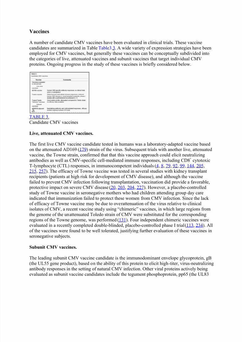
8/10/2019 Neuropathogenesis of Congenital Cytomegalovirus Infection
http://slidepdf.com/reader/full/neuropathogenesis-of-congenital-cytomegalovirus-infection 28/43
Vaccines
A number of candidate CMV vaccines have been evaluated in clinical trials. These vaccine
candidates are summarized in Table Table3.3. A wide variety of expression strategies have beenemployed for CMV vaccines, but generally these vaccines can be conceptually subdivided into
the categories of live, attenuated vaccines and subunit vaccines that target individual CMV proteins. Ongoing progress in the study of these vaccines is briefly considered below.
TABLE 3.
Candidate CMV vaccines
Live, attenuated CMV vaccines.
The first live CMV vaccine candidate tested in humans was a laboratory-adapted vaccine basedon the attenuated AD169 (179) strain of the virus. Subsequent trials with another live, attenuated
vaccine, the Towne strain, confirmed that that this vaccine approach could elicit neutralizing
antibodies as well as CMV-specific cell-mediated immune responses, including CD8+ cytotoxic
T-lymphocyte (CTL) responses, in immunocompetent individuals (4, 8, 79, 92, 99, 144, 205, 215, 257). The efficacy of Towne vaccine was tested in several studies with kidney transplant
recipients (patients at high risk for development of CMV disease), and although the vaccine
failed to prevent CMV infection following transplantation, vaccination did provide a favorable, protective impact on severe CMV disease (20, 203, 204, 227). However, a placebo-controlled
study of Towne vaccine in seronegative mothers who had children attending group day care
indicated that immunization failed to protect these women from CMV infection. Since the lackof efficacy of Towne vaccine may be due to overattenuation of the virus relative to clinical
isolates of CMV, a recent vaccine study using “chimeric” vaccines, in which large regions from
the genome of the unattenuated Toledo strain of CMV were substituted for the correspondingregions of the Towne genome, was performed (131). Four independent chimeric vaccines were
evaluated in a recently completed double-blinded, placebo-controlled phase I trial (113, 234). All
of the vaccines were found to be well tolerated, justifying further evaluation of these vaccines in
seronegative subjects.
Subunit CMV vaccines.
The leading subunit CMV vaccine candidate is the immunodominant envelope glycoprotein, gB
(the UL55 gene product), based on the ability of this protein to elicit high-titer, virus-neutralizing
antibody responses in the setting of natural CMV infection. Other viral proteins actively beingevaluated as subunit vaccine candidates include the tegument phosphoprotein, pp65 (the UL83

8/10/2019 Neuropathogenesis of Congenital Cytomegalovirus Infection
http://slidepdf.com/reader/full/neuropathogenesis-of-congenital-cytomegalovirus-infection 29/43
gene product), and IE1 (the UL123 gene product), both of which elicit T-cell responses. The
current status of individual subunit vaccine candidates is summarized below (Table (Table33).
(i) Adjuvanted protein vaccines.
The formulation of CMV gB currently being utilized in clinical vaccine trials is a recombinant protein expressed in Chinese hamster ovary (CHO) cells (250, 251). In a phase I randomized,
double-blind, placebo-controlled trial in adults, gB vaccine, combined either with a novel
adjuvant, MF59, or alum adjuvant, elicited levels of virus-neutralizing antibody that exceededthose observed in CMV-seropositive control subjects (197). In all subsequent studies to date (86,
176), the safety profile of the vaccine has been very favorable, with injection site discomfort
being the only significant adverse event observed. There is currently a phase III efficacy study ofgB/MF59 vaccine ongoing in young, CMV-seronegative women who are vaccinated postpartum
(289). These women are at high risk for primary CMV infection following delivery. Since these
women commonly become pregnant again soon after delivery, this study will be of great
importance to the prevention of congenital CMV infection in subsequent deliveries.
(ii) DNA vaccines.
DNA vaccines developed for CMV (265) have focused on the gB, IE1, and pp65 genes as the
candidate target immunogens. There are currently phase I clinical trials under way of both a bivalent CMV DNA vaccine candidate, using plasmid DNA encoding pp65 and gB, and atrivalent vaccine candidate, which also includes a third plasmid encoding the IE1 gene product,
developed and produced by Vical Vaccines (240, 277). The vaccines are formulated using the
poloxamer adjuvant CRL1005 and benzalkonium chloride. A study is currently ongoing with the bivalent DNA vaccine in a hematopoietic stem cell transplant population. Both donors and
recipients are being vaccinated, with the goal of reducing CMV disease, viral load, and use of
antiviral therapies in the posttransplant period. The long-term role of these vaccines for potentialcontrol of congenital CMV infection in women of child-bearing age remains to be determined, asdoes the ultimate acceptability of DNA vaccines for clinical use.
(iii) Vectored vaccines.
Another approach to CMV subunit vaccination is utilization of a “vectored” vaccine, in whichthe gene product of interest is expressed in the context of a nonreplicating (usually viral) vector.
Once example of such a vaccine vector is a canarypox vector known as ALVAC, an attenuated
poxvirus that replicates productively in avian species but abortively in mammalian cells.
ALVAC expressing CMV gB has been studied in several clinical trials (7, 30). An ALVACvaccine expressing pp65, the major CTL target in naturally CMV seropositive persons, has also
been evaluated in human trials. The ALVAC expression vector has been found to be safe and
well tolerated, although the expression strategy does not appear to offer advantages inimmunogenicity compared to other approaches. Other vectored CMV vaccine expression
strategies include an approach based on the modified vaccinia virus Ankara (279) and a
Venezuelan equine encephalitis virus-vectored CMV vaccine (235), manufactured by AlphaVaxVaccines. Both approaches show promise, and the Venezuelan equine encephalitis virus-

8/10/2019 Neuropathogenesis of Congenital Cytomegalovirus Infection
http://slidepdf.com/reader/full/neuropathogenesis-of-congenital-cytomegalovirus-infection 30/43
vectored approach provided protection in an animal model of congenital CMV infection (see
below), but these vaccines have not yet been tested in human clinical trials.
Preclinical vaccine approaches.
Other vaccine approaches have been suggested for development of a CMV vaccine, and these arethe subject of a recent review (226). These potential strategies are listed in Table Table3.3. CMV
glycoproteins other than gB have been considered for vaccine development, including the gcII
complex, consisting of gN (UL73) and gM (UL100), and the gcIII complex, consisting of gH(UL75), gO (UL74), and gL (UL115). Vaccines based on nonstructural proteins that are highly
conserved among the CMVs, including the DNA polymerase and helicase genes, show promise
in murine models. Dense bodies, which are enveloped, replication-defective particles formedduring replication of CMVs in cell culture, are capable of inducing virus-neutralizing antibodies
and T-cell responses after immunization of mice (226). The use of synthetic peptides comprising
immunodominant cytotoxic T-cell epitopes has been advocated, although such vaccines would
have to be tailored for donor-recipient pairs based on HLA genetics. Viral genomes cloned in
Escherichia coli as bacterial artificial chromosomes provide the opportunity to generaterecombinant, “designer” vaccines with specific genomic deletions or insertions that could modify
the immune response or improve the safety profile of the candidate vaccine. A “prime- boost”approach, in which priming with DNA vaccination is followed by boosting with formalin-
inactivated viral particles, elicits high levels of neutralizing antibodies as well as CD8+ T-cell
responses. These approaches deserve further attention in preclinical study and in phase I clinical
trials. The importance of identifying the maternal immune factors that protect the developingfetal CNS cannot be overstated, and this should be a key priority in future vaccine design.
Validation of vaccines for congenital CMV in animal models.
Prioritization of vaccine candidates for clinical trials can be based on results identified in theguinea pig model of congenital CMV infection. In light of the ability of GPCMV to cross the placenta and infect the pup in utero, the guinea pig provides an ideal small animal model for the
study of vaccines designed to protect the fetus. A live, attenuated GPCMV vaccine, as well as a
partially purified, soluble envelope vaccine, administered with Freund's adjuvant, was able toshow protection against acute viremia and death and to reduce the incidence and extent of
maternal and fetal infection (31). Adjuvanted glycoprotein vaccines, generated by
immunoaffinity purification, have also been shown to protect newborn pups against congenitalinfection and disease (40, 110). Molecular cloning techniques allowed extension of these studies
to generation of cloned, recombinant subunit vaccine candidates in a number of expression
systems. Subunit vaccines based on the GPCMV homolog of gB are capable of inducing
neutralizing antibody responses when administered either as DNA vaccines or as adjuvanted, purified glcyoprotein vaccines expressed in baculovirus (231, 232). Efficacy analyses against
congenital GPCMV have been performed with these vaccines. A DNA gB subunit vaccine was
able to reduce congenital infection rates following third-trimester challenge of vaccinated,
pregnant guinea pigs. In a group of 26 live-born pups in the control group, the total congenitalinfection rate was 77% (20/26). In contrast, in the gB-vaccinated group the total congenital
infection rate was 41% (11/27) ( P < 0.05 versus control group). This was the first study
indicating that a DNA vaccine was capable of providing protection against congenital CMV
8/10/2019 Neuropathogenesis of Congenital Cytomegalovirus Infection
http://slidepdf.com/reader/full/neuropathogenesis-of-congenital-cytomegalovirus-infection 31/43
infection (231), and these data provide support for the ongoing development and testing of DNA
vaccines for clinical trials. A three-dose series of purified, recombinant gB, expressed and
purified from baculovirus, was studied following administration with either Freund's adjuvant oralum-based adjuvant (233). Among 36 live-born pups in the Freund's adjuvant group, 8 (22%)
had congenital GPCMV infection, compared to 15/32 pups (47%) with in the alum adjuvant
group ( P < 0.05 versus Freund's adjuvant). The basis for the improved protection in the Freund'sadjuvant group was the induction of higher virus-neutralizing antibody titers, an observation thatmay be relevant to clinical trials of gB vaccine.
Recently, the UL83 (pp65) homolog of CMV, GP83, was also evaluated as a vaccine against
congenital CMV infection and disease in the guinea pig model. This vaccine was developed from
a propagation-defective, single-cycle RNA replicon vector system, based on an attenuated strain
of the alphavirus Venezuelan equine encephalitis virus. This expression system was used to produce virus-like replicon particles (VRPs) expressing GP83. Subsequent vaccination with
VRP-GP83 induced antibodies and CD4+ and CD8
+ T-cell responses in GPCMV-seronegative
female guinea pigs. Guinea pigs immunized with VRP-GP83 vaccine were bred for pregnancy
and subsequent GPCMV challenge during the early third trimester. Dams vaccinated with VRP-GP83 had improved pregnancy outcomes compared with controls, supporting the concept that T-
cell-mediated immune responses directed against a CMV matrix protein can protect againstcongenital CMV infection and disease.
Although these studies shed light on mechanisms that protect the fetus, there is a paucity of
information about how vaccination in animal models can protect against neuropathogenesis. Thisis a high-priority area for future research in the guinea pig model. Assessments of vaccine-
mediated protection against SHNL and neuropathology in this and other congenital infection
models will be of great importance in clarifying which human CMV vaccines strategies should be emphasized in clinical trials. An important issue to clarify in future clinical trials will be the
impact of reinfection in women with preconception immunity on the incidence and morbidity of
congenital CMV infection. A fundamental and unresolved quandary is that if vaccines induce
immunity but preexisting natural immunity does not fully protect women from CMV reinfectionand the fetal consequences of these infections, could vaccines ever be effective in eradication of
congenital CMV disease? The answer may lie in how the vaccine would be applied in clinical
practice. As suggested by some authorities, the force of infection of CMV is low, and a universalimmunization program could eliminate CMV infection from a community, even if the vaccine
was only modestly effective (103) Thus, the issue may be not the immunogenicity of the vaccine
but the degree to which CMV vaccination could confer herd immunity. If herd immunity in turndecreases the prevalence of CMV infection in the population, then the incidence of congenital
CMV infection would be predicted to be substantially reduced. This was the effect observed
following adoption of universal immunization against rubella (103), which led to the virtual
elimination of congenital rubella syndrome.
Go to:
SUMMARY AND PERSPECTIVES

8/10/2019 Neuropathogenesis of Congenital Cytomegalovirus Infection
http://slidepdf.com/reader/full/neuropathogenesis-of-congenital-cytomegalovirus-infection 32/43
Congential CMV infection is the predominant cause of developmental neurological disabilities in
the United States. It most frequently begins as a primary infection in the pregnant mother, who
may be exposed to several risk factors, including her own CMV-infected children who shedinfectious virus. Prior exposure to CMV markedly reduces the possibility of fetal infection but
does not preclude the virus from crossing the placenta to infect the fetus. Not only does the
infected placenta serve as an amplifying reservoir, but CMV infection may also cause placentalinsufficiency that result in fetal pathologies. Gestational age at infection greatly influencesneurological outcomes in the fetus. However, the combination of factors that render the
developing brain susceptible to CMV infection is poorly understood.
CMV infection of the fetus may alter the “normal blueprint” of the developing brain, thus
resulting in long-term neurological sequelae. Studies that investigate mechanisms that alter
developmental paradigms will help elucidate CMV neuropathogenesis and explain the clinicaloutcomes of congenital CMV infection. Neural stem cells in the fetal brain appear to be the
predominant cell type affected during development by CMV. There is an abundance of neural
stem cells in the fetal brain, and their increased susceptibility to viral infection explains the
predominance of nerurological sequelae associated with congenital CMV infection. Some possible mechanisms of developmental disruption due to CMV infection include (i) the loss of
neural stem cells or intermediate progenitors, the building blocks of the developing brain, due toinfection, which may have potential effects on brain size and maturation (165); (ii) alterations instem cell migration and fate of cell differentiation, processes which are critical for normal
pattering and function of neural structures; (iii) infection of astroglia, which may disrupt their
normal supportive functions in the brain that are a critical for neural circuitry guidance, synapticintegration, and development of the functionally intergrated mature neuron (160); and (iv)
alterations in the microenvironment of the developing brain due to cytokines and soluble factors
generated by resident glial cells (microglia and astrocytes) and infiltrating immune cells, which
may result in neurotoxicity (68) and altered neuronal physiology (116) and induce abnormaldevelopmental cues in the fetal brain (Fig. (Fig.4).4). All these mechanisms may potentially
affect the developmental patterning of the brain.
FIG. 4.
Schematic representation of neuropathogenic mechanisms during congenital CMV infection.
CMV brain infection may have multifaceted influences on the developing brain. Neural stem
cells (in blue), found along the lateral ventricular wall of the brain, are ...
Antiviral drugs are available for treatment of congenital CMV infection, and there is evidencethat therapy ameliorates the severity of one of the CNS complications of infection, SNHL. Long-
term neurodevelopmental follow-up studies should further clarify the value of antiviral therapy
in congenitally infected infants. Uncontrolled studies of therapy in utero with CMV immune
globulin have suggested an impact on neuropathogenesis, and controlled trials should beconducted with pregnant women. Finally, CMV vaccines may hold the greatest promise in
reducing the neurodevelopmental consequences of congenital infection, although the immune

8/10/2019 Neuropathogenesis of Congenital Cytomegalovirus Infection
http://slidepdf.com/reader/full/neuropathogenesis-of-congenital-cytomegalovirus-infection 33/43
correlates of protection of the fetus remain incompletely defined. Preclinical study with relevant
animal models may provide insights into strategies that bear further testing in human clinical
trials. Until more-effective interventions are available, better education of women of child- bearing age will be important. Only with increased public awareness of the urgency of this
problem can the societal and political forces necessary to effect changes be marshaled. These
changes include increased funding for the study of the pathogenesis of congenital CMV and anincreased sense of urgency in conducting clinical vaccine trials. Web-based information from both the Centers for Disease Control (http://www.cdc.gov/cmv/) and parent support groups
founded by individuals who have children afflicted with congenital CMV
(http://www.cmvfoundation.org) can be an effective conduit of knowledge and perspective thathelp to increase pubic awareness of this urgent problem.
Go to:
ACKNOWLEDGMENTS
This work was supported by NIH grants NS038836, HD038416, and HD044864.
Go to:
REFERENCES
1. Abbott, N. J., L. Ronnback, and E. Hansson. 2006. Astrocyte-endothelial interactions at the
blood-brain barrier. Nat. Rev. Neurosci. 7:41-53. [PubMed]
2. Adler, B., L. Scrivano, Z. Ruzcics, B. Rupp, C. Sinzger, and U. Koszinowski. 2006. Role of
human cytomegalovirus UL131A in cell type-specific virus entry and release. J. Gen. Virol.
87:2451-2460. [PubMed] 3. Adler, S. P., J. W. Finney, A. M. Manganello, and A. M. Best. 2004. Prevention of child-to-
mother transmission of cytomegalovirus among pregnant women. J. Pediatr. 145:485-491.[PubMed]
4. Adler, S. P., S. H. Hempfling, S. E. Starr, S. A. Plotkin, and S. Riddell. 1998. Safety and
immunogenicity of the Towne strain cytomegalovirus vaccine. Pediatr. Infect. Dis. J. 17:200-
206. [PubMed] 5. Adler, S. P., and G. Nigro. 2006. Interrupting intrauterine transmission of cytomegalovirus.
Rev. Med. Virol. 16:69-71. [PubMed]
6. Adler, S. P., G. Nigro, and L. Pereira. 2007. Recent advances in the prevention and treatment
of congenital cytomegalovirus infections. Semin. Perinatol. 31:10-18. [PubMed] 7. Adler, S. P., S. A. Plotkin, E. Gonczol, M. Cadoz, C. Meric, J. B. Wang, P. Dellamonica, A.
M. Best, J. Zahradnik, S. Pincus, K. Berencsi, W. I. Cox, and Z. Gyulai. 1999. A canarypox
vector expressing cytomegalovirus (CMV) glycoprotein B primes for antibody responses to alive attenuated CMV vaccine (Towne). J. Infect. Dis. 180:843-846. [PubMed]
8. Adler, S. P., S. E. Starr, S. A. Plotkin, S. H. Hempfling, J. Buis, M. L. Manning, and A. M.
Best. 1995. Immunity induced by primary human cytomegalovirus infection protects againstsecondary infection among women of childbearing age. J. Infect. Dis. 171:26-32. [PubMed]
8/10/2019 Neuropathogenesis of Congenital Cytomegalovirus Infection
http://slidepdf.com/reader/full/neuropathogenesis-of-congenital-cytomegalovirus-infection 34/43
9. Akter, P., C. Cunningham, B. P. McSharry, A. Dolan, C. Addison, D. J. Dargan, A. F. Hassan-
Walker, V. C. Emery, P. D. Griffiths, G. W. Wilkinson, and A. J. Davison. 2003. Two novel
spliced genes in human cytomegalovirus. J. Gen. Virol. 84:1117-1122. [PubMed] 10. Anastassova-Kristeva, M. 2003. The origin and development of the immune system with a
view to stem cell therapy. J. Hematother. Stem Cell Res. 12:137-154. [PubMed]
11. Ancora, G., M. Lanari, T. Lazzarotto, V. Venturi, E. Tridapalli, F. Sandri, M. Menarini, E.Ferretti, and G. Faldella. 2007. Cranial ultrasound scanning and prediction of outcome innewborns with congenital cytomegalovirus infection. J. Pediatr. 150:157-161. [PubMed]
12. Andriesse, G. I., A. J. Weersink, and J. de Boer. 2006. Visual impairment and deafness in
young children: consider the diagnosis of congenital infection with cytomegalovirus, even yearsafter birth. Arch. Ophthalmol. 124:743. [PubMed]
13. Arav-Boger, R., C. A. Battaglia, T. Lazzarotto, L. Gabrielli, J. C. Zong, G. S. Hayward, M.
Diener-West, and M. P. Landini. 2006. Cytomegalovirus (CMV)-encoded UL144 (truncated
tumor necrosis factor receptor) and outcome of congenital CMV infection. J. Infect. Dis.194:464-473. [PubMed]
14. Arnoult, D., L. M. Bartle, A. Skaletskaya, D. Poncet, N. Zamzami, P. U. Park, J. Sharpe, R.
J. Youle, and V. S. Goldmacher. 2004. Cytomegalovirus cell death suppressor vMIA blocksBax- but not Bak-mediated apoptosis by binding and sequestering Bax at mitochondria. Proc.
Natl. Acad. Sci. USA 101:7988-7993. [PMC free article] [PubMed]
15. Ashkenazi, A. 2002. Targeting death and decoy receptors of the tumour-necrosis factor
superfamily. Nat. Rev. Cancer 2:420-430. [PubMed] 16. Attard-Montalto, S. P., M. C. English, L. Stimmler, and G. J. Snodgrass. 1993. Ganciclovir
treatment of congenital cytomegalovirus infection: a report of two cases. Scand. J. Infect. Dis.
25:385-388. [PubMed] 17. Bachor, E., H. Sudhoff, R. Litschel, and C. S. Karmody. 2000. The pathology of the temporal
bones of a child with acquired cytomegalovirus infection: studies by light microscopy,
immunohistochemistry and polymerase-chain reaction. Int. J. Pediatr. Otorhinolaryngol. 55:215-
224. [PubMed] 18. Bakardjiev, A. I., B. A. Stacy, S. J. Fisher, and D. A. Portnoy. 2004. Listeriosis in the
pregnant guinea pig: a model of vertical transmission. Infect. Immun. 72:489-497. [PMC free
article] [PubMed] 19. Bakardjiev, A. I., B. A. Stacy, and D. A. Portnoy. 2005. Growth of Listeria monocytogenes
in the guinea pig placenta and role of cell-to-cell spread in fetal infection. J. Infect. Dis.
191:1889-1897. [PubMed] 20. Balfour, H. H., Jr., G. W. Sachs, P. Welo, R. C. Gehrz, R. L. Simmons, and J. S. Najarian.
1984. Cytomegalovirus vaccine in renal transplant candidates: progress report of a randomized,
placebo-controlled, double-blind trial. Birth Defects Orig. Artic. Ser. 20:289-304. [PubMed]
21. Bantug, G. R., D. Cekinovic, R. Bradford, T. Koontz, S. Jonjic, and W. J. Britt. 2008. CD8+T lymphocytes control murine cytomegalovirus replication in the central nervous system of
newborn animals. J. Immunol. 181:2111-2123. [PMC free article] [PubMed]
22. Barkovich, A. J., and N. Girard. 2003. Fetal brain infections. Childs Nerv. Syst. 19:501-507.
[PubMed] 23. Barkovich, A. J., and C. E. Lindan. 1994. Congenital cytomegalovirus infection of the brain:
imaging analysis and embryologic considerations. AJNR Am. J. Neuroradiol. 15:703-715.
[PubMed]
8/10/2019 Neuropathogenesis of Congenital Cytomegalovirus Infection
http://slidepdf.com/reader/full/neuropathogenesis-of-congenital-cytomegalovirus-infection 35/43
24. Baron, M. H., and S. T. Fraser. 2005. The specification of early hematopoiesis in the
mammal. Curr. Opin. Hematol. 12:217-221. [PubMed]
25. Bass, K. E., H. Li, S. P. Hawkes, E. Howard, E. Bullen, T. K. Vu, M. McMaster, M.Janatpour, and S. J. Fisher. 1997. Tissue inhibitor of metalloproteinase-3 expression is
upregulated during human cytotrophoblast invasion in vitro. Dev. Genet. 21 :61-67. [PubMed]
26. Bauer, P. W., M. Parizi-Robinson, P. S. Roland, and S. Yegappan. 2005. Cytomegalovirus inthe perilymphatic fluid. Laryngoscope 115:223-225. [PubMed] 27. Benedict, C. A. 2003. Viruses and the TNF-related cytokines, an evolving battle. Cytokine
Growth Factor Rev. 14:349-357. [PubMed]
28. Benedict, C. A., P. S. Norris, and C. F. Ware. 2002. To kill or be killed: viral evasion ofapoptosis. Nat. Immunol. 3:1013-1018. [PubMed]
29. Bentz, G. L., M. Jarquin-Pardo, G. Chan, M. S. Smith, C. Sinzger, and A. D. Yurochko.
2006. Human cytomegalovirus (HCMV) infection of endothelial cells promotes naive monocyte
extravasation and transfer of productive virus to enhance hematogenous dissemination ofHCMV. J. Virol. 80:11539-11555. [PMC free article] [PubMed]
30. Bernstein, D. I., M. R. Schleiss, K. Berencsi, E. Gonczol, M. Dickey, P. Khoury, M. Cadoz,
C. Meric, J. Zahradnik, A. M. Duliege, and S. Plotkin. 2002. Effect of previous or simultaneousimmunization with canarypox expressing cytomegalovirus (CMV) glycoprotein B (gB) on
response to subunit gB vaccine plus MF59 in healthy CMV-seronegative adults. J. Infect. Dis.
185:686-690. [PubMed]
31. Bia, F. J., B. P. Griffith, M. Tarsio, and G. D. Hsiung. 1980. Vaccination for the preventionof maternal and fetal infection with guinea pig cytomegalovirus. J. Infect. Dis. 142:732-738.
[PubMed]
32. Bigger, J. E., M. Tanigawa, M. Zhang, and S. S. Atherton. 2000. Murine cytomegalovirusinfection causes apoptosis of uninfected retinal cells. Investig. Ophthalmol. Vis. Sci. 41:2248-
2254. [PubMed]
33. Boppana, S. B., and W. J. Britt. 1995. Antiviral antibody responses and intrauterine
transmission after primary maternal cytomegalovirus infection. J. Infect. Dis. 171:1115-1121.[PubMed]
34. Boppana, S. B., K. B. Fowler, W. J. Britt, S. Stagno, and R. F. Pass. 1999. Symptomatic
congenital cytomegalovirus infection in infants born to mothers with preexisting immunity tocytomegalovirus. Pediatrics 104:55-60. [PubMed]
35. Boppana, S. B., K. B. Fowler, R. F. Pass, L. B. Rivera, R. D. Bradford, F. D. Lakeman, and
W. J. Britt. 2005. Congenital cytomegalovirus infection: association between virus burden ininfancy and hearing loss. J. Pediatr. 146:817-823. [PubMed]
36. Boppana, S. B., K. B. Fowler, Y. Vaid, G. Hedlund, S. Stagno, W. J. Britt, and R. F. Pass.
1997. Neuroradiographic findings in the newborn period and long-term outcome in children with
symptomatic congenital cytomegalovirus infection. Pediatrics 99:409-414. [PubMed] 37. Boppana, S. B., J. Miller, and W. J. Britt. 1996. Transplacentally acquired antiviral
antibodies and outcome in congenital human cytomegalovirus infection. Viral Immunol. 9:211-
218. [PubMed]
38. Boppana, S. B., R. F. Pass, and W. J. Britt. 1993. Virus-specific antibody responses inmothers and their newborn infants with asymptomatic congenital cytomegalovirus infections. J.
Infect. Dis. 167:72-77. [PubMed]
8/10/2019 Neuropathogenesis of Congenital Cytomegalovirus Infection
http://slidepdf.com/reader/full/neuropathogenesis-of-congenital-cytomegalovirus-infection 36/43
39. Boppana, S. B., L. B. Rivera, K. B. Fowler, M. Mach, and W. J. Britt. 2001. Intrauterine
transmission of cytomegalovirus to infants of women with preconceptional immunity. N. Engl. J.
Med. 344:1366-1371. [PubMed] 40. Bourne, N., M. R. Schleiss, F. J. Bravo, and D. I. Bernstein. 2001. Preconception
immunization with a cytomegalovirus (CMV) glycoprotein vaccine improves pregnancy
outcome in a guinea pig model of congenital CMV infection. J. Infect. Dis. 183:59-64.[PubMed] 41. Bresnahan, W. A., I. Boldogh, E. A. Thompson, and T. Albrecht. 1996. Human
cytomegalovirus inhibits cellular DNA synthesis and arrests productively infected cells in late
G1. Virology 224:150-160. [PubMed] 42. Britt, W. J. 1996. Human cytomegalovirus infections during pregnancy, p. 327-343,
Baillière's clinical infectious diseases, vol. 3. Baillière Tindall, Oxford, United Kingdom.
43. Brune, W., M. Nevels, and T. Shenk. 2003. Murine cytomegalovirus m41 open reading
frame encodes a Golgi-localized antiapoptotic protein. J. Virol. 77:11633-11643. [PMC freearticle] [PubMed]
44. Cannon, M. J., and K. F. Davis. 2005. Washing our hands of the congenital cytomegalovirus
disease epidemic. BMC Public Health 5:70. [PMC free article] [PubMed] 45. Carson, M. J., J. M. Doose, B. Melchior, C. D. Schmid, and C. C. Ploix. 2006. CNS immune
privilege: hiding in plain sight. Immunol. Rev. 213:48-65. [PMC free article] [PubMed]
46. Castillo, J. P., and T. F. Kowalik. 2002. Human cytomegalovirus immediate early proteins
and cell growth control. Gene 290:19-34. [PubMed] 47. Chaouat, G., N. Ledee-Bataille, and S. Dubanchet. 2007. Immune cells in uteroplacental
tissues throughout pregnancy: a brief review. Reprod. Biomed. Online 14:256-266. [PubMed]
48. Cheeran, M. C., G. Gekker, S. Hu, X. Min, D. Cox, and J. R. Lokensgard. 2004. Intracerebralinfection with murine cytomegalovirus induces CXCL10 and is restricted by adoptive transfer of
splenocytes. J. Neurovirol. 10:152-162. [PubMed]
49. Cheeran, M. C., G. Gekker, S. Hu, J. M. Palmquist, and J. R. Lokensgard. 2005. T cell-
mediated restriction of intracerebral murine cytomegalovirus infection displays dependence upon perforin but not interferon-gamma. J. Neurovirol. 11:274-280. [PubMed]
50. Cheeran, M. C., S. Hu, G. Gekker, and J. R. Lokensgard. 2000. Decreased cytomegalovirus
expression following proinflammatory cytokine treatment of primary human astrocytes. J.Immunol. 164:926-933. [PubMed]
51. Cheeran, M. C., S. Hu, H. T. Ni, W. Sheng, J. M. Palmquist, P. K. Peterson, and J. R.
Lokensgard. 2005. Neural precursor cell susceptibility to human cytomegalovirus diverges alongglial or neuronal differentiation pathways. J. Neurosci. Res. 82:839-850. [PubMed]
52. Cheeran, M. C., S. Hu, J. M. Palmquist, T. Bakken, G. Gekker, and J. R. Lokensgard. 2007.
Dysregulated interferon-gamma responses during lethal cytomegalovirus brain infection of IL-
10-deficient mice. Virus Res. 130:96-102. [PMC free article] [PubMed] 53. Cheeran, M. C., S. Hu, W. S. Sheng, P. K. Peterson, and J. R. Lokensgard. 2003. CXCL10
production from cytomegalovirus-stimulated microglia is regulated by both human and viral
interleukin-10. J. Virol. 77:4502-4515. [PMC free article] [PubMed]
54. Cheeran, M. C., S. Hu, S. L. Yager, G. Gekker, P. K. Peterson, and J. R. Lokensgard. 2001.Cytomegalovirus induces cytokine and chemokine production differentially in microglia and
astrocytes: antiviral implications. J. Neurovirol. 7:135-147. [PubMed]
55. Chiou, S. H., J. H. Liu, W. M. Hsu, S. S. Chen, S. Y. Chang, L. J. Juan, J. C. Lin, Y. T.Yang, W. W. Wong, C. Y. Liu, Y. S. Lin, W. T. Liu, and C. W. Wu. 2001. Up-regulation of Fas

8/10/2019 Neuropathogenesis of Congenital Cytomegalovirus Infection
http://slidepdf.com/reader/full/neuropathogenesis-of-congenital-cytomegalovirus-infection 37/43
ligand expression by human cytomegalovirus immediate-early gene product 2: a novel
mechanism in cytomegalovirus-induced apoptosis in human retina. J. Immunol. 167:4098-4103.
[PubMed] 56. Chiou, S. H., Y. P. Yang, J. C. Lin, C. H. Hsu, H. C. Jhang, Y. T. Yang, C. H. Lee, L. L. Ho,
W. M. Hsu, H. H. Ku, S. J. Chen, S. S. Chen, M. D. Chang, C. W. Wu, and L. J. Juan. 2006. The
immediate early 2 protein of human cytomegalovirus (HCMV) mediates the apoptotic control inHCMV retinitis through up-regulation of the cellular FLICE-inhibitory protein expression. J.Immunol. 177:6199-6206. [PubMed]
57. Choi, C., and E. N. Benveniste. 2004. Fas ligand/Fas system in the brain: regulator of
immune and apoptotic responses. Brain Res. Brain Res. Rev. 44:65-81. [PubMed] 58. Chou, S., N. S. Lurain, K. D. Thompson, R. C. Miner, and W. L. Drew. 2003. Viral DNA
polymerase mutations associated with drug resistance in human cytomegalovirus. J. Infect. Dis.
188:32-39. [PubMed]
59. Cinatl, J., Jr., R. Blaheta, M. Bittoova, M. Scholz, S. Margraf, J. U. Vogel, J. Cinatl, and H.W. Doerr. 2000. Decreased neutrophil adhesion to human cytomegalovirus-infected retinal
pigment epithelial cells is mediated by virus-induced up-regulation of Fas ligand independent of
neutrophil apoptosis. J. Immunol. 165:4405-4413. [PubMed] 60. Crowley, B. 2002. Ganciclovir treatment of symptomatic congenital cytomegalovirus
infection. J. Antimicrob. Chemother. 50:435-436. [PubMed]
61. Dahle, A. J., K. B. Fowler, J. D. Wright, S. B. Boppana, W. J. Britt, and R. F. Pass. 2000.
Longitudinal investigation of hearing disorders in children with congenital cytomegalovirus. J.Am. Acad. Audiol. 11:283-290. [PubMed]
62. Davis, L. E., L. G. Johnsson, and M. Kornfeld. 1981. Cytomegalovirus labyrinthitis in an
infant: morphological, virological, and immunofluorescent studies. J. Neuropathol. Exp. Neurol.40:9-19. [PubMed]
63. Davison, A. J., A. Dolan, P. Akter, C. Addison, D. J. Dargan, D. J. Alcendor, D. J. McGeoch,
and G. S. Hayward. 2003. The human cytomegalovirus genome revisited: comparison with the
chimpanzee cytomegalovirus genome. J. Gen. Virol. 84:17-28. [PubMed] 64. DeBiasi, R. L., B. K. Kleinschmidt-DeMasters, S. Richardson-Burns, and K. L. Tyler. 2002.
Central nervous system apoptosis in human herpes simplex virus and cytomegalovirus
encephalitis. J. Infect. Dis. 186:1547-1557. [PubMed] 65. de Graaf-Peters, V. B., and M. Hadders-Algra. 2006. Ontogeny of the human central nervous
system: what is happening when? Early Hum. Dev. 82:257-266. [PubMed]
66. Demmler, G. J. 1994. Congenital cytomegalovirus infection. Semin. Pediatr. Neurol. 1:36-42. [PubMed]
67. D'Erchia, A. M., C. Gissi, G. Pesole, C. Saccone, and U. Arnason. 1996. The guinea-pig is
not a rodent. Nature 381:597-600. [PubMed]
68. Deshpande, M., J. Zheng, K. Borgmann, R. Persidsky, L. Wu, C. Schellpeper, and A.Ghorpade. 2005. Role of activated astrocytes in neuronal damage: potential links to HIV-1-
associated dementia. Neurotox. Res. 7:183-192. [PubMed]
69. Donnelly, T. M., and C. J. Brown. 2004. Guinea pig and chinchilla care and husbandry. Vet.
Clin. N. Am. Exot. Anim. Pract. 7:351-373. [PubMed] 70. Edington, N., S. Broad, A. E. Wrathall, and J. T. Done. 1988. Superinfection with porcine
cytomegalovirus initiating transplacental infection. Vet. Microbiol. 16:189-193. [PubMed]

8/10/2019 Neuropathogenesis of Congenital Cytomegalovirus Infection
http://slidepdf.com/reader/full/neuropathogenesis-of-congenital-cytomegalovirus-infection 38/43
71. Eggers, M., K. Radsak, G. Enders, and M. Reschke. 2001. Use of recombinant glycoprotein
antigens gB and gH for diagnosis of primary human cytomegalovirus infection during
pregnancy. J. Med. Virol. 63:135-142. [PubMed] 72. Elbou Ould, M. A., D. Luton, M. Yadini, B. Pedron, Y. Aujard, E. Jacqz-Aigrain, F.
Jacquemard, and G. Sterkers. 2004. Cellular immune response of fetuses to cytomegalovirus.
Pediatr. Res. 55:280-286. [PubMed] 73. Eudy, J. D., M. D. Weston, S. Yao, D. M. Hoover, H. L. Rehm, M. Ma-Edmonds, D. Yan, I.Ahmad, J. J. Cheng, C. Ayuso, C. Cremers, S. Davenport, C. Moller, C. B. Talmadge, K. W.
Beisel, M. Tamayo, C. C. Morton, A. Swaroop, W. J. Kimberling, and J. Sumegi. 1998.
Mutation of a gene encoding a protein with extracellular matrix motifs in Usher syndrome typeIIa. Science 280:1753-1757. [PubMed]
74. Fagerheim, T., O. Nilssen, P. Raeymaekers, V. Brox, T. Moum, H. H. Elverland, E. Teig, H.
H. Omland, G. K. Fostad, and L. Tranebjaerg. 1996. Identification of a new locus for autosomal
dominant non-syndromic hearing impairment (DFNA7) in a large Norwegian family. Hum. Mol.Genet. 5:1187-1191. [PubMed]
75. Fan-Havard, P., M. C. Nahata, and M. T. Brady. 1989. Ganciclovir — a review of
pharmacology, therapeutic efficacy and potential use for treatment of congenitalcytomegalovirus infections. J. Clin. Pharm. Ther. 14:329-340. [PubMed]
76. Fischler, B., T. H. Casswall, P. Malmborg, and A. Nemeth. 2002. Ganciclovir treatment in
infants with cytomegalovirus infection and cholestasis. J. Pediatr. Gastroenterol. Nutr. 34:154-
157. [PubMed] 77. Fish, K. N., C. Soderberg-Naucler, L. K. Mills, S. Stenglein, and J. A. Nelson. 1998. Human
cytomegalovirus persistently infects aortic endothelial cells. J. Virol. 72:5661-5668. [PMC free
article] [PubMed] 78. Fisher, S., O. Genbacev, E. Maidji, and L. Pereira. 2000. Human cytomegalovirus infection
of placental cytotrophoblasts in vitro and in utero: implications for transmission and
pathogenesis. J. Virol. 74:6808-6820. [PMC free article] [PubMed]
79. Fleisher, G. R., S. E. Starr, H. M. Friedman, and S. A. Plotkin. 1982. Vaccination of pediatricnurses with live attenuated cytomegalovirus. Am. J. Dis. Child. 136:294-296. [PubMed]
80. Fortunato, E. A., M. L. Dell'Aquila, and D. H. Spector. 2000. Specific chromosome 1 breaks
induced by human cytomegalovirus. Proc. Natl. Acad. Sci. USA 97:853-858. [PMC free article] [PubMed]
81. Fowler, K. B., A. J. Dahle, S. B. Boppana, and R. F. Pass. 1999. Newborn hearing screening:
will children with hearing loss caused by congenital cytomegalovirus infection be missed? J.Pediatr. 135:60-64. [PubMed]
82. Fowler, K. B., F. P. McCollister, A. J. Dahle, S. Boppana, W. J. Britt, and R. F. Pass. 1997.
Progressive and fluctuating sensorineural hearing loss in children with asymptomatic congenital
cytomegalovirus infection. J. Pediatr. 130:624-630. [PubMed] 83. Fowler, K. B., and R. F. Pass. 2006. Risk factors for congenital cytomegalovirus infection in
the offspring of young women: exposure to young children and recent onset of sexual activity.
Pediatrics 118:e286-e292. [PubMed]
84. Fowler, K. B., S. Stagno, and R. F. Pass. 1993. Maternal age and congenital cytomegalovirusinfection: screening of two diverse newborn populations, 1980-1990. J. Infect. Dis. 168 :552-556.
[PubMed]
85. Fowler, K. B., S. Stagno, and R. F. Pass. 2003. Maternal immunity and prevention ofcongenital cytomegalovirus infection. JAMA. 289:1008-1011. [PubMed]
8/10/2019 Neuropathogenesis of Congenital Cytomegalovirus Infection
http://slidepdf.com/reader/full/neuropathogenesis-of-congenital-cytomegalovirus-infection 39/43
86. Frey, S. E., C. Harrison, R. F. Pass, E. Yang, D. Boken, R. E. Sekulovich, S. Percell, A. E.
Izu, S. Hirabayashi, R. L. Burke, and A. M. Duliege. 1999. Effects of antigen dose and
immunization regimens on antibody responses to a cytomegalovirus glycoprotein B subunitvaccine. J. Infect. Dis. 180:1700-1703. [PubMed]
87. Fryer, J. F., P. D. Griffiths, V. C. Emery, and D. A. Clark. 2004. Susceptibility of porcine
cytomegalovirus to antiviral drugs. J. Antimicrob. Chemother. 53:975-980. [PubMed] 88. Fukuda, S., E. M. Keithley, and J. P. Harris. 1988. Experimental cytomegalovirus infection:viremic spread to the inner ear. Am. J. Otolaryngol. 9:135-141. [PubMed]
89. Gage, F. H. 2000. Mammalian neural stem cells. Science 287:1433-1438. [PubMed]
90. Galea, I., I. Bechmann, and V. H. Perry. 2007. What is immune privilege (not)? TrendsImmunol. 28:12-18. [PubMed]
91. Gaytant, M. A., E. A. Steegers, B. A. Semmekrot, H. M. Merkus, and J. M. Galama. 2002.
Congenital cytomegalovirus infection: review of the epidemiology and outcome. Obstet.
Gynecol. Surv. 57:245-256. [PubMed] 92. Gehrz, R. C., W. R. Christianson, K. M. Linner, K. E. Groth, and H. H. Balfour, Jr. 1980.
Cytomegalovirus vaccine. Specific humoral and cellular immune responses in human volunteers.
Arch. Intern. Med. 140:936-939. [PubMed] 93. Gerna, G., E. Percivalle, D. Lilleri, L. Lozza, C. Fornara, G. Hahn, F. Baldanti, and M. G.
Revello. 2005. Dendritic-cell infection by human cytomegalovirus is restricted to strains carrying
functional UL131-128 genes and mediates efficient viral antigen presentation to CD8+ T cells. J.
Gen. Virol. 86:275-284. [PubMed] 94. Gibson, L., S. Dooley, S. Trzmielina, M. Somasundaran, D. Fisher, M. G. Revello, and K.
Luzuriaga. 2007. Cytomegalovirus (CMV) IE1- and pp65-specific CD8+ T cell responses
broaden over time after primary CMV infection in infants. J. Infect. Dis. 195:1789-1798.[PubMed]
95. Gibson, L., G. Piccinini, D. Lilleri, M. G. Revello, Z. Wang, S. Markel, D. J. Diamond, and
K. Luzuriaga. 2004. Human cytomegalovirus proteins pp65 and immediate early protein 1 are
common targets for CD8+ T cell responses in children with congenital or postnatal humancytomegalovirus infection. J. Immunol. 172:2256-2264. [PubMed]
96. Goldmacher, V. S. 2005. Cell death suppression by cytomegaloviruses. Apoptosis 10:251-
265. [PubMed] 97. Goldmacher, V. S., L. M. Bartle, A. Skaletskaya, C. A. Dionne, N. L. Kedersha, C. A. Vater,
J. W. Han, R. J. Lutz, S. Watanabe, E. D. Cahir McFarland, E. D. Kieff, E. S. Mocarski, and T.
Chittenden. 1999. A cytomegalovirus-encoded mitochondria-localized inhibitor of apoptosisstructurally unrelated to Bcl-2. Proc. Natl. Acad. Sci. USA 96:12536-12541. [PMC free article]
[PubMed]
98. Gonczol, E., P. W. Andrews, and S. A. Plotkin. 1984. Cytomegalovirus replicates in
differentiated but not in undifferentiated human embryonal carcinoma cells. Science 224:159-161. [PubMed]
99. Gonczol, E., J. Ianacone, G. Furlini, W. Ho, and S. A. Plotkin. 1989. Humoral immune
response to cytomegalovirus Towne vaccine strain and to Toledo low-passage strain. J. Infect.
Dis. 159:851-859. [PubMed] 100. Grassi, M. P., F. Clerici, C. Perin, A. D'Arminio Monforte, L. Vago, M. Borella, R.
Boldorini, and A. Mangoni. 1998. Microglial nodular encephalitis and ventriculoencephalitis due
to cytomegalovirus infection in patients with AIDS: two distinct clinical patterns. Clin. Infect.Dis. 27:504-508. [PubMed]

8/10/2019 Neuropathogenesis of Congenital Cytomegalovirus Infection
http://slidepdf.com/reader/full/neuropathogenesis-of-congenital-cytomegalovirus-infection 40/43
101. Griffith, B. P., H. L. Lucia, and G. D. Hsiung. 1982. Brain and visceral involvement during
congenital cytomegalovirus infection of guinea pigs. Pediatr. Res. 16:455-459. [PubMed]
102. Griffiths, P. D. 2002. The treatment of cytomegalovirus infection. J. Antimicrob.Chemother. 49:243-253. [PubMed]
103. Griffiths, P. D., A. McLean, and V. C. Emery. 2001. Encouraging prospects for
immunisation against primary cytomegalovirus infection. Vaccine 19:1356-1362. [PubMed] 104. Haginoya, K., T. Ohura, K. Kon, T. Yagi, Y. Sawaishi, K. K. Ishii, T. Funato, S. Higano, S.Takahashi, and K. Iinuma. 2002. Abnormal white matter lesions with sensorineural hearing loss
caused by congenital cytomegalovirus infection: retrospective diagnosis by PCR using Guthrie
cards. Brain Dev. 24:710-714. [PubMed] 105. Hahn, G., R. Jores, and E. S. Mocarski. 1998. Cytomegalovirus remains latent in a common
precursor of dendritic and myeloid cells. Proc. Natl. Acad. Sci. USA 95:3937-3942. [PMC free
article] [PubMed]
106. Hahn, G., M. G. Revello, M. Patrone, E. Percivalle, G. Campanini, A. Sarasini, M. Wagner,A. Gallina, G. Milanesi, U. Koszinowski, F. Baldanti, and G. Gerna. 2004. Human
cytomegalovirus UL131-128 genes are indispensable for virus growth in endothelial cells and
virus transfer to leukocytes. J. Virol. 78:10023-10033. [PMC free article] [PubMed] 107. Halwachs-Baumann, G. 2006. The congenital cytomegalovirus infection: virus-host
interaction for defense and transmission. Curr. Pharm. Biotechnol. 7:303-312. [PubMed]
108. Han, G. P., L. Li, I. Kosugi, H. Kawasaki, T. Tsuchida, K. Miura, and Y. Tsutsui. 2007.
Enhancement of susceptibility of adult mouse brain to cytomegalovirus infection by infusion ofepidermal growth factor. J. Neurosci. Res. 85:2981-2990. [PubMed]
109. Harris, J. P., N. K. Woolf, A. F. Ryan, D. M. Butler, and D. D. Richman. 1984.
Immunologic and electrophysiological response to cytomegaloviral inner ear infection in theguinea pig. J. Infect. Dis. 150:523-530. [PubMed]
110. Harrison, C. J., W. J. Britt, N. M. Chapman, J. Mullican, and S. Tracy. 1995. Reduced
congenital cytomegalovirus (CMV) infection after maternal immunization with a guinea pig
CMV glycoprotein before gestational primary CMV infection in the guinea pig model. J. Infect.Dis. 172:1212-1220. [PubMed]
111. Hartley, J. W., W. P. Rowe, and R. J. Huebner. 1957. Serial propagation of the guinea pig
salivary gland virus in tissue culture. Proc. Soc. Exp. Biol. Med. 96:281-285. [PubMed] 112. Hassan, J., S. Dooley, and W. Hall. 2007. Immunological response to cytomegalovirus in
congenitally infected neonates. Clin. Exp. Immunol. 147:465-471. [PMC free article] [PubMed]
113. Heineman, T. C., M. Schleiss, D. I. Bernstein, R. R. Spaete, L. Yan, G. Duke, M. Prichard,Z. Wang, Q. Yan, M. A. Sharp, N. Klein, A. M. Arvin, and G. Kemble. 2006. A phase 1 study of
4 live, recombinant human cytomegalovirus Towne/Toledo chimeric vaccines. J. Infect. Dis.
193:1350-1360. [PubMed]
114. Hertel, L., S. Chou, and E. S. Mocarski. 2007. Viral and cell cycle-regulated kinases incytomegalovirus-induced pseudomitosis and replication. PLoS Pathog. 3:e6. [PMC free article]
[PubMed]
115. Hicks, T., K. Fowler, M. Richardson, A. Dahle, L. Adams, and R. Pass. 1993. Congenital
cytomegalovirus infection and neonatal auditory screening. J. Pediatr. 123:779-782. [PubMed] 116. Ho, W. S., and A. N. van den Pol. 2007. Bystander attenuation of neuronal and astrocyte
intercellular communication by murine cytomegalovirus infection of glia. J. Virol. 81:7286-
7292. [PMC free article] [PubMed]
8/10/2019 Neuropathogenesis of Congenital Cytomegalovirus Infection
http://slidepdf.com/reader/full/neuropathogenesis-of-congenital-cytomegalovirus-infection 41/43
117. Hocker, J. R., L. N. Cook, G. Adams, and G. P. Rabalais. 1990. Ganciclovir therapy of
congenital cytomegalovirus pneumonia. Pediatr. Infect. Dis. J. 9:743-745. [PubMed]
118. Holt, P. G., and C. A. Jones. 2000. The development of the immune system during pregnancy and early life. Allergy 55:688-697. [PubMed]
119. Huang, D., Y. Han, M. R. Rani, A. Glabinski, C. Trebst, T. Sorensen, M. Tani, J. Wang, P.
Chien, S. O'Bryan, B. Bielecki, Z. L. Zhou, S. Majumder, and R. M. Ransohoff. 2000.Chemokines and chemokine receptors in inflammation of the nervous system: manifold roles andexquisite regulation. Immunol. Rev. 177:52-67. [PubMed]
120. Ishiwata, M., S. Baba, M. Kawashima, I. Kosugi, H. Kawasaki, M. Kaneta, T. Tsuchida, S.
Kozuma, and Y. Tsutsui. 2006. Differential expression of the immediate-early 2 and 3 proteins indeveloping mouse brains infected with murine cytomegalovirus. Arch. Virol. 151:2181-2196.
[PubMed]
121. Jamieson, D. J., J. E. Ellis, D. B. Jernigan, and T. A. Treadwell. 2006. Emerging infectious
disease outbreaks: old lessons and new challenges for obstetrician-gynecologists. Am. J. Obstet.Gynecol. 194:1546-1555. [PubMed]
122. Jarvis, M. A., and J. A. Nelson. 2002. Human cytomegalovirus persistence and latency in
endothelial cells and macrophages. Curr. Opin. Microbiol. 5:403-407. [PubMed] 123. Jault, F. M., J. M. Jault, F. Ruchti, E. A. Fortunato, C. Clark, J. Corbeil, D. D. Richman, and
D. H. Spector. 1995. Cytomegalovirus infection induces high levels of cyclins, phosphorylated
Rb, and p53, leading to cell cycle arrest. J. Virol. 69:6697-6704. [PMC free article] [PubMed]
124. Junker, A. K., D. Matheson, A. J. Tingle, and E. E. Thomas. 1991. Immune responses afterganciclovir and immunoglobulin therapy of infants. Pediatr. Infect. Dis. J. 10:256-258.
[PubMed]
125. Kashiwai, A., N. Kawamura, C. Kadota, and Y. Tsutsui. 1992. Susceptibility of mouseembryo to murine cytomegalovirus infection in early and mid-gestation stages. Arch. Virol.
127:37-48. [PubMed]
126. Katano, H., Y. Sato, Y. Tsutsui, T. Sata, A. Maeda, N. Nozawa, N. Inoue, Y. Nomura, and
T. Kurata. 2007. Pathogenesis of cytomegalovirus-associated labyrinthitis in a guinea pig model.Microbes Infect. 9:183-191. [PubMed]
127. Kaufmann, P., and M. Davidoff. 1977. The guinea-pig placenta. Adv. Anat. Embryol. Cell
Biol. 53:5-91. [PubMed] 128. Kawasaki, H., I. Kosugi, Y. Arai, and Y. Tsutsui. 2002. The amount of immature glial cells
in organotypic brain slices determines the susceptibility to murine cytomegalovirus infection.
Lab Investig. 82:1347-1358. [PubMed] 129. Keithley, E. M., N. K. Woolf, and J. P. Harris. 1989. Development of morphological and
physiological changes in the cochlea induced by cytomegalovirus. Laryngoscope 99:409-414.
[PubMed]
130. Keller, M. J., A. W. Wu, J. I. Andrews, P. W. McGonagill, E. E. Tibesar, and J. L. Meier.2007. Reversal of human cytomegalovirus major immediate-early enhancer/promoter silencing
in quiescently infected cells via the cyclic AMP signaling pathway. J. Virol. 81:6669-6681.
[PMC free article] [PubMed]
131. Kemble, G., G. Duke, R. Winter, and R. Spaete. 1996. Defined large-scale alterations of thehuman cytomegalovirus genome constructed by cotransfection of overlapping cosmids. J. Virol.
70:2044-2048. [PMC free article] [PubMed]
132. Kenneson, A., and M. J. Cannon. 2007. Review and meta-analysis of the epidemiology ofcongenital cytomegalovirus (CMV) infection. Rev. Med. Virol. 17:253-276. [PubMed]

8/10/2019 Neuropathogenesis of Congenital Cytomegalovirus Infection
http://slidepdf.com/reader/full/neuropathogenesis-of-congenital-cytomegalovirus-infection 42/43
133. Kimberlin, D. W. 2002. Antiviral therapy for cytomegalovirus infections in pediatric
patients. Semin. Pediatr. Infect. Dis. 13:22-30. [PubMed]
134. Kimberlin, D. W., C. Y. Lin, P. J. Sanchez, G. J. Demmler, W. Dankner, M. Shelton, R. F.Jacobs, W. Vaudry, R. F. Pass, J. M. Kiell, S. J. Soong, and R. J. Whitley. 2003. Effect of
ganciclovir therapy on hearing in symptomatic congenital cytomegalovirus disease involving the
central nervous system: a randomized, controlled trial. J. Pediatr. 143:16-25. [PubMed] 135. Kondo, K., H. Kaneshima, and E. S. Mocarski. 1994. Human cytomegalovirus latentinfection of granulocyte-macrophage progenitors. Proc. Natl. Acad. Sci. USA 91:11879-11883.
[PMC free article] [PubMed]
136. Kondo, K., J. Xu, and E. S. Mocarski. 1996. Human cytomegalovirus latent gene expressionin granulocyte-macrophage progenitors in culture and in seropositive individuals. Proc. Natl.
Acad. Sci. USA 93:11137-11142. [PMC free article] [PubMed]
137. Koontz, T., M. Bralic, J. Tomac, E. Pernjak-Pugel, G. Bantug, S. Jonjic, and W. J. Britt.
2008. Altered development of the brain after focal herpesvirus infection of the central nervoussystem. J. Exp. Med. 205:423-435. [PMC free article] [PubMed]
138. Kossmann, T., M. C. Morganti-Kossmann, J. M. Orenstein, W. J. Britt, S. M. Wahl, and P.
D. Smith. 2003. Cytomegalovirus production by infected astrocytes correlates with transforminggrowth factor-beta release. J. Infect. Dis. 187:534-541. [PubMed]
139. Kosugi, I., H. Kawasaki, Y. Arai, and Y. Tsutsui. 2002. Innate immune responses to
cytomegalovirus infection in the developing mouse brain and their evasion by virus-infected
neurons. Am. J. Pathol. 161:919-928. [PMC free article] [PubMed] 140. Kosugi, I., H. Kawasaki, T. Tsuchida, and Y. Tsutsui. 2005. Cytomegalovirus infection
inhibits the expression of N-methyl-d-aspartate receptors in the developing mouse hippocampus
and primary neuronal cultures. Acta Neuropathol. 109:475-482. [PubMed] 141. Kosugi, I., Y. Shinmura, R. Y. Li, S. Aiba-Masago, S. Baba, K. Miura, and Y. Tsutsui.
1998. Murine cytomegalovirus induces apoptosis in non-infected cells of the developing mouse
brain and blocks apoptosis in primary neuronal culture. Acta Neuropathol. 96:239-247.
[PubMed] 142. Kylat, R. I., E. N. Kelly, and E. L. Ford-Jones. 2006. Clinical findings and adverse outcome
in neonates with symptomatic congenital cytomegalovirus (SCCMV) infection. Eur. J. Pediatr.
165:773-778. [PubMed] 143. LaFemina, R., and G. S. Hayward. 1986. Constitutive and retinoic acid-inducible expression
of cytomegalovirus immediate-early genes in human teratocarcinoma cells. J. Virol. 58:434-440.
[PMC free article] [PubMed] 144. La Rosa, C., Z. Wang, S. F. Lacey, S. F. Markel, M. C. Sharma, J. Martinez, M. M.
Lalimarmo, and D. J. Diamond. 2005. Characterization of host immunity to cytomegalovirus
pp150 (UL32). Hum. Immunol. 66:116-126. [PubMed]
145. Lathey, J. L., and S. A. Spector. 1991. Unrestricted replication of human cytomegalovirusin hydrocortisone-treated macrophages. J. Virol. 65:6371-6375. [PMC free article] [PubMed]
146. Lathey, J. L., C. A. Wiley, M. A. Verity, and J. A. Nelson. 1990. Cultured human brain
capillary endothelial cells are permissive for infection by human cytomegalovirus. Virology
176:266-273. [PubMed] 147. La Torre, R., G. Nigro, M. Mazzocco, A. M. Best, and S. P. Adler. 2006. Placental
enlargement in women with primary maternal cytomegalovirus infection is associated with fetal
and neonatal disease. Clin. Infect. Dis. 43:994-1000. [PubMed]
8/10/2019 Neuropathogenesis of Congenital Cytomegalovirus Infection
http://slidepdf.com/reader/full/neuropathogenesis-of-congenital-cytomegalovirus-infection 43/43
148. Lazzarotto, T., P. Spezzacatena, P. Pradelli, D. A. Abate, S. Varani, and M. P. Landini.
1997. Avidity of immunoglobulin G directed against human cytomegalovirus during primary and
secondary infections in immunocompetent and immunocompromised subjects. Clin. Diagn. Lab.Immunol. 4:469-473. [PMC free article] [PubMed]
149. Lazzarotto, T., S. Varani, B. Guerra, A. Nicolosi, M. Lanari, and M. P. Landini. 2000.
Prenatal indicators of congenital cytomegalovirus infection. J. Pediatr. 137:90-95. [PubMed] 150. Leiser, R., and P. Kaufmann. 1994. Placental structure: in a comparative aspect. Exp. Clin.Endocrinol. 102:122-134. [PubMed]
151. Li, R. Y., and Y. Tsutsui. 2000. Growth retardation and microcephaly induced in mice by
placental infection with murine cytomegalovirus. Teratology 62:79-85. [PubMed] 152. Lim, K. H., Y. Zhou, M. Janatpour, M. McMaster, K. Bass, S. H. Chun, and S. J. Fisher.
1997. Human cytotrophoblast differentiation/invasion is abnormal in pre-eclampsia. Am. J.
Pathol. 151:1809-1818. [PMC free article] [PubMed]
153. Littler, E., A. D. Stuart, and M. S. Chee. 1992. Human cytomegalovirus UL97 open readingframe encodes a protein that phosphorylates the antiviral nucleoside analogue ganciclovir.
Nature 358:160-162. [PubMed]
154. Lockridge, K. M., G. Sequar, S. S. Zhou, Y. Yue, C. P. Mandell, and P. A. Barry. 1999.Pathogenesis of experimental rhesus cytomegalovirus infection. J. Virol. 73:9576-9583. [PMC
free article] [PubMed]
155. Loh, H. S., M. L. Mohd-Azmi, K. Y. Lai, A. R. Sheikh-Omar, and M. Zamri-Saad. 2003.
Characterization of a novel rat cytomegalovirus (RCMV) infecting placenta-uterus of Rattusrattus diardii. Arch. Virol. 148:2353-2367. [PubMed]
156. Loh, H. S., M. A. Mohd-Lila, S. O. Abdul-Rahman, and L. J. Kiew. 2006. Pathogenesis and
vertical transmission of a transplacental rat cytomegalovirus. Virol. J. 3:42. [PMC free article] [PubMed]
157. Lokensgard, J. R., M. C. Cheeran, G. Gekker, S. Hu, C. C. Chao, and P. K. Peterson. 1999.
Human cytomegalovirus replication and modulation of apoptosis in astrocytes. J. Hum. Virol.
2:91-101. [PubMed] 158. Lokensgard, J. R., M. C. Cheeran, S. Hu, G. Gekker, and P. K. Peterson. 2002. Glial cell
responses to herpesvirus infections: role in defense and immunopathogenesis. J. Infect. Dis.
186(Suppl. 2):S171-S179. [PubMed] 159. Lu, M., and T. Shenk. 1996. Human cytomegalovirus infection inhibits cell cycle
progression at multiple points, including the transition from G1 to S. J. Virol. 70:8850-8857.
[PMC free article] [PubMed] 160. Ma, D. K., G. L. Ming, and H. Song. 2005. Glial influences on neural stem cell
development: cellular niches for adult neurogenesis. Curr. Opin. Neurobiol. 15:514-520.
[PubMed]