new concepts in cancer biomarkers: circulating … · international journal of molecular sciences...
TRANSCRIPT

International Journal of
Molecular Sciences
Review
New Concepts in Cancer Biomarkers: CirculatingmiRNAs in Liquid Biopsies
Erika Larrea 1,†, Carla Sole 1,†, Lorea Manterola 1, Ibai Goicoechea 1, María Armesto 1,María Arestin 1, María M. Caffarel 1,2, Angela M. Araujo 1, María Araiz 3,Marta Fernandez-Mercado 1,‡ and Charles H. Lawrie 1,2,4,*,‡
1 Molecular Oncology, Biodonostia Research Institute, 20014 San Sebastián, Spain;[email protected] (E.L.); [email protected] (C.S.);[email protected] (L.M.); [email protected] (I.G.);[email protected] (M.Arm.); [email protected] (M.Are.);[email protected] (M.M.C.); [email protected] (A.M.A.);[email protected] (M.F.-M.)
2 IKERBASQUE, Basque Foundation for Science, 48013 Bilbao, Spain3 Hematology Department, Donostia Hospital, 20014 San Sebastián, Spain;
[email protected] Nuffield Department of Clinical Laboratory Sciences, University of Oxford, Oxford OX3 9DU, UK* Correspondence: [email protected]; Tel.: +34-943-006138; Fax: +34-943-006250† These authors contributed equally to this work.‡ Joint senior authors.
Academic Editor: William Chi-shing ChoReceived: 13 February 2016; Accepted: 18 April 2016; Published: 27 April 2016
Abstract: The effective and efficient management of cancer patients relies upon early diagnosisand/or the monitoring of treatment, something that is often difficult to achieve using standard tissuebiopsy techniques. Biological fluids such as blood hold great possibilities as a source of non-invasivecancer biomarkers that can act as surrogate markers to biopsy-based sampling. The non-invasivenature of these “liquid biopsies” ultimately means that cancer detection may be earlier and that theability to monitor disease progression and/or treatment response represents a paradigm shift in thetreatment of cancer patients. Below, we review one of the most promising classes of circulating cancerbiomarkers: microRNAs (miRNAs). In particular, we will consider their history, the controversysurrounding their origin and biology, and, most importantly, the hurdles that remain to be overcomeif they are really to become part of future clinical practice.
Keywords: miRNAs; cfmiRNAs; liquid biopsies
1. Introduction
Cancer represents the leading cause of morbidity and mortality worldwide, with approximately14 million new cases and 8.2 million cancer related deaths in 2012, and this number is predicted torise by approximately 70% over the next two decades according to the World Health Organization [1].The effective and efficient management of cancer patients relies upon both early diagnosis and thefrequent monitoring of patient response to treatment.
The current gold standard of cancer diagnosis is the histological examination of tissue, obtainedeither by radiologically guided biopsy or surgical excision. However, these procedures are invasive,expensive, and not without risk to the patient. They also take time and need to be consistentlyevaluated by expert pathologists. Therefore, there is a clear clinical need for alternative diagnostictechniques. In particular, the use of biological fluids such as blood as a source of non-invasivebiomarkers of cancer has raised a great deal of interest [2]. So-called “liquidbiopsies” hold great clinical
Int. J. Mol. Sci. 2016, 17, 627; doi:10.3390/ijms17050627 www.mdpi.com/journal/ijms

Int. J. Mol. Sci. 2016, 17, 627 2 of 42
promise, as their non-invasive nature allows for rapid, economical, and repeat sampling—features thatpermit their use in screening programs and for the close monitoring of treatment response and diseaseprogression, allowing for earlier intervention and dynamic treatment management. Furthermore,there is an increasing awareness of the genetic heterogeneity of tumors and a realization that tissuebiopsies may miss this diversity. Liquid biopsies in contrast can capture the entire genetic panoramaof the tumoral landscape. Consequently, this technology has the potential to radically improve currenttreatment regimens and therefore the outcome of cancer patients, allowing for a personalized approachto be taken for each patient. Although the majority of liquid biopsy research to date has focusedupon the isolation of circulating tumor cells (CTCs), these cells are relatively rare and require sensitivecollection and enrichment technology. Increasingly, the focus of liquid biopsy studies is shiftingtowards circulating (or cell-free) nucleic acids (cfDNA/RNA) as being easier to collect and analyze.There has been a particular interest in circulating cell-free microRNAs (cfmiRNAs), the subject ofthis review. For a wider overview of circulating nucleic acids as cancer biomarkers—in particular,mRNA and non-coding RNAs other than microRNAs (miRNAs)—we refer the reader to our previousarticle [2].
The history of circulating (blood) nucleic acids goes back to a finding in 1947 by Mandel andMetais of RNA and DNA in the plasma of healthy and sick individuals [3]. Remarkably, this reportpredates the realization that DNA was the molecule responsible for inheritance and the discovery ofthe double helix structure by Watson and Crick. It was not until the 1960s when scientific interestwas aroused by the finding of cfDNA in patients with the autoimmune diseases, systemic lupuserythematosus (SLE) [4] and rheumatoid arthritis [5]. However, it was not until 1977 when thepotential of cfDNA as cancer biomarkers was postulated—when Leon et al. reported elevated levels ofcirculating cfDNA in pancreatic cancer patients [6]. After that, in 1994, cancer-specific DNA mutationsin NRAS (myelodysplastic syndrome (MDS)) [7] and KRAS (pancreatic cancer) [8] were found in theblood of cancer patients. cfRNA, in contrast to cfDNA, was not identified until 1999, when Lo et al.first identified (viral) cfRNA in the blood of nasopharyngeal carcinoma patients [9]. Several yearslater, in 2007, our group reported the presence of miRNAs in the blood of lymphoma patients [10];the following year, it was demonstrated that miRNAs could be useful as non-invasive biomarkers ofcancer [11,12].
miRNAs are endogenous, small (18–24 nt), non-coding (nc) RNA molecules that regulateeukaryotic gene expression post-transcriptionally. miRNAs were unknown in science until justover 20 years ago, and, even then, were not formally recognized until 2001 [13]. There are nowover 2500 human miRNAs that have been identified [14], and it is believed that nearly two thirdsof all human genes are directly targeted by miRNAs [15]. miRNAs have been shown to play keyregulatory roles in virtually every aspect of biology [16], including in the pathogenesis of cancer, andare aberrantly expressed in many diseases (Figure 1). Indeed, there is now compelling evidence thatmiRNAs regulate all aspects of the so-called “hallmarks of cancer” that enable tumor growth andmetastatic dissemination [17,18] (Figure 2).
The field of circulating miRNAs has generated a great deal of interest and has been growingat an exponential rate with more than 2000 publications now published on the subject (source:PubMed; Figure 1), and many conferences and commercial entities are involved in this area. Below,we discuss some of the controversies behind the origin of these molecules and their possible functions.We also review some of the major evidence to suggest their potential as cancer biomarkers, but, mostimportantly, we discuss some the barriers that are still to be overcome if these molecules are to becomea part of routine clinical practice.

Int. J. Mol. Sci. 2016, 17, 627 3 of 42
Int. J. Mol. Sci. 2016, 17, 627 3 of 40
Figure 1. Chronological timeline of key discoveries in the microRNA (miRNA) field and their relevance to cancer. The overlapping plot depicts the number of PubMed-indexed publications for miRNA (dark blue) or miRNA related to cancer (light blue).
Figure 2. Selected circulating cell-free microRNAs (cfmiRNAs) and their functional role in the hallmarks of cancer.The figure lists some examples of biomarker cfmiRNAs (with a focus on the ones described in this review) that regulate genes involved in the different hallmarks of cancer as defined by Hanahan and Weinberg [18].
Figure 1. Chronological timeline of key discoveries in the microRNA (miRNA) field and their relevanceto cancer. The overlapping plot depicts the number of PubMed-indexed publications for miRNA (darkblue) or miRNA related to cancer (light blue).
Int. J. Mol. Sci. 2016, 17, 627 3 of 40
Figure 1. Chronological timeline of key discoveries in the microRNA (miRNA) field and their relevance to cancer. The overlapping plot depicts the number of PubMed-indexed publications for miRNA (dark blue) or miRNA related to cancer (light blue).
Figure 2. Selected circulating cell-free microRNAs (cfmiRNAs) and their functional role in the hallmarks of cancer.The figure lists some examples of biomarker cfmiRNAs (with a focus on the ones described in this review) that regulate genes involved in the different hallmarks of cancer as defined by Hanahan and Weinberg [18].
Figure 2. Selected circulating cell-free microRNAs (cfmiRNAs) and their functional role in the hallmarksof cancer.The figure lists some examples of biomarker cfmiRNAs (with a focus on the ones described inthis review) that regulate genes involved in the different hallmarks of cancer as defined by Hanahanand Weinberg [18].

Int. J. Mol. Sci. 2016, 17, 627 4 of 42
1.1. Origin of Extracellular miRNAs
There are several different hypotheses that have been proposed to explain the presence ofcirculating miRNAs in biological fluids [19–21]. These include the passive release of miRNAs frombroken cells after tissue injury, cell apoptosis or necrosis, chronic inflammation, and from cells witha short half-life such as platelets [22–24]. For example, specific miRNAs are elevated in blood aftermyocardial infarction [24–26] or hepatobiliar injury [27]. An alternative hypothesis, though notmutually exclusive, is that miRNAs are actively secreted from cells either shuttled via microvesiclessuch as exosomes or shedding vesicles [12,28–30], or directly in complex with RNA-binding proteins orlipoproteins such as nucleophosmin (NPM1) [31], high-density lipoprotein (HDL) [32], or Argonauteproteins [22,33].
There is some controversy as to which of these represent the true origin of cfmiRNA, or atleast the relative proportion of the different routes; until fairly recently, it was believed that mostcirculating miRNAs were derived from cell-derived vesicles [34].This has been contested by at leasttwo independent reports that suggest that more than 90% of the miRNAs in blood are membrane-freeand associated with Ago proteins [22,33]. Irrespective of their origin, the composition of cfmiRNAsappears to differ greatly from their respective donor cells [35]. In fact, some secreted miRNAs arenotpresent at all in the parental cells [30].
1.2. Cell–Cell Communication (Hormone-Like Molecules)
Aside from their (passive) role as biomarkers, there has been a great deal of interest in thefunction of cfmiRNAs and in particular their ability to act as signaling molecules that potentially allowtumor cells to modify the bodies response to its own advantage. The first evidence that extracellularmiRNAs could act as signaling molecules was discovered in plants in 1996 [36]. There is now emergingevidence of human miRNAs acting in a similar fashion either as paracrine signalers or even as systemiccommunicators between cells in an endocrine manner (in a hormone-like way) [20,37]. A numberof facts support this possibility: miRNAs appear to be selectively packaged and secreted [31,38];extracellular miRNAs are protected from RNases either by lipoprotein or protein carriers or bymicrovesicle membranes [33]; and circulating miRNAs are able to alter gene expression in recipientcells and mediate functional changes in them [30,35,39,40]. The first indication that miRNAs couldshuttle between cells via exosomes was demonstrated in mast cells [29]. Later, the transfer of miRNASbetween different cell types (embryonic stem cells and fibroblasts) was demonstrated [41]. Morerecently, exosomal miRNA has been shown to be able to modulate inmunological response throughmodification of the gene expression of antigen presenting cells (APC) by T-cells, B-cells, and dendriticcell-derived miRNAs [35].
Multiple studies suggest that cfmiRNAs could play a role in cancer biology through tumor-derivedexosomal miRNA modulating non-tumor cells to the ultimate benefit of the tumor. For example,exosomal-cfmiRNAs have been demonstrated to modulate chemosensitivity [42], angiogenesis, andcell invasiveness [43–46]. While this is potentially a fascinating phenomena, this is still a contentiousissue, and it is worth remembering that the few studies carried out to date have been almost exclusivelyin vitro. Finally, although Ago2-boundmiRNAs appear to form the majority of cfmiRNA, there is noevidence (or known mechanism) for the active release of vesicle-free AGO2-miRNA complexes inmammals, nor any indication of Ago-2 surface receptors for the uptake by recipient cells. Therefore, thephysiological relevance of cfmiRNA as an intercellular signaling mechanism remains to be determined.
2. miRNAs as Cancer Biomarkers
According to the National Cancer Institute, a biomarker is defined as “a biological molecule foundin blood, other body fluids, or tissues that are a sign of a normal or abnormal process, or of a conditionor disease.” In cancer, they can be divided into three general categories: (1) diagnostic biomarkers,which are used for a differential diagnosis; (2) prognostic biomarkers, which can distinguish tumors

Int. J. Mol. Sci. 2016, 17, 627 5 of 42
with a good outcome from those with a bad outcome; and (3) predictive biomarkers, which arefor assessing whether a treatment is likely to be effective for a particular patient or not. An idealbiomarker should have a high specificity, sensitivity, and predictive power. miRNAs have a numberof intrinsic characteristics that make them attractive as biomarkers. Firstly, they are highly specific,and it has been shown that miRNA expression profiles differ between cancer types according todiagnosis and the developmental stage of the tumor, with a greater resolution than traditional geneexpression analysis [47]. Secondly, unlike other RNA classes, miRNAs are remarkably stable andtherefore can be robustly measured not only in biological fluids but also from routinely preparedformalin-fixed paraffin-embedded (FFPE) material [48]. Indeed, unlike other RNA species, miRNAsappear resistant to boiling, pH changes, repeated freeze-thawing cycles, and fragmentation by chemicalor enzymes [12,20,49]. It should be noted, however, that cfmiRNAs are not themselves intrinsicallyresilient to RNase or any other treatment; rather, they are protected by their lipidic or protein-basedcarrier [12,50,51]. As a result of these characteristics, the use of cfmiRNAs as biomarkers—and inparticular as cancer biomarkers—has generated a plethora of publications over the last few years.Due to the limitations of space, we will not attempt to review all of these but instead discuss themore robust studies that identify common cfmiRNA biomarkers in multiple studies. More often thannot, these biomarker miRNAs are themselves intimately involved in cancer pathology, as shown inTable 1, which includes their respective experimentally validated targets. While it may be tempting tospeculate that these miRNAs may have the same effect while in circulation as intracellularly, there isno evidence that this is indeed the case.
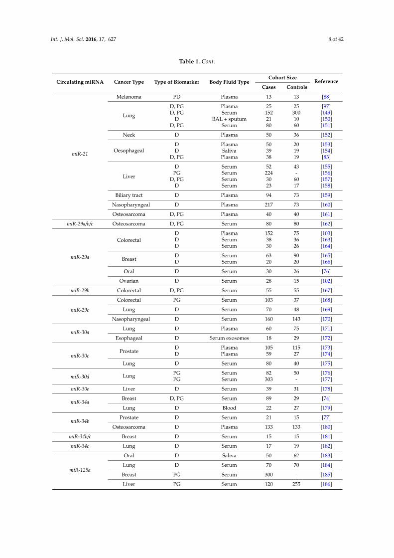
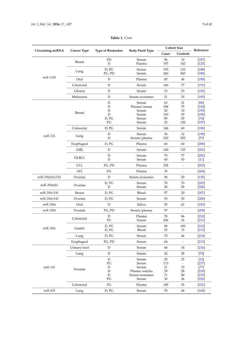
Table 1. Examples of deregulated levels of circulating miRNAs in various malignancies proposed tohave either diagnostic and/or prognostic value. MDS: Myelodysplastic syndrome; HCC: Hepatocellularcarcinoma; MM: Multiple myeloma; DLBCL: Diffuse large B-cell lymphoma; HL: Hodgkin lymphoma;CNS: Central nervous system; AML: Acute myeloid leukemia; CLL: Chronic lymphocytic leukemia;ATL: Adult T-cell leukemia; BAL: Bronchoalveolar lavage; D: Diagnostic; PG: Prognostic; and PD:Predictive of response.
Circulating miRNA Cancer Type Type of Biomarker Body Fluid TypeCohort Size
ReferenceCases Controls
let-7a
Prostate D Blood 75 27 [52]
ColorectalD Serum exosomes 88 11 [53]D Plasma 51 26 [54]
MDS PG Plasma 50 76 [55]
Gastric D Plasma 69 30 [56]
let-7a/b Lung D, PG Serum/plasma 220 220 [57]
let-7bHCC D, PG Serum 120 30 [58]
Ovarian D Serum 18 12 [59]
let-7cLung D Plasma 20 360 [60]
Breast D Serum 90 64 [61]
let-7c/i/f Gastric D Serum 214 424 [62]
let-7d Prostate PG Plasma 50 10 [63]
let-7eMM PG Serum 121 30 [64]
Thyroid D Serum 95 44 [65]
let-7f
Ovarian D, PG Plasma 360 200 [66]
Colorectal D Feces 51 26 [54]
Lung D, PG Plasma vesicles 106 68 [67]
HCC D, PG Serum 90 60 [68]

Int. J. Mol. Sci. 2016, 17, 627 6 of 42
Table 1. Cont.
Circulating miRNA Cancer Type Type of Biomarker Body Fluid TypeCohort Size
ReferenceCases Controls
let-7iLung PG Serum 10 20 [69]
Ovarian D Serum/plasma 25 25 [70]
miR-10
BreastD, PG Serum 113 - [71]
PG Serum 89 29 [72]PG Cerebrospinal 16 15 [73]
Lung D Serum 42 28 [74]PG Cerebrospinal 28 15 [73]
Glioblastoma D Cerebrospinal 19 15 [73]
Oesophageal D Serum 50 50 [75]
miR-16
Oral D Serum 30 26 [76]
ProstateD Serum 21 15 [77]D Serum 73 20 [78]
Breast D Serum 76 76 [79]
Osteosarcoma D Serum 20 20 [80]
GastricD, PG Plasma 30 18 [81]D, PG Serum 50 47 [82]
Liver D Serum 90 60 [68]
Oesophageal D, PG Plasma 38 19 [83]
miR-17
Gastric D, PG Serum 50 47 [82]
Liver D Serum 90 60 [68]
Oesophageal D, PG Plasma 38 19 [83]
miR-18a
Oesophageal D Serum 106 54 [84]
Gastric D Plasma 104 65 [85]
Breast D, PD Serum 108 75 [86]
Colorectal D Stool 198 198 [87]
miR-17/19a Melanoma PD Plasma 13 13 [88]
miR-19a
BreastPD Serum 30 38 [89]D Serum 63 21 [90]
D, PG Serum 113 - [71]
Bladder D Plasma 50 50 [91]
Colorectal D, PG Serum 90 12 [92]
MM D,PG,PD Serum 108 56 [93]
Lung D, PG Serum 201 103 [94]
miR-19bGastric D, PG Plasma 30 18 [81]
Lung D, PG Serum 94 94 [95]
miR-20a
Prostate D Plasma 82 - [96]
Lung D Plasma 126 60 [97]
Osteosarcoma D Serum 20 20 [80]
Colorectal D Feces 397 198 [98]
Esophageal D Plasma 70 40 [99]
miR-17/-92Colorectal PD Serum 37 7 [100]
Colorectal D Plasma 90 90 [101]
miR-92 Ovarian D Serum 28 15 [102]
miR-92aColorectal D Plasma 152 75 [103]
Leukemia D Plasma 77 16 [104]

Int. J. Mol. Sci. 2016, 17, 627 7 of 42
Table 1. Cont.
Circulating miRNA Cancer Type Type of Biomarker Body Fluid TypeCohort Size
ReferenceCases Controls
miR-92a/b Prostate D Serum 21 15 [77]
miR-106a
Gastric D, PG Plasma 48 22 [105]
ColorectalD Feces 117 107 [106]D Plasma 100 79 [107]
PG Serum 175 130 [108]
miR-17/-106a/b Gastric D Plasma 69 30 [56]
miR-17/-106b Gastric D Serum 72 36 [109]
miR-106b
LiverD Plasma 47 61 [110]D Serum exosomes 20 40 [111]
Breast D, PG Plasma 173 50 [112]
Gastric D, PG Plasma 20 20 [113]
Bladder D Urine 112 78 [114]
Ovarian D Serum 31 31 [115]
miR-21
DLBCL
D Serum 60 43 [11]D,PD,PG Serum 112 45 [116]
D Serum 60 43 [11]D,PD,PG Serum 62 50 [117]
HL D Plasma 42 20 [118]
CNSlymphoma D, PG Serum 37 53 [119]
Breast
D Plasma 14 8 [120]D, PG Serum 62 10 [121]D, PG Serum 30 60 [122]D, PG Serum 50 82 [123]
PG Serum 326 223 [124]D Plasma 114 116 [125]D Urine 24 24 [126]
PG Serum 113 - [71]
Gastric
D Plasma 69 30 [56]D, PG Plasma 42 - [127]
PG Serum 103 - [128]PG Serum 79 - [129]PG Serum 64 64 [130]PG Plasma 69 - [131]D Serum 50 50 [132]
GlioblastomaPG Serum 30 30 [39]D Plasma 10 10 [133]
OvarianD Serum 28 15 [102]
D, PG Serum 94 40 [134]D Serum 60 10 [135]
Pancreatic
D Plasma 49 36 [136]D, PG Plasma 32 30 [137]
D Plasma 24 24 [138]D Stool 30 15 [139]D Serum 22 14 [140]D Plasma 30 26 [141]D Saliva 7 4 [142]
ProstatePD Plasma 82 - [96]PD Serum 56 - [143]
Colorectal
PD Serum 37 7 [100]D, PG Serum 40 40 [144]
D Serum 160 77 [144]D, PG Serum 186 96 [145]
D Serum 200 130 [146]PG Serum 102 - [147]D Serum 56 197 [148]
Int. J. Mol. Sci. 2016, 17, 627 8 of 42
Table 1. Cont.
Circulating miRNA Cancer Type Type of Biomarker Body Fluid TypeCohort Size
ReferenceCases Controls
miR-21
Melanoma PD Plasma 13 13 [88]
Lung
D, PG Plasma 25 25 [97]D, PG Serum 152 300 [149]
D BAL + sputum 21 10 [150]D, PG Serum 80 60 [151]
Neck D Plasma 50 36 [152]
OesophagealD Plasma 50 20 [153]D Saliva 39 19 [154]
D, PG Plasma 38 19 [83]
Liver
D Serum 52 43 [155]PG Serum 224 - [156]
D, PG Serum 30 60 [157]D Serum 23 17 [158]
Biliary tract D Plasma 94 73 [159]
Nasopharyngeal D Plasma 217 73 [160]
Osteosarcoma D, PG Plasma 40 40 [161]
miR-29a/b/c Osteosarcoma D, PG Serum 80 80 [162]
miR-29a
ColorectalD Plasma 152 75 [103]D Serum 38 36 [163]D Serum 30 26 [164]
BreastD Serum 63 90 [165]D Serum 20 20 [166]
Oral D Serum 30 26 [76]
Ovarian D Serum 28 15 [102]
miR-29b Colorectal D, PG Serum 55 55 [167]
miR-29c
Colorectal PG Serum 103 37 [168]
Lung D Serum 70 48 [169]
Nasopharyngeal D Serum 160 143 [170]
miR-30aLung D Plasma 60 75 [171]
Esophageal D Serum exosomes 18 29 [172]
miR-30cProstate
D Plasma 105 115 [173]D Plasma 59 27 [174]
Lung D Serum 80 40 [175]
miR-30d Lung PG Serum 82 50 [176]PG Serum 303 - [177]
miR-30e Liver D Serum 39 31 [178]
miR-34aBreast D, PG Serum 89 29 [74]
Lung D Blood 22 27 [179]
miR-34bProstate D Serum 21 15 [77]
Osteosarcoma D Plasma 133 133 [180]
miR-34b/c Breast D Serum 15 15 [181]
miR-34c Lung D Serum 17 19 [182]
miR-125a
Oral D Saliva 50 62 [183]
Lung D Serum 70 70 [184]
Breast PG Serum 300 - [185]
Liver PG Serum 120 255 [186]
Int. J. Mol. Sci. 2016, 17, 627 9 of 42
Table 1. Cont.
Circulating miRNA Cancer Type Type of Biomarker Body Fluid TypeCohort Size
ReferenceCases Controls
miR-125b
BreastPD Serum 56 10 [187]D Plasma 197 142 [125]
Lung D, PG Serum 193 110 [188]PG, PD Serum 260 260 [189]
Oral D Plasma 85 46 [190]
Colorectal D Serum 160 77 [191]
Glioma D Serum 33 33 [192]
Melanoma D Serum exosomes 21 35 [193]
miR-155
Breast
D Serum 63 21 [90]D Plasma/serum 184 75 [194]D Serum 20 10 [195]D Serum 103 55 [196]
D, PG Serum 89 29 [74]PG Serum 32 120 [197]
Colorectal D, PG Serum 146 60 [198]
Lung D Serum 36 32 [199]D Serum/plasma 220 220 [57]
Esophageal D, PG Plasma 60 60 [200]
AML D Serum 140 135 [201]
DLBCLD Serum 75 77 [202]D Serum 60 43 [11]
CLL PG, PD Plasma 228 - [203]
ATL PG Plasma 35 - [204]
miR-200a/b/c/141 Ovarian D Serum exosomes 50 20 [135]
miR-200a/b/c OvarianD, PG Serum 70 70 [205]
D Serum 28 28 [206]
miR-200c/141 Breast D, PG Blood 57 20 [207]
miR-200c/141 Ovarian D, PG Serum 93 50 [208]
miR-200a Oral D Saliva 50 62 [183]
miR-200b Prostate PG, PD Serum/plasma 97 - [209]
miR-200c
ColorectalD Plasma 78 86 [210]
PG Serum 206 24 [211]
GastricD, PG Serum 98 100 [212]D, PG Blood 52 15 [213]
Lung D, PG Serum 70 44 [214]
Esophageal PG, PD Serum 64 - [215]
miR-141
Urinary tract D Serum 44 34 [216]
Lung D Serum 42 28 [74]
Prostate
D Serum 25 25 [12]PG Serum 113 - [217]D Serum 21 15 [77]D Plasma vesicles 78 28 [218]D Serum exosomes 71 80 [219]
PG Serum 30 26 [220]
Colorectal PG Plasma 185 76 [221]
miR-429 Lung D, PG Serum 70 48 [169]

Int. J. Mol. Sci. 2016, 17, 627 10 of 42
Table 1. Cont.
Circulating miRNA Cancer Type Type of Biomarker Body Fluid TypeCohort Size
ReferenceCases Controls
miR-210
DLBCL D Serum 60 43 [11]
PancreaticD Plasma 49 36 [136]D Plasma 22 25 [222]
RenalD Serum 78 42 [223]D Serum 34 23 [224]
Breast PG, PD Plasma 69 43 [225]
PancreaticD Saliva 7 4 [142]
D, PG Pancreatic juice 6 6 [226]D Plasma 30 26 [141]
BladderD, PG Serum 168 104 [227]
D Urine 94 56 [228]
Glioma D, PG Serum 136 50 [229]
Liver PD, PG Serum 113 39 [230]
miR-221
ColorectalD Plasma 103 37 [231]D Stool 198 198 [87]
Prostate PG Plasma 82 - [96]
Leukemia D Plasma 79 37 [232]
Liver D Serum 20 40 [111]
Larynx D Plasma 30 30 [233]
Glioma D, PG Plasma 50 51 [234]
Melanoma D, PG Serum 72 54 [235]
Renal PG Plasma 77 - [236]
miR-375
Breast PG Serum 68 - [237]
Lung D, PG Plasma 217 217 [238]PG Serum 113 - [217]
Prostate
PG Serum 47 72 [218]PG Serum 84 - [239]PG Plasma 100 - [240]D Plasma 78 28 [218]
OesophagealD Plasma 38 19 [83]
PG Serum 194 94 [241]D Plasma 50 20 [153]
Liver D Serum 78 156 [242]
Colorectal D Plasma 88 40 [243]
2.1. let-7 Family (let-7a, -7b, -7c, -7e, -7f, -7i)
There are 13 different let-7 family members in humans: let-7a-1, 7a-2, 7a-3, 7b, 7c, 7d, 7e, 7f-1, 7f-2, 7g,7i, miR-98, and miR-202 [244]. Differential expression of let-7 family members has been described to bedownregulated in a wide variety of cancers such as melanoma, pancreatic cancer, prostate cancer, andsarcoma, although some, including lymphoma, mesothelioma, and breast cancer, have been shown tobe upregulated; thus, the let-7 family is generally regarded as a tumor suppressor [245]. let-7 has beenshown to be a direct regulator of some important oncogenes, such as the three RAS genes [246,247],HMGA2 [248,249], STAT3 [250], UHRF2 [251], and MYC [252–254]; additionally, let-7 family targetscell cycle and cell proliferation genes [255–257]; finally, apoptosis is also shown to be regulated bylet-7 family, through CASP3 targeting [258]. RNase III nuclease, known to process pre-miRs, was alsoconfirmed as a direct target of the let-7 family, so they might regulate their own processing [259,260].
let-7 family members have been identified differentially expressed and therefore have beenproposed as diagnostic tools in serum/plasma of many cancer types including lung cancer (let-7a,

Int. J. Mol. Sci. 2016, 17, 627 11 of 42
let-7c, let-7f ) [57,60,67], prostate cancer (let-7a) [52], gastric cancer (let-7a, let-7c, let-7i, let-7f ) [56,62],ovarian cancer (let-7b, let-7f, let-7i) [59,66,70], hepatocellular carcinoma (HCC) (let-7b, let-7f ) [58,68],breast cancer (let-7c, let-7b, let-7g) [61,261], acute myeloid leukemia (AML) (let-7b, let-7d) [262], thyroidcarcinoma (let-7e) [65], and colorectal cancer (CRC) (let-7a) [53,54]. Recently, let-7f has also been detectedas deregulated in the feces of CRC patients [54]. However, their potential as prognostic biomarkers hasalso been highlighted in several cancer types such as myelodisplasia (let-7a) [55], lung cancer (let-7b,let-7f, let-7i) [57,67,69], hepatocellular carcinoma (let-7b, let-7f ) [58,68], multiple myeloma (let-7e) [64],prostate cancer (let-7d) [63], ovarian cancer (let-7f ) [66], and breast cancer (miR-202) [263]. In general,low let-7 levels are associated with poor prognosis including overall survival, early recurrence, andtumor size.
2.2. miR-10b
miR-10b acts as a metastasis driver in many different types of cancers such as breast cancer [264],glioma [265], and oesophageal cancer [266], among others, specifically promoting cell mobility andinvasiveness. Validated targets of this miRNA include SDC1 [267], HOXD10 [264,265,268,269],KLF4 [266,270], MICB [271], and CDH1 [272]. It also regulates E2F1-mediated transcriptionthrough p21/CDKN1A regulation [273] and important cell cycle regulators such as BUB1, PLK1, andCCNA2 [274].
Circulating miR-10b levels have been described as being upregulated in patients with ovariancancer [30], lung cancer [29], oesophageal [75], and glioblastoma [73] compared to healthy controls.Consistent with this, miR-10b is increased in plasma from metastatic breast cancer patients [27,28] andin the cerebrospinal fluid of patients with brain metastasis of both breast and lung cancer [73].
2.3. miR-16
miR-16 has an important role in regulating apoptosis in different cancer types including lung,breast, liver, glioblastoma, and squamous cell carcinoma through targeting FEAT/METTL13 [275],RPS6KB1, IGF1R [276], CCND1 [277], BCL2 [278], RECK, and/or SOX6 [279]. This miRNA is also animportant regulator of cell cycle molecules including FG2F, CCNE1, and E2F1 [280–282], as well as cellautophagy (mTORC2) and metastasis (SOX5) [283,284].
Circulating miR-16 has been described significantly differentially expressed in patients comparedwith healthy controls in several cancers: oral cancer [76], breast cancer [79], prostate cancer [36],osteosarcoma [80], gastric cancer [81,82], liver carcinoma [68], and oesophageal carcinoma [83].Furthermore, miR-16 is also associated with prognosis and tumor size in gastric cancer [81,82],hepatocellular carcinoma [68], and esophageal squamous cell carcinoma (ESCC) [83].
2.4. The miR-17~92 Cluster
Over-expression of the miR-17~92 cluster is a key oncogenic event in many cancer types, andoverexpression in murine models result in tumor formation. This cluster is composed of differentmiRNAs: miR-17, miR-18a, miR-19a, miR-19b, miR-20a, miR-92, and miR-106a/b with a variety ofrelated functions, primarily targeting tumor suppressor molecules and pathways such as PTENand RB1 [285,286], and molecules in the TGFβ signaling pathway such as TGFBR2, SMAD2, andSMAD4 [287–289]. These miRNAs also target senescence (p21/CDKN1A) [290,291], metastasis (DLC1and TIMP2) [292–294], cell cycle regulation (E2F family members, RB1 and p21/CDKN1A), andangiogenesis (THBS1 and CTGF) [295–297].
Circulating members of the miR-17~92 cluster have been widely described as being deregulatedin many cancer types including colorectal cancer [56,87,92,98,101,103,105–107,231,298], gastriccancer [56,81,85,109,113], squamous cell carcinoma [84], breast cancer [86,90,114], bladder cancer [91],hematological malignancies [93,104,117,202,299,300], lung cancer [94,95,97], prostate cancer [77],osteosarcoma [80], oesophageal carcinoma [99], ovarian cancer [81,115], and hepatocellularcarcinoma [110,111,301]. They have also been shown to have prognostic value in colorectal
Int. J. Mol. Sci. 2016, 17, 627 12 of 42
cancer [92,100,108], sporadic melanoma [88], breast cancer [71,112], bladder cancer [91], multiplemieloma [93,104,299,300], lung cancer [94], and prostate cancer [96]. Moreover, levels of circulatingmiR-17~92 miRNAs have been associated with the response to chemotherapy (i.e., predictivebiomarkers) in both breast cancer [89] and multiple myeloma [93].
2.5. miR-21
miR-21 acts mainly as an oncogene (“onco-miR”) because most of its target genes are tumorsuppressors. The list of these target genes is extensive, and they are related to all hallmarks ofcancer [302]. One of the principal miR-21 targets is PDCD4, which is a tumor suppressor genethat inhibits PMA-induced neoplastic transformation [303], tumor promotion and progression [304],and invasion and intravasation [305]. miR-21 targets multiple components of TP53, TGFB1, andmitochondrial apoptosis tumor suppressive pathways (including HNRPK and TP63) [306]. Othertargets of miR-21 have been related mainly with apoptosis, cell growth, migration, and invasion,such as BCL2 [307], PTEN [307–309], RECK [310], RHOB [311], and TPM1 [312], among others.
Circulating miR-21 has been described in a lot of different cancers as a diagnostic,predictive, and/or prognosis biomarker. Some of these are hematological cancers [116,117], breastcancer [124,125], gastric cancer [128,130], ovarian cancer [134,135], pancreatic cancer [136,141],colorectal cancer [100,145], lung cancer [97,149], and liver cancer [155,156], among others.
2.6. The miR-29 Family (miR-29a, -29b, and -29c)
The miR-29 family members act as tumor suppressors, and their downregulation is associatedwith many cancer types including leukemia [313–315], melanoma [316], liver cancer [317,318], coloncancer [319], cervical cancer [320], lung cancer [321], and prostate cancer [322]. In many studies,downregulation of miR-29 has correlated with more aggressive forms of cancer and shorter overallsurvival [316,321,323]. It has been demonstrated to directly target genes involved in the control of thecell cycle (CDK6) [319,320,323] and apoptosis (MCL1, BCL2 and FHIT) [315,319,321], as well as genesthat promote cell migration and invasion (LAMC1, CDC42) [322,324]. Furthermore, the miR-29 familytarget genes such as PIK3R1 and CDC42 that normally suppress TP53 [324].
Differential expression of miR-29 family members in plasma/serum has been observed inseveral cancer types. The expression levels of all the miR-29 family members were upregulatedin sera of patients with osteosarcoma. In particular, miR-29a and miR-29b were associated withpoor prognosis [162]. miR-29a has been shown to be upregulated in colorectal cancer andtherefore has been proposed as a potential non-invasive biomarker for early detection of colorectalcancer [101,103,164,231], also involving liver metastasis [163]. It has also been found to be upregulatedin breast cancer [165,166] and downregulated in oral and ovarian cancer, compared with healthycontrols [76,102]. Similarly, serum levels of miR-29b have been proposed as potential biomarkers fordiagnosis and prognosis of colorectal cancer [167], whereas miR-29c could be useful as a predictorof postoperative early relapse [168]. However, it was found to be downregulated in serum ofnasopharyngeal carcinoma patients, compared with controls [170].
2.7. The miR-30 Family (miR-30a, -30b, -30c, -30d, and -30e)
Similar to the miR-29 family, miR-30 family members appear to act primarily as tumorsuppressors in several cancer types such as ovarian cancer, breast cancer, non-small cell lungcancer (NSCLC), and colorectal carcinoma [325–330], although they have also been reported asoncogenes [331]. Several genes have been described to be regulated by the miR-30 family, such as someepithelial-to-mesenchymal transition (EMT)-associated genes [332], anti-apoptotic protein AVEN [333],and DLL4 which has a fundamental role in angiogenesis [334].
Members of the miR-30 family have been identified differentially regulated in body fluids, but theirpotential as biomarkers has mostly been reported in combinations with other miRNAs. For example, ablood test based upon a combination of the levels of five miRNAs including miR-30c has been described

Int. J. Mol. Sci. 2016, 17, 627 13 of 42
to effectively differentiate prostate cancer patients from benign prostatic hyperplasia (BPH) patientsand healthy controls [173]. In addition, the combination of four plasma circulating miRNAs, includingmiR-30c and serum PSA, has a greater potential to be used as a noninvasive diagnostic biomarker forprostate cancer screening than PSA testing alone [174]. Similar studies have been reported for miR-30cand miR-30a-3p in lung adenocarcinoma [171,175], miR-30a in esophageal adenocarcinoma [172], andmiR-30d in lung cancer [176]. In hepatocellular carcinoma, miR-30e has been recently found to bedownregulated in serum when compared with healthy controls [178], and, in lung cancer, high levelsof miR-30d in serum have been associated with a shorter overall survival [176,177].
2.8. The miR-34 Family (miR-34a, -34b, and -34c)
Members of the miR-34 family are well known to regulate cell cycle, senescence, apoptosis,and invasiveness in cancer, and deregulation of miR-34a has been reported in several types ofcancers [335,336]. The miR-34 family targets multiple TP53 inhibitor genes (MDM4, SIRT1, MTA2,HDAC1, and YY1) and promotes proliferation arrest and induction of apoptosis by targeting MYC,CDK6, and MET. These genes encode factors required for G1/S transition (MYC, E2F, CDK4, andCDK6), anti-apoptotic proteins (BCL2, SIRT1), and proteins involved in invasion (MET) [337]. It hasalso been reported to target pluripotency genes such as NANOG, SOX2, and MYCN [338,339] andcomponents of Wnt signaling pathways [340,341] and notch signaling pathways [342,343], whichregulate growth, epithelial–mesenchymal transition (EMT), and metastasis.
Elevated levels of miR-34a in serum can discriminate between breast cancer patients and healthycontrols, and are also associated with the presence of overt metastasis [72,237]. High levels ofcirculating miR-34a have also been observed in ovarian and lung cancer [72,74,179], and miR-34b hasbeen found to be upregulated in serum from prostate cancer patients [77]. In osteosarcoma patients,miR-34b levels were found to be downregulated when compared with controls, and these expressionlevels were significantly decreased in the metastatic patients [180]. Similarly, downregulation ofcirculating miR-34c in serum of NSCLC patients and miR-34b/c in serum of breast cancer patients hasbeen reported and might have potential as biomarkers for the diagnosis of these pathologies [181,182].
2.9. The miR-125 Family (miR-125a and -125b)
miR-125 has been shown to act as a tumor-suppressor in several cancers including ovariancancer [313,344], bladder cancer [345], breast cancer [346,347], hepatocellular carcinoma [348–350],melanoma [351], cutaneous squamous cell carcinoma [352], and osteosarcoma [353]. miR-125 targetsseveral genes associated with carcinogenesis such as transcription factors (STAT3 and E2F3) [345,353],matrix-metalloprotease (MMP11 and MMP13) [348,352], members of the BCL2 family [354,355], andgrowth factors (VEGFA) [348].
Deregulated levels of miR-125a were present in the saliva of oral squamous cell carcinoma (OSCC)patients and in serum of NSCLC patients compared with healthy controls [183,184]. In a similarway, miR-125b levels were significantly lower in glioma patients and in serum-derived exosomes ofmelanoma patients [192,193]. In addition, low circulating levels of miR-125a have been associated withpoor prognosis in both breast cancer and hepatocellular carcinoma [185,186]. In contrast, miR-125bwas found to be upregulated in the plasma and serum of metastatic prostate cancer patients [356],breast cancer [125], OSCC [190], colorectal cancer [191], and NSCLC [188], in comparison with healthycontrols, and to be associated with poor prognostic outcome and chemotherapeutic resistance in thiscancer [187–189].
2.10. miR-155
miR-155 is involved in both physiological (hematopoiesis andimmune response) and pathologicalprocesses. The oncogenic role of miR-155 is well established in both hematological malignances aswell as solid cancers such as breast cancer, where its overexpression is generally correlated with poorprognosis [357,358]. Validated miR-155 target genes are present in multiple pathways associated with
Int. J. Mol. Sci. 2016, 17, 627 14 of 42
cancer and cancer progression, including EMT (SMAD5), proliferation (SOCS1, INPP5D, and CSF1R),block of differentiation (SPI1, CEBPB), and apoptosis (CASP3, FADD, APAF1, and FOXO3A) [359–367].
In many studies, differentially expressed levels of circulating miR-155 have been identified,including breast cancer [90,194–196], colorectal cancer [198], lung cancer [57,199], AML [201],diffuse large B-cell lymphoma (DLBCL) [11,202], and esophageal cancer [200], making it a potentialnon-invasive diagnostic biomarker for early detections in these pathologies. In fact, a biosensor formiR-155 detection in plasma has recently been developed for the diagnosis of breast cancer [368].Elevated levels of miR-155 are also related to overt metastasis in breast cancer [72,197], and these highlevels have also been identified not only in blood but also in the urine of breast cancer patients [126].In addition, miR-155 has been also suggested as a prognostic biomarker in chronic lymphocytic leukemia(CLL) and adult T-cell leukemia (ATL) [203,204], and as a predictive biomarker to response to therapyin CLL [203].
2.11. The miR-200 Family (miR-200a, -200b, -200c, -141, and -429)
The miR-200 family is believed to play crucial roles in both cancer initiation and metastasis—inparticular, in epithelial-mesenchymal transition (EMT)—primarily through the targeting of ZEB1 andZEB2 transcription factors [369,370]. It has also been associated with angiogenesis by the targeting ofVEGFA and VEGF receptors [371,372] and pro-angiogenic ligands such as CXCL8 and CXCL1 [373].
Elevated serum levels of miR-200a, miR-200b, miR-200c, and miR-141 have been suggested asgood biomarkers for diagnosis and prognosis in ovarian cancer [205,206,208], and serum levels ofmiR-429 were associated with poor overall survival in NSCLC [169]. In addition, elevated levelsof circulating miR-141 have been identified to show diagnostic potential in patients with upperurinary tract urothelial cancer [216], lung cancer [74], prostate cancer [12,217–220], breast cancer [207],and bladder cancer [374]. In breast and bladder cancer, this upregulation is also associated withprognosis. Furthermore, miR-200c was found significantly elevated in the plasma of patients withcolorectal cancer [210], gastric cancer [212,213], and breast cancer [207], and this upregulation wasassociated with poor prognosis. In colorectal cancer, it has also been identified as a metastasis predictivebiomarker [211] as well as miR-141 [221]. Similarly, miR-200c can be useful to predict prognosis inNSCLC [214] and in esophageal cancer. In the latest research, the serum level of miR-200c, as well asmiR-200b, can be useful for predicting response to chemotherapy [215], and also has prognostic valuein prostate cancer and predictive value in docetaxel chemotherapy outcomes [209].
2.12. miR-210
miR-210 is strongly linked with the hypoxic pathway and angiogenesis through the targeting ofEFNA3 [375,376], VEGF [377], and STAT3 [378]. miR-210 also acts upon cell cycle and apoptoticpathways by targeting E2F3, MNT [379–381], FGFRL1 [382], BCL2 [383,384], and STAT3 [378].Furthermore, miR-210 can inhibit DNA damage repair genes such as RAD52 [385] and oncogenessuch as HOXA1 [386].
Circulating miR-210 levels have been shown to have diagnostic value in DLBCL [11], pancreaticcancer [136,141,142,222,226], bladder cancer [227,228], glioma [229], liver carcinoma [230], and renalcarcinoma [223,224] and with the presence of metastasis in patients with breast cancer [225] andpancreatic cancer [226]. miR-210 has been also correlated with sensitivity to treatment in breast cancerand with prognosis in patients with breast cancer [225], pancreatic cancer [226], bladder cancer [227],and liver carcinoma [230].
2.13. miR-221/-222
miR-221 and its paralogue miR-222 are known to target angiogenesis by direct interactionwith KIT [387,388], PTEN [389], TIMP3, ADAM10, and ADAM17 [390] and by indirectlyregulating endothelial nitric oxide synthase expression [387,391]. miR-221/-222 have also beendescribed as regulators of cell proliferation via the targeting of SEMA3B [392], IRF2, SOCS3 [393],

Int. J. Mol. Sci. 2016, 17, 627 15 of 42
p27/CDKN1B [394,395], HECTD2, RAB1A [396], β-catenin/CTNNB1, TGFB1 [397], ADAM17, ITGB4,and STAT3 [398]. Other pathways regulated by miR-221/-222 include apoptosis and metastasis viaPTEN [389], IRF2, SOCS3 [393], BBC3 [399], SEMA3B [392], HECTD2, RAB1A [396], ADAM17, ITGB4,STAT5A [398], and Ecm29/KIAA0368 [400]; in resistance to chemotherapy in some type of cancersthrough PTEN [389] and β-catenin/CTNNB1 [397] regulation.
Circulating miR-221/-222 levels have been identified as diagnostic markers in prostate cancer [96],colorectal carcinoma [87,231], NK/T-cell lymphoma [104], liver carcinoma [111], larynx cancer [233],glioma [234], and melanoma [235]. They also have prognostic value in glioma [234], melanoma [235],prostate cancer [96], and renal carcinoma [236].
2.14. miR-375
miR-375 is a tumor suppressor miRNA that has been described in different kind of cancers, whereit targets genes related to proliferation and apoptosis (JAK2, PDK1, 14-3-3ζ, IGF1R, KLF4, KLF5, survivin,ERBB2, PIK3CA, MTDH, YAP1, CIP2A/KIAA1524, MTDH, and BCL2) [401–414] as well as metastasis(IGF1R, CLDN1, CIP2A/KIAA1524, and BCL2) [404,412,414,415] and mediates resistance to therapy(IGF1R, TP53, and PHLPP1) [416–418]. Furthermore, miR-375 is involved in epithelial to mesenchymaltransition in breast cancer [419] and targets ATG7 inhibiting autophagy and impairing the viability ofcells under hypoxic conditions in liver cancer [420].
Circulating miR-375 has been identified as a diagnostic biomarker in oesophagealcarcinoma [83,153], liver cancer [242], colorectal cancer [243], and lung cancer [421]. Additionally,in prostate cancer [218,239,240], lung cancer [238], and oesophageal carcinoma [241], miR-375 has beenshown to have prognostic value.
Int. J. Mol. Sci. 2016, 17, 627 15 of 40
proliferation via the targeting of SEMA3B [392], IRF2, SOCS3 [393], p27/CDKN1B [394,395], HECTD2, RAB1A [396], β-catenin/CTNNB1, TGFB1 [397], ADAM17, ITGB4, and STAT3 [398]. Other pathways regulated by miR-221/-222 include apoptosis and metastasis via PTEN [389], IRF2, SOCS3 [393], BBC3 [399], SEMA3B [392], HECTD2, RAB1A [396], ADAM17, ITGB4, STAT5A [398], and Ecm29/KIAA0368 [400]; in resistance to chemotherapy in some type of cancers through PTEN [389] and β-catenin/CTNNB1 [397] regulation.
Circulating miR-221/-222 levels have been identified as diagnostic markers in prostate cancer [96], colorectal carcinoma [87,231], NK/T-cell lymphoma [104], liver carcinoma [111], larynx cancer [233], glioma [234], and melanoma [235]. They also have prognostic value in glioma [234], melanoma [235], prostate cancer [96], and renal carcinoma [236].
2.14. miR-375
miR-375 is a tumor suppressor miRNA that has been described in different kind of cancers, where it targets genes related to proliferation and apoptosis (JAK2, PDK1, 14-3-3ζ, IGF1R, KLF4, KLF5, survivin, ERBB2, PIK3CA, MTDH, YAP1, CIP2A/KIAA1524, MTDH, and BCL2) [401–414] as well as metastasis (IGF1R, CLDN1, CIP2A/KIAA1524, and BCL2) [404,412,414,415] and mediates resistance to therapy (IGF1R, TP53, and PHLPP1) [416–418]. Furthermore, miR-375 is involved in epithelial to mesenchymal transition in breast cancer [419] and targets ATG7 inhibiting autophagy and impairing the viability of cells under hypoxic conditions in liver cancer [420].
Circulating miR-375 has been identified as a diagnostic biomarker in oesophageal carcinoma [83,153], liver cancer [242], colorectal cancer [243], and lung cancer [421]. Additionally, in prostate cancer [218,239,240], lung cancer [238], and oesophageal carcinoma [241], miR-375 has been shown to have prognostic value.
Figure 3. Origin of extracellular RNA. Several hypotheses have been proposed to explain the presence of miRNA in biological fluids, including the passive release of miRNA from broken cells and tissues and the active secretion from cells in microvesicles or conjugated to RNA-binding proteins. Cell-free miRNA can be detected in different body fluids including plasma, serum, saliva, tears, urine, amniotic fluid, colostrum, breast milk, bronchial lavage, cerebrospinal fluid, peritoneal fluid, pleural fluid, and seminal fluid and also in feces. Ago 1–4: Argonaute proteins 1–4; LDL: Low-density lipoprotein; HDL: High-density lipoprotein; MVB: Multivesicular body.
3. Extracellular miRNAs in Other Biological Fluids
In addition to blood, other biological fluids such as urine, saliva, cerebrospinal fluid, vitreous humor of the eye, breast milk, seminal fluid, and tears have been studied as potential sources of miRNA biomarkers [422,423] (Figure 3). The majority of these studies concern tumor types associated with the source of the biological fluids. For example, saliva has been studied in head and
Figure 3. Origin of extracellular RNA. Several hypotheses have been proposed to explain the presenceof miRNA in biological fluids, including the passive release of miRNA from broken cells and tissuesand the active secretion from cells in microvesicles or conjugated to RNA-binding proteins. Cell-freemiRNA can be detected in different body fluids including plasma, serum, saliva, tears, urine, amnioticfluid, colostrum, breast milk, bronchial lavage, cerebrospinal fluid, peritoneal fluid, pleural fluid, andseminal fluid and also in feces. Ago 1–4: Argonaute proteins 1–4; LDL: Low-density lipoprotein; HDL:High-density lipoprotein; MVB: Multivesicular body.
3. Extracellular miRNAs in Other Biological Fluids
In addition to blood, other biological fluids such as urine, saliva, cerebrospinal fluid, vitreoushumor of the eye, breast milk, seminal fluid, and tears have been studied as potential sources of miRNAbiomarkers [422,423] (Figure 3). The majority of these studies concern tumor types associated with

Int. J. Mol. Sci. 2016, 17, 627 16 of 42
the source of the biological fluids. For example, saliva has been studied in head and neck squamouscell carcinoma [183,424–427], tumors of the parotid gland [183,424,428], esophageal cancer [154], andpancreatic cancer [142,429]. Urine is another well studied source of cfmiRNAs associated with cancerin particular urological cancers including prostate and bladder cancer (reviewed in [430]). In addition,several studies have looked at the potential of urine for miRNA biomarkers in ovarian, breast, andliver cancer [126,431,432]. miRNAs in cerebrospinal fluid have been described as potential biomarkersfor the diagnosis and monitoring of disease in brain tumors such as glioblastoma but also in CNSlymphomas and in brain metastases of non-neuronal origin [73,433–436]. In a similar vein, miRNAs, inthe vitreous humor of the eye, have been identified in ocular cancers including vitreoretinal lymphomaor uveal melanoma [437,438]. It also has been suggested that the miRNA profile of breast milk couldbe a more sensitive biomarker for breast cancer than blood-associated miRNAs [439] and that seminalfluid-associated miRNAs can serve as biomarkers of prostate cancer [440].
4. Discussion
Challenges in Studying cfmiRNA
A major obstacle to the translation of cfmiRNAs from laboratory studies into the clinic is thelack of consistent and robust results with many apparently contradictory reports in the literature.A likely reason for this lack of reproducibility is that there are very few multi-center studies, andcohorts are often insufficiently powered. Another confounding factor is the fact that there is a highdegree of inter-individual variability in the levels of cfmiRNAs, even when focusing only on healthypopulations [441]. Moreover, there is a technical source of variation between studies, such as thestarting material used for the experiments (e.g., the purification of cells, the cell types, the controlpopulations used, the RNA extraction method, etc.), the technological platforms (e.g., microarray,qRT-PCR vs. next generation sequencing (NGS) etc.), and the differing statistical methodologies used.
The blood collection and processing represent critical points of variability in cfmiRNA studies.In the first instance, miRNA contamination can occur at the venopuncture site itself [442]. Afterextraction, the elapsed time between blood collection and processing should be minimized to preventlysis and cellular contamination, which can be a major source of variability between samples [443–446].In addition, the choice of anti-coagulant used in plasma collection can influence downstream detectiontechnologies, such as qRT-PCR and heparin-coated tubes, should be avoided [447]. Another majorsource of difference in cfmiRNA profiles comes from the choice of whether to use serum or plasma,and whether to purify exosomes or use whole serum/plasma [448–451].
The choice of RNA purification procedure can also critically affect the results of cfmiRNA studiesand should be considered carefully in terms of experimental design. For example, small RNA moleculeswith low GC content are known to be selectively lost during Trizol-based extraction (the most popularmethod) when present in low concentrations, such as in biological fluids, and thus should be avoidedif possible; specific commercially available kits should be used instead [452]. Many researchersuse non-human miRNAs (e.g., C. elegans sequences) as spike-in controls to control for variabilitybetween the miRNA extraction efficiency between samples [12]. Another important issue is that itis almost impossible to accurately quantify RNA in samples from biological fluids due to the lowquantities of RNA present and the high levels of contaminating salts and protein that can interfere withspectrophotometric measurement. Therefore, studies often use fixed volumes of plasma to standardize,even if it is evident that they may contain different amounts of RNA [453].
There are many different methods available to measure cfmiRNAs, including qRT-PCR(LNA-based, Taqman or other proprietary technologies), digital PCR (dPCR), microarrays, and nextgeneration sequencing (NGS) techniques. The choice of platform depends largely on the experimentaldesign required (Figure 4). Importantly, it should be borne in mind that the choice of techniquecan massively influence measurements; indeed, several studies show a lack of concordance betweenplatforms when using the same sample source [454,455].

Int. J. Mol. Sci. 2016, 17, 627 17 of 42Int. J. Mol. Sci. 2016, 17, 627 17 of 40
Figure 4. Comparison of methods commonly used to study extracellular RNA. Color code indicates the relative feasibility of that particular technique based on a given feature, from light blue (more feasible), through turquoise, to dark blue (less feasible). Data analysis: Easy (feasible in any molecular biology lab), Moderate (various software platforms available), Difficult (requires advanced computational infrastructure).
Another challenging issue in cfmiRNA studies is the lack of consensus about a suitable endogenous reference to use in biological fluids, as the small nucleolar RNAs (snoRNAs) generally used as reference genes in miRNA cell-based studies is not present in biological fluids due to degradation [456,457]. As an alternative, individual miRNAs themselves are frequently used [11,12,458]. However, it has been shown that the expression levels of the most commonly used housekeeping miRNAs in cfmiRNA studies vary significantly between samples depending upon the pathology that is being studied [77,459]. Therefore, miRNAs to be used as reference genes have to be chosen with care, determining empirically for each experiment which miRNAs are more stable (using geNorm and/or NormFinder algorithms), an approach taken by some studies [458,459] but not always possible when sample volumes are limited. Alternatively, a more economical option is to include at least two (preferably three) miRNAs as reference controls for cfmiRNAs studies.
5. Conclusions
It is clear that there is a great deal of interest in liquid biopsies, and in cfmiRNA in particular, as a viable alternative to tissue-based sampling in the clinic. Such an approach would bring a fundamental change to cancer patient management by allowing repeated sampling for treatment response monitoring, an assessment of tumor heterogeneity, and even cancer screening programs. cfmiRNAs are particularly attractive candidates for non-invasive cancer biomarkers due to their surprising degree of stability in biological fluids; as we have outlined above, there is now a wealth of literature to suggest that this class of molecules holds great clinical promise. The caveat is that, as a very recently discovered field, there appears to be little agreement between seemingly identical studies, presumably due to many different factors outlined above between studies. In others words,
Figure 4. Comparison of methods commonly used to study extracellular RNA. Color code indicatesthe relative feasibility of that particular technique based on a given feature, from light blue (morefeasible), through turquoise, to dark blue (less feasible). Data analysis: Easy (feasible in anymolecular biology lab), Moderate (various software platforms available), Difficult (requires advancedcomputational infrastructure).
Another challenging issue in cfmiRNA studies is the lack of consensus about a suitableendogenous reference to use in biological fluids, as the small nucleolar RNAs (snoRNAs) generallyused as reference genes in miRNA cell-based studies is not present in biological fluids dueto degradation [456,457]. As an alternative, individual miRNAs themselves are frequentlyused [11,12,458]. However, it has been shown that the expression levels of the most commonly usedhousekeeping miRNAs in cfmiRNA studies vary significantly between samples depending upon thepathology that is being studied [77,459]. Therefore, miRNAs to be used as reference genes have to bechosen with care, determining empirically for each experiment which miRNAs are more stable (usinggeNorm and/or NormFinder algorithms), an approach taken by some studies [458,459] but not alwayspossible when sample volumes are limited. Alternatively, a more economical option is to include atleast two (preferably three) miRNAs as reference controls for cfmiRNAs studies.
5. Conclusions
It is clear that there is a great deal of interest in liquid biopsies, and in cfmiRNA in particular,as a viable alternative to tissue-based sampling in the clinic. Such an approach would bring afundamental change to cancer patient management by allowing repeated sampling for treatmentresponse monitoring, an assessment of tumor heterogeneity, and even cancer screening programs.cfmiRNAs are particularly attractive candidates for non-invasive cancer biomarkers due to theirsurprising degree of stability in biological fluids; as we have outlined above, there is now a wealth ofliterature to suggest that this class of molecules holds great clinical promise. The caveat is that, as avery recently discovered field, there appears to be little agreement between seemingly identical studies,

Int. J. Mol. Sci. 2016, 17, 627 18 of 42
presumably due to many different factors outlined above between studies. In others words, there is aclear need of setting standardized approaches to be put into practicein future cfmiRNA biomarkerstudies if these molecules are to ever make their way into routine clinical practice.
Acknowledgments: We apologize to the authors of the many studies that were not included in this review becauseof space limitations. Charles H. Lawrie and his research is supported by grants from the Ikerbasque Foundationfor Science, the Starmer-Smith Memorial Fund, the Ministerio de Economía y Competitividad of the SpanishCentral Government and FEDER funds (PI12/00663, PIE13/00048, DTS14/00109, PI15/00275), the Consejerías deIndustriaComercio y Sanidad of the Basque Government, the Asociación Española Contra el Cancer (AECC), andthe DiputaciónForal de Guipuzcoa (DFG). Marta Fernández-Mercado acknowledges support from AECC andDFG. Ibai Goicoechea also acknowledges support from AECC. Maria M. Caffarel acknowledges support fromthe Ikerbasque Foundation for Science and the Ministerio de Economía y Competitividad of Spanish CentralGovernment. Erika Larrea also acknowledges support from the Ministerio de Economía y Competitividad of theSpanish Central Government.
Author Contributions: Erika Larrea and Carla Sole wrote most part of the manuscript, including literature updateand critically reviewed and approved the final version of the manuscript. Lorea Manterola, María Armesto,María Arestin, María M. Caffarel, Angela M. Araujo and María Araiz wrote part of the manuscript and criticallyreviewed and approved the final version of the manuscript. Ibai Goicoechea wrote part of the manuscript, drewa figure and critically reviewed and approved the final version of the manuscript. Marta Fernandez-Mercadodesigned structure and contents of the manuscript, wrote part of the manuscript, drew figures and criticallyreviewed and approved the final version of the manuscript; and Charles H. Lawrie designed structure andcontents of the manuscript, wrote part of the manuscript and critically reviewed and approved the final version ofthe manuscript.
Conflicts of Interest: The authors declare no conflict of interest.
References
1. World Health Organization. Available online: http://www.who.int/mediacentre/factsheets/fs297/en/(accessed on 10 January 2016).
2. Pathak, A.K.; Bhutani, M.; Kumar, S.; Mohan, A.; Guleria, R. Circulating cell-free DNA in plasma/serum oflung cancer patients as a potential screening and prognostic tool. Clin. Chem. 2006, 52, 1833–1842. [PubMed]
3. Mandel, P.; Metais, P. Les acides nucléiques du plasma sanguin chez l’homme. C. R. Acad. Sci. Paris 1948,142, 241–243.
4. Tan, E.M.; Schur, P.H.; Carr, R.I.; Kunkel, H.G. Deoxybonucleic acid (DNA) and antibodies to DNA in theserum of patients with systemic lupus erythematosus. J. Clin. Investig. 1966, 45, 1732–1740. [CrossRef][PubMed]
5. Ayala, W.; Moore, L.V.; Hess, E.L. The purple color reaction given by diphenylamine reagent. I. With normaland rheumatic fever sera. J. Clin. Investig. 1951, 30, 781–785. [CrossRef] [PubMed]
6. Leon, S.A.; Shapiro, B.; Sklaroff, D.M.; Yaros, M.J. Free DNA in the serum of cancer patients and the effect oftherapy. Cancer Res. 1977, 37, 646–650. [PubMed]
7. Vasioukhin, V.; Anker, P.; Maurice, P.; Lyautey, J.; Lederrey, C.; Stroun, M. Point mutations of the N-ras genein the blood plasma DNA of patients with myelodysplastic syndrome or acute myelogenous leukaemia.Br. J. Haematol. 1994, 86, 774–779. [CrossRef] [PubMed]
8. Sorenson, G.D.; Pribish, D.M.; Valone, F.H.; Memoli, V.A.; Bzik, D.J.; Yao, S.L. Soluble normal and mutatedDNA sequences from single-copy genes in human blood. Cancer Epidemiol. Biomark. Prev. 1994, 3, 67–71.
9. Lo, K.W.; Lo, Y.M.; Leung, S.F.; Tsang, Y.S.; Chan, L.Y.; Johnson, P.J.; Hjelm, N.M.; Lee, J.C.; Huang, D.P.Analysis of cell-free Epstein-Barr virus associated RNA in the plasma of patients with nasopharyngealcarcinoma. Clin. Chem. 1999, 45, 1292–1294. [PubMed]
10. Lawrie, C.H. MicroRNA expression in lymphoma. Expert Opin. Biol. Ther. 2007, 7, 1363–1374. [CrossRef][PubMed]
11. Lawrie, C.H.; Gal, S.; Dunlop, H.M.; Pushkaran, B.; Liggins, A.P.; Pulford, K.; Banham, A.H.; Pezzella, F.;Boultwood, J.; Wainscoat, J.S.; et al. Detection of elevated levels of tumour-associated microRNAs in serumof patients with diffuse large B-cell lymphoma. Br. J. Haematol. 2008, 141, 672–675. [CrossRef] [PubMed]
12. Mitchell, P.S.; Parkin, R.K.; Kroh, E.M.; Fritz, B.R.; Wyman, S.K.; Pogosova-Agadjanyan, E.L.; Peterson, A.;Noteboom, J.; O’Briant, K.C.; Allen, A.; et al. Circulating microRNAs as stable blood-based markers forcancer detection. Proc. Natl. Acad. Sci. USA 2008, 105, 10513–10518. [CrossRef] [PubMed]

Int. J. Mol. Sci. 2016, 17, 627 19 of 42
13. Lee, R.C.; Ambros, V. An extensive class of small RNAs in Caenorhabditis elegans. Science 2001, 294, 862–864.[CrossRef] [PubMed]
14. Griffiths-Jones, S.; Grocock, R.J.; van Dongen, S.; Bateman, A.; Enright, A.J. miRBase: MicroRNA sequences,targets and gene nomenclature. Nucleic Acids Res. 2006, 34, D140–D144. [CrossRef] [PubMed]
15. Friedman, R.C.; Farh, K.K.; Burge, C.B.; Bartel, D.P. Most mammalian mRNAs are conserved targets ofmicroRNAs. Genome Res. 2009, 19, 92–105. [CrossRef] [PubMed]
16. Kim, V.N. MicroRNA biogenesis: Coordinated cropping and dicing. Nat. Rev. Mol. Cell Biol. 2005, 6, 376–385.[CrossRef] [PubMed]
17. Hanahan, D.; Weinberg, R.A. The hallmarks of cancer. Cell 2000, 100, 57–70. [CrossRef]18. Hanahan, D.; Weinberg, R.A. Hallmarks of cancer: The next generation. Cell 2011, 144, 646–674. [CrossRef]
[PubMed]19. Turchinovich, A.; Weiz, L.; Burwinkel, B. Extracellular miRNAs: The mystery of their origin and function.
Trends Biochem. Sci. 2012, 37, 460–465. [CrossRef] [PubMed]20. Cortez, M.A.; Bueso-Ramos, C.; Ferdin, J.; Lopez-Berestein, G.; Sood, A.K.; Calin, G.A. MicroRNAs in body
fluids—The mix of hormones and biomarkers. Nat. Rev. Clin. Oncol. 2011, 8, 467–477. [CrossRef] [PubMed]21. Witwer, K.W. Circulating microRNA biomarker studies: Pitfalls and potential solutions. Clin. Chem. 2015, 61,
56–63. [CrossRef] [PubMed]22. Turchinovich, A.; Weiz, L.; Langheinz, A.; Burwinkel, B. Characterization of extracellular circulating
microRNA. Nucleic Acids Res. 2011, 39, 7223–7233. [CrossRef] [PubMed]23. Laterza, O.F.; Lim, L.; Garrett-Engele, P.W.; Vlasakova, K.; Muniappa, N.; Tanaka, W.K.; Johnson, J.M.;
Sina, J.F.; Fare, T.L.; Sistare, F.D.; et al. Plasma microRNAs as sensitive and specific biomarkers of tissueinjury. Clin. Chem. 2009, 55, 1977–1983. [CrossRef] [PubMed]
24. Corsten, M.F.; Dennert, R.; Jochems, S.; Kuznetsova, T.; Devaux, Y.; Hofstra, L.; Wagner, D.R.; Staessen, J.A.;Heymans, S.; Schroen, B. Circulating microRNA-208b and microRNA-499 reflect myocardial damage incardiovascular disease. Circ. Cardiovasc. Genet. 2010, 3, 499–506. [CrossRef] [PubMed]
25. Gholamin, S.; Pasdar, A.; Khorrami, M.S.; Mirzaei, H.; Mirzaei, H.R.; Salehi, R.; Ferns, G.A.;Ghayour-Mobarhan, M.; Avan, A. The potential for circulating microRNAs in the diagnosis of myocardialinfarction: A novel approach to disease diagnosis and treatment. Curr. Pharm. Des. 2016, 22, 397–403.[CrossRef] [PubMed]
26. Sayed, A.S.; Xia, K.; Yang, T.L.; Peng, J. Circulating microRNAs: A potential role in diagnosis and prognosisof acute myocardial infarction. Dis. Markers 2013, 35, 561–566. [CrossRef] [PubMed]
27. Anker, P.; Mulcahy, H.; Stroun, M. Circulating nucleic acids in plasma and serum as a noninvasiveinvestigation for cancer: Time for large-scale clinical studies? Int. J. Cancer 2003, 103, 149–152. [CrossRef][PubMed]
28. Hunter, M.P.; Ismail, N.; Zhang, X.; Aguda, B.D.; Lee, E.J.; Yu, L.; Xiao, T.; Schafer, J.; Lee, M.L.;Schmittgen, T.D.; et al. Detection of microRNA expression in human peripheral blood microvesicles.PLoS ONE 2008, 3, e3694. [CrossRef] [PubMed]
29. Ismail, N.; Wang, Y.; Dakhlallah, D.; Moldovan, L.; Agarwal, K.; Batte, K.; Shah, P.; Wisler, J.; Eubank, T.D.;Tridandapani, S.; et al. Macrophage microvesicles induce macrophage differentiation and miR-223 transfer.Blood 2013, 121, 984–995. [CrossRef] [PubMed]
30. Valadi, H.; Ekstrom, K.; Bossios, A.; Sjostrand, M.; Lee, J.J.; Lotvall, J.O. Exosome-mediated transfer ofmRNAs and microRNAs is a novel mechanism of genetic exchange between cells. Nat. Cell Biol. 2007, 9,654–659. [CrossRef] [PubMed]
31. Wang, K.; Zhang, S.; Weber, J.; Baxter, D.; Galas, D.J. Export of microRNAs and microRNA-protective proteinby mammalian cells. Nucleic Acids Res. 2010, 38, 7248–7259. [CrossRef] [PubMed]
32. Vickers, K.C.; Palmisano, B.T.; Shoucri, B.M.; Shamburek, R.D.; Remaley, A.T. MicroRNAs are transportedin plasma and delivered to recipient cells by high-density lipoproteins. Nat. Cell Biol. 2011, 13, 423–433.[CrossRef] [PubMed]
33. Arroyo, J.D.; Chevillet, J.R.; Kroh, E.M.; Ruf, I.K.; Pritchard, C.C.; Gibson, D.F.; Mitchell, P.S.; Bennett, C.F.;Pogosova-Agadjanyan, E.L.; Stirewalt, D.L.; et al. Argonaute2 complexes carry a population of circulatingmicroRNAs independent of vesicles in human plasma. Proc. Natl. Acad. Sci. USA 2011, 108, 5003–5008.[CrossRef] [PubMed]

Int. J. Mol. Sci. 2016, 17, 627 20 of 42
34. Gallo, A.; Tandon, M.; Alevizos, I.; Illei, G.G. The majority of microRNAs detectable in serum and saliva isconcentrated in exosomes. PLoS ONE 2012, 7, e30679. [CrossRef] [PubMed]
35. Mittelbrunn, M.; Gutierrez-Vazquez, C.; Villarroya-Beltri, C.; Gonzalez, S.; Sanchez-Cabo, F.; Gonzalez, M.A.;Bernad, A.; Sanchez-Madrid, F. Unidirectional transfer of microRNA-loaded exosomes from T cells toantigen-presenting cells. Nat. Commun. 2011, 2, 282. [CrossRef] [PubMed]
36. Baulcombe, D.C. RNA as a target and an initiator of post-transcriptional gene silencing in transgenic plants.Plant Mol. Biol. 1996, 32, 79–88. [CrossRef] [PubMed]
37. Turchinovich, A.; Samatov, T.R.; Tonevitsky, A.G.; Burwinkel, B. Circulating miRNAs: Cell–cellcommunication function? Front. Genet. 2013, 4, 119. [CrossRef] [PubMed]
38. Thery, C. Exosomes: Secreted vesicles and intercellular communications. F1000 Biol. Rep. 2011, 3, 15.[CrossRef] [PubMed]
39. Skog, J.; Wurdinger, T.; van Rijn, S.; Meijer, D.H.; Gainche, L.; Sena-Esteves, M.; Curry, W.T., Jr.; Carter, B.S.;Krichevsky, A.M.; Breakefield, X.O. Glioblastoma microvesicles transport RNA and proteins that promotetumour growth and provide diagnostic biomarkers. Nat. Cell Biol. 2008, 10, 1470–1476. [CrossRef] [PubMed]
40. Pegtel, D.M.; Cosmopoulos, K.; Thorley-Lawson, D.A.; van Eijndhoven, M.A.; Hopmans, E.S.;Lindenberg, J.L.; de Gruijl, T.D.; Wurdinger, T.; Middeldorp, J.M. Functional delivery of viral miRNAsvia exosomes. Proc. Natl. Acad. Sci. USA 2010, 107, 6328–6333. [CrossRef] [PubMed]
41. Yuan, A.; Farber, E.L.; Rapoport, A.L.; Tejada, D.; Deniskin, R.; Akhmedov, N.B.; Farber, D.B. Transfer ofmicroRNAs by embryonic stem cell microvesicles. PLoS ONE 2009, 4, e4722. [CrossRef] [PubMed]
42. Mao, L.; Li, J.; Chen, W.X.; Cai, Y.Q.; Yu, D.D.; Zhong, S.L.; Zhao, J.H.; Zhou, J.W.; Tang, J.H. Exosomesdecrease sensitivity of breast cancer cells to adriamycin by delivering microRNAs. Tumour Biol. 2015.[CrossRef] [PubMed]
43. De Souza, P.S.; Faccion, R.S.; Bernardo, P.S.; Maia, R.C. Membrane microparticles: Shedding new light intocancer cell communication. Cancer Res. Clin. Oncol. 2015. [CrossRef] [PubMed]
44. Maushagen, R.; Pries, R.; Wollenberg, B. Chemotherapy with paclitaxel leads to microRNA release. HNO2015, 63, 792–796. [CrossRef] [PubMed]
45. Hannafon, B.N.; Carpenter, K.J.; Berry, W.L.; Janknecht, R.; Dooley, W.C.; Ding, W.Q. Exosome-mediatedmicroRNA signaling from breast cancer cells is altered by the anti-angiogenesis agent docosahexaenoic acid(DHA). Mol. Cancer 2015, 14, 133. [CrossRef] [PubMed]
46. Gai, C.; Carpanetto, A.; Deregibus, M.C.; Camussi, G. Extracellular vesicle-mediated modulation ofangiogenesis. Histol. Histopathol. 2015, 34, 11708.
47. Lu, J.; Getz, G.; Miska, E.A.; Alvarez-Saavedra, E.; Lamb, J.; Peck, D.; Sweet-Cordero, A.; Ebert, B.L.;Mak, R.H.; Ferrando, A.A.; et al. MicroRNA expression profiles classify human cancers. Nature 2005, 435,834–838. [CrossRef] [PubMed]
48. Lawrie, C.H.; Soneji, S.; Marafioti, T.; Cooper, C.D.; Palazzo, S.; Paterson, J.C.; Cattan, H.; Enver, T.; Mager, R.;Boultwood, J.; et al. MicroRNA expression distinguishes between germinal center B cell–like and activated Bcell–like subtypes of diffuse large B cell lymphoma. Int. J. Cancer 2007, 121, 1156–1161. [CrossRef] [PubMed]
49. Mo, M.H.; Chen, L.; Fu, Y.; Wang, W.; Fu, S.W. Cell-free Circulating miRNA Biomarkers in Cancer. J. Cancer2012, 3, 432–448. [CrossRef] [PubMed]
50. Chen, X.; Ba, Y.; Ma, L.; Cai, X.; Yin, Y.; Wang, K.; Guo, J.; Zhang, Y.; Chen, J.; Guo, X.; et al. Characterizationof microRNAs in serum: A novel class of biomarkers for diagnosis of cancer and other diseases. Cell Res.2008, 18, 997–1006. [CrossRef] [PubMed]
51. Kosaka, N.; Iguchi, H.; Ochiya, T. Circulating microRNA in body fluid: A new potential biomarker for cancerdiagnosis and prognosis. Cancer Sci. 2010, 101, 2087–2092. [CrossRef] [PubMed]
52. Kelly, B.D.; Miller, N.; Sweeney, K.J.; Durkan, G.C.; Rogers, E.; Walsh, K.; Kerin, M.J. A Circulating MicroRNASignature as a Biomarker for Prostate Cancer in a High Risk Group. J. Clin. Med. 2015, 4, 1369–1379.[CrossRef] [PubMed]
53. Ogata-Kawata, H.; Izumiya, M.; Kurioka, D.; Honma, Y.; Yamada, Y.; Furuta, K.; Gunji, T.; Ohta, H.;Okamoto, H.; Sonoda, H.; et al. Circulating exosomal microRNAs as biomarkers of colon cancer. PLoS ONE2014, 9, e92921.

Int. J. Mol. Sci. 2016, 17, 627 21 of 42
54. Ghanbari, R.; Mosakhani, N.; Sarhadi, V.K.; Armengol, G.; Nouraee, N.; Mohammadkhani, A.; Khorrami, S.;Arefian, E.; Paryan, M.; Malekzadeh, R.; et al. Simultaneous Underexpression of let-7a-5p and let-7f-5pmicroRNAs in Plasma and Stool Samples from Early Stage Colorectal Carcinoma. Biomark. Cancer 2015, 7,39–48. [PubMed]
55. Zuo, Z.; Calin, G.A.; de Paula, H.M.; Medeiros, L.J.; Fernandez, M.H.; Shimizu, M.; Garcia-Manero, G.;Bueso-Ramos, C.E. Circulating microRNAs let-7a and miR-16 predict progression-free survival and overallsurvival in patients with myelodysplastic syndrome. Blood 2011, 118, 413–415. [CrossRef] [PubMed]
56. Tsujiura, M.; Ichikawa, D.; Komatsu, S.; Shiozaki, A.; Takeshita, H.; Kosuga, T.; Konishi, H.; Morimura, R.;Deguchi, K.; Fujiwara, H.; et al. Circulating microRNAs in plasma of patients with gastric cancers. Br. J.Cancer 2010, 102, 1174–1179. [CrossRef] [PubMed]
57. Heegaard, N.H.; Schetter, A.J.; Welsh, J.A.; Yoneda, M.; Bowman, E.D.; Harris, C.C. Circulating micro-RNAexpression profiles in early stage nonsmall cell lung cancer. Int. J. Cancer 2012, 130, 1378–1386. [CrossRef][PubMed]
58. Hung, C.H.; Hu, T.H.; Lu, S.N.; Kuo, F.Y.; Chen, C.H.; Wang, J.H.; Huang, C.M.; Lee, C.M.; Lin, C.Y.; Yen, Y.H.;et al. Circulating microRNAs as biomarkers for diagnosis of early hepatocellular carcinoma associated withhepatitis B virus. Int. J. Cancer 2016, 138, 714–720. [CrossRef] [PubMed]
59. Chung, Y.W.; Bae, H.S.; Song, J.Y.; Lee, J.K.; Lee, N.W.; Kim, T.; Lee, K.W. Detection of microRNA as novelbiomarkers of epithelial ovarian cancer from the serum of ovarian cancer patients. Int. J. Gynecol. Cancer2013, 23, 673–679. [CrossRef] [PubMed]
60. Dou, H.; Wang, Y.; Su, G.; Zhao, S. Decreased plasma let-7c and miR-152 as noninvasive biomarker fornon-small-cell lung cancer. Int. J. Clin. Exp. Med. 2015, 8, 9291–9298. [PubMed]
61. Li, X.X.; Gao, S.Y.; Wang, P.Y.; Zhou, X.; Li, Y.J.; Yu, Y.; Yan, Y.F.; Zhang, H.H.; Lv, C.J.; Zhou, H.H.; et al.Reduced expression levels of let-7c in human breast cancer patients. Oncol. Lett. 2015, 9, 1207–1212.[CrossRef] [PubMed]
62. Liu, W.J.; Xu, Q.; Sun, L.P.; Dong, Q.G.; He, C.Y.; Yuan, Y. Expression of serum let-7c, let-7i, and let-7fmicroRNA with its target gene, pepsinogen C, in gastric cancer and precancerous disease. Tumour Biol. 2015,36, 3337–3343. [CrossRef] [PubMed]
63. Ali, S.; Almhanna, K.; Chen, W.; Philip, P.A.; Sarkar, F.H. Differentially expressed miRNAs in the plasma mayprovide a molecular signature for aggressive pancreatic cancer. Am. J. Transl. Res. 2010, 3, 28–47. [PubMed]
64. Kubiczkova, L.; Kryukov, F.; Slaby, O.; Dementyeva, E.; Jarkovsky, J.; Nekvindova, J.; Radova, L.;Greslikova, H.; Kuglik, P.; Vetesnikova, E.; et al. Circulating serum microRNAs as novel diagnostic andprognostic biomarkers for multiple myeloma and monoclonal gammopathy of undetermined significance.Haematologica 2014, 99, 511–518. [CrossRef] [PubMed]
65. Yu, S.; Liu, Y.; Wang, J.; Guo, Z.; Zhang, Q.; Yu, F.; Zhang, Y.; Huang, K.; Li, Y.; Song, E.; et al.Circulating microRNA profiles as potential biomarkers for diagnosis of papillary thyroid carcinoma. J. Clin.Endocrinol. Metab. 2012, 97, 2084–2092. [CrossRef] [PubMed]
66. Zheng, H.; Zhang, L.; Zhao, Y.; Yang, D.; Song, F.; Wen, Y.; Hao, Q.; Hu, Z.; Zhang, W.; Chen, K. PlasmamiRNAs as diagnostic and prognostic biomarkers for ovarian cancer. PLoS ONE 2013, 8, e77853. [CrossRef][PubMed]
67. Silva, J.; Garcia, V.; Zaballos, A.; Provencio, M.; Lombardia, L.; Almonacid, L.; Garcia, J.M.; Dominguez, G.;Pena, C.; Diaz, R.; et al. Vesicle-related microRNAs in plasma of nonsmall cell lung cancer patients andcorrelation with survival. Eur. Respir. J. 2011, 37, 617–623. [CrossRef] [PubMed]
68. Ge, W.; Yu, D.C.; Li, Q.G.; Chen, X.; Zhang, C.Y.; Ding, Y.T. Expression of serum miR-16, let-7f, and miR-21 inpatients with hepatocellular carcinoma and their clinical significances. Clin. Lab. 2014, 60, 427–434. [PubMed]
69. Huang, J.; Wu, J.; Li, Y.; Li, X.; Yang, T.; Yang, Q.; Jiang, Y. Deregulation of serum microRNA expression isassociated with cigarette smoking and lung cancer. BioMed Res. Int. 2014, 2014, 364316. [CrossRef] [PubMed]
70. Langhe, R.; Norris, L.; Saadeh, F.A.; Blackshields, G.; Varley, R.; Harrison, A.; Gleeson, N.; Spillane, C.;Martin, C.; O'Donnell, D.M.; et al. A novel serum microRNA panel to discriminate benign from malignantovarian disease. Cancer Lett. 2015, 356, 628–636. [CrossRef] [PubMed]
71. Anfossi, S.; Giordano, A.; Gao, H.; Cohen, E.N.; Tin, S.; Wu, Q.; Garza, R.J.; Debeb, B.G.; Alvarez, R.H.;Valero, V.; et al. High serum miR-19a levels are associated with inflammatory breast cancer and are predictiveof favorable clinical outcome in patients with metastatic HER2+ inflammatory breast cancer. PLoS ONE 2014,9, e83113. [CrossRef] [PubMed]

Int. J. Mol. Sci. 2016, 17, 627 22 of 42
72. Roth, C.; Rack, B.; Muller, V.; Janni, W.; Pantel, K.; Schwarzenbach, H. Circulating microRNAs as blood-basedmarkers for patients with primary and metastatic breast cancer. Breast Cancer Res. 2010, 12, R90. [CrossRef][PubMed]
73. Teplyuk, N.M.; Mollenhauer, B.; Gabriely, G.; Giese, A.; Kim, E.; Smolsky, M.; Kim, R.Y.; Saria, M.G.;Pastorino, S.; Kesari, S.; et al. MicroRNAs in cerebrospinal fluid identify glioblastoma and metastatic braincancers and reflect disease activity. Neuro Oncol. 2012, 14, 689–700. [CrossRef] [PubMed]
74. Roth, C.; Kasimir-Bauer, S.; Pantel, K.; Schwarzenbach, H. Screening for circulating nucleic acids and caspaseactivity in the peripheral blood as potential diagnostic tools in lung cancer. Mol. Oncol. 2011, 5, 281–291.[CrossRef] [PubMed]
75. Xu, H.; Yao, Y.; Meng, F.; Qian, X.; Jiang, X.; Li, X.; Gao, Z.; Gao, L. Predictive Value of Serum miR-10b,miR-29c, and miR-205 as Promising Biomarkers in Esophageal Squamous Cell Carcinoma Screening.Medicine (Baltimore) 2015, 94, e1558. [CrossRef] [PubMed]
76. Maclellan, S.A.; Lawson, J.; Baik, J.; Guillaud, M.; Poh, C.F.; Garnis, C. Differential expression of miRNAs inthe serum of patients with high-risk oral lesions. Cancer Med. 2012, 1, 268–274. [CrossRef] [PubMed]
77. Lodes, M.J.; Caraballo, M.; Suciu, D.; Munro, S.; Kumar, A.; Anderson, B. Detection of cancer with serummiRNAs on an oligonucleotide microarray. PLoS ONE 2009, 4, e6229. [CrossRef] [PubMed]
78. Mahn, R.; Heukamp, L.C.; Rogenhofer, S.; von Ruecker, A.; Muller, S.C.; Ellinger, J. Circulating microRNAs(miRNA) in serum of patients with prostate cancer. Urology 2011, 77, 1265.e9–1265.e16. [CrossRef] [PubMed]
79. Hu, Z.; Dong, J.; Wang, L.E.; Ma, H.; Liu, J.; Zhao, Y.; Tang, J.; Chen, X.; Dai, J.; Wei, Q.; et al. Serum microRNAprofiling and breast cancer risk: The use of miR-484/191 as endogenous controls. Carcinogenesis 2012, 33,828–834. [CrossRef] [PubMed]
80. Li, H.; Zhang, K.; Liu, L.H.; Ouyang, Y.; Guo, H.B.; Zhang, H.; Bu, J.; Xiao, T. MicroRNA screening identifiescirculating microRNAs as potential biomarkers for osteosarcoma. Oncol. Lett. 2015, 10, 1662–1668. [CrossRef][PubMed]
81. Zhang, J.; Song, Y.; Zhang, C.; Zhi, X.; Fu, H.; Ma, Y.; Chen, Y.; Pan, F.; Wang, K.; Ni, J.; et al. CirculatingMiR-16–5p and MiR-19b-3p as Two Novel Potential Biomarkers to Indicate Progression of Gastric Cancer.Theranostics 2015, 5, 733–745. [CrossRef]
82. Wang, H.; Wang, L.; Wu, Z.; Sun, R.; Jin, H.; Ma, J.; Liu, L.; Ling, R.; Yi, J.; Wang, L.; et al. Three dysregulatedmicroRNAs in serum as novel biomarkers for gastric cancer screening. Med. Oncol. 2014, 31, 298. [CrossRef][PubMed]
83. Li, B.X.; Yu, Q.; Shi, Z.L.; Li, P.; Fu, S. Circulating microRNAs in esophageal squamous cell carcinoma:Association with locoregional staging and survival. Int. J. Clin. Exp. Med. 2015, 8, 7241–7250.
84. Hirajima, S.; Komatsu, S.; Ichikawa, D.; Takeshita, H.; Konishi, H.; Shiozaki, A.; Morimura, R.; Tsujiura, M.;Nagata, H.; Kawaguchi, T.; et al. Clinical impact of circulating miR-18a in plasma of patients with oesophagealsquamous cell carcinoma. Br. J. Cancer 2013, 108, 1822–1829. [CrossRef]
85. Tsujiura, M.; Komatsu, S.; Ichikawa, D.; Shiozaki, A.; Konishi, H.; Takeshita, H.; Moriumura, R.; Nagata, H.;Kawaguchi, T.; Hirajima, S.; et al. Circulating miR-18a in plasma contributes to cancer detection andmonitoring in patients with gastric cancer. Gastric Cancer 2015, 18, 271–279. [CrossRef]
86. Kodahl, A.R.; Lyng, M.B.; Binder, H.; Cold, S.; Gravgaard, K.; Knoop, A.S.; Ditzel, H.J. Novel circulatingmicroRNA signature as a potential non-invasive multi-marker test in ER-positive early-stage breast cancer:A case control study. Mol. Oncol. 2014, 8, 874–883. [CrossRef] [PubMed]
87. Yau, T.O.; Wu, C.W.; Dong, Y.; Tang, C.M.; Ng, S.S.; Chan, F.K.; Sung, J.J.; Yu, J. microRNA-221 andmicroRNA-18a identification in stool as potential biomarkers for the non-invasive diagnosis of colorectalcarcinoma. Br. J. Cancer 2014, 111, 1765–1771. [CrossRef] [PubMed]
88. Pfeffer, S.R.; Grossmann, K.F.; Cassidy, P.B.; Yang, C.H.; Fan, M.; Kopelovich, L.; Leachman, S.A.; Pfeffer, L.M.Detection of Exosomal miRNAs in the Plasma of Melanoma Patients. J. Clin. Med. 2015, 4, 2012–2027.[CrossRef] [PubMed]
89. Li, Q.; Liu, M.; Ma, F.; Luo, Y.; Cai, R.; Wang, L.; Xu, N.; Xu, B. Circulating miR-19a and miR-205 in serummay predict the sensitivity of luminal A subtype of breast cancer patients to neoadjuvant chemotherapywith epirubicin plus paclitaxel. PLoS ONE 2014, 9, e104870. [CrossRef] [PubMed]
90. Sochor, M.; Basova, P.; Pesta, M.; Dusilkova, N.; Bartos, J.; Burda, P.; Pospisil, V.; Stopka, T. OncogenicmicroRNAs: miR-155, miR-19a, miR-181b, and miR-24 enable monitoring of early breast cancer in serum.BMC Cancer 2014, 14, 448. [CrossRef]

Int. J. Mol. Sci. 2016, 17, 627 23 of 42
91. Feng, Y.; Liu, J.; Kang, Y.; He, Y.; Liang, B.; Yang, P.; Yu, Z. miR-19a acts as an oncogenic microRNA and isup-regulated in bladder cancer. J. Exp. Clin. Cancer Res. 2014, 33, 67. [CrossRef] [PubMed]
92. Matsumura, T.; Sugimachi, K.; Iinuma, H.; Takahashi, Y.; Kurashige, J.; Sawada, G.; Ueda, M.; Uchi, R.;Ueo, H.; Takano, Y.; et al. Exosomal microRNA in serum is a novel biomarker of recurrence in humancolorectal cancer. Br. J. Cancer 2015, 113, 275–281. [CrossRef] [PubMed]
93. Hao, M.; Zang, M.; Wendlandt, E.; Xu, Y.; An, G.; Gong, D.; Li, F.; Qi, F.; Zhang, Y.; Yang, Y.; et al. Lowserum miR-19a expression as a novel poor prognostic indicator in multiple myeloma. Int. J. Cancer 2015, 136,1835–1844. [CrossRef]
94. Lin, Q.; Chen, T.; Lin, Q.; Lin, G.; Lin, J.; Chen, G.; Guo, L. Serum miR-19a expression correlates with worseprognosis of patients with non-small cell lung cancer. J. Surg. Oncol. 2013, 107, 767–771. [CrossRef] [PubMed]
95. Wu, C.; Cao, Y.; He, Z.; He, J.; Hu, C.; Duan, H.; Jiang, J. Serum levels of miR-19b and miR-146a as prognosticbiomarkers for non-small cell lung cancer. Tohoku J. Exp. Med. 2014, 232, 85–95. [CrossRef] [PubMed]
96. Shen, J.; Hruby, G.W.; McKiernan, J.M.; Gurvich, I.; Lipsky, M.J.; Benson, M.C.; Santella, R.M. Dysregulation ofcirculating microRNAs and prediction of aggressive prostate cancer. Prostate 2012, 72, 1469–1477. [CrossRef][PubMed]
97. Geng, Q.; Fan, T.; Zhang, B.; Wang, W.; Xu, Y.; Hu, H. Five microRNAs in plasma as novel biomarkers forscreening of early-stage non-small cell lung cancer. Respir. Res. 2014, 15, 149. [CrossRef] [PubMed]
98. Yau, T.O.; Wu, C.W.; Tang, C.M.; Chen, Y.; Fang, J.; Dong, Y.; Liang, Q.; Ng, S.S.; Chan, F.K.; Sung, J.J.; et al.MicroRNA-20a in human faeces as a non-invasive biomarker for colorectal cancer. Oncotarget 2016, 7,1559–1568. [PubMed]
99. He, F.C.; Meng, W.W.; Qu, Y.H.; Zhou, M.X.; He, J.; Lv, P.; Ming, L. Expression of circulating microRNA-20aand let-7a in esophageal squamous cell carcinoma. World J. Gastroenterol. 2015, 21, 4660–4665. [PubMed]
100. Conev, N.V.; Donev, I.S.; Konsoulova-Kirova, A.A.; Chervenkov, T.G.; Kashlov, J.K.; Ivanov, K.D. Serumexpression levels of miR-17, miR-21, and miR-92 as potential biomarkers for recurrence after adjuvantchemotherapy in colon cancer patients. Biosci. Trends 2016, 9, 393–401. [CrossRef] [PubMed]
101. Ng, E.K.; Chong, W.W.; Jin, H.; Lam, E.K.; Shin, V.Y.; Yu, J.; Poon, T.C.; Ng, S.S.; Sung, J.J. Differentialexpression of microRNAs in plasma of patients with colorectal cancer: A potential marker for colorectalcancer screening. Gut 2009, 58, 1375–1381. [CrossRef] [PubMed]
102. Resnick, K.E.; Alder, H.; Hagan, J.P.; Richardson, D.L.; Croce, C.M.; Cohn, D.E. The detection of differentiallyexpressed microRNAs from the serum of ovarian cancer patients using a novel real-time PCR platform.Gynecol. Oncol. 2009, 112, 55–59. [CrossRef] [PubMed]
103. Huang, Z.; Huang, D.; Ni, S.; Peng, Z.; Sheng, W.; Du, X. Plasma microRNAs are promising novel biomarkersfor early detection of colorectal cancer. Int. J. Cancer 2010, 127, 118–126. [CrossRef] [PubMed]
104. Tanaka, M.; Oikawa, K.; Takanashi, M.; Kudo, M.; Ohyashiki, J.; Ohyashiki, K.; Kuroda, M. Down-regulationof miR-92 in human plasma is a novel marker for acute leukemia patients. PLoS ONE 2009, 4, e5532.[CrossRef] [PubMed]
105. Yuan, R.; Wang, G.; Zhi, Q.; Chen, H.; Han, Y.; Wang, B.; Kou, Z.; Hu, H.; Guo, Z.; Xue, X.; et al. Up-regulatedcirculating miR-106a by DNA methylation promised a potential diagnostic and prognostic marker for gastriccancer. Anticancer Agents Med. Chem. 2015. PMID:26179261.
106. Koga, Y.; Yamazaki, N.; Yamamoto, Y.; Yamamoto, S.; Saito, N.; Kakugawa, Y.; Otake, Y.; Matsumoto, M.;Matsumura, Y. Fecal miR-106a is a useful marker for colorectal cancer patients with false-negative results inimmunochemical fecal occult blood test. Cancer Epidemiol. Biomark. Prev. 2013, 22, 1844–1852. [CrossRef][PubMed]
107. Chen, W.Y.; Zhao, X.J.; Yu, Z.F.; Hu, F.L.; Liu, Y.P.; Cui, B.B.; Dong, X.S.; Zhao, Y.S. The potential of plasmamiRNAs for diagnosis and risk estimation of colorectal cancer. Int. J. Clin. Exp. Pathol. 2015, 8, 7092–7101.[PubMed]
108. Li, J.; Liu, Y.; Wang, C.; Deng, T.; Liang, H.; Wang, Y.; Huang, D.; Fan, Q.; Wang, X.; Ning, T.; et al. SerummiRNA expression profile as a prognostic biomarker of stage II/III colorectal adenocarcinoma. Sci. Rep.2015, 5, 12921. [CrossRef] [PubMed]
109. Zeng, Q.; Jin, C.; Chen, W.; Xia, F.; Wang, Q.; Fan, F.; Du, J.; Guo, Y.; Lin, C.; Yang, K.; et al. Downregulationof serum miR-17 and miR-106b levels in gastric cancer and benign gastric diseases. Chin. J. Cancer Res. 2014,26, 711–716. [PubMed]
Int. J. Mol. Sci. 2016, 17, 627 24 of 42
110. Jiang, L.; Li, X.; Cheng, Q.; Zhang, B.H. Plasma microRNA might as a potential biomarker for hepatocellularcarcinoma and chronic liver disease screening. Tumour Biol. 2015, 36, 7167–7174. [CrossRef] [PubMed]
111. Sohn, W.; Kim, J.; Kang, S.H.; Yang, S.R.; Cho, J.Y.; Cho, H.C.; Shim, S.G.; Paik, Y.H. Serum exosomalmicroRNAs as novel biomarkers for hepatocellular carcinoma. Exp. Mol. Med. 2015, 47, e184. [CrossRef][PubMed]
112. Zheng, R.; Pan, L.; Gao, J.; Ye, X.; Chen, L.; Zhang, X.; Tang, W.; Zheng, W. Prognostic value of miR-106bexpression in breast cancer patients. J. Surg. Res. 2015, 195, 158–165. [CrossRef] [PubMed]
113. Zhang, R.; Wang, W.; Li, F.; Zhang, H.; Liu, J. MicroRNA-106b~25 expressions in tumor tissues and plasmaof patients with gastric cancers. Med. Oncol. 2014, 31, 243. [CrossRef] [PubMed]
114. Zhou, X.; Zhang, X.; Yang, Y.; Li, Z.; Du, L.; Dong, Z.; Qu, A.; Jiang, X.; Li, P.; Wang, C. Urinary cell-freemicroRNA-106b as a novel biomarker for detection of bladder cancer. Med. Oncol. 2014, 31, 197. [CrossRef][PubMed]
115. Ji, T.; Zheng, Z.G.; Wang, F.M.; Xu, L.J.; Li, L.F.; Cheng, Q.H.; Guo, J.F.; Ding, X.F. Differential microRNAexpression by Solexa sequencing in the sera of ovarian cancer patients. Asian Pac. J. Cancer Prev. 2014, 15,1739–1743. [CrossRef] [PubMed]
116. Li, J.; Fu, R.; Yang, L.; Tu, W. miR-21 expression predicts prognosis in diffuse large B-cell lymphoma. Int. J.Clin. Exp. Pathol. 2015, 8, 15019–15024. [PubMed]
117. Chen, W.; Wang, H.; Chen, H.; Liu, S.; Lu, H.; Kong, D.; Huang, X.; Kong, Q.; Lu, Z. Clinical significance anddetection of microRNA-21 in serum of patients with diffuse large B-cell lymphoma in Chinese population.Eur. J. Haematol. 2014, 92, 407–412. [CrossRef] [PubMed]
118. Jones, K.; Nourse, J.P.; Keane, C.; Bhatnagar, A.; Gandhi, M.K. Plasma microRNA are disease responsebiomarkers in classical Hodgkin lymphoma. Clin. Cancer Res. 2014, 20, 253–264. [CrossRef] [PubMed]
119. Mao, X.; Sun, Y.; Tang, J. Serum miR-21 is a diagnostic and prognostic marker of primary central nervoussystem lymphoma. Neurol. Sci. 2014, 35, 233–238. [CrossRef] [PubMed]
120. Kumar, S.; Keerthana, R.; Pazhanimuthu, A.; Perumal, P. Overexpression of circulating miRNA-21 andmiRNA-146a in plasma samples of breast cancer patients. Indian J. Biochem. Biophys. 2013, 50, 210–214.[PubMed]
121. Asaga, S.; Kuo, C.; Nguyen, T.; Terpenning, M.; Giuliano, A.E.; Hoon, D.S. Direct serum assay formicroRNA-21 concentrations in early and advanced breast cancer. Clin. Chem. 2011, 57, 84–91. [CrossRef][PubMed]
122. Toraih, E.A.; Mohammed, E.A.; Farrag, S.; Ramsis, N.; Hosny, S. Pilot Study of Serum MicroRNA-21 as aDiagnostic and Prognostic Biomarker in Egyptian Breast Cancer Patients. Mol. Diagn. Ther. 2015, 19, 179–190.[CrossRef] [PubMed]
123. Usmani, A.; Shoro, A.A.; Memon, Z.; Hussain, M.; Rehman, R. Diagnostic, prognostic and predictive valueof MicroRNA-21 in breast cancer patients, their daughters and healthy individuals. Am. J. Cancer Res. 2015,5, 2484–2490. [PubMed]
124. Wang, G.; Wang, L.; Sun, S.; Wu, J.; Wang, Q. Quantitative measurement of serum microRNA-21 expressionin relation to breast cancer metastasis in Chinese females. Ann. Lab. Med. 2015, 35, 226–232. [CrossRef][PubMed]
125. Matamala, N.; Vargas, M.T.; Gonzalez-Campora, R.; Minambres, R.; Arias, J.I.; Menendez, P.; Andres-Leon, E.;Gomez-Lopez, G.; Yanowsky, K.; Calvete-Candenas, J.; et al. Tumor microRNA expression profiling identifiescirculating microRNAs for early breast cancer detection. Clin. Chem. 2015, 61, 1098–1106. [CrossRef][PubMed]
126. Erbes, T.; Hirschfeld, M.; Rucker, G.; Jaeger, M.; Boas, J.; Iborra, S.; Mayer, S.; Gitsch, G.; Stickeler, E. Feasibilityof urinary microRNA detection in breast cancer patients and its potential as an innovative non-invasivebiomarker. BMC Cancer 2015, 15, 193. [CrossRef] [PubMed]
127. Ma, G.J.; Gu, R.M.; Zhu, M.; Wen, X.; Li, J.T.; Zhang, Y.Y.; Zhang, X.M.; Chen, S.Q. Plasma post-operativemiR-21 expression in the prognosis of gastric cancers. Asian Pac. J. Cancer Prev. 2013, 14, 7551–7554.[CrossRef] [PubMed]
128. Song, J.; Bai, Z.; Zhang, J.; Meng, H.; Cai, J.; Deng, W.; Bi, J.; Ma, X.; Zhang, Z. Serum microRNA-21 levels arerelated to tumor size in gastric cancer patients but cannot predict prognosis. Oncol. Lett. 2013, 6, 1733–1737.[PubMed]

Int. J. Mol. Sci. 2016, 17, 627 25 of 42
129. Kim, S.Y.; Jeon, T.Y.; Choi, C.I.; Kim, D.H.; Kim, D.H.; Kim, G.H.; Ryu, D.Y.; Lee, B.E.; Kim, H.H. Validationof circulating miRNA biomarkers for predicting lymph node metastasis in gastric cancer. J. Mol. Diagn. 2013,15, 661–669. [CrossRef] [PubMed]
130. Shiotani, A.; Murao, T.; Kimura, Y.; Matsumoto, H.; Kamada, T.; Kusunoki, H.; Inoue, K.; Uedo, N.; Iishi, H.;Haruma, K. Identification of serum miRNAs as novel non-invasive biomarkers for detection of high risk forearly gastric cancer. Br. J. Cancer 2013, 109, 2323–2330. [CrossRef] [PubMed]
131. Komatsu, S.; Ichikawa, D.; Tsujiura, M.; Konishi, H.; Takeshita, H.; Nagata, H.; Kawaguchi, T.; Hirajima, S.;Arita, T.; Shiozaki, A.; et al. Prognostic impact of circulating miR-21 in the plasma of patients with gastriccarcinoma. Anticancer Res. 2013, 33, 271–276. [PubMed]
132. Wu, J.; Li, G.; Wang, Z.; Yao, Y.; Chen, R.; Pu, X.; Wang, J. Circulating MicroRNA-21 Is a Potential DiagnosticBiomarker in Gastric Cancer. Dis. Markers 2015, 2015, 435656. [CrossRef] [PubMed]
133. Ilhan-Mutlu, A.; Wagner, L.; Wohrer, A.; Furtner, J.; Widhalm, G.; Marosi, C.; Preusser, M. PlasmaMicroRNA-21 concentration may be a useful biomarker in glioblastoma patients. Cancer Investig. 2012, 30,615–621. [CrossRef] [PubMed]
134. Xu, Y.Z.; Xi, Q.H.; Ge, W.L.; Zhang, X.Q. Identification of serum microRNA-21 as a biomarker for earlydetection and prognosis in human epithelial ovarian cancer. Asian Pac. J. Cancer Prev. 2013, 14, 1057–1060.[CrossRef] [PubMed]
135. Taylor, D.D.; Gercel-Taylor, C. MicroRNA signatures of tumor-derived exosomes as diagnostic biomarkers ofovarian cancer. Gynecol. Oncol. 2008, 110, 13–21. [CrossRef] [PubMed]
136. Wang, J.; Chen, J.; Chang, P.; LeBlanc, A.; Li, D.; Abbruzzesse, J.L.; Frazier, M.L.; Killary, A.M.; Sen, S.MicroRNAs in plasma of pancreatic ductal adenocarcinoma patients as novel blood-based biomarkers ofdisease. Cancer Prev. Res. (Phila) 2009, 2, 807–813. [CrossRef] [PubMed]
137. Abue, M.; Yokoyama, M.; Shibuya, R.; Tamai, K.; Yamaguchi, K.; Sato, I.; Tanaka, N.; Hamada, S.;Shimosegawa, T.; Sugamura, K.; et al. Circulating miR-483-3p and miR-21 is highly expressed in plasma ofpancreatic cancer. Int. J. Oncol. 2015, 46, 539–547. [CrossRef] [PubMed]
138. Ali, S.; Dubaybo, H.; Brand, R.E.; Sarkar, F.H. Differential Expression of MicroRNAs in Tissues and PlasmaCo-exists as a Biomarker for Pancreatic Cancer. J. Cancer Sci. Ther. 2015, 7, 336–346. [CrossRef] [PubMed]
139. Yang, J.Y.; Sun, Y.W.; Liu, D.J.; Zhang, J.F.; Li, J.; Hua, R. MicroRNAs in stool samples as potential screeningbiomarkers for pancreatic ductal adenocarcinoma cancer. Am. J. Cancer Res. 2014, 4, 663–673. [PubMed]
140. Que, R.; Ding, G.; Chen, J.; Cao, L. Analysis of serum exosomal microRNAs and clinicopathologic features ofpatients with pancreatic adenocarcinoma. World J. Surg. Oncol. 2013, 11, 219. [CrossRef] [PubMed]
141. Pan, W.; Tang, W.; Yuan, W.; Yu, Q.; Zuo, W.; Xu, C.; Ma, J. Expression and clinical significance of plasmasmall RNA in patients with pancreatic cancer. Zhonghua Zhong Liu Za Zhi 2014, 36, 351–354. [PubMed]
142. Humeau, M.; Vignolle-Vidoni, A.; Sicard, F.; Martins, F.; Bournet, B.; Buscail, L.; Torrisani, J.; Cordelier, P.Salivary MicroRNA in Pancreatic Cancer Patients. PLoS ONE 2015, 10, e0130996. [CrossRef] [PubMed]
143. Zhang, H.L.; Yang, L.F.; Zhu, Y.; Yao, X.D.; Zhang, S.L.; Dai, B.; Zhu, Y.P.; Shen, Y.J.; Shi, G.H.; Ye, D.W. SerummiRNA-21: Elevated levels in patients with metastatic hormone-refractory prostate cancer and potentialpredictive factor for the efficacy of docetaxel-based chemotherapy. Prostate 2011, 71, 326–331. [CrossRef][PubMed]
144. Basati, G.; Emami Razavi, A.; Abdi, S.; Mirzaei, A. Elevated level of microRNA-21 in the serum of patientswith colorectal cancer. Med. Oncol. 2014, 31, 205. [CrossRef] [PubMed]
145. Toiyama, Y.; Takahashi, M.; Hur, K.; Nagasaka, T.; Tanaka, K.; Inoue, Y.; Kusunoki, M.; Boland, C.R.; Goel, A.Serum miR-21 as a diagnostic and prognostic biomarker in colorectal cancer. J. Natl. Cancer Inst. 2013, 105,849–859. [CrossRef] [PubMed]
146. Liu, G.H.; Zhou, Z.G.; Chen, R.; Wang, M.J.; Zhou, B.; Li, Y.; Sun, X.F. Serum miR-21 and miR-92a asbiomarkers in the diagnosis and prognosis of colorectal cancer. Tumour Biol. 2013, 34, 2175–2181. [CrossRef][PubMed]
147. Menendez, P.; Padilla, D.; Villarejo, P.; Palomino, T.; Nieto, P.; Menendez, J.M.; Rodriguez-Montes, J.A.Prognostic implications of serum microRNA-21 in colorectal cancer. J. Surg. Oncol. 2013, 108, 369–373.[CrossRef] [PubMed]
148. Beckett, E.L.; Martin, C.; Choi, J.H.; King, K.; Niblett, S.; Boyd, L.; Duesing, K.; Yates, Z.; Veysey, M.;Lucock, M. Folate status, folate-related genes and serum miR-21 expression: Implications for miR-21 as abiomarker. BBA Clin. 2015, 4, 45–51. [CrossRef] [PubMed]

Int. J. Mol. Sci. 2016, 17, 627 26 of 42
149. Yang, J.S.; Li, B.J.; Lu, H.W.; Chen, Y.; Lu, C.; Zhu, R.X.; Liu, S.H.; Yi, Q.T.; Li, J.; Song, C.H. Serum miR-152,miR-148a, miR-148b, and miR-21 as novel biomarkers in non-small cell lung cancer screening. Tumour Biol.2015, 36, 3035–3042. [CrossRef] [PubMed]
150. Kim, J.O.; Gazala, S.; Razzak, R.; Guo, L.; Ghosh, S.; Roa, W.H.; Bedard, E.L. Non-small cell lung cancerdetection using microRNA expression profiling of bronchoalveolar lavage fluid and sputum. Anticancer Res.2015, 35, 1873–1880. [PubMed]
151. Zhao, W.; Zhao, J.J.; Zhang, L.; Xu, Q.F.; Zhao, Y.M.; Shi, X.Y.; Xu, A.G. Serum miR-21 level: A potentialdiagnostic and prognostic biomarker for non-small cell lung cancer. Int. J. Clin. Exp. Med. 2015, 8,14759–14763. [PubMed]
152. Hsu, C.M.; Lin, P.M.; Wang, Y.M.; Chen, Z.J.; Lin, S.F.; Yang, M.Y. Circulating miRNA is a novel marker forhead and neck squamous cell carcinoma. Tumour Biol. 2012, 33, 1933–1942. [CrossRef] [PubMed]
153. Komatsu, S.; Ichikawa, D.; Takeshita, H.; Tsujiura, M.; Morimura, R.; Nagata, H.; Kosuga, T.; Iitaka, D.;Konishi, H.; Shiozaki, A.; et al. Circulating microRNAs in plasma of patients with oesophageal squamouscell carcinoma. Br. J. Cancer 2011, 105, 104–111. [CrossRef] [PubMed]
154. Xie, Z.; Chen, G.; Zhang, X.; Li, D.; Huang, J.; Yang, C.; Zhang, P.; Qin, Y.; Duan, Y.; Gong, B.; et al.Salivary microRNAs as promising biomarkers for detection of esophageal cancer. PLoS ONE 2013, 8, e57502.[CrossRef] [PubMed]
155. Zhuang, C.; Jiang, W.; Huang, D.; Xu, L.; Yang, Q.; Zheng, L.; Wang, X.; Hu, L. Serum miR-21, miR-26aand miR-101 as potential biomarkers of hepatocellular carcinoma. Clin. Res. Hepatol. Gastroenterol. 2015.[CrossRef] [PubMed]
156. Yin, J.; Bai, Z.; Song, J.; Yang, Y.; Wang, J.; Han, W.; Zhang, J.; Meng, H.; Ma, X.; Yang, Y.; et al. Differentialexpression of serum miR-126, miR-141 and miR-21 as novel biomarkers for early detection of liver metastasisin colorectal cancer. Chin. J. Cancer Res. 2014, 26, 95–103. [PubMed]
157. Wang, H.; Hou, L.; Li, A.; Duan, Y.; Gao, H.; Song, X. Expression of serum exosomal microRNA-21 in humanhepatocellular carcinoma. BioMed Res. Int. 2014, 2014, 864894. [CrossRef] [PubMed]
158. Amr, K.S.; Ezzat, W.M.; Elhosary, Y.A.; Hegazy, A.E.; Fahim, H.H.; Kamel, R.R. The potential role of miRNAs21 and 199-a in early diagnosis of hepatocellular carcinoma. Gene 2016, 575, 66–70. [CrossRef] [PubMed]
159. Kishimoto, T.; Eguchi, H.; Nagano, H.; Kobayashi, S.; Akita, H.; Hama, N.; Wada, H.; Kawamoto, K.;Tomokuni, A.; Tomimaru, Y.; et al. Plasma miR-21 is a novel diagnostic biomarker for biliary tract cancer.Cancer Sci. 2013, 104, 1626–1631. [CrossRef] [PubMed]
160. Liu, X.; Luo, H.N.; Tian, W.D.; Lu, J.; Li, G.; Wang, L.; Zhang, B.; Liang, B.J.; Peng, X.H.; Lin, S.X.;et al. Diagnostic and prognostic value of plasma microRNA deregulation in nasopharyngeal carcinoma.Cancer Biol. Ther. 2013, 14, 1133–1142. [CrossRef] [PubMed]
161. Ouyang, L.; Liu, P.; Yang, S.; Ye, S.; Xu, W.; Liu, X. A three-plasma miRNA signature serves as novelbiomarkers for osteosarcoma. Med. Oncol. 2013, 30, 340. [CrossRef] [PubMed]
162. Hong, Q.; Fang, J.; Pang, Y.; Zheng, J. Prognostic value of the microRNA-29 family in patients with primaryosteosarcomas. Med. Oncol. 2014, 31, 37. [CrossRef] [PubMed]
163. Wang, L.G.; Gu, J. Serum microRNA-29a is a promising novel marker for early detection of colorectal livermetastasis. Cancer Epidemiol. 2012, 36, e61–e67. [CrossRef] [PubMed]
164. Brunet Vega, A.; Pericay, C.; Moya, I.; Ferrer, A.; Dotor, E.; Pisa, A.; Casalots, A.; Serra-Aracil, X.; Oliva, J.C.;Ruiz, A.; et al. microRNA expression profile in stage III colorectal cancer: Circulating miR-18a and miR-29aas promising biomarkers. Oncol. Rep. 2013, 30, 320–326. [CrossRef] [PubMed]
165. Wu, Q.; Wang, C.; Lu, Z.; Guo, L.; Ge, Q. Analysis of serum genome-wide microRNAs for breast cancerdetection. Clin. Chim. Acta 2012, 413, 1058–1065. [CrossRef] [PubMed]
166. Wu, Q.; Lu, Z.; Li, H.; Lu, J.; Guo, L.; Ge, Q. Next-generation sequencing of microRNAs for breast cancerdetection. J. Biomed. Biotechnol. 2011, 2011, 597145. [CrossRef] [PubMed]
167. Basati, G.; Razavi, A.E.; Pakzad, I.; Malayeri, F.A. Circulating levels of the miRNAs, miR-194, and miR-29b,as clinically useful biomarkers for colorectal cancer. Tumour Biol. 2015. [CrossRef] [PubMed]
168. Yang, I.P.; Tsai, H.L.; Huang, C.W.; Huang, M.Y.; Hou, M.F.; Juo, S.H.; Wang, J.Y. The functional significanceof microRNA-29c in patients with colorectal cancer: A potential circulating biomarker for predicting earlyrelapse. PLoS ONE 2013, 8, e66842. [CrossRef] [PubMed]

Int. J. Mol. Sci. 2016, 17, 627 27 of 42
169. Zhu, W.; He, J.; Chen, D.; Zhang, B.; Xu, L.; Ma, H.; Liu, X.; Zhang, Y.; Le, H. Expression of miR-29c, miR-93,and miR-429 as potential biomarkers for detection of early stage non-small lung cancer. PLoS ONE 2014, 9,e87780. [CrossRef] [PubMed]
170. Zeng, X.; Xiang, J.; Wu, M.; Xiong, W.; Tang, H.; Deng, M.; Li, X.; Liao, Q.; Su, B.; Luo, Z.; et al. CirculatingmiR-17, miR-20a, miR-29c, and miR-223 combined as non-invasive biomarkers in nasopharyngeal carcinoma.PLoS ONE 2012, 7, e46367. [CrossRef] [PubMed]
171. Cazzoli, R.; Buttitta, F.; di Nicola, M.; Malatesta, S.; Marchetti, A.; Rom, W.N.; Pass, H.I. MicroRNAs derivedfrom circulating exosomes as noninvasive biomarkers for screening and diagnosing lung cancer. J. Thorac.Oncol. 2013, 8, 1156–1162. [CrossRef] [PubMed]
172. Chiam, K.; Wang, T.; Watson, D.I.; Mayne, G.C.; Irvine, T.S.; Bright, T.; Smith, L.; White, I.A.;Bowen, J.M.; Keefe, D.; et al. Circulating Serum Exosomal miRNAs As Potential Biomarkers for EsophagealAdenocarcinoma. J. Gastrointest. Surg. 2015, 19, 1208–1215. [CrossRef] [PubMed]
173. Chen, Z.H.; Zhang, G.L.; Li, H.R.; Luo, J.D.; Li, Z.X.; Chen, G.M.; Yang, J. A panel of five circulatingmicroRNAs as potential biomarkers for prostate cancer. Prostate 2012, 72, 1443–1452. [CrossRef] [PubMed]
174. Kachakova, D.; Mitkova, A.; Popov, E.; Popov, I.; Vlahova, A.; Dikov, T.; Christova, S.; Mitev, V.; Slavov, C.;Kaneva, R. Combinations of serum prostate-specific antigen and plasma expression levels of let-7c, miR-30c,miR-141, and miR-375 as potential better diagnostic biomarkers for prostate cancer. DNA Cell Biol. 2015, 34,189–200. [CrossRef] [PubMed]
175. Rani, S.; Gately, K.; Crown, J.; O’Byrne, K.; O’Driscoll, L. Global analysis of serum microRNAs as potentialbiomarkers for lung adenocarcinoma. Cancer Biol. Ther. 2013, 14, 1104–1112. [CrossRef] [PubMed]
176. Le, H.B.; Zhu, W.Y.; Chen, D.D.; He, J.Y.; Huang, Y.Y.; Liu, X.G.; Zhang, Y.K. Evaluation of dynamic changeof serum miR-21 and miR-24 in pre- and post-operative lung carcinoma patients. Med. Oncol. 2012, 29,3190–3197. [CrossRef] [PubMed]
177. Hu, Z.; Chen, X.; Zhao, Y.; Tian, T.; Jin, G.; Shu, Y.; Chen, Y.; Xu, L.; Zen, K.; Zhang, C.; et al. SerummicroRNA signatures identified in a genome-wide serum microRNA expression profiling predict survival ofnon-small-cell lung cancer. J. Clin. Oncol. 2010, 28, 1721–1726. [CrossRef] [PubMed]
178. Bhattacharya, S.; Steele, R.; Shrivastava, S.; Chakraborty, S.; di Bisceglie, A.M.; Ray, R.B. Serum miR-30e andmiR-223 as Novel Noninvasive Biomarkers for Hepatocellular Carcinoma. Am. J. Pathol. 2016, 186, 242–247.[CrossRef] [PubMed]
179. Franchina, T.; Amodeo, V.; Bronte, G.; Savio, G.; Ricciardi, G.R.; Picciotto, M.; Russo, A.; Giordano, A.;Adamo, V. Circulating miR-22, miR-24 and miR-34a as novel predictive biomarkers to pemetrexed-basedchemotherapy in advanced non-small cell lung cancer. J. Cell. Physiol. 2014, 229, 97–99. [CrossRef] [PubMed]
180. Tian, Q.; Jia, J.; Ling, S.; Liu, Y.; Yang, S.; Shao, Z. A causal role for circulating miR-34b in osteosarcoma.Eur. J. Surg. Oncol. 2014, 40, 67–72. [CrossRef] [PubMed]
181. Liu, X.; Feng, J.; Tang, L.; Liao, L.; Xu, Q.; Zhu, S. The regulation and function of miR-21-FOXO3a-miR-34b/csignaling in breast cancer. Int. J. Mol. Sci. 2015, 16, 3148–3162. [CrossRef] [PubMed]
182. Liu, F.; Wang, X.; Li, J.; Gu, K.; Lv, L.; Zhang, S.; Che, D.; Cao, J.; Jin, S.; Yu, Y. MiR-34c-3p functions as atumour suppressor by inhibiting eIF4E expression in non-small cell lung cancer. Cell Prolif. 2015, 48, 582–592.[CrossRef] [PubMed]
183. Park, N.J.; Zhou, H.; Elashoff, D.; Henson, B.S.; Kastratovic, D.A.; Abemayor, E.; Wong, D.T. SalivarymicroRNA: Discovery, characterization, and clinical utility for oral cancer detection. Clin. Cancer Res. 2009,15, 5473–5477. [CrossRef] [PubMed]
184. Wang, R.J.; Zheng, Y.H.; Wang, P.; Zhang, J.Z. Serum miR-125a-5p, miR-145 and miR-146a as diagnosticbiomarkers in non-small cell lung cancer. Int. J. Clin. Exp. Pathol. 2015, 8, 765–771. [PubMed]
185. Hsieh, T.H.; Hsu, C.Y.; Tsai, C.F.; Long, C.Y.; Chai, C.Y.; Hou, M.F.; Lee, J.N.; Wu, D.C.; Wang, S.C.; Tsai, E.M.miR-125a-5p is a prognostic biomarker that targets HDAC4 to suppress breast tumorigenesis. Oncotarget2015, 6, 494–509. [CrossRef] [PubMed]
186. Zheng, J.; Zhou, Z.; Xu, Z.; Li, G.; Dong, P.; Chen, Z.; Lin, D.; Chen, B.; Yu, F. Serum microRNA-125a-5p, auseful biomarker in liver diseases, correlates with disease progression. Mol. Med. Rep. 2015, 12, 1584–1590.[CrossRef] [PubMed]
187. Wang, H.; Tan, G.; Dong, L.; Cheng, L.; Li, K.; Wang, Z.; Luo, H. Circulating miR-125b as a marker predictingchemoresistance in breast cancer. PLoS ONE 2012, 7, e34210. [CrossRef] [PubMed]

Int. J. Mol. Sci. 2016, 17, 627 28 of 42
188. Yuxia, M.; Zhennan, T.; Wei, Z. Circulating miR-125b is a novel biomarker for screening non-small-cell lungcancer and predicts poor prognosis. J. Cancer Res. Clin. Oncol. 2012, 138, 2045–2050. [CrossRef] [PubMed]
189. Cui, E.H.; Li, H.J.; Hua, F.; Wang, B.; Mao, W.; Feng, X.R.; Li, J.Y.; Wang, X. Serum microRNA 125b as adiagnostic or prognostic biomarker for advanced NSCLC patients receiving cisplatin-based chemotherapy.Acta Pharmacol. Sin. 2013, 34, 309–313. [CrossRef] [PubMed]
190. Gu, W.L.; Ye, D.X.; Wu, J.J. Expression and clinical significance of plasma microRNA-125b level in patientswith oral squamous cell carcinoma. Shanghai Kou Qiang Yi Xue 2015, 24, 71–75. [PubMed]
191. Yamada, A.; Horimatsu, T.; Okugawa, Y.; Nishida, N.; Honjo, H.; Ida, H.; Kou, T.; Kusaka, T.; Sasaki, Y.;Yagi, M.; et al. Serum miR-21, miR-29a, and miR-125b Are Promising Biomarkers for the Early Detection ofColorectal Neoplasia. Clin. Cancer Res. 2015, 21, 4234–4242. [CrossRef] [PubMed]
192. Wei, X.; Chen, D.; Lv, T.; Li, G.; Qu, S. Serum MicroRNA-125b as a Potential Biomarker for Glioma Diagnosis.Mol. Neurobiol. 2016, 53, 163–170. [CrossRef] [PubMed]
193. Alegre, E.; Sanmamed, M.F.; Rodriguez, C.; Carranza, O.; Martin-Algarra, S.; Gonzalez, A. Study ofcirculating microRNA-125b levels in serum exosomes in advanced melanoma. Arch. Pathol. Lab. Med.2014, 138, 828–832. [CrossRef] [PubMed]
194. Wang, F.; Hou, J.; Jin, W.; Li, J.; Yue, Y.; Jin, H.; Wang, X. Increased circulating microRNA-155 as a potentialbiomarker for breast cancer screening: A meta-analysis. Molecules 2014, 19, 6282–6293. [CrossRef] [PubMed]
195. Liu, J.; Mao, Q.; Liu, Y.; Hao, X.; Zhang, S.; Zhang, J. Analysis of miR-205 and miR-155 expression in theblood of breast cancer patients. Chin. J. Cancer Res. 2013, 25, 46–54. [PubMed]
196. Sun, Y.; Wang, M.; Lin, G.; Sun, S.; Li, X.; Qi, J.; Li, J. Serum microRNA-155 as a potential biomarker to trackdisease in breast cancer. PLoS ONE 2012, 7, e47003. [CrossRef] [PubMed]
197. Eichelser, C.; Flesch-Janys, D.; Chang-Claude, J.; Pantel, K.; Schwarzenbach, H. Deregulated serumconcentrations of circulating cell-free microRNAs miR-17, miR-34a, miR-155, and miR-373 in human breastcancer development and progression. Clin. Chem. 2013, 59, 1489–1496. [CrossRef] [PubMed]
198. Lv, Z.C.; Fan, Y.S.; Chen, H.B.; Zhao, D.W. Investigation of microRNA-155 as a serum diagnostic andprognostic biomarker for colorectal cancer. Tumour Biol. 2015, 36, 1619–1625. [CrossRef] [PubMed]
199. Gao, F.; Chang, J.; Wang, H.; Zhang, G. Potential diagnostic value of miR-155 in serum from lungadenocarcinoma patients. Oncol. Rep. 2014, 31, 351–357. [PubMed]
200. Liu, R.; Liao, J.; Yang, M.; Shi, Y.; Peng, Y.; Wang, Y.; Pan, E.; Guo, W.; Pu, Y.; Yin, L. Circulating miR-155expression in plasma: A potential biomarker for early diagnosis of esophageal cancer in humans. J. Toxicol.Environ. Health A 2012, 75, 1154–1162. [CrossRef] [PubMed]
201. Zhi, F.; Cao, X.; Xie, X.; Wang, B.; Dong, W.; Gu, W.; Ling, Y.; Wang, R.; Yang, Y.; Liu, Y. Identification ofcirculating microRNAs as potential biomarkers for detecting acute myeloid leukemia. PLoS ONE 2013, 8,e56718. [CrossRef] [PubMed]
202. Fang, C.; Zhu, D.X.; Dong, H.J.; Zhou, Z.J.; Wang, Y.H.; Liu, L.; Fan, L.; Miao, K.R.; Liu, P.; Xu, W.; et al.Serum microRNAs are promising novel biomarkers for diffuse large B cell lymphoma. Ann. Hematol. 2012,91, 553–559. [CrossRef] [PubMed]
203. Ferrajoli, A.; Shanafelt, T.D.; Ivan, C.; Shimizu, M.; Rabe, K.G.; Nouraee, N.; Ikuo, M.; Ghosh, A.K.; Lerner, S.;Rassenti, L.Z.; et al. Prognostic value of miR-155 in individuals with monoclonal B-cell lymphocytosis andpatients with B chronic lymphocytic leukemia. Blood 2013, 122, 1891–1899. [CrossRef] [PubMed]
204. Ishihara, K.; Sasaki, D.; Tsuruda, K.; Inokuchi, N.; Nagai, K.; Hasegawa, H.; Yanagihara, K.; Kamihira, S.Impact of miR-155 and miR-126 as novel biomarkers on the assessment of disease progression and prognosisin adult T-cell leukemia. Cancer Epidemiol. 2012, 36, 560–565. [CrossRef] [PubMed]
205. Zuberi, M.; Mir, R.; Das, J.; Ahmad, I.; Javid, J.; Yadav, P.; Masroor, M.; Ahmad, S.; Ray, P.C.; Saxena, A.Expression of serum miR-200a, miR-200b, and miR-200c as candidate biomarkers in epithelial ovarian cancerand their association with clinicopathological features. Clin. Transl. Oncol. 2015, 17, 779–787. [CrossRef][PubMed]
206. Kan, C.W.; Hahn, M.A.; Gard, G.B.; Maidens, J.; Huh, J.Y.; Marsh, D.J.; Howell, V.M. Elevated levels ofcirculating microRNA-200 family members correlate with serous epithelial ovarian cancer. BMC Cancer 2012,12, 627. [CrossRef] [PubMed]
207. Antolin, S.; Calvo, L.; Blanco-Calvo, M.; Santiago, M.P.; Lorenzo-Patino, M.J.; Haz-Conde, M.; Santamarina, I.;Figueroa, A.; Anton-Aparicio, L.M.; Valladares-Ayerbes, M. Circulating miR-200c and miR-141 and outcomesin patients with breast cancer. BMC Cancer 2015, 15, 297. [CrossRef] [PubMed]
Int. J. Mol. Sci. 2016, 17, 627 29 of 42
208. Gao, Y.C.; Wu, J. MicroRNA-200c and microRNA-141 as potential diagnostic and prognostic biomarkers forovarian cancer. Tumour Biol. 2015, 36, 4843–4850. [CrossRef] [PubMed]
209. Lin, H.M.; Castillo, L.; Mahon, K.L.; Chiam, K.; Lee, B.Y.; Nguyen, Q.; Boyer, M.J.; Stockler, M.R.;Pavlakis, N.; Marx, G.; et al. Circulating microRNAs are associated with docetaxel chemotherapy outcome incastration-resistant prostate cancer. Br. J. Cancer 2014, 110, 2462–2471. [CrossRef] [PubMed]
210. Zhang, G.J.; Zhou, T.; Liu, Z.L.; Tian, H.P.; Xia, S.S. Plasma miR-200c and miR-18a as potential biomarkersfor the detection of colorectal carcinoma. Mol. Clin. Oncol. 2013, 1, 379–384. [PubMed]
211. Toiyama, Y.; Hur, K.; Tanaka, K.; Inoue, Y.; Kusunoki, M.; Boland, C.R.; Goel, A. Serum miR-200c is a novelprognostic and metastasis-predictive biomarker in patients with colorectal cancer. Ann. Surg. 2014, 259,735–743. [CrossRef] [PubMed]
212. Zhang, H.P.; Sun, F.B.; Li, S.J. Serum miR-200c expression level as a prognostic biomarker for gastric cancer.Genet. Mol. Res. 2015, 14, 15913–15920. [CrossRef] [PubMed]
213. Valladares-Ayerbes, M.; Reboredo, M.; Medina-Villaamil, V.; Iglesias-Diaz, P.; Lorenzo-Patino, M.J.; Haz, M.;Santamarina, I.; Blanco, M.; Fernandez-Tajes, J.; Quindos, M.; et al. Circulating miR-200c as a diagnostic andprognostic biomarker for gastric cancer. J. Transl. Med. 2012, 10, 186. [CrossRef] [PubMed]
214. Liu, X.G.; Zhu, W.Y.; Huang, Y.Y.; Ma, L.N.; Zhou, S.Q.; Wang, Y.K.; Zeng, F.; Zhou, J.H.; Zhang, Y.K. Highexpression of serum miR-21 and tumor miR-200c associated with poor prognosis in patients with lung cancer.Med. Oncol. 2012, 29, 618–626. [CrossRef] [PubMed]
215. Tanaka, K.; Miyata, H.; Yamasaki, M.; Sugimura, K.; Takahashi, T.; Kurokawa, Y.; Nakajima, K.; Takiguchi, S.;Mori, M.; Doki, Y. Circulating miR-200c levels significantly predict response to chemotherapy and prognosisof patients undergoing neoadjuvant chemotherapy for esophageal cancer. Ann. Surg. Oncol. 2013, 20,S607–S615. [CrossRef] [PubMed]
216. Kriebel, S.; Schmidt, D.; Holdenrieder, S.; Goltz, D.; Kristiansen, G.; Moritz, R.; Fisang, C.; Muller, S.C.;Ellinger, J. Analysis of tissue and serum microRNA expression in patients with upper urinary tract urothelialcancer. PLoS ONE 2015, 10, e0117284. [CrossRef] [PubMed]
217. Brase, J.C.; Johannes, M.; Schlomm, T.; Falth, M.; Haese, A.; Steuber, T.; Beissbarth, T.; Kuner, R.; Sultmann, H.Circulating miRNAs are correlated with tumor progression in prostate cancer. Int. J. Cancer 2011, 128,608–616. [CrossRef] [PubMed]
218. Bryant, R.J.; Pawlowski, T.; Catto, J.W.; Marsden, G.; Vessella, R.L.; Rhees, B.; Kuslich, C.; Visakorpi, T.;Hamdy, F.C. Changes in circulating microRNA levels associated with prostate cancer. Br. J. Cancer. 2012, 106,768–774. [CrossRef] [PubMed]
219. Li, Z.; Ma, Y.Y.; Wang, J.; Zeng, X.F.; Li, R.; Kang, W.; Hao, X.K. Exosomal microRNA-141 is upregulated inthe serum of prostate cancer patients. Oncol. Targets Ther. 2016, 9, 139–148.
220. Zhang, H.L.; Qin, X.J.; Cao, D.L.; Zhu, Y.; Yao, X.D.; Zhang, S.L.; Dai, B.; Ye, D.W. An elevated serum miR-141level in patients with bone-metastatic prostate cancer is correlated with more bone lesions. Asian J. Androl.2013, 15, 231–235. [CrossRef] [PubMed]
221. Cheng, H.; Zhang, L.; Cogdell, D.E.; Zheng, H.; Schetter, A.J.; Nykter, M.; Harris, C.C.; Chen, K.;Hamilton, S.R.; Zhang, W. Circulating plasma miR-141 is a novel biomarker for metastatic colon cancer andpredicts poor prognosis. PLoS ONE 2011, 6, e17745. [CrossRef] [PubMed]
222. Ho, A.S.; Huang, X.; Cao, H.; Christman-Skieller, C.; Bennewith, K.; Le, Q.T.; Koong, A.C. CirculatingmiR-210 as a Novel Hypoxia Marker in Pancreatic Cancer. Transl. Oncol. 2010, 3, 109–113. [CrossRef][PubMed]
223. Zhao, A.; Li, G.; Peoc’h, M.; Genin, C.; Gigante, M. Serum miR-210 as a novel biomarker for moleculardiagnosis of clear cell renal cell carcinoma. Exp. Mol. Pathol. 2013, 94, 115–120. [CrossRef] [PubMed]
224. Iwamoto, H.; Kanda, Y.; Sejima, T.; Osaki, M.; Okada, F.; Takenaka, A. Serum miR-210 as a potentialbiomarker of early clear cell renal cell carcinoma. Int. J. Oncol. 2014, 44, 53–58. [PubMed]
225. Jung, E.J.; Santarpia, L.; Kim, J.; Esteva, F.J.; Moretti, E.; Buzdar, A.U.; di Leo, A.; Le, X.F.; Bast, R.C., Jr.;Park, S.T.; et al. Plasma microRNA 210 levels correlate with sensitivity to trastuzumab and tumor presencein breast cancer patients. Cancer 2012, 118, 2603–2614. [CrossRef] [PubMed]
226. Wang, J.; Raimondo, M.; Guha, S.; Chen, J.; Diao, L.; Dong, X.; Wallace, M.B.; Killary, A.M.; Frazier, M.L.;Woodward, T.A.; et al. Circulating microRNAs in Pancreatic Juice as Candidate Biomarkers of PancreaticCancer. J. Cancer 2014, 5, 696–705. [CrossRef] [PubMed]

Int. J. Mol. Sci. 2016, 17, 627 30 of 42
227. Yang, Y.; Qu, A.; Liu, J.; Wang, R.; Liu, Y.; Li, G.; Duan, W.; Fang, Q.; Jiang, X.; Wang, L.; et al. Serum miR-210Contributes to Tumor Detection, Stage Prediction and Dynamic Surveillance in Patients with Bladder Cancer.PLoS ONE 2015, 10, e0135168. [CrossRef] [PubMed]
228. Eissa, S.; Matboli, M.; Essawy, N.O.; Kotb, Y.M. Integrative functional genetic-epigenetic approach forselecting genes as urine biomarkers for bladder cancer diagnosis. Tumour Biol. 2015, 36, 9545–9552. [CrossRef][PubMed]
229. Lai, N.S.; Wu, D.G.; Fang, X.G.; Lin, Y.C.; Chen, S.S.; Li, Z.B.; Xu, S.S. Serum microRNA-210 as a potentialnoninvasive biomarker for the diagnosis and prognosis of glioma. Br. J. Cancer 2015, 112, 1241–1246.[CrossRef] [PubMed]
230. Zhan, M.; Li, Y.; Hu, B.; He, X.; Huang, J.; Zhao, Y.; Fu, S.; Lu, L. Serum microRNA-210 as a predictivebiomarker for treatment response and prognosis in patients with hepatocellular carcinoma undergoingtransarterial chemoembolization. J. Vasc. Int. Radiol. 2014, 25, 1279–1287. [CrossRef] [PubMed]
231. Pu, X.X.; Huang, G.L.; Guo, H.Q.; Guo, C.C.; Li, H.; Ye, S.; Ling, S.; Jiang, L.; Tian, Y.; Lin, T.Y. CirculatingmiR-221 directly amplified from plasma is a potential diagnostic and prognostic marker of colorectal cancerand is correlated with p53 expression. J. Gastroenterol. Hepatol. 2010, 25, 1674–1680. [CrossRef] [PubMed]
232. Guo, H.Q.; Huang, G.L.; Guo, C.C.; Pu, X.X.; Lin, T.Y. Diagnostic and prognostic value of circulating miR-221for extranodal natural killer/T-cell lymphoma. Dis. Markers 2010, 29, 251–258. [CrossRef] [PubMed]
233. Yilmaz, S.S.; Guzel, E.; Karatas, O.F.; Yilmaz, M.; Creighton, C.J.; Ozen, M. MiR-221 as a pre- andpostoperative plasma biomarker for larynx cancer patients. Laryngoscope 2015, 125, E377–E381. [CrossRef][PubMed]
234. Zhang, R.; Pang, B.; Xin, T.; Guo, H.; Xing, Y.; Xu, S.; Feng, B.; Liu, B.; Pang, Q. Plasma miR-221/222 Familyas Novel Descriptive and Prognostic Biomarkers for Glioma. Mol. Neurobiol. 2015. [CrossRef] [PubMed]
235. Li, P.; He, Q.Y.; Luo, C.Q.; Qian, L.Y. Circulating miR-221 expression level and prognosis of cutaneousmalignant melanoma. Med. Sci. Monit. 2014, 20, 2472–2477. [PubMed]
236. Teixeira, A.L.; Ferreira, M.; Silva, J.; Gomes, M.; Dias, F.; Santos, J.I.; Mauricio, J.; Lobo, F.; Medeiros, R.Higher circulating expression levels of miR-221 associated with poor overall survival in renal cell carcinomapatients. Tumour Biol. 2014, 35, 4057–4066. [CrossRef] [PubMed]
237. Wu, X.; Somlo, G.; Yu, Y.; Palomares, M.R.; Li, A.X.; Zhou, W.; Chow, A.; Yen, Y.; Rossi, J.J.; Gao, H.; et al.De novo sequencing of circulating miRNAs identifies novel markers predicting clinical outcome of locallyadvanced breast cancer. J. Transl. Med. 2012, 10, 42. [CrossRef] [PubMed]
238. Yu, H.; Jiang, L.; Sun, C.; Guo, L.; Lin, M.; Huang, J.; Zhu, L. Decreased circulating miR-375: A potentialbiomarker for patients with non-small-cell lung cancer. Gene 2014, 534, 60–65. [CrossRef] [PubMed]
239. Nguyen, H.C.; Xie, W.; Yang, M.; Hsieh, C.L.; Drouin, S.; Lee, G.S.; Kantoff, P.W. Expression differences ofcirculating microRNAs in metastatic castration resistant prostate cancer and low-risk, localized prostatecancer. Prostate 2013, 73, 346–354. [CrossRef] [PubMed]
240. Huang, X.; Yuan, T.; Liang, M.; Du, M.; Xia, S.; Dittmar, R.; Wang, D.; See, W.; Costello, B.A.; Quevedo, F.; et al.Exosomal miR-1290 and miR-375 as prognostic markers in castration-resistant prostate cancer. Eur. Urol.2015, 67, 33–41. [CrossRef] [PubMed]
241. Wu, C.; Li, M.; Hu, C.; Duan, H. Clinical significance of serum miR-223, miR-25 and miR-375 in patients withesophageal squamous cell carcinoma. Mol. Biol. Rep. 2014, 41, 1257–1266. [CrossRef] [PubMed]
242. Yin, J.; Hou, P.; Wu, Z.; Wang, T.; Nie, Y. Circulating miR-375 and miR-199a-3p as potential biomarkers forthe diagnosis of hepatocellular carcinoma. Tumour Biol. 2015, 36, 4501–4507. [CrossRef] [PubMed]
243. Xu, L.; Li, M.; Wang, M.; Yan, D.; Feng, G.; An, G. The expression of microRNA-375 in plasma and tissue ismatched in human colorectal cancer. BMC Cancer 2014, 14, 714. [CrossRef] [PubMed]
244. Roush, S.; Slack, F.J. The let-7 family of microRNAs. Trends Cell Biol. 2008, 18, 505–516. [CrossRef] [PubMed]245. Boyerinas, B.; Park, S.M.; Hau, A.; Murmann, A.E.; Peter, M.E. The role of let-7 in cell differentiation and
cancer. Endocr. Relat. Cancer 2010, 17, F19–F36. [CrossRef] [PubMed]246. Johnson, S.M.; Grosshans, H.; Shingara, J.; Byrom, M.; Jarvis, R.; Cheng, A.; Labourier, E.; Reinert, K.L.;
Brown, D.; Slack, F.J. RAS is regulated by the let-7 microRNA family. Cell 2005, 120, 635–647. [CrossRef][PubMed]
247. Kumar, M.S.; Erkeland, S.J.; Pester, R.E.; Chen, C.Y.; Ebert, M.S.; Sharp, P.A.; Jacks, T. Suppression ofnon-small cell lung tumor development by the let-7 microRNA family. Proc. Natl. Acad. Sci. USA 2008, 105,3903–3908. [CrossRef] [PubMed]
Int. J. Mol. Sci. 2016, 17, 627 31 of 42
248. Mayr, C.; Hemann, M.T.; Bartel, D.P. Disrupting the pairing between let-7 and Hmga2 enhances oncogenictransformation. Science 2007, 315, 1576–1579. [CrossRef] [PubMed]
249. Lee, Y.S.; Dutta, A. The tumor suppressor microRNA let-7 represses the HMGA2 oncogene. Genes Dev. 2007,21, 1025–1030. [CrossRef] [PubMed]
250. Wang, Y.; Lu, Y.; Toh, S.T.; Sung, W.K.; Tan, P.; Chow, P.; Chung, A.Y.; Jooi, L.L.; Lee, C.G. Lethal-7 isdown-regulated by the hepatitis B virus x protein and targets signal transducer and activator of transcription3. J. Hepatol. 2010, 53, 57–66. [CrossRef] [PubMed]
251. He, X.; Duan, C.; Chen, J.; Ou-Yang, X.; Zhang, Z.; Li, C.; Peng, H. Let-7a elevates p21(WAF1) levels bytargeting of NIRF and suppresses the growth of A549 lung cancer cells. FEBS Lett. 2009, 583, 3501–3507.[CrossRef] [PubMed]
252. Sampson, V.B.; Rong, N.H.; Han, J.; Yang, Q.; Aris, V.; Soteropoulos, P.; Petrelli, N.J.; Dunn, S.P.; Krueger, L.J.MicroRNA let-7a down-regulates MYC and reverts MYC-induced growth in Burkitt lymphoma cells.Cancer Res. 2007, 67, 9762–9770. [CrossRef] [PubMed]
253. Akao, Y.; Nakagawa, Y.; Naoe, T. let-7 microRNA functions as a potential growth suppressor in human coloncancer cells. Biol. Pharm. Bull. 2006, 29, 903–906. [CrossRef] [PubMed]
254. Kim, H.H.; Kuwano, Y.; Srikantan, S.; Lee, E.K.; Martindale, J.L.; Gorospe, M. HuR recruits let-7/RISC torepress c-Myc expression. Genes Dev. 2009, 23, 1743–1748. [CrossRef] [PubMed]
255. Legesse-Miller, A.; Elemento, O.; Pfau, S.J.; Forman, J.J.; Tavazoie, S.; Coller, H.A. Let-7 Overexpression leadsto an increased fraction of cells in G2/M, direct down-regulation of Cdc34, and stabilization of Wee1 kinasein primary fibroblasts. J. Biol. Chem. 2009, 284, 6605–6609. [CrossRef] [PubMed]
256. Johnson, C.D.; Esquela-Kerscher, A.; Stefani, G.; Byrom, M.; Kelnar, K.; Ovcharenko, D.; Wilson, M.; Wang, X.;Shelton, J.; Shingara, J.; et al. The let-7 microRNA represses cell proliferation pathways in human cells.Cancer Res. 2007, 67, 7713–7722. [CrossRef] [PubMed]
257. Schultz, J.; Lorenz, P.; Gross, G.; Ibrahim, S.; Kunz, M. MicroRNA let-7b targets important cell cycle moleculesin malignant melanoma cells and interferes with anchorage-independent growth. Cell Res. 2008, 18, 549–557.[CrossRef] [PubMed]
258. Tsang, W.P.; Kwok, T.T. Let-7a microRNA suppresses therapeutics-induced cancer cell death by targetingcaspase-3. Apoptosis 2008, 13, 1215–1222. [CrossRef] [PubMed]
259. Tokumaru, S.; Suzuki, M.; Yamada, H.; Nagino, M.; Takahashi, T. Let-7 regulates Dicer expression andconstitutes a negative feedback loop. Carcinogenesis 2008, 29, 2073–2077. [CrossRef] [PubMed]
260. Forman, J.J.; Legesse-Miller, A.; Coller, H.A. A search for conserved sequences in coding regions revealsthat the let-7 microRNA targets Dicer within its coding sequence. Proc. Natl. Acad. Sci. USA 2008, 105,14879–14884. [CrossRef] [PubMed]
261. Cookson, V.J.; Bentley, M.A.; Hogan, B.V.; Horgan, K.; Hayward, B.E.; Hazelwood, L.D.; Hughes, T.A.Circulating microRNA profiles reflect the presence of breast tumours but not the profiles of microRNAswithin the tumours. Cell. Oncol. 2012, 35, 301–308. [CrossRef] [PubMed]
262. Fayyad-Kazan, H.; Bitar, N.; Najar, M.; Lewalle, P.; Fayyad-Kazan, M.; Badran, R.; Hamade, E.; Daher, A.;Hussein, N.; ElDirani, R.; et al. Circulating miR-150 and miR-342 in plasma are novel potential biomarkersfor acute myeloid leukemia. J. Transl. Med. 2013, 11, 31. [CrossRef] [PubMed]
263. Joosse, S.A.; Muller, V.; Steinbach, B.; Pantel, K.; Schwarzenbach, H. Circulating cell-free cancer-testisMAGE-A RNA, BORIS RNA, let-7b and miR-202 in the blood of patients with breast cancer and benignbreast diseases. Br. J. Cancer 2014, 111, 909–917. [CrossRef] [PubMed]
264. Ma, L.; Teruya-Feldstein, J.; Weinberg, R.A. Tumour invasion and metastasis initiated by microRNA-10b inbreast cancer. Nature 2007, 449, 682–688. [CrossRef] [PubMed]
265. Sun, L.; Yan, W.; Wang, Y.; Sun, G.; Luo, H.; Zhang, J.; Wang, X.; You, Y.; Yang, Z.; Liu, N. MicroRNA-10binduces glioma cell invasion by modulating MMP-14 and uPAR expression via HOXD10. Brain Res. 2011,1389, 9–18. [CrossRef] [PubMed]
266. Tian, Y.; Luo, A.; Cai, Y.; Su, Q.; Ding, F.; Chen, H.; Liu, Z. MicroRNA-10b promotes migration and invasionthrough KLF4 in human esophageal cancer cell lines. J. Biol. Chem. 2010, 285, 7986–7994. [CrossRef][PubMed]
Int. J. Mol. Sci. 2016, 17, 627 32 of 42
267. Ibrahim, S.A.; Yip, G.W.; Stock, C.; Pan, J.W.; Neubauer, C.; Poeter, M.; Pupjalis, D.; Koo, C.Y.; Kelsch, R.;Schule, R.; et al. Targeting of syndecan-1 by microRNA miR-10b promotes breast cancer cell motility andinvasiveness via a Rho-GTPase- and E-cadherin-dependent mechanism. Int. J. Cancer 2012, 131, E884–E896.[CrossRef] [PubMed]
268. Liao, C.G.; Kong, L.M.; Zhou, P.; Yang, X.L.; Huang, J.G.; Zhang, H.L.; Lu, N. miR-10b is overexpressed inhepatocellular carcinoma and promotes cell proliferation, migration and invasion through RhoC, uPAR andMMPs. J. Transl. Med. 2014, 12, 234. [CrossRef] [PubMed]
269. Liu, Z.; Zhu, J.; Cao, H.; Ren, H.; Fang, X. miR-10b promotes cell invasion through RhoC-AKT signalingpathway by targeting HOXD10 in gastric cancer. Int. J. Oncol. 2012, 40, 1553–1560. [PubMed]
270. Ma, Z.; Chen, Y.; Min, L.; Li, L.; Huang, H.; Li, J.; Yan, Q.; Song, P.; Dai, L.; Yao, X. Augmented miR-10bexpression associated with depressed expression of its target gene KLF4 involved in gastric carcinoma. Int. J.Clin. Exp. Pathol. 2015, 8, 5071–5079. [PubMed]
271. Tsukerman, P.; Stern-Ginossar, N.; Gur, C.; Glasner, A.; Nachmani, D.; Bauman, Y.; Yamin, R.; Vitenshtein, A.;Stanietsky, N.; Bar-Mag, T.; et al. miR-10b downregulates the stress-induced cell surface molecule MICB, acritical ligand for cancer cell recognition by natural killer cells. Cancer Res. 2012, 72, 5463–5472. [CrossRef][PubMed]
272. Zhang, L.; Sun, J.; Wang, B.; Ren, J.C.; Su, W.; Zhang, T. MicroRNA-10b Triggers the Epithelial-MesenchymalTransition (EMT) of Laryngeal Carcinoma Hep-2 Cells by Directly Targeting the E-cadherin. Appl. Biochem.Biotechnol. 2015, 176, 33–44. [CrossRef] [PubMed]
273. Teplyuk, N.M.; Uhlmann, E.J.; Wong, A.H.; Karmali, P.; Basu, M.; Gabriely, G.; Jain, A.; Wang, Y.;Chiocca, E.A.; Stephens, R.; et al. MicroRNA-10b inhibition reduces E2F1-mediated transcription andmiR-15/16 activity in glioblastoma. Oncotarget 2015, 6, 3770–3783. [CrossRef] [PubMed]
274. Biagioni, F.; Bossel Ben-Moshe, N.; Fontemaggi, G.; Canu, V.; Mori, F.; Antoniani, B.; di Benedetto, A.;Santoro, R.; Germoni, S.; de Angelis, F.; et al. miR-10b*, a master inhibitor of the cell cycle, is down-regulatedin human breast tumours. EMBO Mol. Med. 2012, 4, 1214–1229. [CrossRef] [PubMed]
275. Liang, H.; Fu, Z.; Jiang, X.; Wang, N.; Wang, F.; Wang, X.; Zhang, S.; Wang, Y.; Yan, X.; Guan, W.X.; et al.miR-16 promotes the apoptosis of human cancer cells by targeting FEAT. BMC Cancer 2015, 15, 448. [CrossRef][PubMed]
276. Chen, L.; Wang, Q.; Wang, G.D.; Wang, H.S.; Huang, Y.; Liu, X.M.; Cai, X.H. miR-16 inhibits cell proliferationby targeting IGF1R and the Raf1-MEK1/2-ERK1/2 pathway in osteosarcoma. FEBS Lett. 2013, 587, 1366–1372.[CrossRef] [PubMed]
277. Cai, C.K.; Zhao, G.Y.; Tian, L.Y.; Liu, L.; Yan, K.; Ma, Y.L.; Ji, Z.W.; Li, X.X.; Han, K.; Gao, J.; et al. miR-15a andmiR-16–1 downregulate CCND1 and induce apoptosis and cell cycle arrest in osteosarcoma. Oncol. Rep.2012, 28, 1764–1770. [PubMed]
278. Yang, T.Q.; Lu, X.J.; Wu, T.F.; Ding, D.D.; Zhao, Z.H.; Chen, G.L.; Xie, X.S.; Li, B.; Wei, Y.X.; Guo, L.C.;et al. MicroRNA-16 inhibits glioma cell growth and invasion through suppression of BCL2 and the nuclearfactor-κB1/MMP9 signaling pathway. Cancer Sci. 2014, 105, 265–271. [CrossRef] [PubMed]
279. Zhu, Y.; Xia, Y.; Niu, H.; Chen, Y. miR-16 induced the suppression of cell apoptosis while promoteproliferation in esophageal squamous cell carcinoma. Cell. Physiol. Biochem. 2014, 33, 1340–1348. [CrossRef][PubMed]
280. He, Q.; Ren, X.; Chen, J.; Li, Y.; Tang, X.; Wen, X.; Yang, X.; Zhang, J.; Wang, Y.; Ma, J.; et al. miR-16 targetsfibroblast growth factor 2 to inhibit NPC cell proliferation and invasion via PI3K/AKT and MAPK signalingpathways. Oncotarget 2015. [CrossRef]
281. Zubillaga-Guerrero, M.I.; Alarcon-Romero Ldel, C.; Illades-Aguiar, B.; Flores-Alfaro, E.; Bermudez-Morales, V.H.;Deas, J.; Peralta-Zaragoza, O. MicroRNA miR-16-1 regulates CCNE1 (cyclin E1) gene expression in humancervical cancer cells. Int. J. Clin. Exp. Med. 2015, 8, 15999–16006. [PubMed]
282. Ofir, M.; Hacohen, D.; Ginsberg, D. miR-15 and miR-16 are direct transcriptional targets of E2F1 that limitE2F-induced proliferation by targeting cyclin E. Mol. Cancer Res. 2011, 9, 440–447. [CrossRef] [PubMed]
283. Huang, N.; Wu, J.; Qiu, W.; Lyu, Q.; He, J.; Xie, W.; Xu, N.; Zhang, Y. miR-15a and miR-16 induce autophagyand enhance chemosensitivity of Camptothecin. Cancer Biol. Ther. 2015, 16, 941–948. [CrossRef] [PubMed]
284. Renjie, W.; Haiqian, L. miR-132, miR-15a and miR-16 synergistically inhibit pituitary tumor cell proliferation,invasion and migration by targeting Sox5. Cancer Lett. 2015, 356, 568–578. [CrossRef] [PubMed]

Int. J. Mol. Sci. 2016, 17, 627 33 of 42
285. Lewis, B.P.; Shih, I.H.; Jones-Rhoades, M.W.; Bartel, D.P.; Burge, C.B. Prediction of mammalian microRNAtargets. Cell 2003, 115, 787–798. [CrossRef]
286. Takakura, S.; Mitsutake, N.; Nakashima, M.; Namba, H.; Saenko, V.A.; Rogounovitch, T.I.; Nakazawa, Y.;Hayashi, T.; Ohtsuru, A.; Yamashita, S. Oncogenic role of miR-17-92 cluster in anaplastic thyroid cancer cells.Cancer Sci. 2008, 99, 1147–1154. [CrossRef] [PubMed]
287. Dews, M.; Fox, J.L.; Hultine, S.; Sundaram, P.; Wang, W.; Liu, Y.Y.; Furth, E.; Enders, G.H.; El-Deiry, W.;Schelter, J.M.; et al. The myc-miR-17~92 axis blunts TGFβ signaling and production of multipleTGF{beta}-dependent antiangiogenic factors. Cancer Res. 2010, 70, 8233–8246. [CrossRef] [PubMed]
288. Mestdagh, P.; Bostrom, A.K.; Impens, F.; Fredlund, E.; van Peer, G.; de Antonellis, P.; von Stedingk, K.;Ghesquiere, B.; Schulte, S.; Dews, M.; et al. The miR-17–92 microRNA cluster regulates multiple componentsof the TGF-β pathway in neuroblastoma. Mol. Cell 2010, 40, 762–773. [CrossRef] [PubMed]
289. Petrocca, F.; Vecchione, A.; Croce, C.M. Emerging role of miR-106b-25/miR-17–92 clusters in the control oftransforming growth factor beta signaling. Cancer Res. 2008, 68, 8191–8194. [CrossRef] [PubMed]
290. Sokolova, V.; Fiorino, A.; Zoni, E.; Crippa, E.; Reid, J.F.; Gariboldi, M.; Pierotti, M.A. The Effects of miR-20aon p21: Two Mechanisms Blocking Growth Arrest in TGF-β-Responsive Colon Carcinoma. J. Cell. Physiol.2015, 230, 3105–3114. [CrossRef] [PubMed]
291. Hong, L.; Lai, M.; Chen, M.; Xie, C.; Liao, R.; Kang, Y.J.; Xiao, C.; Hu, W.Y.; Han, J.; Sun, P. The miR-17–92cluster of microRNAs confers tumorigenicity by inhibiting oncogene-induced senescence. Cancer Res. 2010,70, 8547–8557. [CrossRef] [PubMed]
292. Zhang, G.J.; Li, J.S.; Zhou, H.; Xiao, H.X.; Li, Y.; Zhou, T. MicroRNA-106b promotes colorectal cancer cellmigration and invasion by directly targeting DLC1. J. Exp. Clin. Cancer Res. 2015, 34, 73. [CrossRef][PubMed]
293. Li, P.; Xu, Q.; Zhang, D.; Li, X.; Han, L.; Lei, J.; Duan, W.; Ma, Q.; Wu, Z.; Wang, Z. Upregulated miR-106aplays an oncogenic role in pancreatic cancer. FEBS Lett. 2014, 588, 705–712. [CrossRef] [PubMed]
294. Wang, Z.; Wang, B.; Shi, Y.; Xu, C.; Xiao, H.L.; Ma, L.N.; Xu, S.L.; Yang, L.; Wang, Q.L.; Dang, W.Q.; et al.Oncogenic miR-20a and miR-106a enhance the invasiveness of human glioma stem cells by directly targetingTIMP-2. Oncogene 2015, 34, 1407–1419. [CrossRef] [PubMed]
295. Woods, K.; Thomson, J.M.; Hammond, S.M. Direct regulation of an oncogenic micro-RNA cluster by E2Ftranscription factors. J. Biol. Chem. 2007, 282, 2130–2134. [CrossRef] [PubMed]
296. Prasad, R.; Katiyar, S.K. Down-regulation of miRNA-106b inhibits growth of melanoma cells by promotingG1-phase cell cycle arrest and reactivation of p21/WAF1/Cip1 protein. Oncotarget 2014, 5, 10636–10649.[CrossRef] [PubMed]
297. Dews, M.; Homayouni, A.; Yu, D.; Murphy, D.; Sevignani, C.; Wentzel, E.; Furth, E.E.; Lee, W.M.; Enders, G.H.;Mendell, J.T.; et al. Augmentation of tumor angiogenesis by a Myc-activated microRNA cluster. Nat. Genet.2006, 38, 1060–1065. [CrossRef] [PubMed]
298. Zheng, G.; Du, L.; Yang, X.; Zhang, X.; Wang, L.; Yang, Y.; Li, J.; Wang, C. Serum microRNA panel asbiomarkers for early diagnosis of colorectal adenocarcinoma. Br. J. Cancer 2014, 111, 1985–1992. [CrossRef][PubMed]
299. Ohyashiki, K.; Umezu, T.; Yoshizawa, S.; Ito, Y.; Ohyashiki, M.; Kawashima, H.; Tanaka, M.; Kuroda, M.;Ohyashiki, J.H. Clinical impact of down-regulated plasma miR-92a levels in non-Hodgkin’s lymphoma.PLoS ONE 2011, 6, e16408. [CrossRef] [PubMed]
300. Yoshizawa, S.; Ohyashiki, J.H.; Ohyashiki, M.; Umezu, T.; Suzuki, K.; Inagaki, A.; Iida, S.; Ohyashiki, K.Downregulated plasma miR-92a levels have clinical impact on multiple myeloma and related disorders.Blood Cancer J. 2012, 2, e53. [CrossRef] [PubMed]
301. Wen, Y.; Han, J.; Chen, J.; Dong, J.; Xia, Y.; Liu, J.; Jiang, Y.; Dai, J.; Lu, J.; Jin, G.; et al. Plasma miRNAs as earlybiomarkers for detecting hepatocellular carcinoma. Int. J. Cancer 2015, 137, 1679–1690. [CrossRef] [PubMed]
302. Buscaglia, L.E.; Li, Y. Apoptosis and the target genes of microRNA-21. Chin. J. Cancer 2011, 30, 371–380.[CrossRef] [PubMed]
303. Cmarik, J.L.; Min, H.; Hegamyer, G.; Zhan, S.; Kulesz-Martin, M.; Yoshinaga, H.; Matsuhashi, S.;Colburn, N.H. Differentially expressed protein Pdcd4 inhibits tumor promoter-induced neoplastictransformation. Proc. Natl. Acad. Sci. USA 1999, 96, 14037–14042. [CrossRef] [PubMed]
304. Jansen, A.P.; Camalier, C.E.; Colburn, N.H. Epidermal expression of the translation inhibitor programmedcell death 4 suppresses tumorigenesis. Cancer Res. 2005, 65, 6034–6041. [CrossRef] [PubMed]

Int. J. Mol. Sci. 2016, 17, 627 34 of 42
305. Leupold, J.H.; Yang, H.S.; Colburn, N.H.; Asangani, I.; Post, S.; Allgayer, H. Tumor suppressor Pdcd4 inhibitsinvasion/intravasation and regulates urokinase receptor (u-PAR) gene expression via Sp-transcriptionfactors. Oncogene 2007, 26, 4550–4562. [CrossRef] [PubMed]
306. Papagiannakopoulos, T.; Shapiro, A.; Kosik, K.S. MicroRNA-21 targets a network of key tumor-suppressivepathways in glioblastoma cells. Cancer Res. 2008, 68, 8164–8172. [CrossRef] [PubMed]
307. Wickramasinghe, N.S.; Manavalan, T.T.; Dougherty, S.M.; Riggs, K.A.; Li, Y.; Klinge, C.M. Estradioldownregulates miR-21 expression and increases miR-21 target gene expression in MCF-7 breast cancercells. Nucleic Acids Res. 2009, 37, 2584–2595. [PubMed]
308. Meng, F.; Henson, R.; Wehbe-Janek, H.; Ghoshal, K.; Jacob, S.T.; Patel, T. MicroRNA-21 regulates expressionof the PTEN tumor suppressor gene in human hepatocellular cancer. Gastroenterology 2007, 133, 647–658.[CrossRef] [PubMed]
309. He, C.; Dong, X.; Zhai, B.; Jiang, X.; Dong, D.; Li, B.; Jiang, H.; Xu, S.; Sun, X. miR-21 mediates sorafenibresistance of hepatocellular carcinoma cells by inhibiting autophagy via the PTEN/Akt pathway. Oncotarget2015, 6, 28867–28881. [PubMed]
310. Gabriely, G.; Wurdinger, T.; Kesari, S.; Esau, C.C.; Burchard, J.; Linsley, P.S.; Krichevsky, A.M. MicroRNA21 promotes glioma invasion by targeting matrix metalloproteinase regulators. Mol. Cell. Biol. 2008, 28,5369–5380. [CrossRef] [PubMed]
311. Sabatel, C.; Malvaux, L.; Bovy, N.; Deroanne, C.; Lambert, V.; Gonzalez, M.L.; Colige, A.; Rakic, J.M.; Noel, A.;Martial, J.A.; et al. MicroRNA-21 exhibits antiangiogenic function by targeting RhoB expression in endothelialcells. PLoS ONE 2011, 6, e16979. [CrossRef] [PubMed]
312. Zhu, S.; Si, M.L.; Wu, H.; Mo, Y.Y. MicroRNA-21 targets the tumor suppressor gene tropomyosin 1 (TPM1).J. Biol. Chem. 2007, 282, 14328–14336. [CrossRef] [PubMed]
313. Garzon, R.; Heaphy, C.E.; Havelange, V.; Fabbri, M.; Volinia, S.; Tsao, T.; Zanesi, N.; Kornblau, S.M.;Marcucci, G.; Calin, G.A.; et al. MicroRNA 29b functions in acute myeloid leukemia. Blood 2009, 114,5331–5341. [CrossRef] [PubMed]
314. Oliveira, L.H.; Schiavinato, J.L.; Fraguas, M.S.; Lucena-Araujo, A.R.; Haddad, R.; Araujo, A.G.;Dalmazzo, L.F.; Rego, E.M.; Covas, D.T.; Zago, M.A.; et al. Potential roles of microRNA-29a in the molecularpathophysiology of T-cell acute lymphoblastic leukemia. Cancer Sci. 2015, 106, 1264–1277. [CrossRef][PubMed]
315. Xu, L.; Xu, Y.; Jing, Z.; Wang, X.; Zha, X.; Zeng, C.; Chen, S.; Yang, L.; Luo, G.; Li, B.; et al. Altered expressionpattern of miR-29a, miR-29b and the target genes in myeloid leukemia. Exp. Hematol. Oncol. 2014, 3, 17.[CrossRef] [PubMed]
316. Nguyen, T.; Kuo, C.; Nicholl, M.B.; Sim, M.S.; Turner, R.R.; Morton, D.L.; Hoon, D.S. Downregulationof microRNA-29c is associated with hypermethylation of tumor-related genes and disease outcome incutaneous melanoma. Epigenetics 2011, 6, 388–394. [CrossRef] [PubMed]
317. Lin, L.L.; Wang, W.; Hu, Z.; Wang, L.W.; Chang, J.; Qian, H. Negative feedback of miR-29 family TET1involves in hepatocellular cancer. Med. Oncol. 2014, 31, 291. [CrossRef] [PubMed]
318. Xiong, Y.; Fang, J.H.; Yun, J.P.; Yang, J.; Zhang, Y.; Jia, W.H.; Zhuang, S.M. Effects of microRNA-29on apoptosis, tumorigenicity, and prognosis of hepatocellular carcinoma. Hepatology 2010, 51, 836–845.[CrossRef] [PubMed]
319. Inoue, A.; Yamamoto, H.; Uemura, M.; Nishimura, J.; Hata, T.; Takemasa, I.; Ikenaga, M.; Ikeda, M.;Murata, K.; Mizushima, T.; et al. MicroRNA-29b is a Novel Prognostic Marker in Colorectal Cancer. Ann. Surg.Oncol. 2015, 22, 1410–1418. [CrossRef] [PubMed]
320. Li, Y.; Wang, F.; Xu, J.; Ye, F.; Shen, Y.; Zhou, J.; Lu, W.; Wan, X.; Ma, D.; Xie, X. Progressive miRNA expressionprofiles in cervical carcinogenesis and identification of HPV-related target genes for miR-29. J. Pathol. 2011,224, 484–495. [CrossRef] [PubMed]
321. Wu, D.W.; Hsu, N.Y.; Wang, Y.C.; Lee, M.C.; Cheng, Y.W.; Chen, C.Y.; Lee, H. c-Myc suppressesmicroRNA-29b to promote tumor aggressiveness and poor outcomes in non-small cell lung cancer bytargeting FHIT. Oncogene 2015, 34, 2072–2082. [CrossRef] [PubMed]
322. Nishikawa, R.; Goto, Y.; Kojima, S.; Enokida, H.; Chiyomaru, T.; Kinoshita, T.; Sakamoto, S.; Fuse, M.;Nakagawa, M.; Naya, Y.; et al. Tumor-suppressive microRNA-29s inhibit cancer cell migration and invasionvia targeting LAMC1 in prostate cancer. Int. J. Oncol. 2014, 45, 401–410. [CrossRef] [PubMed]
Int. J. Mol. Sci. 2016, 17, 627 35 of 42
323. Zhao, J.J.; Lin, J.; Lwin, T.; Yang, H.; Guo, J.; Kong, W.; Dessureault, S.; Moscinski, L.C.; Rezania, D.;Dalton, W.S.; et al. microRNA expression profile and identification of miR-29 as a prognostic marker andpathogenetic factor by targeting CDK6 in mantle cell lymphoma. Blood 2010, 115, 2630–2639. [CrossRef][PubMed]
324. Park, S.Y.; Lee, J.H.; Ha, M.; Nam, J.W.; Kim, V.N. MiR-29 miRNAs activate p53 by targeting p85 α andCDC42. Nat. Struct. Mol. Biol. 2009, 16, 23–29. [CrossRef] [PubMed]
325. Ye, Z.; Zhao, L.; Li, J.; Chen, W.; Li, X. MiR-30d Blocked Transforming Growth Factor β1-InducedEpithelial-Mesenchymal Transition by Targeting Snail in Ovarian Cancer Cells. Int. J. Gynecol. Cancer2015, 25, 1574–1581. [CrossRef] [PubMed]
326. Zhang, N.; Wang, X.; Huo, Q.; Sun, M.; Cai, C.; Liu, Z.; Hu, G.; Yang, Q. MicroRNA-30a suppresses breasttumor growth and metastasis by targeting metadherin. Oncogene 2014, 33, 3119–3128. [CrossRef] [PubMed]
327. Jiang, B.Y.; Zhang, X.C.; Su, J.; Meng, W.; Yang, X.N.; Yang, J.J.; Zhou, Q.; Chen, Z.Y.; Chen, Z.H.; Xie, Z.; et al.BCL11A overexpression predicts survival and relapse in non-small cell lung cancer and is modulated bymicroRNA-30a and gene amplification. Mol. Cancer 2013, 12, 61. [CrossRef] [PubMed]
328. Zhong, M.; Bian, Z.; Wu, Z. miR-30a suppresses cell migration and invasion through downregulation ofPIK3CD in colorectal carcinoma. Cell. Physiol. Biochem. 2013, 31, 209–218. [CrossRef] [PubMed]
329. Zhong, K.; Chen, K.; Han, L.; Li, B. MicroRNA-30b/c inhibits non-small cell lung cancer cell proliferation bytargeting Rab18. BMC Cancer 2014, 14, 703. [CrossRef] [PubMed]
330. Zhang, Q.; Tang, Q.; Qin, D.; Yu, L.; Huang, R.; Lv, G.; Zou, Z.; Jiang, X.C.; Zou, C.; Liu, W.; et al. Role ofmicroRNA 30a targeting insulin receptor substrate 2 in colorectal tumorigenesis. Mol. Cell. Biol. 2015, 35,988–1000. [CrossRef] [PubMed]
331. Wang, T.; Li, F.; Tang, S. MiR-30a upregulates BCL2A1, IER3 and cyclin D2 expression by targeting FOXL2.Oncol. Lett. 2015, 9, 967–971. [PubMed]
332. Kao, C.J.; Martiniez, A.; Shi, X.B.; Yang, J.; Evans, C.P.; Dobi, A.; deVere White, R.W.; Kung, H.J. miR-30 asa tumor suppressor connects EGF/Src signal to ERG and EMT. Oncogene 2014, 33, 2495–2503. [CrossRef][PubMed]
333. Ouzounova, M.; Vuong, T.; Ancey, P.B.; Ferrand, M.; Durand, G.; Le-Calvez Kelm, F.; Croce, C.; Matar, C.;Herceg, Z.; Hernandez-Vargas, H. MicroRNA miR-30 family regulates non-attachment growth of breastcancer cells. BMC Genom. 2013, 14, 139. [CrossRef] [PubMed]
334. Bridge, G.; Monteiro, R.; Henderson, S.; Emuss, V.; Lagos, D.; Georgopoulou, D.; Patient, R.; Boshoff, C. ThemicroRNA-30 family targets DLL4 to modulate endothelial cell behavior during angiogenesis. Blood 2012,120, 5063–5072. [CrossRef] [PubMed]
335. Maroof, H.; Salajegheh, A.; Smith, R.A.; Lam, A.K. Role of microRNA-34 family in cancer with particularreference to cancer angiogenesis. Exp. Mol. Pathol. 2014, 97, 298–304. [CrossRef] [PubMed]
336. Ghandadi, M.; Sahebkar, A. MicroRNA-34a and its target genes: Key factors in cancer multidrug resistance.Curr. Pharm. Des. 2016, 22, 933–939. [CrossRef] [PubMed]
337. Navarro, F.; Lieberman, J. miR-34 and p53: New Insights into a Complex Functional Relationship. PLoS ONE2015, 10, e0132767. [CrossRef] [PubMed]
338. Choi, Y.J.; Lin, C.P.; Ho, J.J.; He, X.; Okada, N.; Bu, P.; Zhong, Y.; Kim, S.Y.; Bennett, M.J.; Chen, C.; et al.miR-34 miRNAs provide a barrier for somatic cell reprogramming. Nat. Cell Biol. 2011, 13, 1353–1360.[CrossRef] [PubMed]
339. De Antonellis, P.; Medaglia, C.; Cusanelli, E.; Andolfo, I.; Liguori, L.; de Vita, G.; Carotenuto, M.; Bello, A.;Formiggini, F.; Galeone, A.; et al. miR-34a targeting of Notch ligand delta-like 1 impairs CD15+/CD133+
tumor-propagating cells and supports neural differentiation in medulloblastoma. PLoS ONE 2011, 6, e24584.[CrossRef] [PubMed]
340. Cha, Y.H.; Kim, N.H.; Park, C.; Lee, I.; Kim, H.S.; Yook, J.I. MiRNA-34 intrinsically links p53 tumor suppressorand Wnt signaling. Cell Cycle 2012, 11, 1273–1281. [CrossRef] [PubMed]
341. Siemens, H.; Jackstadt, R.; Hunten, S.; Kaller, M.; Menssen, A.; Gotz, U.; Hermeking, H. miR-34 and SNAILform a double-negative feedback loop to regulate epithelial-mesenchymal transitions. Cell Cycle 2011, 10,4256–4271. [CrossRef] [PubMed]
342. Brzozowa, M.; Mielanczyk, L.; Michalski, M.; Malinowski, L.; Kowalczyk-Ziomek, G.; Helewski, K.;Harabin-Slowinska, M.; Wojnicz, R. Role of Notch signaling pathway in gastric cancer pathogenesis.Contemp. Oncol. (Pozn) 2013, 17, 1–5. [CrossRef] [PubMed]
Int. J. Mol. Sci. 2016, 17, 627 36 of 42
343. Bae, Y.; Yang, T.; Zeng, H.C.; Campeau, P.M.; Chen, Y.; Bertin, T.; Dawson, B.C.; Munivez, E.; Tao, J.; Lee, B.H.miRNA-34c regulates Notch signaling during bone development. Hum. Mol. Genet. 2012, 21, 2991–3000.[CrossRef] [PubMed]
344. Guan, Y.; Yao, H.; Zheng, Z.; Qiu, G.; Sun, K. miR-125b targets BCL3 and suppresses ovarian cancerproliferation. Int. J. Cancer 2011, 128, 2274–2283. [CrossRef] [PubMed]
345. Huang, L.; Luo, J.; Cai, Q.; Pan, Q.; Zeng, H.; Guo, Z.; Dong, W.; Huang, J.; Lin, T. MicroRNA-125bsuppresses the development of bladder cancer by targeting E2F3. Int. J. Cancer 2011, 128, 1758–1769.[CrossRef] [PubMed]
346. Li, W.; Duan, R.; Kooy, F.; Sherman, S.L.; Zhou, W.; Jin, P. Germline mutation of microRNA-125a is associatedwith breast cancer. J. Med. Genet. 2009, 46, 358–360. [CrossRef] [PubMed]
347. Zhou, M.; Liu, Z.; Zhao, Y.; Ding, Y.; Liu, H.; Xi, Y.; Xiong, W.; Li, G.; Lu, J.; Fodstad, O.; et al. MicroRNA-125bconfers the resistance of breast cancer cells to paclitaxel through suppression of pro-apoptotic Bcl-2 antagonistkiller 1 (Bak1) expression. J. Biol. Chem. 2010, 285, 21496–21507. [CrossRef] [PubMed]
348. Bi, Q.; Tang, S.; Xia, L.; Du, R.; Fan, R.; Gao, L.; Jin, J.; Liang, S.; Chen, Z.; Xu, G.; et al. Ectopic expression ofMiR-125a inhibits the proliferation and metastasis of hepatocellular carcinoma by targeting MMP11 andVEGF. PLoS ONE 2012, 7, e40169. [CrossRef] [PubMed]
349. Liang, L.; Wong, C.M.; Ying, Q.; Fan, D.N.; Huang, S.; Ding, J.; Yao, J.; Yan, M.; Li, J.; Yao, M.; et al.MicroRNA-125b suppressesed human liver cancer cell proliferation and metastasis by directly targetingoncogene LIN28B2. Hepatology 2010, 52, 1731–1740. [CrossRef] [PubMed]
350. Jia, H.Y.; Wang, Y.X.; Yan, W.T.; Li, H.Y.; Tian, Y.Z.; Wang, S.M.; Zhao, H.L. MicroRNA-125b functions asa tumor suppressor in hepatocellular carcinoma cells. Int. J. Mol. Sci. 2012, 13, 8762–8774. [CrossRef][PubMed]
351. Kappelmann, M.; Kuphal, S.; Meister, G.; Vardimon, L.; Bosserhoff, A.K. MicroRNA miR-125b controlsmelanoma progression by direct regulation of c-Jun protein expression. Oncogene 2013, 32, 2984–2991.[CrossRef] [PubMed]
352. Xu, N.; Zhang, L.; Meisgen, F.; Harada, M.; Heilborn, J.; Homey, B.; Grander, D.; Stahle, M.; Sonkoly, E.;Pivarcsi, A. MicroRNA-125b down-regulates matrix metallopeptidase 13 and inhibits cutaneous squamouscell carcinoma cell proliferation, migration, and invasion. J. Biol. Chem. 2012, 287, 29899–29908. [CrossRef][PubMed]
353. Liu, L.H.; Li, H.; Li, J.P.; Zhong, H.; Zhang, H.C.; Chen, J.; Xiao, T. miR-125b suppresses the proliferation andmigration of osteosarcoma cells through down-regulation of STAT3. Biochem. Biophys. Res. Commun. 2011,416, 31–38. [CrossRef] [PubMed]
354. Gong, J.; Zhang, J.P.; Li, B.; Zeng, C.; You, K.; Chen, M.X.; Yuan, Y.; Zhuang, S.M. MicroRNA-125b promotesapoptosis by regulating the expression of Mcl-1, Bcl-w and IL-6R. Oncogene 2013, 32, 3071–3079. [CrossRef][PubMed]
355. Zhao, A.; Zeng, Q.; Xie, X.; Zhou, J.; Yue, W.; Li, Y.; Pei, X. MicroRNA-125b induces cancer cell apoptosisthrough suppression of Bcl-2 expression. J. Genet. Genom. 2012, 39, 29–35. [CrossRef] [PubMed]
356. Pang, Y.; Young, C.Y.; Yuan, H. MicroRNAs and prostate cancer. Acta Biochim. Biophys. Sin. (Shanghai) 2010,42, 363–369. [CrossRef] [PubMed]
357. Mashima, R. Physiological roles of miR-155. Immunology 2015, 145, 323–333. [CrossRef] [PubMed]358. Seddiki, N.; Brezar, V.; Ruffin, N.; Levy, Y.; Swaminathan, S. Role of miR-155 in the regulation of lymphocyte
immune function and disease. Immunology 2014, 142, 32–38. [CrossRef] [PubMed]359. George, J.; Lewis, M.G.; Renne, R.; Mattapallil, J.J. Suppression of transforming growth factor beta receptor 2
and Smad5 is associated with high levels of microRNA miR-155 in the oral mucosa during chronic simianimmunodeficiency virus infection. J. Virol. 2015, 89, 2972–2978. [CrossRef] [PubMed]
360. Yao, R.; Ma, Y.L.; Liang, W.; Li, H.H.; Ma, Z.J.; Yu, X.; Liao, Y.H. MicroRNA-155 modulates Treg and Th17cells differentiation and Th17 cell function by targeting SOCS1. PLoS ONE 2012, 7, e46082. [CrossRef][PubMed]
361. Xue, H.; Hua, L.M.; Guo, M.; Luo, J.M. SHIP1 is targeted by miR-155 in acute myeloid leukemia. Oncol. Rep.2014, 32, 2253–2259. [CrossRef] [PubMed]
362. Salemi, D.; Cammarata, G.; Agueli, C.; Augugliaro, L.; Corrado, C.; Bica, M.G.; Raimondo, S.; Marfia, A.;Randazzo, V.; Dragotto, P.; et al. miR-155 regulative network in FLT3 mutated acute myeloid leukemia.Leuk. Res. 2015, 39, 883–896. [CrossRef] [PubMed]

Int. J. Mol. Sci. 2016, 17, 627 37 of 42
363. Wei, Y.; Zhu, M.; Corbalan-Campos, J.; Heyll, K.; Weber, C.; Schober, A. Regulation of Csf1r and Bcl6 inmacrophages mediates the stage-specific effects of microRNA-155 on atherosclerosis. Arterioscler. Thromb.Vasc. Biol. 2015, 35, 796–803. [CrossRef] [PubMed]
364. De Santis, R.; Liepelt, A.; Mossanen, J.C.; Dueck, A.; Simons, N.; Mohs, A.; Trautwein, C.; Meister, G.;Marx, G.; Ostareck-Lederer, A.; et al. miR-155 targets Caspase-3 mRNA in activated macrophages. RNA Biol.2016, 13, 43–58. [CrossRef] [PubMed]
365. Wang, H.Q.; Yu, X.D.; Liu, Z.H.; Cheng, X.; Samartzis, D.; Jia, L.T.; Wu, S.X.; Huang, J.; Chen, J.; Luo, Z.J.Deregulated miR-155 promotes Fas-mediated apoptosis in human intervertebral disc degeneration bytargeting FADD and caspase-3. J. Pathol. 2011, 225, 232–242. [CrossRef] [PubMed]
366. Zang, Y.S.; Zhong, Y.F.; Fang, Z.; Li, B.; An, J. miR-155 inhibits the sensitivity of lung cancer cells to cisplatinvia negative regulation of Apaf-1 expression. Cancer Gene Ther. 2012, 19, 773–778. [CrossRef] [PubMed]
367. Ling, N.; Gu, J.; Lei, Z.; Li, M.; Zhao, J.; Zhang, H.T.; Li, X. MicroRNA-155 regulates cell proliferation andinvasion by targeting FOXO3a in glioma. Oncol. Rep. 2013, 30, 2111–2118. [PubMed]
368. Azimzadeh, M.; Rahaie, M.; Nasirizadeh, N.; Ashtari, K.; Naderi-Manesh, H. An electrochemicalnanobiosensor for plasma miRNA-155, based on graphene oxide and gold nanorod, for early detection ofbreast cancer. Biosens. Bioelectron. 2016, 77, 99–106. [CrossRef] [PubMed]
369. Paik, W.H.; Song, B.J.; Kim, H.W.; Kim, H.R.; Hwang, J.H. MicroRNA-200c as a Prognostic Biomarker forPancreatic Cancer. Korean J. Gastroenterol. 2015, 66, 215–220. [CrossRef] [PubMed]
370. Zaravinos, A. The Regulatory Role of MicroRNAs in EMT and Cancer. J. Oncol. 2015, 2015, 865816. [CrossRef][PubMed]
371. Choi, Y.C.; Yoon, S.; Jeong, Y.; Yoon, J.; Baek, K. Regulation of vascular endothelial growth factor signalingby miR-200b. Mol. Cells 2011, 32, 77–82. [CrossRef] [PubMed]
372. McArthur, K.; Feng, B.; Wu, Y.; Chen, S.; Chakrabarti, S. MicroRNA-200b regulates vascular endothelialgrowth factor-mediated alterations in diabetic retinopathy. Diabetes 2011, 60, 1314–1323. [CrossRef] [PubMed]
373. Pecot, C.V.; Rupaimoole, R.; Yang, D.; Akbani, R.; Ivan, C.; Lu, C.; Wu, S.; Han, H.D.; Shah, M.Y.;Rodriguez-Aguayo, C.; et al. Tumour angiogenesis regulation by the miR-200 family. Nat. Commun.2013, 4, 2427. [CrossRef] [PubMed]
374. Wang, X.L.; Xie, H.Y.; Zhu, C.D.; Zhu, X.F.; Cao, G.X.; Chen, X.H.; Xu, H.F. Increased miR-141 expression isassociated with diagnosis and favorable prognosis of patients with bladder cancer. Tumour Biol. 2015, 36,877–883. [CrossRef] [PubMed]
375. Fasanaro, P.; D’Alessandra, Y.; di Stefano, V.; Melchionna, R.; Romani, S.; Pompilio, G.; Capogrossi, M.C.;Martelli, F. MicroRNA-210 modulates endothelial cell response to hypoxia and inhibits the receptor tyrosinekinase ligand Ephrin-A3. J. Biol. Chem. 2008, 283, 15878–15883. [CrossRef] [PubMed]
376. Pulkkinen, K.; Malm, T.; Turunen, M.; Koistinaho, J.; Yla-Herttuala, S. Hypoxia induces microRNA miR-210in vitro and in vivo ephrin-A3 and neuronal pentraxin 1 are potentially regulated by miR-210. FEBS Lett.2008, 582, 2397–2401. [CrossRef] [PubMed]
377. Foekens, J.A.; Sieuwerts, A.M.; Smid, M.; Look, M.P.; de Weerd, V.; Boersma, A.W.; Klijn, J.G.;Wiemer, E.A.; Martens, J.W. Four miRNAs associated with aggressiveness of lymph node-negative, estrogenreceptor-positive human breast cancer. Proc. Natl. Acad. Sci. USA 2008, 105, 13021–13026. [CrossRef][PubMed]
378. Okamoto, M.; Nasu, K.; Abe, W.; Aoyagi, Y.; Kawano, Y.; Kai, K.; Moriyama, M.; Narahara, H. EnhancedmiR-210 expression promotes the pathogenesis of endometriosis through activation of signal transducer andactivator of transcription 3. Hum. Reprod. 2015, 30, 632–641. [CrossRef] [PubMed]
379. Zhang, Z.; Sun, H.; Dai, H.; Walsh, R.M.; Imakura, M.; Schelter, J.; Burchard, J.; Dai, X.; Chang, A.N.;Diaz, R.L.; et al. MicroRNA miR-210 modulates cellular response to hypoxia through the MYC antagonistMNT. Cell Cycle 2009, 8, 2756–2768. [CrossRef] [PubMed]
380. Giannakakis, A.; Sandaltzopoulos, R.; Greshock, J.; Liang, S.; Huang, J.; Hasegawa, K.; Li, C.;O’Brien-Jenkins, A.; Katsaros, D.; Weber, B.L.; et al. miR-210 links hypoxia with cell cycle regulationand is deleted in human epithelial ovarian cancer. Cancer Biol. Ther. 2008, 7, 255–264. [CrossRef] [PubMed]
381. Nakada, C.; Tsukamoto, Y.; Matsuura, K.; Nguyen, T.L.; Hijiya, N.; Uchida, T.; Sato, F.; Mimata, H.; Seto, M.;Moriyama, M. Overexpression of miR-210, a downstream target of HIF1α, causes centrosome amplificationin renal carcinoma cells. J. Pathol. 2011, 224, 280–288. [CrossRef] [PubMed]

Int. J. Mol. Sci. 2016, 17, 627 38 of 42
382. Tsuchiya, S.; Fujiwara, T.; Sato, F.; Shimada, Y.; Tanaka, E.; Sakai, Y.; Shimizu, K.; Tsujimoto, G. MicroRNA-210regulates cancer cell proliferation through targeting fibroblast growth factor receptor-like 1 (FGFRL1). J. Biol.Chem. 2011, 286, 420–428. [CrossRef] [PubMed]
383. Qiu, J.; Zhou, X.Y.; Zhou, X.G.; Cheng, R.; Liu, H.Y.; Li, Y. Neuroprotective effects of microRNA-210 againstoxygen-glucose deprivation through inhibition of apoptosis in PC12 cells. Mol. Med. Rep. 2013, 7, 1955–1959.[CrossRef] [PubMed]
384. Chio, C.C.; Lin, J.W.; Cheng, H.A.; Chiu, W.T.; Wang, Y.H.; Wang, J.J.; Hsing, C.H.; Chen, R.M. MicroRNA-210targets antiapoptotic Bcl-2 expression and mediates hypoxia-induced apoptosis of neuroblastoma cells.Arch. Toxicol. 2013, 87, 459–468. [CrossRef] [PubMed]
385. Crosby, M.E.; Kulshreshtha, R.; Ivan, M.; Glazer, P.M. MicroRNA regulation of DNA repair gene expressionin hypoxic stress. Cancer Res. 2009, 69, 1221–1229. [CrossRef] [PubMed]
386. Huang, X.; Ding, L.; Bennewith, K.L.; Tong, R.T.; Welford, S.M.; Ang, K.K.; Story, M.; Le, Q.T.; Giaccia, A.J.Hypoxia-inducible miR-210 regulates normoxic gene expression involved in tumor initiation. Mol. Cell 2009,35, 856–867. [CrossRef] [PubMed]
387. Urbich, C.; Kuehbacher, A.; Dimmeler, S. Role of microRNAs in vascular diseases, inflammation, andangiogenesis. Cardiovasc. Res. 2008, 79, 581–588. [CrossRef] [PubMed]
388. Poliseno, L.; Tuccoli, A.; Mariani, L.; Evangelista, M.; Citti, L.; Woods, K.; Mercatanti, A.; Hammond, S.;Rainaldi, G. MicroRNAs modulate the angiogenic properties of HUVECs. Blood 2006, 108, 3068–3071.[CrossRef] [PubMed]
389. Ye, X.; Bai, W.; Zhu, H.; Zhang, X.; Chen, Y.; Wang, L.; Yang, A.; Zhao, J.; Jia, L. miR-221 promotestrastuzumab-resistance and metastasis in HER2-positive breast cancers by targeting PTEN. BMB Rep. 2014,47, 268–273. [CrossRef] [PubMed]
390. Garofalo, M.; Di Leva, G.; Romano, G.; Nuovo, G.; Suh, S.S.; Ngankeu, A.; Taccioli, C.; Pichiorri, F.; Alder, H.;Secchiero, P.; et al. miR-221&222 regulate TRAIL resistance and enhance tumorigenicity through PTEN andTIMP3 downregulation. Cancer Cell 2009, 16, 498–509. [PubMed]
391. Suarez, Y.; Fernandez-Hernando, C.; Pober, J.S.; Sessa, W.C. Dicer dependent microRNAs regulate geneexpression and functions in human endothelial cells. Circ. Res. 2007, 100, 1164–1173. [CrossRef] [PubMed]
392. Cai, G.; Qiao, S.; Chen, K. Suppression of miR-221 inhibits glioma cells proliferation and invasion viatargeting SEMA3B. Biol. Res. 2015, 48, 37. [CrossRef] [PubMed]
393. Kneitz, B.; Krebs, M.; Kalogirou, C.; Schubert, M.; Joniau, S.; van Poppel, H.; Lerut, E.; Kneitz, S.; Scholz, C.J.;Strobel, P.; et al. Survival in patients with high-risk prostate cancer is predicted by miR-221, which regulatesproliferation, apoptosis, and invasion of prostate cancer cells by inhibiting IRF2 and SOCS3. Cancer Res.2014, 74, 2591–2603. [CrossRef] [PubMed]
394. Zhong, C.; Ding, S.; Xu, Y.; Huang, H. MicroRNA-222 promotes human non-small cell lung cancer H460growth by targeting p27. Int. J. Clin. Exp. Med. 2015, 8, 5534–5540. [PubMed]
395. Yang, C.J.; Shen, W.G.; Liu, C.J.; Chen, Y.W.; Lu, H.H.; Tsai, M.M.; Lin, S.C. MiR-221 and miR-222 expressionincreased the growth and tumorigenesis of oral carcinoma cells. J. Oral Pathol. Med. 2011, 40, 560–566.[CrossRef] [PubMed]
396. Sun, T.; Wang, X.; He, H.H.; Sweeney, C.J.; Liu, S.X.; Brown, M.; Balk, S.; Lee, G.S.; Kantoff, P.W. miR-221promotes the development of androgen independence in prostate cancer cells via downregulation of HECTD2and RAB1A. Oncogene 2014, 33, 2790–2800. [CrossRef] [PubMed]
397. Rao, X.; Di Leva, G.; Li, M.; Fang, F.; Devlin, C.; Hartman-Frey, C.; Burow, M.E.; Ivan, M.; Croce, C.M.;Nephew, K.P. MicroRNA-221/222 confers breast cancer fulvestrant resistance by regulating multiplesignaling pathways. Oncogene 2011, 30, 1082–1097. [CrossRef] [PubMed]
398. Dentelli, P.; Traversa, M.; Rosso, A.; Togliatto, G.; Olgasi, C.; Marchio, C.; Provero, P.; Lembo, A.; Bon, G.;Annaratone, L.; et al. miR-221/222 control luminal breast cancer tumor progression by regulating differenttargets. Cell Cycle 2014, 13, 1811–1826. [CrossRef] [PubMed]
399. Zhang, C.Z.; Zhang, J.X.; Zhang, A.L.; Shi, Z.D.; Han, L.; Jia, Z.F.; Yang, W.D.; Wang, G.X.; Jiang, T.;You, Y.P.; et al. miR-221 and miR-222 target PUMA to induce cell survival in glioblastoma. Mol. Cancer 2010,9, 229. [CrossRef] [PubMed]

Int. J. Mol. Sci. 2016, 17, 627 39 of 42
400. Goto, Y.; Kojima, S.; Nishikawa, R.; Kurozumi, A.; Kato, M.; Enokida, H.; Matsushita, R.; Yamazaki, K.;Ishida, Y.; Nakagawa, M.; et al. MicroRNA expression signature of castration-resistant prostate cancer: ThemicroRNA-221/222 cluster functions as a tumour suppressor and disease progression marker. Br. J. Cancer2015, 113, 1055–1065. [CrossRef] [PubMed]
401. Ding, L.; Xu, Y.; Zhang, W.; Deng, Y.; Si, M.; Du, Y.; Yao, H.; Liu, X.; Ke, Y.; Si, J.; et al. miR-375 frequentlydownregulated in gastric cancer inhibits cell proliferation by targeting JAK2. Cell Res. 2010, 20, 784–793.[CrossRef] [PubMed]
402. Tsukamoto, Y.; Nakada, C.; Noguchi, T.; Tanigawa, M.; Nguyen, L.T.; Uchida, T.; Hijiya, N.; Matsuura, K.;Fujioka, T.; Seto, M.; et al. MicroRNA-375 is downregulated in gastric carcinomas and regulates cell survivalby targeting PDK1 and 14-3-3zeta. Cancer Res. 2010, 70, 2339–2349. [CrossRef] [PubMed]
403. Song, S.D.; Zhou, J.; Zhou, J.; Zhao, H.; Cen, J.N.; Li, D.C. MicroRNA-375 targets the3-phosphoinositide-dependent protein kinase-1 gene in pancreatic carcinoma. Oncol. Lett. 2013, 6, 953–959.[PubMed]
404. Luo, J.; Wu, J.; Li, Z.; Qin, H.; Wang, B.; Wong, T.S.; Yang, W.; Fu, Q.L.; Lei, W. miR-375 suppresses IGF1Rexpression and contributes to inhibition of cell progression in laryngeal squamous cell carcinoma. BioMed Res.Int. 2014, 2014, 374598. [CrossRef] [PubMed]
405. Mao, Q.; Quan, T.; Luo, B.; Guo, X.; Liu, L.; Zheng, Q. MiR-375 targets KLF4 and impacts the proliferation ofcolorectal carcinoma. Tumour Biol. 2015. [CrossRef] [PubMed]
406. Shi, W.; Yang, J.; Li, S.; Shan, X.; Liu, X.; Hua, H.; Zhao, C.; Feng, Z.; Cai, Z.; Zhang, L.; et al. Potentialinvolvement of miR-375 in the premalignant progression of oral squamous cell carcinoma mediated viatranscription factor KLF5. Oncotarget 2015, 6, 40172–40185. [PubMed]
407. Wang, X.Z.; Hang, Y.K.; Liu, J.B.; Hou, Y.Q.; Wang, N.; Wang, M.J. Over-expression of microRNA-375 inhibitspapillary thyroid carcinoma cell proliferation and induces cell apoptosis by targeting ERBB2. J. Pharmacol.Sci. 2016, 130, 78–84. [CrossRef] [PubMed]
408. Shen, Z.Y.; Zhang, Z.Z.; Liu, H.; Zhao, E.H.; Cao, H. miR-375 inhibits the proliferation of gastric cancer cellsby repressing ERBB2 expression. Exp. Ther. Med. 2014, 7, 1757–1761. [CrossRef] [PubMed]
409. Wang, Y.; Tang, Q.; Li, M.; Jiang, S.; Wang, X. MicroRNA-375 inhibits colorectal cancer growth by targetingPIK3CA. Biochem. Biophys. Res. Commun. 2014, 444, 199–204. [CrossRef] [PubMed]
410. He, J.; Cao, Y.; Su, T.; Jiang, Y.; Jiang, L.; Zhou, W.; Zhang, C.; Wang, W.; Ning, G. Downregulation of miR-375in aldosterone-producing adenomas promotes tumour cell growth via MTDH. Clin. Endocrinol. (Oxf.) 2015,83, 581–589. [CrossRef] [PubMed]
411. Hudson, J.; Duncavage, E.; Tamburrino, A.; Salerno, P.; Xi, L.; Raffeld, M.; Moley, J.; Chernock, R.D.Overexpression of miR-10a and miR-375 and downregulation of YAP1 in medullary thyroid carcinoma.Exp. Mol. Pathol. 2013, 95, 62–67. [CrossRef] [PubMed]
412. Jung, H.M.; Patel, R.S.; Phillips, B.L.; Wang, H.; Cohen, D.M.; Reinhold, W.C.; Chang, L.J.; Yang, L.J.;Chan, E.K. Tumor suppressor miR-375 regulates MYC expression via repression of CIP2A coding sequencethrough multiple miRNA-mRNA interactions. Mol. Biol. Cell 2013, 24, 1638–1648. [CrossRef] [PubMed]
413. He, X.X.; Chang, Y.; Meng, F.Y.; Wang, M.Y.; Xie, Q.H.; Tang, F.; Li, P.Y.; Song, Y.H.; Lin, J.S. MicroRNA-375targets AEG-1 in hepatocellular carcinoma and suppresses liver cancer cell growth in vitro and in vivo.Oncogene 2012, 31, 3357–3369. [CrossRef] [PubMed]
414. Zaharie, F.; Muresan, M.S.; Petrushev, B.; Berce, C.; Gafencu, G.A.; Selicean, S.; Jurj, A.; Cojocneanu-Petric, R.;Lisencu, C.I.; Pop, L.A.; et al. Exosome-Carried microRNA-375 Inhibits Cell Progression and Disseminationvia Bcl-2 Blocking in Colon Cancer. J. Gastroint. Liver Dis. 2015, 24, 435–443.
415. Yoda, S.; Soejima, K.; Hamamoto, J.; Yasuda, H.; Nakayama, S.; Satomi, R.; Terai, H.; Ikemura, S.; Sato, T.;Naoki, K.; et al. Claudin-1 is a novel target of miR-375 in non-small-cell lung cancer. Lung Cancer 2014, 85,366–372. [CrossRef] [PubMed]
416. Ye, X.M.; Zhu, H.Y.; Bai, W.D.; Wang, T.; Wang, L.; Chen, Y.; Yang, A.G.; Jia, L.T. Epigenetic silencing ofmiR-375 induces trastuzumab resistance in HER2-positive breast cancer by targeting IGF1R. BMC Cancer2014, 14, 134. [CrossRef] [PubMed]
417. Liu, Y.; Xing, R.; Zhang, X.; Dong, W.; Zhang, J.; Yan, Z.; Li, W.; Cui, J.; Lu, Y. miR-375 targets the p53 gene toregulate cellular response to ionizing radiation and etoposide in gastric cancer cells. DNA Repair. (Amst.)2013, 12, 741–750. [CrossRef] [PubMed]
Int. J. Mol. Sci. 2016, 17, 627 40 of 42
418. Mussnich, P.; Rosa, R.; Bianco, R.; Fusco, A.; D'Angelo, D. miR-199a-5p and miR-375 affect colon cancer cellsensitivity to cetuximab by targeting PHLPP1. Expert Opin. Ther. Targets 2015, 19, 1017–1026. [CrossRef][PubMed]
419. Hong, S.; Noh, H.; Teng, Y.; Shao, J.; Rehmani, H.; Ding, H.F.; Dong, Z.; Su, S.B.; Shi, H.; Kim, J.; et al. SHOX2is a direct miR-375 target and a novel epithelial-to-mesenchymal transition inducer in breast cancer cells.Neoplasia 2014, 16, 279–290. [CrossRef] [PubMed]
420. Chang, Y.; Yan, W.; He, X.; Zhang, L.; Li, C.; Huang, H.; Nace, G.; Geller, D.A.; Lin, J.; Tsung, A. miR-375inhibits autophagy and reduces viability of hepatocellular carcinoma cells under hypoxic conditions.Gastroenterology 2012, 143, 177–187. [CrossRef] [PubMed]
421. Szigeti, A.; Tatrai, E.; Szamosi, A.; Vargha, P.; Nagy, Z.Z.; Nemeth, J.; DeBuc, D.C.; Somfai, G.M. Amorphological study of retinal changes in unilateral amblyopia using optical coherence tomography imagesegmentation. PLoS ONE 2014, 9, e88363. [CrossRef] [PubMed]
422. Park, N.J.; Zhou, X.; Yu, T.; Brinkman, B.M.; Zimmermann, B.G.; Palanisamy, V.; Wong, D.T. Characterizationof salivary RNA by cDNA library analysis. Arch. Oral Biol. 2007, 52, 30–35. [CrossRef] [PubMed]
423. Weber, J.A.; Baxter, D.H.; Zhang, S.; Huang, D.Y.; Huang, K.H.; Lee, M.J.; Galas, D.J.; Wang, K. The microRNAspectrum in 12 body fluids. Clin. Chem. 2010, 56, 1733–1741. [CrossRef] [PubMed]
424. Liu, C.J.; Lin, S.C.; Yang, C.C.; Cheng, H.W.; Chang, K.W. Exploiting salivary miR-31 as a clinical biomarkerof oral squamous cell carcinoma. Head Neck 2012, 34, 219–224. [CrossRef] [PubMed]
425. Momen-Heravi, F.; Trachtenberg, A.J.; Kuo, W.P.; Cheng, Y.S. Genomewide Study of Salivary MicroRNAs forDetection of Oral Cancer. J. Dent. Res. 2014, 93, 86S–93S. [CrossRef] [PubMed]
426. Zahran, F.; Ghalwash, D.; Shaker, O.; Al-Johani, K.; Scully, C. Salivary microRNAs in oral cancer. Oral Dis.2015, 21, 739–747. [CrossRef] [PubMed]
427. Duz, M.B.; Karatas, O.F.; Guzel, E.; Turgut, N.F.; Yilmaz, M.; Creighton, C.J.; Ozen, M. Identification ofmiR-139-5p as a saliva biomarker for tongue squamous cell carcinoma: A pilot study. Cell. Oncol. (Dordr.)2015. [CrossRef] [PubMed]
428. Matse, J.H.; Yoshizawa, J.; Wang, X.; Elashoff, D.; Bolscher, J.G.; Veerman, E.C.; Bloemena, E.; Wong, D.T.Discovery and prevalidation of salivary extracellular microRNA biomarkers panel for the noninvasivedetection of benign and malignant parotid gland tumors. Clin. Cancer Res. 2013, 19, 3032–3038. [CrossRef][PubMed]
429. Xie, Z.; Yin, X.; Gong, B.; Nie, W.; Wu, B.; Zhang, X.; Huang, J.; Zhang, P.; Zhou, Z.; Li, Z. Salivary microRNAsshow potential as a noninvasive biomarker for detecting resectable pancreatic cancer. Cancer Prev. Res. (Phila)2015, 8, 165–173. [CrossRef] [PubMed]
430. Huang, X.; Liang, M.; Dittmar, R.; Wang, L. Extracellular microRNAs in urologic malignancies: Chances andchallenges. Int. J. Mol. Sci. 2013, 14, 14785–14799. [CrossRef] [PubMed]
431. Zhou, J.; Gong, G.; Tan, H.; Dai, F.; Zhu, X.; Chen, Y.; Wang, J.; Liu, Y.; Chen, P.; Wu, X.; et al. UrinarymicroRNA-30a-5p is a potential biomarker for ovarian serous adenocarcinoma. Oncol. Rep. 2015, 33,2915–2923. [CrossRef] [PubMed]
432. Abdalla, M.A.; Haj-Ahmad, Y. Promising Candidate Urinary MicroRNA Biomarkers for the Early Detectionof Hepatocellular Carcinoma among High-Risk Hepatitis C Virus Egyptian Patients. J. Cancer 2012, 3, 19–31.[CrossRef] [PubMed]
433. Machida, A.; Ohkubo, T.; Yokota, T. Circulating microRNAs in the cerebrospinal fluid of patients with braindiseases. Methods Mol. Biol. 2013, 1024, 203–209. [PubMed]
434. Baraniskin, A.; Kuhnhenn, J.; Schlegel, U.; Chan, A.; Deckert, M.; Gold, R.; Maghnouj, A.; Zollner, H.;Reinacher-Schick, A.; Schmiegel, W.; et al. Identification of microRNAs in the cerebrospinal fluid as markerfor primary diffuse large B-cell lymphoma of the central nervous system. Blood 2011, 117, 3140–3146.[CrossRef] [PubMed]
435. Baraniskin, A.; Kuhnhenn, J.; Schlegel, U.; Maghnouj, A.; Zollner, H.; Schmiegel, W.; Hahn, S.; Schroers, R.Identification of microRNAs in the cerebrospinal fluid as biomarker for the diagnosis of glioma. Neuro Oncol.2012, 14, 29–33. [CrossRef] [PubMed]
436. Baraniskin, A.; Kuhnhenn, J.; Schlegel, U.; Schmiegel, W.; Hahn, S.; Schroers, R. MicroRNAs in cerebrospinalfluid as biomarker for disease course monitoring in primary central nervous system lymphoma. J. Neurooncol.2012, 109, 239–244. [CrossRef] [PubMed]

Int. J. Mol. Sci. 2016, 17, 627 41 of 42
437. Tuo, J.; Shen, D.; Yang, H.H.; Chan, C.C. Distinct microRNA-155 expression in the vitreous of patients withprimary vitreoretinal lymphoma and uveitis. Am. J. Ophthalmol. 2014, 157, 728–734. [CrossRef] [PubMed]
438. Ragusa, M.; Caltabiano, R.; Russo, A.; Puzzo, L.; Avitabile, T.; Longo, A.; Toro, M.D.; di Pietro, C.; Purrello, M.;Reibaldi, M. MicroRNAs in vitreus humor from patients with ocular diseases. Mol. Vis. 2013, 19, 430–440.[PubMed]
439. Gu, Y.Q.; Gong, G.; Xu, Z.L.; Wang, L.Y.; Fang, M.L.; Zhou, H.; Xing, H.; Wang, K.R.; Sun, L. miRNA profilingreveals a potential role of milk stasis in breast carcinogenesis. Int. J. Mol. Med. 2014, 33, 1243–1249. [CrossRef][PubMed]
440. Selth, L.A.; Roberts, M.J.; Chow, C.W.; Marshall, V.R.; Doi, S.A.; Vincent, A.D.; Butler, L.M.; Lavin, M.F.;Tilley, W.D.; Gardiner, R.A. Human seminal fluid as a source of prostate cancer-specific microRNAbiomarkers. Endocr. Relat. Cancer 2014, 21, L17–L21. [CrossRef] [PubMed]
441. Margue, C.; Reinsbach, S.; Philippidou, D.; Beaume, N.; Walters, C.; Schneider, J.G.; Nashan, D.; Behrmann, I.;Kreis, S. Comparison of a healthy miRNome with melanoma patient miRNomes: Are microRNAs suitableserum biomarkers for cancer? Oncotarget 2015, 6, 12110–12127. [CrossRef] [PubMed]
442. Witwer, K.W.; Buzas, E.I.; Bemis, L.T.; Bora, A.; Lasser, C.; Lotvall, J.; Nolte-’t Hoen, E.N.; Piper, M.G.;Sivaraman, S.; Skog, J.; et al. Standardization of sample collection, isolation and analysis methods inextracellular vesicle research. J. Extracell. Vesicles 2013, 2, 20360. [CrossRef] [PubMed]
443. McDonald, J.S.; Milosevic, D.; Reddi, H.V.; Grebe, S.K.; Algeciras-Schimnich, A. Analysis of circulatingmicroRNA: Preanalytical and analytical challenges. Clin. Chem. 2011, 57, 833–840. [CrossRef] [PubMed]
444. Kirschner, M.B.; Kao, S.C.; Edelman, J.J.; Armstrong, N.J.; Vallely, M.P.; van Zandwijk, N.; Reid, G.Haemolysis during sample preparation alters microRNA content of plasma. PLoS ONE 2011, 6, e24145.[CrossRef] [PubMed]
445. Pritchard, C.C.; Kroh, E.; Wood, B.; Arroyo, J.D.; Dougherty, K.J.; Miyaji, M.M.; Tait, J.F.; Tewari, M. Bloodcell origin of circulating microRNAs: A cautionary note for cancer biomarker studies. Cancer Prev. Res. (Phila)2012, 5, 492–497. [CrossRef] [PubMed]
446. Kannan, M.; Atreya, C. Differential profiling of human red blood cells during storage for 52 selectedmicroRNAs. Transfusion 2010, 50, 1581–1588. [CrossRef] [PubMed]
447. Al-Soud, W.A.; Radstrom, P. Purification and characterization of PCR-inhibitory components in blood cells.J. Clin. Microbiol. 2001, 39, 485–493. [CrossRef] [PubMed]
448. Heneghan, H.M.; Miller, N.; Kerin, M.J. Circulating miRNA signatures: Promising prognostic tools for cancer.J. Clin. Oncol. 2010, 28, e573–e574. [CrossRef] [PubMed]
449. Gourzones, C.; Ferrand, F.R.; Amiel, C.; Verillaud, B.; Barat, A.; Guerin, M.; Gattolliat, C.H.; Gelin, A.; Klibi, J.;Chaaben, A.B.; et al. Consistent high concentration of the viral microRNA BART17 in plasma samples fromnasopharyngeal carcinoma patients—Evidence of non-exosomal transport. Virol. J. 2013, 10, 119. [CrossRef][PubMed]
450. Hu, J.; Wang, Z.; Tan, C.J.; Liao, B.Y.; Zhang, X.; Xu, M.; Dai, Z.; Qiu, S.J.; Huang, X.W.; Sun, J.; et al. PlasmamicroRNA, a potential biomarker for acute rejection after liver transplantation. Transplantation 2013, 95,991–999. [CrossRef] [PubMed]
451. Cheng, H.H.; Yi, H.S.; Kim, Y.; Kroh, E.M.; Chien, J.W.; Eaton, K.D.; Goodman, M.T.; Tait, J.F.; Tewari, M.;Pritchard, C.C. Plasma processing conditions substantially influence circulating microRNA biomarker levels.PLoS ONE 2013, 8, e64795. [CrossRef] [PubMed]
452. Kim, Y.K.; Yeo, J.; Kim, B.; Ha, M.; Kim, V.N. Short structured RNAs with low GC content are selectively lostduring extraction from a small number of cells. Mol. Cell 2012, 46, 893–895. [CrossRef] [PubMed]
453. Whitney, A.R.; Diehn, M.; Popper, S.J.; Alizadeh, A.A.; Boldrick, J.C.; Relman, D.A.; Brown, P.O. Individualityand variation in gene expression patterns in human blood. Proc. Natl. Acad. Sci. USA 2003, 100, 1896–1901.[CrossRef] [PubMed]
454. Watson, A.K.; Witwer, K.W. Do platform-specific factors explain microRNA profiling disparities? Clin. Chem.2012, 58, 472–474. [CrossRef] [PubMed]
455. Dickinson, B.; Zhang, Y.; Petrick, J.S.; Heck, G.; Ivashuta, S.; Marshall, W.S. Lack of detectable oralbioavailability of plant microRNAs after feeding in mice. Nat. Biotechnol. 2013, 31, 965–967. [CrossRef][PubMed]
456. Haider, B.A.; Baras, A.S.; McCall, M.N.; Hertel, J.A.; Cornish, T.C.; Halushka, M.K. A critical evaluation ofmicroRNA biomarkers in non-neoplastic disease. PLoS ONE 2014, 9, e89565. [CrossRef] [PubMed]

Int. J. Mol. Sci. 2016, 17, 627 42 of 42
457. Xiang, M.; Zeng, Y.; Yang, R.; Xu, H.; Chen, Z.; Zhong, J.; Xie, H.; Xu, Y.; Zeng, X. U6 is not a suitableendogenous control for the quantification of circulating microRNAs. Biochem. Biophys. Res. Commun. 2014,454, 210–214. [CrossRef] [PubMed]
458. McDermott, A.M.; Kerin, M.J.; Miller, N. Identification and validation of miRNAs as endogenous controlsfor RQ-PCR in blood specimens for breast cancer studies. PLoS ONE 2013, 8, e83718. [CrossRef] [PubMed]
459. Murata, K.; Furu, M.; Yoshitomi, H.; Ishikawa, M.; Shibuya, H.; Hashimoto, M.; Imura, Y.; Fujii, T.;Ito, H.; Mimori, T.; et al. Comprehensive microRNA analysis identifies miR-24 and miR-125a-5p as plasmabiomarkers for rheumatoid arthritis. PLoS ONE 2013, 8, e69118. [CrossRef] [PubMed]
© 2016 by the authors; licensee MDPI, Basel, Switzerland. This article is an open accessarticle distributed under the terms and conditions of the Creative Commons Attribution(CC-BY) license (http://creativecommons.org/licenses/by/4.0/).