new mri approaches in myeloma - lymphomapet.com · new mri approaches in myeloma alain rahmouni...
TRANSCRIPT

New MRI approaches in
myeloma
Alain RahmouniService d’Imagerie Médicale
Centre Hospitalo-Universitaire Henri Mondor-Assistance Publique-Hôpitaux de Paris et Université Paris Est-Créteil,

Plan• Whole-Body MR imaging
• Biological aspects
• DCE MR imaging
• Whole Body DCE WB MR imaging
• Whole Body DWI imaging

MRI in Myeloma• Standard MRI – direct, high contrast visualization
of bone marrow: best imaging technique for detection
61y female patient
Durie/Salmon 1975
Durie/Salmon PLUS 2006
?Rahmouni et al. AJR 1993,
Baur-Melnyk A et al. EJR2005
T1WI T2WI

Plan• Whole-Body MR imaging
• Biological aspects
• DCE MR imaging
• WB DCE WB MR imaging
• WB DWI imaging
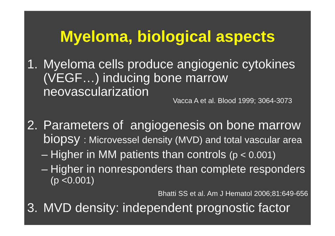
Myeloma, biological aspects
1. Myeloma cells produce angiogenic cytokines (VEGF…) inducing bone marrow neovascularization
2. Parameters of angiogenesis on bone marrow biopsy : Microvessel density (MVD) and total vascular area
– Higher in MM patients than controls (p < 0.001)
– Higher in nonresponders than complete responders (p <0.001)
3. MVD density: independent prognostic factorBhatti SS et al. Am J Hematol 2006;81:649-656
Vacca A et al. Blood 1999; 3064-3073

Plan• Whole-Body MR imaging
• Biological aspects
• DCE MR imaging
• WB DCE MR imaging
• WB DWI imaging
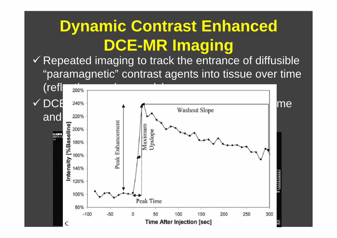
Dynamic Contrast Enhanced DCE-MR Imaging
�Repeated imaging to track the entrance of diffusible “paramagnetic” contrast agents into tissue over time (reflecting angiogenesis)
�DCE parameters are related to flow, blood volume and capillary permeability

DCE-MR Imaging
Norsas-Garcia S et al. J Magn Reson Imaging 2005.Rahmouni A et al. Radiology 2003.
Low infiltration
high infiltration
� Infiltration grade /MVD/disease activity (serum markers)
before after
�Treatment response
** But only a single segment!2D turboFLASH sequence
single or 11 slices
Plan• Whole-Body MR imaging
• Biological aspects
• DCE MR imaging
• Whole Body DCE Whole-Body MR imaging
• WB DWI imaging

Cover most of the bone marrow spaceWhere?Which planes ?How many stations in total?WB temporal resolution (per repetition)? Factor of acceleration-parallel imaging?Resolution, number of slicesSequence, 2D vs. 3D?How long the duration? When to inject?
DCE WB MR Imaging

DCE WB MR Imaging

WB 5-stations DCE-MRI
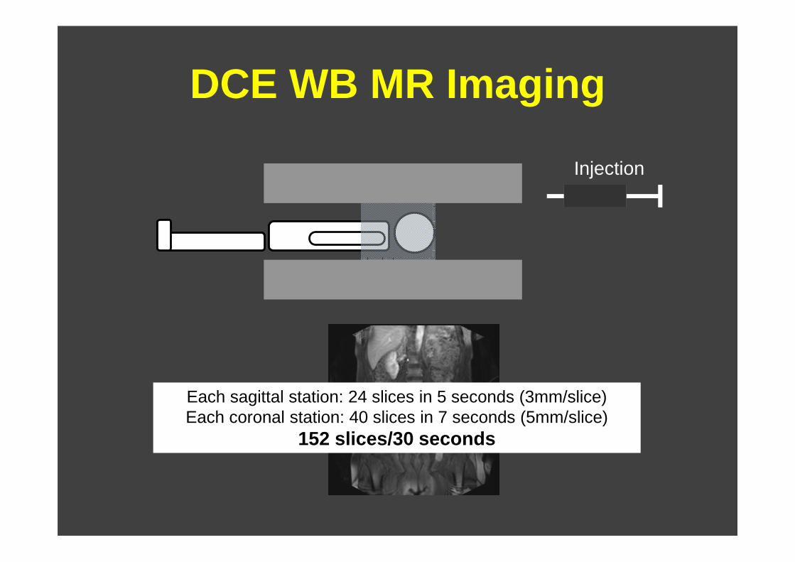
DCE WB MR Imaging
Injection
Each sagittal station: 24 slices in 5 seconds (3mm/slice)Each coronal station: 40 slices in 7 seconds (5mm/slice)
152 slices/30 seconds

T2 TSE FST1 SE
Example: 64 ans / baseline 1ère répétition2ème répétition3ème répétition4ème répétition5ème répétition6ème répétition7ème répétition

3D Isotropic 2mm Coronal acquisition360 images in 39 sec
Recent Improvement

Without
Diffuse infiltration
Before Gd After Gd 1st repetition
With
Diffuse infiltration
Before Gd After Gd 1st repetition

Focal lesion, Before Treatment

Focal lesion After Treatment, GR
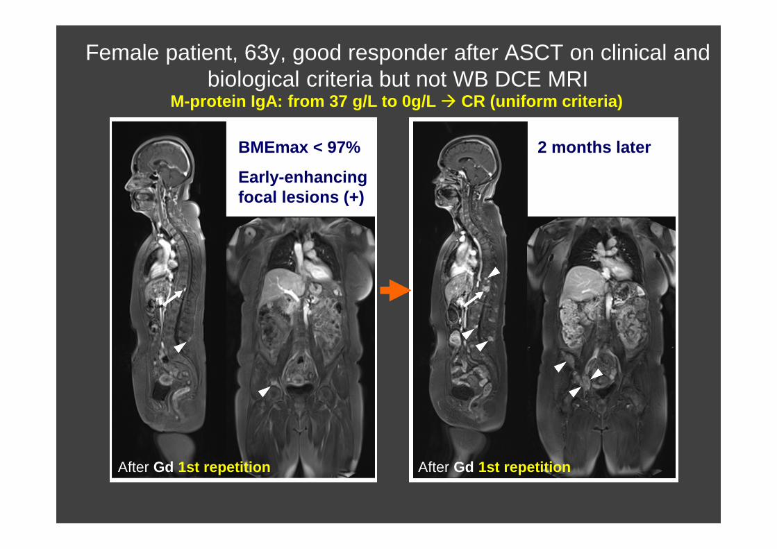
Female patient, 63y, good responder after ASCT on clinical andbiological criteria but not WB DCE MRI
M-protein IgA: from 37 g/L to 0g/L � CR (uniform criteria)
BMEmax < 97%
Early-enhancing focal lesions (+)
After Gd 1st repetition After Gd 1st repetition
2 months later

Plan• Whole-Body MR imaging
• Biological aspects
• DCE MR imaging
• WB DCE WB MR imaging
• WB DWI imaging

21
Diffusion
Stejskal and Tanner (1965)Koh DM et al. AJR 2007
*

22
Restriction (tumor)Low ADC
No restrictionHigh ADC
H2O
H2O
H2O
vessel
cell

23
No restriction: ADC is high Restriction: ADC is low
vessel
cell

24
Restriction (tumor)No restriction
H2O
H2O
H2O
vessel
cell

Before IV GADO After IV GADO
Before Treatment

After Treatment, GR

b50
b800
Before Treatment

b50
b800
After Treatment, GR

29
Apparent Diffusion Coefficient: ADC
• b (s/mm2) determines diffusion-weighting• ADC can be calculated with ≥ 2 data points with different b values = (1/b1-b0) ln (S[b1]/S[b0]) mm2/s
Koh DM et al. AJR 2007Radiology 1988;168:497-505

FLEmax = 376 %
D=1 mm2/sec
D*= 37,7 mm2/sec
F= 15,9%
ADC= 1,3 mm2/sec
FLEmax = 110 %
D=1,7 mm2/sec
D*= 1,4 mm2/sec
F= 1,6%
ADC= 1,7 mm2/sec
Initial Post-CTb (sec/mm2)
Ln
(S/S0)
b800 initial
b800 post-CT

Plan• Whole-Body MR imaging
• Biological aspects
• DCE MR imaging
• WB DCE WB MR imaging
• WB DWI imaging
Detection of Extra-medullaryDisease and other lesions

35 y/o man, non-secretory MMpost autologous stem cell transplant one year ago
Newly-onset low back pain

Diffuse marrow infiltration + hepatic nodules(incidental findings)

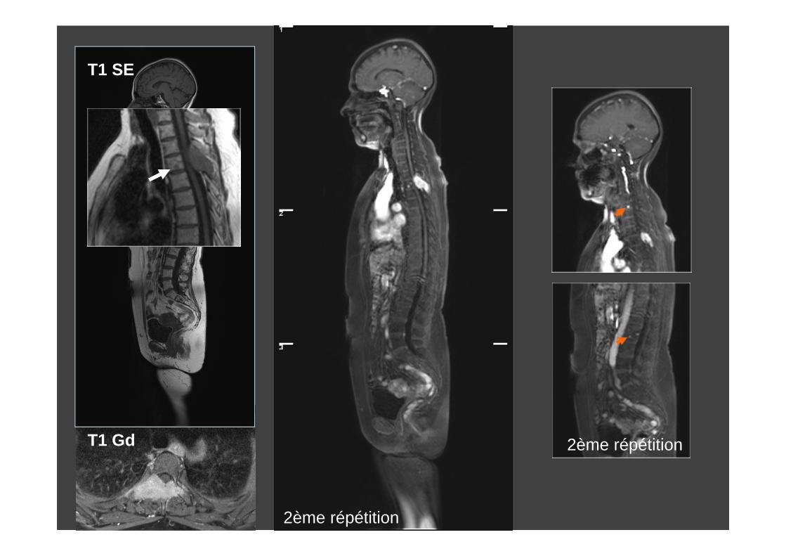
57y/o woman, MM with right sacral mass. Bone marrow transplant 2 years
ago
Follow-up exam, clinically mild right shoulder pain
1ère répétition2ème répétition3ème répétition4ème répétition5ème répétition6ème répétition7ème répétition
2ème répétition
2ème répétition
T1 SE
T1 Gd

60y/o man, MM after 4 cyclesIncidental findings


Conclusions
• What’s new?• Local functional MR techniques, i.e dynamic
contrast enhanced reflecting angiogenesis and diffusion weighted imaging reflecting cellularity can be now applied at a whole body scale
• Better characterization and understanding of myeloma lesions also include metabolic imaging using FDG and other tracers