new university of lisbon institute of hygiene and tropical
TRANSCRIPT
New University of Lisbon
Institute of Hygiene and Tropical Medicine
Comparison of the initial Ebola virus disease symptoms and subsequent
sequelae of 10 survivors in the Koinadugu district of Sierra Leone during
the 2014-2015 outbreak
(a pilot study)
Ana Sofia Rodrigues
2017
DISSERTATION FOR THE MASTER DEGREE AWARD IN TROPICAL HEALTH,
TROPICAL HEALTH SPECIALITY, 2015-2016
JANUARY, 2017

New University of Lisbon
Institute of Hygiene and Tropical Medicine
Study comparison of the initial Ebola virus disease symptoms and
subsequent sequelae of 10 survivors in the Koinadugu district of Sierra
Leone during the 2014-2015 outbreak
(a pilot study)
Author: Ana Sofia Rodrigues
Under the orientation of Prof. Dr. Jaime Nina
Dissertation for the Master Degree award in Tropical Health, Tropical Health
Speciality, 2015-2017

i
Acknowledgments
I would like to thank the entire MdM team with whom I worked both directly on the
field and indirectly at the Madrid headquarters and who gave me the permission to write
and defend this dissertation: Nicolás Dotta, Cecilia López, Alvaro Raboso, Sonia
Fernandez, Aleksandra Witkowska and Airam Vadillo, to name a few. Without their
support none of this would have been possible.

ii
Abstract
The 2014-2015 West Africa Ebola Outbreak was the largest the World has ever seen. It
started in December 2013 and was left unnoticed for 3 months, allowing for the virus to
keep spreading uncontrollably and for the outbreak to keep escalating until it was
declared an International emergency in August 2014.
Both short and long term complications have been reported on EVD survivors, ranging
from physical to psychological and social and, in addition, the persistence of EVD in
selected body compartments of the survivors (i.e.: semen) poses a great risk of
reintroduction of the virus in areas where transmission has previously been eliminated.
This study aimed to identify the symptoms presented by 10 non-randomized EVD
survivors both during the acute stage of the disease and months after recovery and
understand if there was any relation between these two stages. The most common
symptoms recorded during the active stage of EVD were weight loss, joint pain and
fever; and months after were headache, fatigue, weakness and back pain. Sixty per cent
of survivors presented months after recovery with one or more of the symptoms they
had during the acute stage of the disease, being headache the most common symptom to
persist, followed by weakness. However, all survivors presented with one or more
symptom months after recovery, regardless of the symptoms existing during the acute
stage of the disease.
The pathogenic and biological events that lead to the development of PEVDS are still
unclear and more studies still need to be done on that subject. However, taking in
consideration a symptomatic approach, this particular study concludes that the severity
of the disease in its acute stage doesn‟t seem to be associated with the severity of the
sequelae, also known as post-EVD syndrome.
Keywords: Ebola virus disease, Ebola sequelae, post-Ebola virus disease syndrome,
outbreak, Sierra Leone.

iii
Sumário
A epidemia pelo vírus Ébola que devastou a África Ocidental em 2014-2015 foi a maior
que o Mundo testemunhou até hoje. Começou em Dezembro de 2013 e permaneuceu
indetectável durante 3 meses, permitindo que o vírus se continuasse a espalhar de forma
descontrolada e para a epidemia escalar até ao ponto em que foi declarada uma
emergencia internacional em Agosto de 2014.
Consequências a curto e longo prazo têm sido documentadas em sobreviventes,
variando desde físicas, a psicológicas e sociais. Além disso, a permanência do vírus em
determinados compartimentos biológicos de sobreviventes (ex: sémen) colocam os
países em alto risco do vírus voltar a ser introduzido em comunidades onde este já foi
eliminado.
Este estudo procurou identificar os sintomas de 10 sobreviventes da doença pelo vírus
Ébola, escolhidos de forma não aleatória, tanto no momento em que estavam com a
doença na sua fase activa como nos meses de convalescença, e verificar se haveria
alguma relação entre os dois. Os sintomas mais comuns durante a fase activa da doença
foram perda de peso, artralgia e febre; e nos meses de convalescença foram cefaleias,
fadiga, astenia e lombalgias. Sessenta por cento dos sobreviventes apresentavam na fase
de convalescença, um ou mais dos sintomas que tinham durante a fase activa da doença,
sendo as cefaleias o sintoma mais comum a persistir, seguido de astenia. No entanto,
todos os sobreviventes apresentavam no mínimo um sintoma meses após a fase activa
da doença, independentemente dos sintomas que haviam desenvolvido na fase activa.
Os eventos biológicos e patogénicos que estão envolvidos no desenvolvimento do
síndrome pós-Ébola ainda não são claros e mais estudos são necessários nesta área. No
entanto, e tendo em consideração uma abordagem sindromática, este estudo em
particular conclui que a gravidade da doença pelo vírus Ébola na sua fase aguda não
parece estar associada com a gravidade das sequelas apresentadas, também conhecidas
como síndrome pós-Ébola.
Palavras-chave: Doença por virus Ébola, sequelas do Ébola, síndrome pós-ebola,
surto, Serra Leoa.
iv
Table of contents
1. Introduction …………………………………………………………..
1.1. Ebola virus disease …………………………………………………
1.2. Post EVD syndrome ………………………………………………..
1.3. EVD outbreaks ……………………………………………………..
1.4. The 2014 West Africa‟s EVD outbreak ……………………………
1.4.1. 2014 West Africa‟s EVD outbreak major events timeline ….
1.4.2. The Ebola Response …………………………………………
1.4.2.1. Case management …………………………………….
1.4.2.2. Case finding, laboratory and contact tracing ……….
1.4.2.3. Safe and dignified burials …………………………….
1.4.2.4. Community engagement and social mobilization ……
1.4.2.5. Other measures ……………………………………….
1.4.5. EVD outbreak in Sierra Leone ………………………………
1.4.5.1. About Sierra Leone …………………………………...
1.4.5.2. The outbreak in Sierra Leone …………………………
1.4.5.3. The outbreak in Nieni Chiefdom, Koinadugu district,
Sierra Leone ……………………………………………………
1.4.5.4. “End of Ebola Outbreak in Sierra Leone” statement …
2. Aims …………………………………………………………………..
3. Materials and Methods ……………………………………………….
3.1. Study design and participants ………………………………………
3.2. Ethical considerations ………………………………………………
3.3. Data collection ……………………………………………………...
3.4. Data analysis ………………………………………………………..
4. Results …………………………………………………………………
5. Discussion ……………………………………………………………..
1
1
13
16
18
20
21
23
29
31
32
33
35
35
37
39
40
43
44
44
44
45
45
46
49

v
6. References …………………………………………………………….
Appendixes
Appendix 1: Dressing and Undressing PPE
Appendix 2: Contact tracing
Appendix 3: Case Pauline
Appendix 4: Ebola Info graphic with the outbreak events timeline
Appendix 5: Cleaning and disinfection in the ETC
Appendix 6: Patient screening in the ETC‟s triage
Appendix 7: Algorithm for admitting a patient in the ETC
Appendix 8: Sierra Leone‟s statistical profile
Appendix 9: Ebola Case Investigation form used at Kumala‟s
Holding Centre
Appendix 10: Table of results: Symptoms of the 10 survivors on
admission to the HC, 8 and 10 months after
56
vi
List of figures
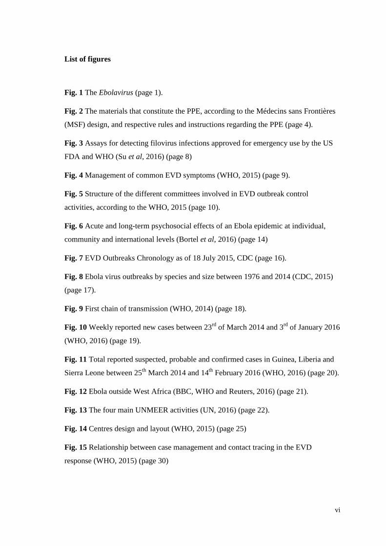
Fig. 1 The Ebolavirus (page 1).
Fig. 2 The materials that constitute the PPE, according to the Médecins sans Frontières
(MSF) design, and respective rules and instructions regarding the PPE (page 4).
Fig. 3 Assays for detecting filovirus infections approved for emergency use by the US
FDA and WHO (Su et al, 2016) (page 8)
Fig. 4 Management of common EVD symptoms (WHO, 2015) (page 9).
Fig. 5 Structure of the different committees involved in EVD outbreak control
activities, according to the WHO, 2015 (page 10).
Fig. 6 Acute and long-term psychosocial effects of an Ebola epidemic at individual,
community and international levels (Bortel et al, 2016) (page 14)
Fig. 7 EVD Outbreaks Chronology as of 18 July 2015, CDC (page 16).
Fig. 8 Ebola virus outbreaks by species and size between 1976 and 2014 (CDC, 2015)
(page 17).
Fig. 9 First chain of transmission (WHO, 2014) (page 18).
Fig. 10 Weekly reported new cases between 23rd
of March 2014 and 3rd
of January 2016
(WHO, 2016) (page 19).
Fig. 11 Total reported suspected, probable and confirmed cases in Guinea, Liberia and
Sierra Leone between 25th
March 2014 and 14th
February 2016 (WHO, 2016) (page 20).
Fig. 12 Ebola outside West Africa (BBC, WHO and Reuters, 2016) (page 21).
Fig. 13 The four main UNMEER activities (UN, 2016) (page 22).
Fig. 14 Centres design and layout (WHO, 2015) (page 25)
Fig. 15 Relationship between case management and contact tracing in the EVD
response (WHO, 2015) (page 30)

vii
Fig. 16 Statistics on Sierra Leone according to the WHO website on May 28th
, 2016
(page 35).
Fig. 17 Flag of Sierra Leone (page 36)
Fig. 18 Coat of arms of Sierra Leone (page 36)
Fig. 19 EVD cases count in Sierra Leone during January 2014 and August 2015,
according to the WHO (2015) (page 38).
Fig. 20 Aerial view from old Kumala (Médicos del Mundo, 2015) (page 40).

viii
List of abbreviations
BBC – British Broadcast Company
CCC – Ebola Community Care Centres
CDC – Centers for Disease Control and
Prevention (United States of America)
CNS – Central Nervous System
CSF – Cerebrospinal fluid
DFID – Department for International
Development
EBOV – Ebolavirus
EVD – Ebola Virus Disease
ETC – Ebola Treatment Centre
HC – Ebola Holding Centre
IPC – Infection prevention and control
MdM – Médicos del Mundo
MoSH – Ministry of Health and
Sanitation
MSF – Médicienes sans Frontiéres
(Doctors Without Borders)
NERC – National Ebola Response
Centre
NGO – Non-Governmental
Organization
ORS – Oral rehydration solution
PEVDS – Post-Ebola virus disease
syndrome
PPE – Personal Protective Equipment
ROA – Regional office for Africa
UK AID – United Kingdom Aid
UN – United Nations
UNMEER – United Nations Mission for
Ebola Emergency Response
US FDA – Food and Drug
Administration of the United States
WASH – Water, Sanitation and
Hygiene
WHO – World Health Organization

Introduction
1
1. INTRODUCTION
The Ebola Virus Disease (EVD) Outbreak that occurred in West Africa during 2014 and
2015 was the largest Ebola epidemic the World has ever known. I got the chance to go
to Sierra Leone, the country with the highest number of reported cases, during the
months of August to October 2015 as nurse working for a Spanish Non-Governmental
Organization – Médicos del Mundo – and work at an Ebola Holding Centre in Kumala,
Koinadugu district. By that time, this area in particular hadn‟t seen an Ebola case in
over 100 days but there was an existing high number of survivors, which led me to this
dissertation where I will compare the symptoms these survivors had upon admission to
the Holding Centre (EVD clinical features) and the complications they had in the
convalescence stage (post-EVD syndrome) to see if there is any correlation between the
two.
1.1. Ebola virus disease
The Ebola Virus Disease is a hemorrhagic fever
caused by a virus from the Filoviridae family,
Ebolavirus (EBOV) genus, that can infect only a
few species of mammals, such as humans, bats,
monkeys and apes.1,3
Within the EBOV genus there are five different
species, four of these capable of causing disease
to humans – Ebola virus (Zaire ebolavirus),
Sudan virus (Sudan ebolavirus), Taï Forest virus
(Taï Forest ebolavirus, formely known as Côte
d’Ivoire ebolavirus), and Bundibugyo virus (Bundibugyo ebolavirus) – all found in the
African continent.1
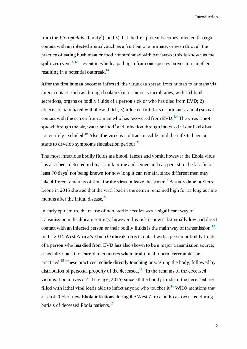
The natural reservoir for this virus is still unknown. However there is evidence that
several mammal species can harbour and transmit the virus and several bat species have
been found to carry filoviruses2, leading researchers to believe that 1) the virus is
animal-borne; 2) that the bats are the most likely reservoirs1 (specifically the fruit bats
Fig. 1 The Ebolavirus.
http://www.cdc.gov/media/dpk/201
4/images/ebola-outbreak/img8.jpg
accessed 30 December 2015.
Introduction
2
from the Pteropodidae family4); and 3) that the first patient becomes infected through
contact with an infected animal, such as a fruit bat or a primate, or even through the
practice of eating bush meat or food contaminated with bat faeces; this is known as the
spillover event 3,13
– event in which a pathogen from one species moves into another,
resulting in a potential outbreak.14
After the first human becomes infected, the virus can spread from human to humans via
direct contact, such as through broken skin or mucous membranes, with 1) blood,
secretions, organs or bodily fluids of a person sick or who has died from EVD; 2)
objects contaminated with these fluids; 3) infected fruit bats or primates; and 4) sexual
contact with the semen from a man who has recovered from EVD.3,4
The virus is not
spread through the air, water or food3 and infection through intact skin is unlikely but
not entirely excluded.19
Also, the virus is not transmissible until the infected person
starts to develop symptoms (incubation period).21
The most infectious bodily fluids are blood, faeces and vomit, however the Ebola virus
has also been detected in breast milk, urine and semen and can persist in the last for at
least 70 days5 not being known for how long it can remain, since different men may
take different amounts of time for the virus to leave the semen.3 A study done in Sierra
Leone in 2015 showed that the viral load in the semen remained high for as long as nine
months after the initial disease.22
In early epidemics, the re-use of non-sterile needles was a significant way of
transmission in healthcare settings; however this risk is now substantially low and direct
contact with an infected person or their bodily fluids is the main way of transmission.13
In the 2014 West Africa‟s Ebola Outbreak, direct contact with a person or bodily fluids
of a person who has died from EVD has also shown to be a major transmission source;
especially since it occurred in countries where traditional funeral ceremonies are
practiced.15
These practices include directly touching or washing the body, followed by
distribution of personal property of the deceased.17
“In the remains of the deceased
victims, Ebola lives on” (Haglage, 2015) since all the bodily fluids of the deceased are
filled with lethal viral loads able to infect anyone who touches it.16
WHO mentions that
at least 20% of new Ebola infections during the West Africa outbreak occurred during
burials of deceased Ebola patients.17

Introduction
3
Aside from funeral practices, healthcare providers and family members caring for EVD
patients are at the highest risk of contracting the disease thus, the healthcare settings are
the places where the disease tends to spread more quickly, especially if the healthcare
providers are not wearing appropriate personal protective equipment (PPE) (Fig.2).
All personnel caring for an Ebola patient must wear full PPE that completely covers
clothing, skin and mucous membranes.8 According to the CDC (2015), the principals
for wearing PPE are as follows:
Dressing:
Following the proper order (appendix 1);
Before entering the patient care area;
Observed and guided by a trained observer.
During patient care:
PPE must remain in place;
Never be adjusted;
Frequent and appropriate disinfection of gloved hands with strong chlorine
solution, especially after contact with bodily fluids;
In case of accidental exposure (breach in PPE) to a potentially contaminated
fluid, the person must immediately move to the Undressing area to remove PPE
and assess the exposure. There should be a facility exposure management plan
that needs to be followed.
Undressing:
To be performed in a designed area for the purpose;
In the presence and guidance of a trained observer and an undressing assistant, if
needed;
PPE must be removed slowly and in the correct sequence (appendix 1) in order
to reduce the possibility of self-contamination.

Introduction
4
Fig. 2 The materials that constitute the PPE, according to the Médecins sans Frontières (MSF)
design, and some rules and instructions regarding the PPE.6
After infection, development of disease is a complex interplay between virus, host and
environment (Goeijenbier et al, 2014).19
Once tissue invasion occurs, through infected fluid that comes in contact with broken
skin or mucosa, the virus tends to invade monocytes, macrophages and dendritic cells
that then migrate to the regional lymph nodes and disseminate.13
The infection of these
cells induces an inflammatory state with high levels of proinflammatory cytokines and
tissue factor, leading to a pro-coagulant state with impaired endothelial barrier function
Introduction
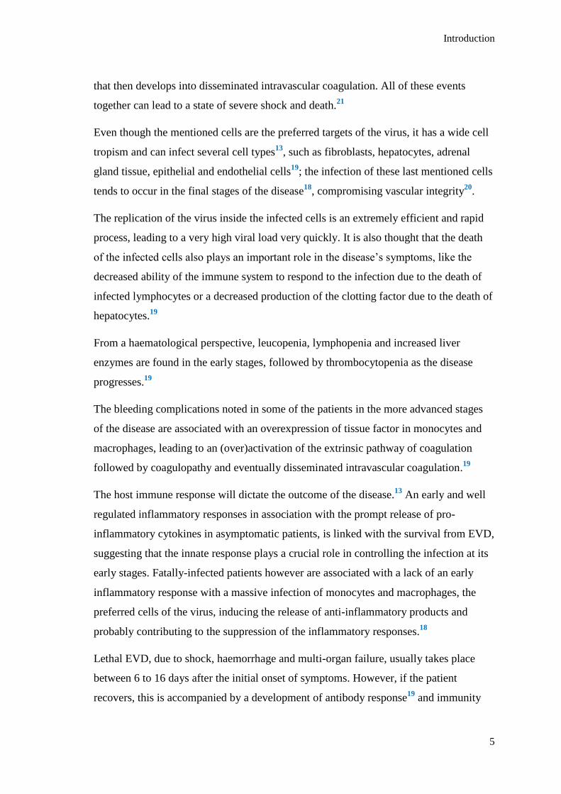
5
that then develops into disseminated intravascular coagulation. All of these events
together can lead to a state of severe shock and death.21
Even though the mentioned cells are the preferred targets of the virus, it has a wide cell
tropism and can infect several cell types13
, such as fibroblasts, hepatocytes, adrenal
gland tissue, epithelial and endothelial cells19
; the infection of these last mentioned cells
tends to occur in the final stages of the disease18
, compromising vascular integrity20
.
The replication of the virus inside the infected cells is an extremely efficient and rapid
process, leading to a very high viral load very quickly. It is also thought that the death
of the infected cells also plays an important role in the disease‟s symptoms, like the
decreased ability of the immune system to respond to the infection due to the death of
infected lymphocytes or a decreased production of the clotting factor due to the death of
hepatocytes.19
From a haematological perspective, leucopenia, lymphopenia and increased liver
enzymes are found in the early stages, followed by thrombocytopenia as the disease
progresses.19
The bleeding complications noted in some of the patients in the more advanced stages
of the disease are associated with an overexpression of tissue factor in monocytes and
macrophages, leading to an (over)activation of the extrinsic pathway of coagulation
followed by coagulopathy and eventually disseminated intravascular coagulation.19
The host immune response will dictate the outcome of the disease.13
An early and well
regulated inflammatory responses in association with the prompt release of pro-
inflammatory cytokines in asymptomatic patients, is linked with the survival from EVD,
suggesting that the innate response plays a crucial role in controlling the infection at its
early stages. Fatally-infected patients however are associated with a lack of an early
inflammatory response with a massive infection of monocytes and macrophages, the
preferred cells of the virus, inducing the release of anti-inflammatory products and
probably contributing to the suppression of the inflammatory responses.18
Lethal EVD, due to shock, haemorrhage and multi-organ failure, usually takes place
between 6 to 16 days after the initial onset of symptoms. However, if the patient
recovers, this is accompanied by a development of antibody response19
and immunity

Introduction
6
from the virus for at least 10 years.24
It‟s unknown whether this immunity could be life-
long or if a new infection with a different species of the EBOV may occur later on.24
Survival from EVD depends on good supportive care and the patient‟s immune
response.24
However the convalescence period is slow and features weight loss and
prostration21
.
Upon infection there‟s an incubation period that ranges from 2 to 21 days, during which
time the individual is not infectious;4 after that, symptoms will start to develop and the
victim becomes more and more infectious as the viral load increases.
The early signs and symptoms are non-specific and can mimic other common tropical
diseases such as malaria, dengue, typhoid fever and other viral infections, which makes
it difficult for an early clinical diagnosis.2,13
These signs and symptoms are:
Fever
Headache
Fatigue
Weakness
Joint and muscle pain
Sore throat
Progressive gastrointestinal symptoms usually develop within 3 to 5 days from initial
symptoms onset: vomiting, diarrhoea, abdominal pain (stomach), nausea, leading to
electrolyte imbalance, intravascular volume depletion and shock. Other symptoms as
skin rash, hiccups, conjunctival infection, respiratory and neurologic finding may also
occur. Hemorrhagic complications appear as a late sign, in less than 20% of patients,
alongside with multiple organ failure, and it is usually associated with a bad prognosis.
Overall, clinical deterioration may progress in a rapid way and may result in death
within 7 to 10 days of symptoms onset.2,4,7,19
However, there‟s a 75% chance of survival
if the patient lives through the second week of infection.13
According to a study done in Sierra Leone between May 25th
and June 18th
of 2014, the
incubation period ranged from 6 to 12 days, the case fatality rate was 74% and the most
common clinical features were: fever (89%), headache (80%), weakness (66%),
dizziness (60%), diarrhoea (51%), abdominal pain (40%) and vomiting (34%). The
Introduction
7
symptoms associated with a fatal outcome were fever, weakness, dizziness and
diarrhoea and also, in patients aged above 45 years old (94%) or patients with 10
million or more EBOV copies per millilitre of blood (also 94%).53
These results are in
accordance with another study also done in Sierra Leone between May 29th
and
December 8th
of 2014 by Lado et al, where the most common clinical features were
fever or history of fever (83%), intense fatigue or weakness (68%), vomiting or nausea
(50%) and diarrhoea (41%).54
Rapid and reliable diagnosis of EVD is essential for appropriate and effective patient
management, hospital or health center infection prevention and control, and
optimization of use of healthcare resources (Martinez et al, 2015).2
Due to the non-specificity of the initial symptoms and on an human epidemic context,
EVD diagnosis is based on the clinical manifestations of the patient and a known
contact history with an infected individual in the 21 days prior to the onset of
symptoms.21
If a person has developed symptoms concordant with an EBOV infection
and there is reason to believe that EVD should be considered, then the individual must
be isolated and public health professionals notified. Blood samples are then collected
for testing and confirmation of infection.1
Some of the investigations used to confirm an EBOV infection are:4
Antibody-capture enzyme-linked immunosorbent assay (ELISA);
Antigen-capture detection tests;
Serum neutralization test;
Reverse transcriptase polymerase chain reaction (RT-PCR) assay;
Electron microscopy;
Virus isolation by cell culture.
These must all be performed in maximum biological containment conditions due to the
extreme biohazard risk of the samples.4
Prior to 2000, the gold standard for EBOV laboratory detection were antigen detection
methods, such as the ELISA, since it has a high sensitivity rate (93%) in the acute stage
of the illness. However, as the disease progresses the antigen levels decline and the
sensitivity of the test lowers within 1-2 weeks after onset of symptoms. Nowadays,

Introduction
8
ELISA has been replaced by RT-PCR (reverse transcription polymerase chain reaction)
testing, allowing a more rapid and portable detection.2
RT-PCR is a rapid and highly sensitive nucleic acid amplification test and has a very
high sensitivity and specificity in detecting the EBOV genome of approximately 100%
and 97% respectively.2
Through the use of RT-PCR the virus can be detected 48 hours after infection19
,
therefore false negative results may occur in the first 2-3 days of the disease since the
molecular assay may not be able to detect the genome in such an early stage2, reason
why it is important to take another blood sample 24 to 48 hours after the first one to
confirm the result.
During the 2014-2015 West Africa‟s outbreak the development of rapid diagnostic tests
has showed viable options for EBOV diagnosis, with suggestions of sensitivity of 100%
and specificity of 90%. Although this is an extremely promising tool it is still not used
in daily practice.2 Some example of this tests are the RealStar Filovirus Screen RT-PCR
Kit 1.0, the ReEBOV Antigen Rapid Test Kit and the Xpert Ebola Assay.55
Fig. 3 Assays for detecting filovirus infections approved for emergency use by the US FDA and
WHO (Su et al, 2016).
Management of EVD is based on early recognition of infection, accompanied by
effective isolation and the provision of the best available symptomatic and supportive
care, including rehydration, maintenance of electrolyte balance, nutrition, pain relief
and effective blood volume, through blood transfusions, if needed. The main goal of
supportive care is to maintain intravascular volume.2,21

Introduction
9
According to the WHO guidelines, treatment of patients with suspected or confirmed
EVD in Ebola Treatment Centres (ETC‟s) had the following principles:
Provide basic care such as food and water;
Patients with fever, especially those with diarrhoea and vomiting, should be
encouraged to drink fluids and as much oral rehydration solution (ORS) as can
be tolerated;
Malaria treatment should be provided to all patients with fever, in accordance
with national guidelines;
Medicines to control symptoms can be given orally (fig. 4). Since injections
would increase the risk of staff infection can only be given by appropriately
trained and assigned staff inside the centre;
If sufficient oversight is available, oral antibiotics may be given to treat apparent
bacterial infections;
Patient‟s temperature should be recorded once a shift with a calibrated infrared
thermometer.46
Fig. 4 Management of common EVD symptoms (WHO, 2015).46
The quality of the care provided is strictly related to the outcome of the disease, for
example, the difficulties that patient‟s face in accessing basic medical care in resource
poor rural settings reflects a higher case-fatality rate of the disease. Also, failure to
Under 6 months 10 mg daily; Over 6 months 20 mg daily for 10-14
days.
Introduction
10
provide full supportive care to the suspected cases1 may result in substandard care for
these, who may later be found to have a treatable disease such as malaria.2,13
Specific antiviral medication is still in experimental stages19
and there are 2 potential
vaccines undergoing human safety testing4, in the meantime and for effective control of
an active EVD outbreak, several measures need to be implemented, with an emphasis to
case management, surveillance and contact tracing, good and safe laboratory service,
safe burials and social mobilization.4
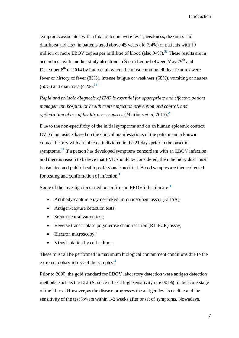
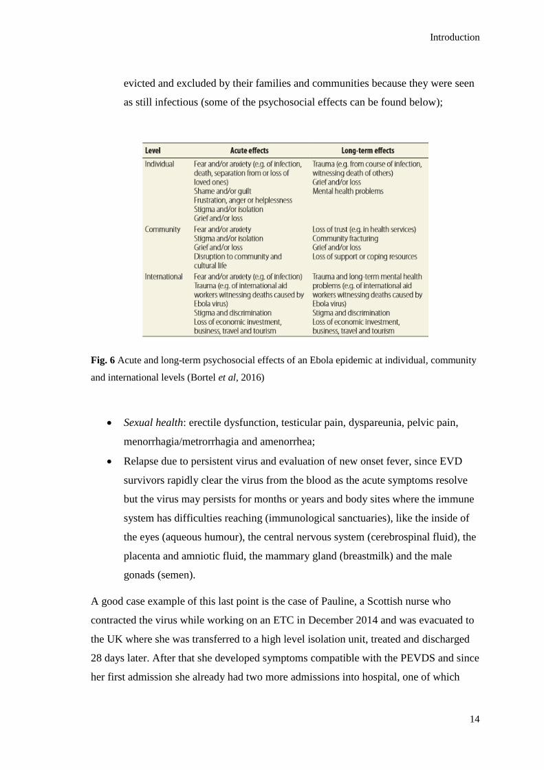
Fig. 5 Structure of the different committees involved in EVD outbreak control activities,
according to the WHO, 2015.26
Good infection control practices, such as disinfection of contaminated areas and objects
(including the patient‟s home and belongings), also plays a vital role in reducing
transmission, plus an attempt on early diagnosis and the use of barriers while
performing patient care, like PPE.13,21
1 Definition found in page 12

Introduction
11
A study done in Kailahun district, Sierra Leone, found that control can be accomplished
by using interventions based on identification and appropriate management of those at
risk of developing EVD.65
During an outbreak, a combination of Ebola-like symptoms with a high-risk exposure –
contact with an EVD patient or body fluids, objects contaminated with body fluids of a
person who has died from EVD, attendance to a funeral, contact with an infected animal
(fruit bats or primates), contact with the semen of an EVD survivor, visit to a local
healthcare or to a known endemic area within the last 21 days – are enough to proceed
with isolation and management protocols. It is however essential that there are sufficient
patient care capacity and staffing in the specialist facilities.19,23,65
Asymptomatic patients with known epidemiological risk factors and persons who may
have come in contact with infected patients (called contacts) are considered to be at risk
of infection and may need monitoring during the duration of the incubation period.25
These should be immediately isolated in a room with private bathroom, all attending
healthcare personnel must wear PPE and all contaminated materials must be treated as
potentially infectious.13
The recognition of these patients is done by a process called
contact tracing2 (appendix 2) that allows a rapid recognition of symptoms with
immediate isolation, testing and provision of care of new cases, thus preventing the
future spread of EBOV.13,25
Contact tracing is strictly connected to surveillance (case finding) and case investigation
measures, since the identification of as EVD case activates a case investigation process
where contacts are identified and thus initiating the process of contact tracing. This can
only be effective if it‟s immediately implemented after case finding and efficiently
managed.
In order for this to happen, strict definitions must be applied stating what constitutes a
“contact”, a “suspected EVD case”, a “probable EVD case” and a “confirmed EVD
case”. The WHO (2015) as defined all of the above as shown on the following table:26
2 This is further discussed in section 1.4.2.2. Contact tracing

Introduction
12
Denomination Definition
Suspected EVD case Any person, alive or dead, suffering or having suffered
from a sudden onset of high fever and having had contact
with a suspected, probable or confirmed Ebola case, or a
dead or sick animal; OR
Any person with sudden onset of high fever and at least
three of the following symptoms: headache, vomiting,
diarrhoea, anorexia/loss of appetite, lethargy, stomach pain,
aching muscles or joints, difficulty swallowing, breathing
difficulties, or hiccup; OR
Any person with unexplained bleeding/haemorrhaging; OR
Any person with sudden, unexplained death.
Probable EVD case Any suspected case evaluated by a clinician, OR
Any person who died from “suspected” EVD and had an
epidemiological link to a confirmed case but was not tested
and did not have laboratory confirmation of disease.
Confirmed EVD case Any suspected or probable cases with a positive laboratory
result.
Contact Any person who has been exposed to a suspected, probable,
or confirmed case of EVD in at least one of the following
ways (including healthcare workers):
Has slept in the same household as a case;
Has had direct physical contact with the case
(alive or dead) during the illness;
Has had direct physical contact with the
(deceased) case at a funeral or during burial
preparation rituals;
Has touched the blood or body fluids (including
urine, faeces, vomit, tears, or sweat) of a case
during their illness;
Has touched the clothes or linens of a case;
A baby who has been breastfed by the case.

Introduction
13
1.2. Post EVD syndrome3
Both short and long term medical problems have been reported in EVD survivors,
including mental and physical symptoms. This is referred to as the Post Ebola virus
disease syndrome (PEVDS).
Definition of an EVD survivor:51
Person with a confirmed positive result on RT-PCR testing for Ebola virus on
any body fluid who subsequently recovered; AND/OR
Person who is IgM and/or IgG positive on serological testing for EVD and has
not been vaccinated against Ebola virus.
According to Gulland (2015) “preliminary data show that people who experienced the
severe form of the acute infection were more likely to have serious chronic problems”
however and for the time being, treatment is still symptomatic and further studies are
still needed to fully understand the long-term effects of EBOV infection and the clinical
spectrum that occur during PEVDS.27,29
Some of the symptoms of PEVDS include: 27,29,51,57,58,61,63
General: fatigue and anorexia;
Musculoskeletal: chronic joint pain, often severe and debilitating;
Ocular: blurred vision, eye pain, redness, dry eyes, sensitivity to light and, more
serious, an inflammatory disease that could lead to blindness if left untreated
called uveitis (seen in 50% of survivors in a study done in Port Loko, Sierra
Leone, between March 7th
, 2015 and April 24th
, 2015);
Auditory: hearing loss and tinnitus;
Abdominal: pain from unknown cause;
Neurological: headache, memory impairment, poor concentration, peripheral
neuropathy, tremor, sleep disturbances, low mood;
Mental health: post-traumatic stress disorder, fear of death, shame,
stigmatization and depression; many survivors were threatened, attacked,
3 For the purposes of this thesis, the EVD sequelae will be focused on the Zaire species of the virus, even
though it has been stated that there are no obvious differences between the species63
Introduction
14
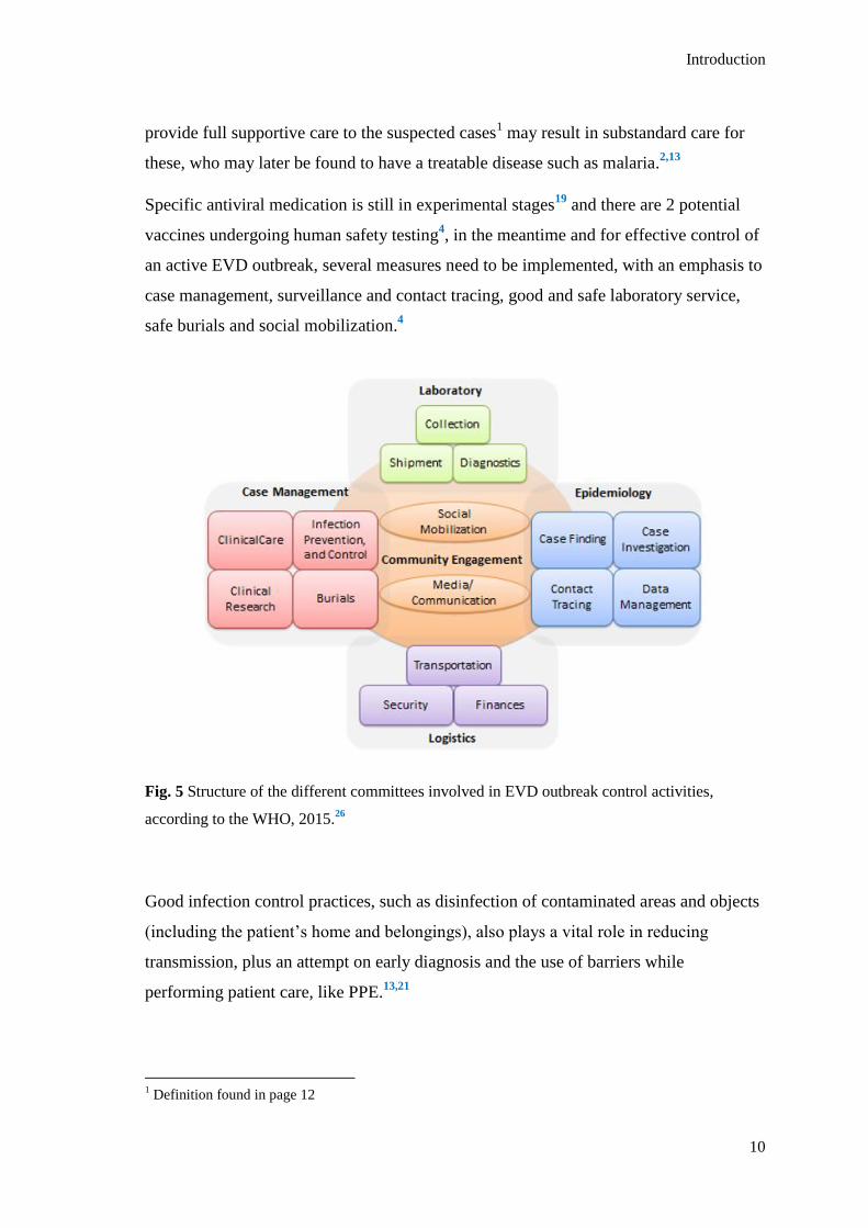
evicted and excluded by their families and communities because they were seen
as still infectious (some of the psychosocial effects can be found below);
Fig. 6 Acute and long-term psychosocial effects of an Ebola epidemic at individual, community
and international levels (Bortel et al, 2016)
Sexual health: erectile dysfunction, testicular pain, dyspareunia, pelvic pain,
menorrhagia/metrorrhagia and amenorrhea;
Relapse due to persistent virus and evaluation of new onset fever, since EVD
survivors rapidly clear the virus from the blood as the acute symptoms resolve
but the virus may persists for months or years and body sites where the immune
system has difficulties reaching (immunological sanctuaries), like the inside of
the eyes (aqueous humour), the central nervous system (cerebrospinal fluid), the
placenta and amniotic fluid, the mammary gland (breastmilk) and the male
gonads (semen).
A good case example of this last point is the case of Pauline, a Scottish nurse who
contracted the virus while working on an ETC in December 2014 and was evacuated to
the UK where she was transferred to a high level isolation unit, treated and discharged
28 days later. After that she developed symptoms compatible with the PEVDS and since
her first admission she already had two more admissions into hospital, one of which

Introduction
15
with a case of meningoencephalitis, from virus that remained in the Central Nervous
System (appendix 3). More details on the case can be found on the case report done by
Michael Jacobs et al, 2016.64
It is still not clear what are the relations between the pathogenesis and biological events
that lead to PEVDS, as is any relation between the severity of the acute disease and the
frequency or severity of sequelae (part of what this study plans to identify).63
According
to Vetter et al (2016), sequelae could be the result of residual dysfunction from direct
viral effects during the acute EVD infection, sustained immune activation, delayed
hypersensibility reaction, molecular mimicry, autoimmune disease, or immune complex
deposition, individually or in combination.
The fact that the virus may persist in selected body compartments of the survivors,
specially the semen, also brings awareness to the possibility of reintroduction of the
virus in areas where the virus has already been eliminated.51
However studies have
shown that even if the virus still remains active in other body fluids (aside from semen),
its infectivity it‟s extremely low at this stage.61,62
“Despite the persistence of Ebola virus
in a few body compartments and recrudescence, other than rare reports of suspected
sexual transmission, there is no conclusive evidence of virus transmission from
convalescence patients“ (Vetter et al, 2016).
After the high amount of sequelae found in this Ebola‟s outbreak survivors and it
became clear how common this syndrome was, the WHO created a plan to follow-up
the survivors for weeks and months following discharge from the ETC. 51
These follow-up visits would take place: 51
2 weeks after discharge;
Every month for 6 months following the 2 weeks;
Every 3 months until completion of one year; and
Continued follow-up as needed after that.
The first visit would include a general medical history and physical examination, with
vital signs recording and nutritional evaluation, plus musculoskeletal, ocular, auditory,
abdominal, neurological, mental health and sexual heath evaluations and a consultation
with a social worker to address issues related to stigma, economic status and
Introduction
16
employment, shelter and food security, dependents, social support, potential substance
misuse or dependency and identification of vulnerable individuals. Routine laboratory
tests would also be done, including full blood counts, creatinine levels, Ebola RT-PCR
testing, plus any other tests that might be needed, such as a malaria rapid diagnostic test.
The following visits would be focused on evaluation the areas relevant to the patient‟s
particular condition, however a complete evaluation, similar to the one done on the first
visit, should be done at least every 3 months during the first year. The follow-up visits
for the male survivors should also be coordinated with visits made for semen testing.51
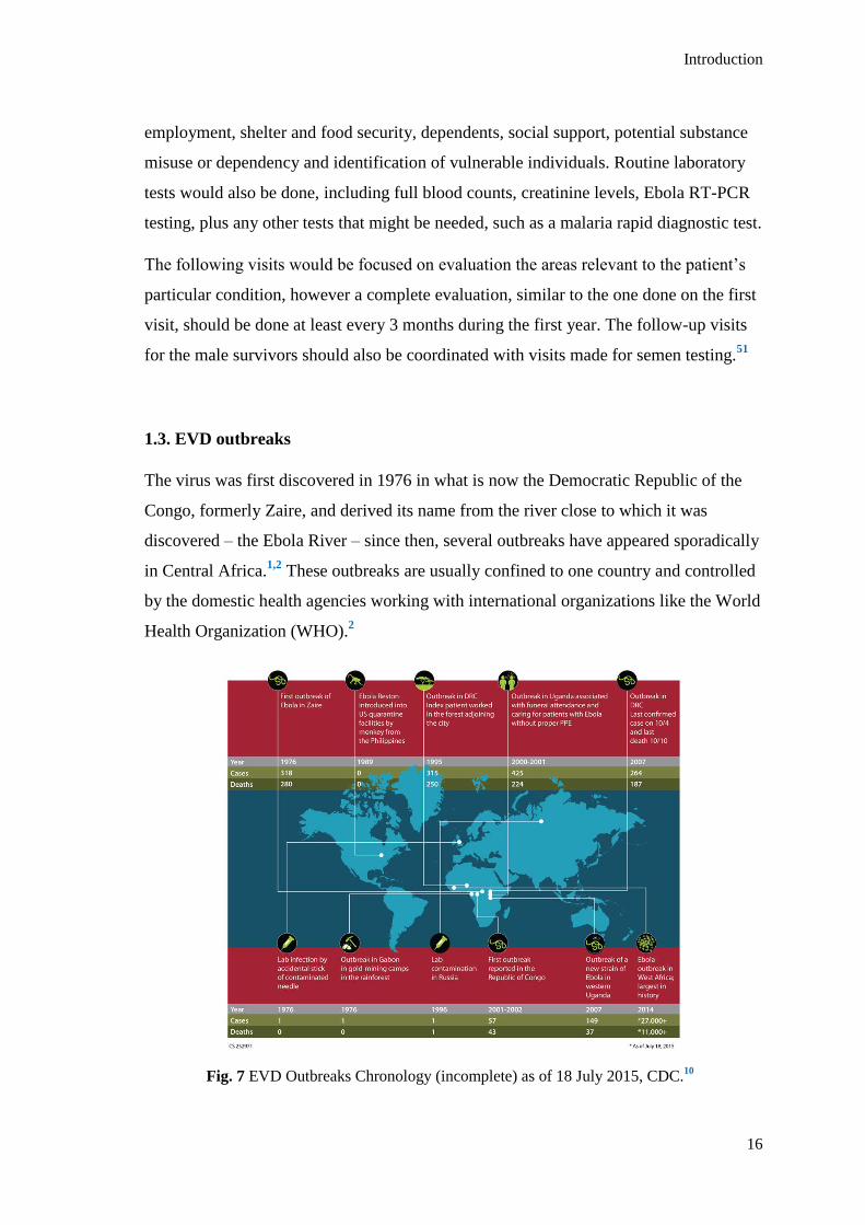
1.3. EVD outbreaks
The virus was first discovered in 1976 in what is now the Democratic Republic of the
Congo, formerly Zaire, and derived its name from the river close to which it was
discovered – the Ebola River – since then, several outbreaks have appeared sporadically
in Central Africa.1,2
These outbreaks are usually confined to one country and controlled
by the domestic health agencies working with international organizations like the World
Health Organization (WHO).2
Fig. 7 EVD Outbreaks Chronology (incomplete) as of 18 July 2015, CDC.10
Introduction
17
To be noted that the map above doesn‟t show all the known outbreaks that happened
between 1976 and 2015. For example, the 1976 South Sudan outbreak, by the Sudan
ebolavirus, that made 284 notified cases, was omitted from this map. 10
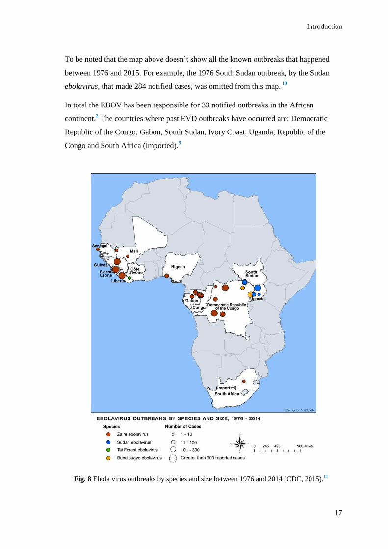
In total the EBOV has been responsible for 33 notified outbreaks in the African
continent.2 The countries where past EVD outbreaks have occurred are: Democratic
Republic of the Congo, Gabon, South Sudan, Ivory Coast, Uganda, Republic of the
Congo and South Africa (imported).9
Fig. 8 Ebola virus outbreaks by species and size between 1976 and 2014 (CDC, 2015).11
(imported)

Introduction
18
1.4. The 2014 West Africa’s EVD outbreak
“The 2014 Ebola outbreak is the largest Ebola outbreak in history and the first Ebola
outbreak in West Africa. This outbreak is the first Ebola epidemic the world has ever
known” (CDC, 2015).12
It started on March 21st 2014, when the Ministry of Health of Guinea notified the WHO
of a rapidly evolving outbreak of EVD. The most affected countries of this outbreak
were Guinea, Liberia and Sierra Leone and the rapid spread of the disease occurred due
to a number of factors including people mobilization and funeral/burial practices.2,74
The total number of notified cases in this outbreak was 28 646 cases, causing 11 323
deaths and making over 10 000 survivors.72
The first case of EVD in this outbreak (index patient) was retrospectively found to be a
2 year old boy from the village of Meliandou, Guinea, who on the 26th
of December,
2013, became ill with fever, melena and vomiting and died 2 days later. It is thought
that the boy became infected by ingesting the meat of an infected wild animal, most
likely, a fruit bat.32,42
Fig. 9 First chain of transmission (WHO, 2014).33

Introduction
19
After his death, the virus remained undetected while creating several chains of fatal
transmission for over 3 months. It was only in March that the local Guinea health
officials, MSF and WHO staff recognised something was happening but were unable to
identify it, therefore, the Ministry of Health of Guinea sent blood samples to the
institute of Pasteur, in Paris, which identified the causative agent as the Zaire
ebolavirus, never before seen in this part of the World.32
Personal protective equipment and medical teams rapidly started arriving in the country,
from both WHO and other Organizations in response to the Outbreak, however, by late
March the virus had entered the capital city of Conakry and there were uncontrollable
numbers of new cases emerging both in the capital and other parts of the country.
Foreign medical teams kept pouring in but the outbreak was out of control.32
In April, the number of new cases was
increasing by 3.75 per day in Guinea
and continued to increase until it
reached 19.07 new cases per day in
December 2014 (peak of the
outbreak).38
Liberia reported its first case on the 30th
of March 2014 and saw a more
attenuated beginning of the outbreak,
recording only an increase of 0.15 new
cases per day in April 2014 but growing
to 47.53 new cases per day by
September 2014 (peak).38,74
Sierra Leone was the most affected the
country of all, beginning the case
reporting on the 25th
of May 2015 and
while in May it was only recording an
increase of 0.44 new cases per day it
reached and astounding peak of 65.23
new cases per day in November
2014.38,74
Fig. 10 Weekly reported new cases between
23rd
of March 2014 and 3rd
of January 2016
(WHO, 2016).39

Introduction
20
Fig. 11 Total reported suspected, probable and confirmed cases in Guinea, Liberia and Sierra
Leone between 25th March 2014 and 14
th February 2016 (WHO, 2016).
40
1.4.1. 2014 West Africa’s EVD outbreak major events timeline
The outbreak was first acknowledged in March, 2014;
By the end of the month it had already spread to Liberia;
It entered Sierra Leone in May;
“In June, the MSF described the Ebola Outbreak as out of control” (BBC, 2016);
By July, two US workers contracted EBOV and had to be evacuated to the USA;
Still in July the virus entered Nigeria and two leading doctors on the subject died
in Liberia and Sierra Leone;
The outbreak is declared as an International Public Health Emergency by the
WHO on the 8th
of August, 2014;
By the end of August the virus enters Senegal and in October, Mali, via
imported cases from Guinea;
Between September and December 2014, two more US worker, plus a Spaniard
and an English workers all are diagnosed with EVD and evacuated to their home
countries;

Introduction
21
Germany, Norway, France, Italy Switzerland and the UK all treat patients who
contract the virus in West Africa;
The outbreaks in Nigeria, Senegal and Mali were of small proportions and all
had been declared Ebola free by January 2015.39,41
A full info graphic with the timeline of the events during this outbreak can be found
on Appendix 4.
Fig. 12 Ebola outside West Africa (BBC, WHO and Reuters, 2016).39
1.4.2 The Ebola Response
After the declaration of the outbreak as an International Public Health Emergency by
the WHO in August, several public health interventions needed to be established,
including: “early identification of cases; appropriate treatment of people with EVD;
physical isolation of cases to reduce further spread; rigorous tracing of contacts; and
Introduction
22
burial practices that were safe in terms of EVD-transmission risk and dignified in terms
of allowing culturally-appropriate grieving” (WHO, 2015), accompanied by strong
social mobilization.46
With this in mind an “Ebola Response Roadmap” document was
created that same month, with the purpose of assisting governments and partners in the
revision and resourcing of country-specific operational plans for Ebola Response, and
the coordination of international support for their full implementation.43
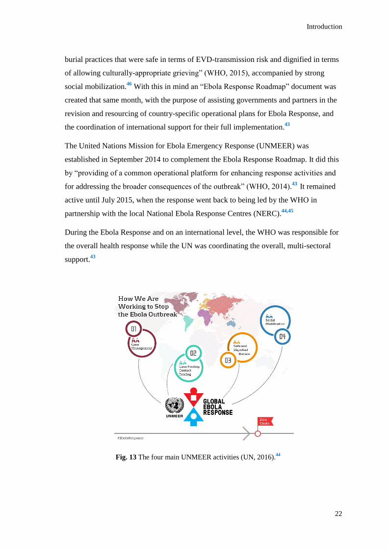
The United Nations Mission for Ebola Emergency Response (UNMEER) was
established in September 2014 to complement the Ebola Response Roadmap. It did this
by “providing of a common operational platform for enhancing response activities and
for addressing the broader consequences of the outbreak” (WHO, 2014).43
It remained
active until July 2015, when the response went back to being led by the WHO in
partnership with the local National Ebola Response Centres (NERC).44,45
During the Ebola Response and on an international level, the WHO was responsible for
the overall health response while the UN was coordinating the overall, multi-sectoral
support.43
Fig. 13 The four main UNMEER activities (UN, 2016).44

Introduction
23
Overall, the Ebola Emergency Response defined five objectives:
1) Stop the outbreak;
2) Treat the infected;
3) Ensure essential services;
4) Preserve stability;
5) Prevent further outbreaks.
The response planned to accomplish the objectives through four main activities:
1) Case management;
2) Case finding, lab and contact tracing;
3) Safe and dignified burials;
4) Community engagement and social mobilization.44
1.4.2.1. Case management
The WHO priority activities in this area were:
Ebola treatment centres with full infection prevention & control (IPC) activities;
Ebola referral/isolation centers (holding centers);
Referral processes for primary health care facilities;
Community-based care supported by intensified IPC and appropriate PPE in
intense transmission areas.43
This was achieved by creating over 60 specialized Ebola Treatment Centres (ETC‟s),
plus over 63 Ebola Community Care Centres (CCC‟S) – that later had the name
changed to Holding Centers (HC‟s) – in the three most-affected countries and by having
more than 40 organizations and 58 foreign medical teams having been deployed with an
estimate of 2 500 international personnel operating on these centers in partnership with
ministries of health and thousands of national staff.
In partnership with other NGO‟s, the WHO was able to provide more than one million
sets of PPE and extensive training for health and front-line workers on infection control
practices, occupational health and safety, clinical management and safe burials.

Introduction
24
This expanded capacity to isolate cases, along with safe and dignified burials and
behavioral changes in communities were key factors in controlling the outbreak, even
though many cases were still not coming forwards for isolation and treatment leading to
Ebola related deaths still occurring in the communities.45
ETC‟s and HC‟s were the places where most of the care for patients with EVD took
place. In these centres, EVD infected people would be isolated and receive basic
curative and palliative care, with access to food, hydration, clean clothes and linen.46
Treatment itself has already been described previously.
The health aid workforce in these centres was formed of both international and local
staff that had been trained for this purpose, including training on the protocols of the
facility, procedures to be followed if exposure occurs by accident and a temperature
check always when entering and leaving the centre (refrain from working when
temperature above 38ºC).46
Both centres had a similar layout: 46
Red zone Green zone
Care of patients suspected or confirmed to
have EVD.
All activities that don‟t pose a risk of EVD
transmission:
Clean and disinfect contaminated objects. - Counselling
Burn waste. - Rest areas for staff and family
Morgue. - Supporting services, such as
administration, stores, pharmacy,
kitchen and laundry for staff‟s PPE.
The movement of staff inside the centre should always be done from clean to more
contaminated areas, including for cleaning purposes (more information on cleaning
done inside the centres can be found on Appendix 5).46

Introduction
25
Entrance to the red zone had to be done through the PPE dressing area and to exit,
through the PPE removal area, thus making sure that every member of staff in the red
area was wearing FULL PPE.46
Patients would enter the red zone through a designated point, different from the staff.46
Fig. 14 Centres design and layout (WHO, 2015)46
There would be hand washing stations on the entrance and exit of every area inside the
centre, both inside the red and green zones and including the entrance/exit of the centre
itself. Hand washing should always be done with soap and water or with an alcohol-

Introduction
26
based handrub, although many centres didn‟t have these resources and so, chlorine
solutions at a concentration of 0.05% (weak solution), applied for a minimum of 40 to
60 seconds were used and considered appropriate.46
Chlorine solutions at a concentration of 0.5% (strong solution) was also available in all
centres (mandatory) and was used to clean all the materials that came in contact with
suspected, probable or confirmed EVD cases, including PPE, the table in the triage area,
the patient‟s area and belongings (ex: plates, utensils, bedpans and waste buckets),
patient‟s latrines and showers, any spills of body fluids and dead bodies.46
Both weak and strong chlorine solutions should be prepared daily in a mixing dedicated
area and following an adequate protocol.46
All solid infected waste, including PPE, had to be incinerated daily in a burn waste or
also called “burning pit” designate for this purpose, which would be located in the red
area and down-wind from the centre.46
Every patient would enter the centre from the Triage area, where a screening/medical
evaluation would be done in the form of an interview and a temperature reading would
be taken with an infrared thermometer (appendix 6). If considered to be a suspected or
probable case of EVD, the patient would be admitted (appendix 7) and grouped into one
of two categories: dry case (fever plus symptoms other than diarrhoea, vomiting or
bleeding) or wet case (with diarrhoea, vomiting or bleeding).46
Patients found not likely to have EVD would be given items such as home kits or
medicines and provided instructions for use and would also be educated on the
transmission and prevention of EVD and when to return to the ETC/HC.46
After admission and from a patient perspective, the only way out of the centre was
either trough the “happy shower” (discharge) or through the morgue.
When recovered, and in order to be eligible to exit the centre (discharge), the patient
would have to fulfil certain criteria:
Patient with fever only and no other symptoms at admission
o No fever for 72 hours and no other symptoms AND

Introduction
27
o Able to eat and carry out daily routine activities such as walking (taking
into account any previous disabilities) and washing themselves
independently.
Patient with fever and other symptoms at admission
o No fever for 72 hours and other symptoms that may be associated with
EVD disappeared for 72 hours AND
o Able to eat and carry out daily routine activities such as walking (taking
into account any previous disabilities) and washing themselves
independently.
If laboratory (PCR) testing is available:
o A negative test on day following onset of fever and symptoms, or later
AND
o A negative test at least 48 hour after the last positive test.46
Leaving the facility was done via the “Happy Shower”, which was a joyful event for
everyone since it meant the person had survived Ebola. All of the patient‟s belongings
were left behind to be incinerated; the person would enter the “Happy Shower”, have a
shower with chlorine solution 0.05% and put on clean clothes before exiting the
facility.56
After discharge, a new stage of the patient‟s recovery would begin and included dealing
with the convalescence stage of the disease, and possible complications that may come
with it, reintegration in the community and the fight against the stigma that still
surrounds Ebola and Ebola survivors.56
On discharge it was also important to advice men to use condoms during sexual
intercourse for at least 3 months, after it became known that the virus could remain
active in the semen; and alert pregnant women for the fact that miscarriage or foetal
death could occur. If so, they should attend an ETC or obstetric clinic equipped with
good infection prevention and control (IPC) practices, including full PPE, for delivery
of the foetus or any further care needed.46
All survivors would also receive education and counselling regarding the possible
sequelae and psycho-social consequences during the convalescence period of the

Introduction
28
disease51
and were also advised to link with their local community engagement staff to
minimize stigma and discrimination.46
If the outcome wasn‟t a discharge, the management of dead bodies and burials in the
centre had to be performed by staff trained in IPC measures and the necessary resources
should be present, such as full PPE, body bags, disinfectant and appropriate
transportation. Preferably this should be done by a burial team (more information on
burial teams on chapter 1.4.2.3.) unless the burial team wasn‟t able to attend the centre
straight away, in which case, and if all the necessary resources and trained staff was
present and could safely perform the disinfection of the body and materials, placement
of the body inside a body bag and movement it to the mortuary area, this could be
performed by trained centre staff, even though the burial itself should and would always
be done by the burial team.46
Despite the number of centres in operation during the peak of the outbreak, the number
of cases was so high that outgrew the capacity of the ETC‟s and some patients remained
at home, placing family members at risk.46 Not only the shortness of beds led to people
staying at home but there were also cases of people who refused to be taken to the ETC
due to the stigma surrounding it. Many believed that people were taken there to die
since everyone that would go wouldn‟t return.59
In Sierra Leone, Ebola Holding Centres were created at the peak of the Ebola outbreak
when ETC‟s couldn‟t‟ provide enough beds for the overwhelming number of new cases
seen. Therefore, these units were created as a temporary place to admit and isolate
suspected EVD cases until the confirmatory diagnostic testing results came. Those
tested positive would then be transferred to the closest ETC and those tested negative
would be discharged or referred to other health care facilities, such as the hospital or
their local health care centre.60
The HC‟s worked in a similar way to the ETC‟s, however, according to Zachariah and
Harries (2015), they were controversial since they could become very overcrowded,
environmental and personal protection measures were limited, and the fear of
nosocomial EVD transmission was prevalent. Also, and because the patient‟s wouldn‟t
be diagnosed or treated for anything else while in the HC‟s, many other diseases
endemic in these areas and whose symptoms mimic the ones from Ebola would go

Introduction
29
unnoticed and could lead to serious or even fatal consequences in the centre while
waiting for the test results, some examples of these are: severe malaria, typhoid fever
and gastroenteritis.
1.4.2.2. Case finding, laboratory and contact tracing
The WHO priority activities in these areas were:
Case diagnosis: by a WHO-recognized laboratory or by an epidemiologic link to
case confirmed by a WHO-recognized laboratory in intense transmission areas;
Surveillance: contact tracing and monitoring, including new transmission
chains.43
To achieve these, over 230 experts were sent to 26 mobile laboratories creating an
ability to test over 750 samples per day and thus, enabling the rapid confirmation of
cases.
Also, over 600 public health experts were deployed to the three most-affected countries
during the course of the outbreak response to assist in surveillance, field epidemiology,
case finding, contact tracing, information management and epidemiological analysis,
which was essential to create and follow chains of transmission and finding new cases
from the contacts lists. Contacts were systematically monitored for 21 days and even
after the last case had been identified, a long period of surveillance was required to
ensure that all chains of transmission were found and there was no re-emergence.45
“Contact tracing is the process of identifying, assessing and managing people who have
been exposed to a disease to prevent onwards transmission. People who have been
exposed to EVD are systematically followed for 21 days (the maximum incubation
period for the disease) from the date of the most recent exposure. This process allows
for rapid identification of people who become symptomatic. Identifying people at the
onset of symptoms and promptly isolating them reduces exposure to other persons,
preventing subsequent EVD infections. Additionally, prompt isolation and admission of
the symptomatic person to a treatment facility decreases the delay to supportive
treatment, which improves the likelihood of survival” (WHO, 2015).26
Introduction
30
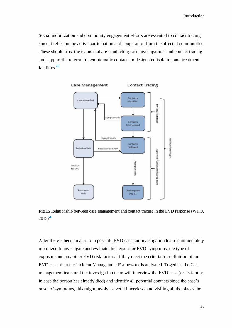
Social mobilization and community engagement efforts are essential to contact tracing
since it relies on the active participation and cooperation from the affected communities.
These should trust the teams that are conducting case investigations and contact tracing
and support the referral of symptomatic contacts to designated isolation and treatment
facilities.26
Fig.15 Relationship between case management and contact tracing in the EVD response (WHO,
2015)26
After there‟s been an alert of a possible EVD case, an Investigation team is immediately
mobilized to investigate and evaluate the person for EVD symptoms, the type of
exposure and any other EVD risk factors. If they meet the criteria for definition of an
EVD case, then the Incident Management Framework is activated. Together, the Case
management team and the investigation team will interview the EVD case (or its family,
in case the person has already died) and identify all potential contacts since the case‟s
onset of symptoms, this might involve several interviews and visiting all the places the

Introduction
31
case went while manifesting EVD symptoms. Failure to identify a single contact may
lead to ongoing EVD transmission.26
All contacts are then personally interviewed and asked about their last interaction with
the case, if no risk of exposure is identified, the person will no longer be considered a
contact, otherwise, they will be educated about the signs and symptoms of EVD and
preventive measures and explained how getting early treatment improves outcome and
reduces the risk of infecting others. In case the contact develops symptoms, they are
instructed to self isolate and notify the team that will then contact the Field
Epidemiologist, who will activate the case management team and the new case will be
transported to a transit/isolation unit for further testing. While that happens, new contact
identification and contact listing is initiated for this new suspected EVD case (appendix
2).26
As a way of following up all contacts, the Contact Follow-up Team should perform
daily visits to every contact on the list. Ideally contact teams should be assigned to the
same contacts for all 21 days of follow-up. During this visits, the contact will be asked
about development of EVD symptoms to him/herself or any other member of the
family. If a new suspected EVD case is found, then the cycle restarts as mentioned
before.26
1.4.2.3. Safe and dignified burials
The WHO priority activities in this area were:
Supervised burials;
Trained and PPE-equipped community burial teams.43
According to the WHO, a single funeral could be linked to 300 or more Ebola cases.30
In 2014, in the district of Moyamba, Sierra Leone, a single traditional funeral led to a
sharp increase in what was previously a low-incidence district.48
This is due to the contact between the mourners and the body and belongings of the
deceased when the family and community members perform religious rites that require

Introduction
32
directly touching or washing the body and when family members distribute personal
property of the diceased.46, 47
Therefore a safe and dignified burial protocol50
and 210 burial teams were created
across the three countries in order to bury everyone suspected or confirmed of having
died from Ebola in a safe and dignified manner. However, despite this measure, unsafe
burials continued to happen throughout the outbreak, especially in Guinea and Sierra
Leone, where some communities believed that there were not enough allowance for
prayer and spirituality during the burial services.45
“Immediate, safe, dignified burials by trained teams with appropriate protective
equipment are critical to interrupt transmission and control Ebola during times of active
community transmission” (Curran et al, 2016).
As an example, in the Sierra Leone‟s Red Cross, each burial team had around 10
people, which included family liason officers, disinfectant sprayers and drivers. The
people working on these teams were people from the community, not medical
professionals. Whenever a call came to attend a burial, a swabbing team from the
ministry of health would go to the site first, to take fluid samples before the burial team
approached, so that the samples could later on be tested and confirmed whether the
person died of Ebola or not. However, every death in the community should be
considered as an Ebola death and handled as such. When the team arrived at the site,
they would first of all, discuss the burial practice with the family, then, put on their
PPE, enter the house, place the body of the deceased in a body bag, place the body in a
coffin previously arranged with the family, sanitize the family‟s environment, remove
PPE, manage waste and perform hand hygiene, transport the coffin or the body bag to
the cemetery, perform the burial at the cemetery following the religious practices
desired by the family and return to the headquarters.49,50
1.4.2.4. Community engagement and social mobilization
The WHO priority activities in these areas were:
Full community engagement in contact tracing and risk mitigation and in
implementing complementary approaches.43

Introduction
33
To achieve this it was essential to build a trust between the local communities and
frontline workers, through dialogue and education of the community. This included
engaging anthropologists with the religious leaders of the community, in order to fight
fear and stigma of the disease, to negotiate alternative religious and cultural practices
and to encourage the communities to seek treatment. The goal was to create a
community engagement model, based on best practice, for the safe and rapid roll out of
Ebola treatment and community care centres.45
The key messages transmitted by the community teams, local organizations and media
were:
Key facts about severity, transmission and importance of early prevention;
Information of how to seek treatment for a person with EVD symptoms, how to
treat a sick family member at home and for those who have full recovered;
How to act if there has been a contact with a person alive or dead with EVD;
Safe burial practices;
Messaged on what practical steps should be taken to stop Ebola in the
community, with focus on effective community mobilization.
These messages were designed to increase the understanding of the EVD and make
people less likely to become ill, to enhance the trust inside the community, to promote
dialogue and community ownership of the response and to minimize psychological
distress. They were selected based on the understanding of their audience and adapted
accordingly.52
“By promoting community approaches and engaging survivors to work alongside other
responders, WHO is helping to minimize stigmatization of communities affected by
Ebola” (WHO, 2015).
1.4.2.5. Other measures
To limit national and international spread of the disease, the WHO implemented short-
term extraordinary measures were:

Introduction
34
Implementation of specific programmes to ensure continuity of essential and
supportive services in containment areas (primary care, food, ...);
If non-essential movement in and out of a containment area is stopped, ensure
that essential movement continues unhindered, such as response providers and
essential services;
To facilitate EVD response, mass gathering should be deferred until intensity of
transmission is reduced;
Prohibit travel of all Ebola cases and contacts (with the exception of medical
evacuation);
Implementations and monitoring of exit screening in international airports,
seaports and major land crossings;
Align practices of all international airlines to the national travel policy.
To ensure essential services and create the foundation for health sector recovery and
strengthening of national core capacities for outbreak response the WHO priority
activities were to:
Establish short-term capacity to address critical gaps in essential services, such
as health, food, education, security, WASH, through national service providers,
NGO‟s, UN agencies, humanitarian organizations and other partners, based on
needs assessment and gaps analysis;
Develop a medium-term investment plan to strengthen health services that
includes syndromic surveillance and laboratory networks to diagnose relevant
pathogens;
Introduce a fast-track training programme for priority health worker gaps,
including surveillance.43
In September 2015, as the number of cases started decreasing, the goal of the response
changed towards achieving and maintaining zero cases with the main objectives at this
phase being:
1. To accurately define and rapidly interrupt all remaining chains of Ebola
transmission

Introduction
35
2. To identify, manage and respond to the consequences of residual Ebola risks.
This last phase of the response (phase 3) aimed to build on the rapid scale-up of
treatment beds, safe and dignified burial teams, and behaviour change capacities during
phase 1 (August-December 2014) and enhanced capacities for case finding, contact
tracing, and community engagement during phase 2 (January-July 2015). It also focused
on incorporating new developments in Ebola control, such as vaccines, diagnostics and
response operations to survivor counselling and care.66
1.4.5. EVD outbreak in Sierra Leone
1.4.5.1. About Sierra Leone
Fig. 16 Statistics on Sierra Leone according to the WHO website, May 28th, 2016.
The Republic of Sierra Leone is a country in the West African coast and part of the 54
countries that make up the whole of the African Continent. It borders Guinea and
Liberia and it has a total surface area of 72 000 km2.67
The country is divided by 3 provinces (Eastern, Northern and Southern), one area
(Western area), 14 districts and 150 chiefdoms.70,71

Introduction
36
37% of its population resides in urban areas with
an expectancy for this number to increase due to
the significant rural to urban migration
happening;
52% of the population is female with an average
fertility rate of 5.1 children per woman; 25% of
the population constitute women of the
reproductive age (15-49); 55% of the population
are adolescents and 20% are infants and children
under 5 years of age;
45% of men and 27% of women are literate;
20 languages in total are spoken in the country,
being English the official language;
More information on the Sierra Leone‟s statistical profile can be found on
appendix 8.67
It‟s one of the poorest countries in the World, ranking180/187 in the United Nations
Programme for Development Human Development Index.73
“The health status of the
people of Sierra Leone is still among the worst in the world. Infant and maternal
mortality rates remain among the highest in the world. According to the Sierra Leone
demographic health survey 2008, life expectancy is 47 years, infant mortality rate is 89
per 1000 live births, under-five mortality rate is 140 per 1000 live births and maternal
mortality ratio is 857 per 100 000 births. Fertility rates are high due to low
contraceptive use prevalence rate.” (ROA, WHO, 2009).
Since the decade of civil war which ended in 2002, that health service delivery in the
country has been a challenge due to the damages it created in the health system. It has
been substantially dependent on external resources for funding, such as the Asian
Development Bank, the Department for International Development (DFID, now known
as UKAID), the United Nations Children‟s Fund, the United Nations Population Fund
and the World Bank; and the weakness of the health system continues to undermine
standards, availability and accessibility of the services provided.67
Fig. 17 Flag
Fig. 18 Coat of arms

Introduction
37
The governmental body responsible for coordinating health interventions, actions and
human workforce in the country is the Ministry of Health and Sanitation (MoHS). It
worked with development partners mainly to implement the “National Health Sector
Strategic Plan”, a 6 year plan created in 2009 to provide a framework for improving the
health of the nation by 2015.67
This plan was mainly focused on fighting HIV, Malaria
and Tuberculosis and supporting maternal and child care (Millennium Development
Goals), leaving very little external aid to support the overall development of the health
system.69
The EVD outbreak greatly disrupted the basic essential (non-Ebola) health services in
the country, exacerbating the existing weaknesses of the health system and making it
even more fragile.68
If a health system is ill-equipped to deal with a disease outbreak or
a catastrophe the affected populations may be extremely vulnerable.69
1.4.5.2. The outbreak in Sierra Leone
A young woman who was admitted into a governmental hospital after a miscarriage on
the 25th
of May, 2014, was the first confirmed Ebola case in Sierra Leone.30,75
According to the WHO, the infection entered the country from the neighbouring country
Guinea via a traditional healer who lived in Sierra Leone and worked in both Sierra
Leone and Guinea. The traditional healer became infected and died. Hundreds of
mourners attended the funeral and it‟s suspected that as many as 365 Ebola deaths are
linked to this event.30
The outbreak started with this young woman in the Kenema district, quickly spreading
to nearby Kailahun district and the Eastern Province adjacent to the epicentre of the
outbreak in Guinea. It reached the capital, Freetown, on the 11th
of July 2014, where it
easily grew into bombastic numbers due to the overcrowded conditions and fluid
population movements.30,70
Sierra Leone declared a state of emergency on the 6th
of August 2014, two days before
the International Public Health Emergency being declared by the WHO, and the
strategic plan of the UNMEER started its implementation in October 2014, same time
by which the Sierra Leone National Ebola Response Centre (NERC) was established to

Introduction
38
coordinate this response via the EVD Response pillars: case management, infection
prevention and control (IPC) and safe burials, surveillance (and contact tracing), social
mobilization and psychosocial support.70,76,78
In total, the epidemic in Sierra Leone saw 14 124 notified cases and 3 956 notified
deaths. It affected 114 of the 150 chiefdoms, reached its peak in the final quarter of
2014 and started to recede in late November 2014.70,72
Fig.19 EVD cases count in Sierra Leone during January 2014 and August 2015 (WHO, 2015).31
According to the Ministry of Health and Sanitation (MoHS) of Sierra Leone (2014), the
challenges that contributed to this massive outbreak included:75
“Inadequate understanding within the communities of the EVD as this is the first
major outbreak reported in Sierra Leone.
Lack of experience among healthcare workers and limited capacities for rapid
response.
High exposure to Ebola virus in the communities through household care and
customary burial procedures. This has resulted in high level of community
deaths leading to panic and anxiety.
Denial, mistrust and rejection of proposed public health interventions arising
from misinterpretation of the cause of the new disease.

Introduction
39
Fear of the disease by frontline health workers leading to either suboptimal care
for patients or substandard implementation of protective measures.
Close community ties and movement within and across borders has led to
difficulties in tracking and following up of contacts for the three countries.
The magnitude and the geographical extent of the EVD outbreak in Sierra Leone
require significant and robust response capacities and structures. This outbreak
poses serious challenges in terms of human capacity, financial, operational and
logistic requirements and threatens national and international heath.”
Fang et al (2016), mentions that the multilayer control interventions placed during the
last quarter of 2014 were associated with 43% reduction of the population-level
transmission risk during intervention phase I and 65% reduction during intervention
phase II; these included the establishment and operation of diagnostic and healthcare
facilities and the national and regional campaigns to improve case isolation and safe
burial (further details about these measures were already mentioned during the Ebola
Response section).
1.4.5.3. The outbreak in Nieni Chiefdom, Koinadugu district, Sierra Leone
Koinadugu was the last District of Sierra Leone to be affected by the epidemic; it
reached the district by October 2014 even after the local chiefs had imposed measures
to prevent the entrance of the virus into the district, such as quarantine, barred travel and
created a system of official distribution vans and trucks to help farmers and traders to
get their products into the neighbouring markets. The hotspot was in Nieni Chiefdom,
being the most affected towns: Kumala, Fankuya and Sumbaria.77
The outbreak in this Chiefdom lasted from October 2014 to Mach 2015. During the first
4 months of the outbreak, Kumala‟s primary school was used as a CCC, named “old
Kumala” (aereal picture below). In total, old Kumala admitted 139 patients and saw 70
confirmed cases. In May 2015, a Holding Centre was created with a 20 bed capacity,
named “new Kumala”.78
New Kumala never saw a confirmed Ebola case until its
closured in October 2015.

Introduction
40
The last confirmed EVD case in Nieni Chiefdom (Kumala‟s CCC) was documented on
the 15th
of March 2015 adding to a total of 108 cases in this area.34
The district itself
saw a total of 229 cases36
and was only declared Ebola free on the 29th
of May 2015,
after having gone 42 days without a recorded case37
.
Fig. 20 Aerial view from old Kumala (Médicos del Mundo, 2015).
1.4.5.4. “End of Ebola Outbreak in Sierra Leone” statement
Delivered by Dr. Anders Nordström, WHO Representative in Sierra Leone on the
7th
of November, 2015, Freetown, Sierra Leone:
“Today, 7 November 2015, the World Health Organization declares the end of the
Ebola outbreak in Sierra Leone.
Introduction
41
Since Sierra Leone recorded the first Ebola case on 24 May 2014, a total number of
8,704 people were infected and 3,589 have died. From those who tragically lost their
lives, 221 of them were healthcare workers. We remember them all today.
We are now moving into a new phase of 90 days enhanced surveillance which will run
until 5 February 2016. This new phase is critical as our goal is to ensure a resilient zero
and that we can detect and respond to any potential Ebola flare ups. This period is about
ensuring that we can consolidate the gains of existing systems to manage future risks.
The World Health Organization commends the Government of Sierra Leone and the
people of Sierra Leone for the significant achievement of ending this Ebola outbreak.
The World had never faced an Ebola outbreak of this scale and magnitude and the
World has neither seen a nation mobilizing its people and resources as Sierra Leone
did. The power of the people of Sierra Leone is the reason why we could put an end to
this outbreak today.
This power of the people and the foundation now in place needs to be further nurtured
and supported in order to build a strong and resilient public health system which stands
ready to contain the next outbreak of a disease, Ebola or any other public health threat.
Under the leadership of the Sierra Leonean Government, an effective response was
initiated to manage the outbreak. The use of rapid response teams and strong
community involvement to identify new cases early and quickly stop any Ebola virus
transmission should continue to be the cornerstone of the national response strategy.
WHO will maintain an enhanced staff presence in Sierra Leone as the response
transitions from outbreak control, to support enhanced vigilance and to the recovery of
essential health services.
The Ebola outbreak has decimated families, the health system, the economy and social
structures. All need to recover and heal.
Introduction
42
WHO is confident that the Government of Sierra Leone together with its national and
international partners will use the foundation already in place; dedicated and trained
health workers; systems for alerts and information management; community
engagement and care for people – to deal with other priority health problems, child
mortality topping the list.
Thank you”32
In January 2016 the NERC seized its operations and its responsibilities were taken over
by the Ministry of Health and Sanitation, the Ministry of Social Welfare, Gender and
Children‟s Affairs.76

Aims
43
2. AIMS
The main goal of this study is to understand if there‟s any relation between the
symptoms during the acute stage of EVD and the sequelae that survivors present moths
after recovery.
Materials and Methods
44
3. MATERIALS AND METHODS
3.1. Study design and participants
This is a case reports study following the symptoms of 10 EVD survivors on the
moment of admission to Kumala‟s Holding Centre and months after.
The population of the study was selected between the 57 Ebola Case Investigation
forms (appendix 9) filled out on time of admission to Kumala‟s Holding Centre between
the dates of 27th
of November 2014 and 14th
of March 2015.
From the 57 admissions there were 15 confirmed cases, of which 11 survived. Since it
was impossible to find information about symptoms months after discharge from one of
the survivors, the population of the study made a total of 10 survivors.
3.2. Ethical considerations
Kumala‟s Holding Centre was created as part of the Ebola Response strategy
implemented in Koinadugu district, Sierra Leone. The NGO Médicos del Mundo –
Spain – were responsible for the case management and running of this centre.
The official owners of the data collected for this study are the Ministry of Health and
Sanitation of Sierra Leone and the Koinadugu District Ebola Response Centre.
Authorization has been granted to MdM by the Ministry of Health and Sanitation of
Sierra Leone to use the data collected from any MdM projects.
Since I was working at Kumala‟s HC as part of this NGO, I requested their
authorization to pursue this study and present it as my Masters of Tropical Health
dissertation, which was granted. It is however to be mentioned that the copyrights of
this study will be shared between myself, the Lisbon School of Hygiene and Tropical
Medicine and Médicos del Mundo – Spain.
None of the survivors name or village is mentioned in the study; instead they have been
numbered at random from 1 to 10. The demographics of the study‟s population will be
mentioned without linking any data to any survivor. The only information linked
directly to the survivor‟s number will be the symptoms they presented on admission to
Materials and Methods
45
the HC, 8 months and 10 months after, since these symptoms are the core of the
presented study.
3.3. Data collection
As mentioned, the symptoms of the 10 survivors upon admission to Kumala‟s HC were
obtained from the Ebola Case Investigation Forms used during triage (a sample of the
form can be found on appendix 9). The symptoms from 8 months after admission were
obtained by the psychosocial team working with EVD survivors in Koinadugu district
as part of the Médicos del Mundo (MdM) team and granted to me upon request. The 10
months data was collected during an Uveitis program held in Kumala during October
2015, where all the survivors from the district gathered and were screened for uveitis
symptoms by an ophthalmologist team and were also seen by a doctor (part of the MdM
team) for a medical check-up post EVD; the symptoms were collected from the form
used by the MdM doctor during this check-up.
3.4. Data analysis
The data analysis will be descriptive statistics, even though the statistics will have little
scientific validity due to the low sample‟s number. Future studies with larger
populations would be needed to confirm or challenge the results obtained in this study.

Results
46
4. RESULTS
From the 10 survivors, six were females and four males with ages between 5 and 47
years old. Seven had a known contact with a person with EVD (dead or alive) in the 30
days that preceded the symptoms upon admission to the HC. The statistical analysis can
be found on the following table.
SEX
- Male
- Female
4 (40%)
6 (60%)
AGE
Mean
AGE GROUPS
- 0-15
- 16-25
- 26-35
- 36-45
- 46-55
25 years old
3 (30%)
3 (30%)
2 (20%)
1 (10%)
1 (10%)
Known contact with an EVD case in the 30 days prior do admission
YES
- Dead
- Alive
NO
7 (70%)
6 (86%)
1 (14%)
3 (30%)
Six out of the ten survivors studied (60%) presented with one or more of the same
symptoms months after admission to the centre that they had when the disease was at its
acute stage. Four still had headaches ten months after, three maintained weakness, one
eye problems, one abdominal pain and another joint pain.
Six survivors (60%) were admitted to KHC presenting severe disease and five survivors
(50%) presented severe sequelae up to 10 months after admission, however, from the
six that presented severe disease during the acute stage, only three of them also
developed severe sequelae.
The following table presents the results regarding the number of symptoms during the
three studied stages. When there are five or more symptoms present at that time the
number is marked in red, to highlight the possible severity of the disease or sequelae.
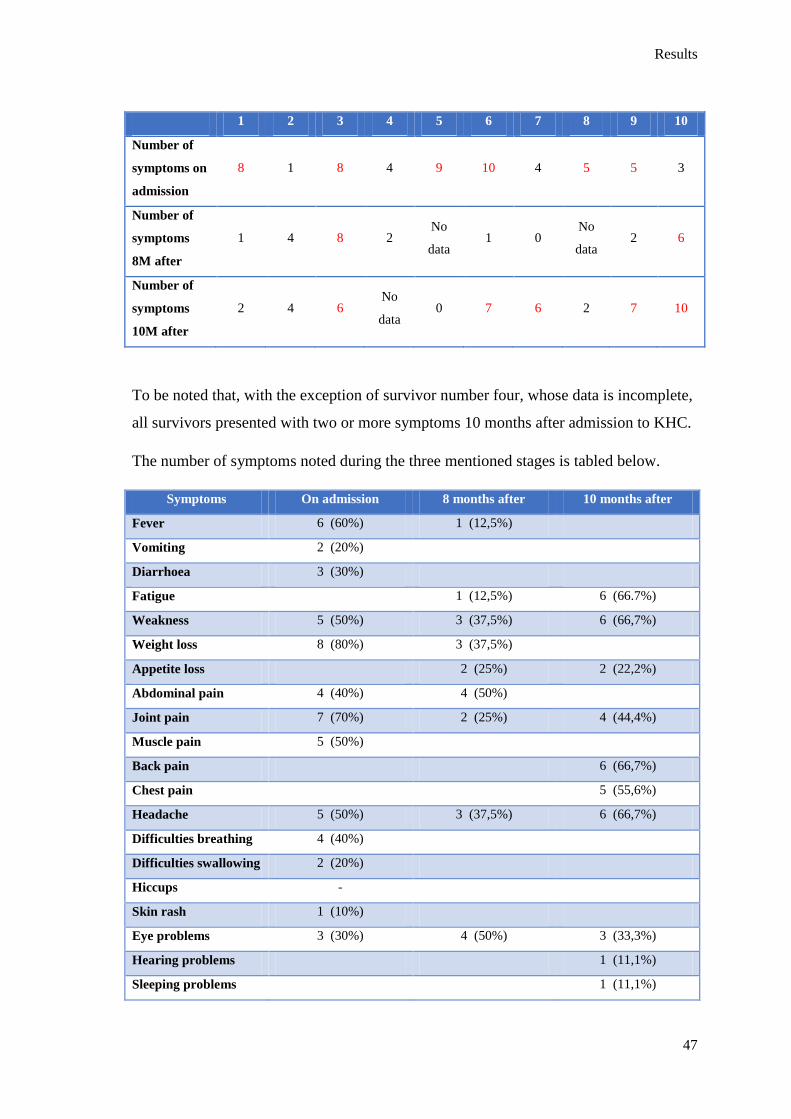
Results
47
1 2 3 4 5 6 7 8 9 10
Number of
symptoms on
admission
8 1 8 4 9 10 4 5 5 3
Number of
symptoms
8M after
1 4 8 2 No
data 1 0
No
data 2 6
Number of
symptoms
10M after
2 4 6 No
data 0 7 6 2 7 10
To be noted that, with the exception of survivor number four, whose data is incomplete,
all survivors presented with two or more symptoms 10 months after admission to KHC.
The number of symptoms noted during the three mentioned stages is tabled below.
Symptoms On admission 8 months after 10 months after
Fever 6 (60%) 1 (12,5%)
Vomiting 2 (20%)
Diarrhoea 3 (30%)
Fatigue 1 (12,5%) 6 (66.7%)
Weakness 5 (50%) 3 (37,5%) 6 (66,7%)
Weight loss 8 (80%) 3 (37,5%)
Appetite loss 2 (25%) 2 (22,2%)
Abdominal pain 4 (40%) 4 (50%)
Joint pain 7 (70%) 2 (25%) 4 (44,4%)
Muscle pain 5 (50%)
Back pain 6 (66,7%)
Chest pain 5 (55,6%)
Headache 5 (50%) 3 (37,5%) 6 (66,7%)
Difficulties breathing 4 (40%)
Difficulties swallowing 2 (20%)
Hiccups -
Skin rash 1 (10%)
Eye problems 3 (30%) 4 (50%) 3 (33,3%)
Hearing problems 1 (11,1%)
Sleeping problems 1 (11,1%)

Results
48
Mood disturbance 2 (22,2%)
Bleeding 2 (20%)
Unfortunately, I was unable to access the data from two survivors 8 months after the
admission to the HC and one other survivor didn‟t show up to the post-EVD mobile
clinic held 10 months later, which is why the percentages for the 8 months after results
are calculated out of eight survivors and for 10 months after out of nine survivors
instead of 10.
A full list of the 10 survivor‟s symptoms on admission to the Holding Centre, 8 months
later and 10 months later can be found in appendix 10.

Discussion
49
5. DISCUSSION
The 2014-2015 West Africa Ebola Outbreak was the largest ever reported in the World.
It started in December 2013 and was left unnoticed for 3 months, allowing for the virus
to keep spreading uncontrollably and for the outbreak to keep escalating until it was
declared an International emergency in August 2014. Despite the large number of
cases/deaths recorded, it is to be noted that underreporting was a big issue, especially in
the first months of the outbreak, having been predicted by the WHO that the real
number of cases during this time was probably 2 to 4 times higher than reported43
.
This was the first ever documented Ebola outbreak outside of Central Africa and it was
unmatched in duration and size. Several factors can be contributed to this, such as
decades of conflict, increasing population growth, poverty, ethnic and linguistic
fragmentation, poor health infrastructures and cultural practices. Due to sociological
and economic factors, there‟s also a large scale movement of populations in the area,
both within and between countries, greatly influencing the spread of the disease.79
As mentioned, the fact that the outbreak affected extremely poor countries, with
severely compromised health systems, lack of knowledge about the virus (and
consequent stigma associated with it) and certain kinds of human-to-human contacts,
based on religious, behavioural and cultural practices, what led to the rapid spread and
increasing fear of the disease. On the other hand, the spread of the virus aggravated the
already weakened health system and the fragile social, political and economic
conditions of these countries, turning the situation into a vicious cycle where urgent
international aid was needed to support the affected countries.
Led and coordinated by the WHO, in partnership with several other UN agencies,
national governments and other partners, an international response was established to
provide the necessary support to the affected countries. This was done by 1)
intensifying response activities, focusing on case management, case finding
(surveillance) and contact tracing, safe and dignifies burials, laboratory and social
mobilization; and by 2) strengthening national capacities, including the creation of
ETC‟s, CCC‟s, HC‟s, burial teams, laboratories and the deployment of large amounts of
international staff to work on the field, not only on the centres (since the number of
local health care staff was already limited before the outbreak and became even more of

Discussion
50
an issue due to the extremely high number of health care workers who were infected
and died of the virus), but also engaging with local communities and workers, leading to
an increasing understanding about the disease itself, it‟s transmission and necessary
infection control strategies that needed to be continuously implemented in order to
reduce transmission, with emphasis on IPC measures and proper use of PPE.
“Achieving real community understanding, ownership and implementation of any
complementary approaches, particularly given the deep-rooted fear and stigmatization
in the affected areas, required sustained mobilization, engagement and dialogue with
community, religious, traditional and other local leaders, woman‟s and young groups, as
well as traditional healers, to build collective trust and confidence in the response
efforts and community action” (WHO, 2014).
Community health care workers played a particularly vital role in “delivering messages,
addressing stigma and implementing complementary approaches to EVD control”
(WHO, 2014), since the fear of this disease led individuals to flee Ebola centres
believing they would die there or simple not seek healthcare and revert to traditional
healers or family members instead. Being an Ebola healthcare worker was, however,
very challenging in such communities since they were awfully stigmatized and rejected
by their communities, believing they were acting as reservoirs for the virus. The same
would be applicable for EVD survivors.79
Fear of stigmatization, alongside burial practices, were probably the main factors that
contributed to the magnitude of this outbreak. Burial practices due to the lack of IPC
measures during the ceremonies and fear of stigmatization since it influenced disease
reporting, pursuing healthcare and overall denial of the disease.
Knowing that the transmission of the virus is done by direct contact and taking into
account the behavioural practices of the affected communities, IPC measures were the
centre of the intensified control strategies set by the Ebola Response Roadmap, in
August 2014, assuming that these would be enough to stop any new transmission within
8 weeks of the index case. It required mobilization of several resources, including
medical staffing, material supplies and other essential needs.

Discussion
51
Some examples of the complementary control activities implemented, aside from the
ones already mentioned, were: 1) all movement of people to and from
contaminated/quarantined areas was limited to response providers and essential
services; 2) mass gatherings were forbidden during the peak of the outbreak and until
the intensity of the virus transmission was reduced, this included closing of schools,
bars, restaurants and even of public Christmas celebrations; 3) mandatory temperature
checks at various checkpoints including hand washing with weak chlorine solution; 4)
implementation of airport, seaport and major land crossings screenings; 5) establishing
short-term capacity to address critical gaps in essential services, including health, food,
education, protection and WASH, by national, international service providers, NGO‟s,
UN agencies, humanitarian organizations and other partners.
Taking into account that it took the affected countries 3 months to realise there was an
Ebola Outbreak devastating their land and the International Health Agencies 5 months
after that to declare it a Global Emergency, it is to be questioned if the activities
developed at that point were enough to fight the alarming way the virus was spreading
at the time. The Ebola Response Roadmap created by the WHO in August 2014 set the
goal “to stop Ebola transmission in affected countries within 6-9 months and prevent
international spread”. This target largely failed, since the end of the outbreak was only
declared almost 15 months after. However, it did manage to significantly slow down the
transmission significantly by January 2015, five months after the Ebola Response
implementation.
From a scientific perspective, the unprecedented high number of cases and subsequent
survivors during this outbreak allowed for a lot of research to be done. Nonetheless, it is
still not enough and a lot of questions still remain regarding EVD and PEVDS.
Both short and long term complications have been reported on EVD survivors, ranging
from physical to psychological and social. In addition, the persistence of EVD in
selected body compartments of the survivors poses a great risk of reintroduction of the
virus in areas where transmission has previously been eliminated.51
The PREVAIL III
study that began in Liberia in June 2015 and that will last for 5 years, is tracking around
1500 EVD survivors and should bring some insight into the long-term consequences of
this infection and also as to how contagious survivors can remain post recovery.

Discussion
52
Preliminary findings are already indicating that 68% of survivors hold neurological
complications, 60% eye problems and 53% musculoskeletal complications, and that
EVD can be found in the semen of male survivors up to 18 months post EVD.84,85
This
is the largest scale survivors study being done and, in comparison with the study
presented on this dissertation, neurological (headaches) and musculoskeletal (mainly
weakness and back pain) were also the more prevalent sequelae found.
From the data collected in this study, the majority of survivors were females (60%),
with an average age of 25 years old (ages ranging from 5 to 47 years) and with 70% of
them referring having had contact with a person with known EVD (dead or alive) in the
30 days prior to admission to the HC. The fact that there were more female survivors
than male is in accordance with a study done by the WHO Ebola Response Team,
mentioned in an article of “The New England Journal of Medicine”, in January 2016,
stating that there is a higher survival rate amongst female individuals83
.
The most common symptoms recorded on admission (during the active stage of EVD),
were weight loss, joint pain and fever; and months after (post EVD) were headache,
fatigue, weakness and back pain (which could also be related to the sample‟s
professions and not the EVD itself, since most of them were farmers and since
musculoskeletal pain is a common complaint in the general population of Sierra Leone).
The psychological complications were not evaluated during this study. However and
taking into account what was mentioned before, alongside previous studies done, it is
clear that “mental health problems are probably the most prevalent” complications
(Marta Lado, as cited by T. Burki, 2016), being depression, anxiety, post-traumatic
stress disorder and survivor guilt some of the most common symptoms noted.84
More
studies would still need to be done on this subject.
Gastrointestinal (GI) symptoms weren‟t very prevalent on these patients upon
admission to the HC, which could be related to the fact that GI symptoms tend to appear
at a more advanced stage of the acute disease and that the survivors studied could‟ve
still been on the initial stage of the disease when admitted. Also, the low frequency of
haemorrhagic symptoms it is in accordance with other studies done, alike in a study
done by Schieffelin et al, published by “The New England Journal of Medicine” in

Discussion
53
October 2014, from the 106 Ebola patients that were studied in Sierra Leone, only 1 had
haemorrhagic symptoms.19,53
Sixty per cent (6 in 10) of survivors presented with one or more of the symptoms they
had during the acute stage of the disease months after recovery, being headache the
most common symptom to persist, followed by weakness. However, all survivors
presented with one or more symptom months after recovery, regardless of the symptoms
existing during the acute stage of the disease (with the exception of one survivor whose
data is incomplete).
A study done by Scott et al, published by the “Emerging Infectious Diseases” in April
2016, also found that all the survivors studied (44 in total) had between one to five post-
Ebola complaints (on average 2), 70% with musculoskeletal pain, 48% with headaches
and 14% with ocular problems86
. This is in keeping with the results found by this study,
although it is to be mentioned that the Scott et al study was done on survivors only 3
weeks after discharged from an ETC.
The pathogenic and biological events that lead to the development of PEVDS are still
unclear and more studies still need to be done on that subject however and taking in
consideration a symptomatic approach, this particular study concludes that the
severity of the disease doesn’t seem to be associated with the severity of the
sequelae (both defined here by the presence of 5 or more symptoms at a time), since
while there were 3 survivors with severe disease that developed severe sequelae, 3
didn‟t; and while 2 survivors with non-severe disease developed severe sequelae other 2
remained non severe throughout.
However, challenges to this study include the fact that I was only able to analyse a small
sample, only 10 survivors, making it difficult to obtain more concrete conclusions.
There‟s also the fact that I didn‟t have access to symptoms from 2 survivors 8 months
after the acute stage of the disease and from 1 survivor 10 months after. And finally, the
fact that I was unable to access the records from what happened to the survivors after
admission to the Holding Centre or after they had been transferred to the ETC, not
allowing me to know how the symptoms developed and making it difficult to compare
how the severity of the disease could be related to the PEVD symptoms. Knowledge of
the viral load during the acute stage of the disease would also be an important piece of

Discussion
54
data information to study to understand if the viral load would have any effect to the
symptoms during PEVDS.
With that in mind, it is difficult to conclude from this study if the symptoms during
the acute stage of the EVD play an important role in the symptoms seen at PEVDS,
more studies overcoming the challenges presented here would be needed.
It would be interesting to continue with studies on EVD survivors, taking into account a
larger sample and including more information, such as the psychological effects
presented by survivors, all the symptoms that patients presented during the acute stage,
from the onset of these until discharge from the ETC, how and when did sequelae
started manifesting, the viral load of the patients during the acute stage of the disease
and when the symptoms are at its worst and, possibly, making a correlation between all
of the above and the remaining of the virus in selected body compartments, such as the
eye and the semen.
In conclusion, and in the words of Anders Nordstrom, WHO‟s representative in Sierra
Leone, as cited by Gullard (2015): “we have never had such large number of survivors.
This is very new. We have a unique and important responsibility to provide care and
support for Ebola survivors trying to restart their normal lives. It is increasingly clear
that emerging from an Ebola treatment unit is just the beginning. The countries affected
by Ebola also have a long road to recovery”.29
In Sierra Leone specifically, the President Ernest Bai Koroma, has promised a
comprehensive package of free healthcare to all EVD survivors. This, however, is still
to be materialized.84
Now that the countries have embarked on the recovery journey post Ebolavirus
Outbreak, focus needs to be held on other health care problems aside from Ebola, this
includes strengthening the current health care systems and restoring essential health
services. With such high resource gaps in the health care systems, how can local
Governments provide proper care for the survivors? Also, failures in health care
systems leads to high mortality from vaccine-preventable and easily-treatable diseases.
Therefore, the health care services of the affected countries must now focus back on

Discussion
55
immunizations and vaccinations; maternal, newborn, child and adolescent health; and
communicable diseases, with an emphasis on Malaria, TB and HIV. 80
Nonetheless, maintaining IPC measures, laboratory diagnosis, surveillance systems,
community engagement, simulations, training, strong workforces and rapid response
teams are also important in this recovery stage, since the possibility of re-introduction
of the virus in the communities by survivors it‟s still a harsh reality and the three Ebola-
impacted countries remain at high risk of additional small outbreaks.81
Since West Africa was declared Ebola-free on the 14th
of January of 2016, with all three
countries having reached zero cases, there have been small flare-ups in Sierra Leone,
Guinea and Liberia and it is expected that more might still happen in the near future.
Prevention and preparedness are essential to protect populations, not only regarding the
residual Ebola risk from survivors, but also of any kind of communicable disease
epidemic that could lead to an outbreak.82
The WHO has created tools such as an
“Ebola Virus Disease Consolidated Preparedness Checklist” that aims to assist countries
in assessing their level of readiness and in identifying concrete actions to be taken to
address identified gaps or a “Hospital preparedness for epidemics” providing
information on how hospitals and health care facilities can accomplish their role in
national and local responses to emergencies.80

56
6. REFERENCES
1. Centers for Disease Control and Prevention (2015), “Ebola (Ebola Virus
Disease)” factsheet, updated 11/08/2015.
http://www.cdc.gov/vhf/ebola/pdf/ebola-factsheet.pdf accessed 27 December
2015.
2. MARTINEZ, Miguel; SALIM, Abdulbaset.; HURTADO, Juan; KILGORE, Paul
(2015); “Ebola Virus Infection: Overview and Update on Prevention and
Treatment”, Infectious Diseases and Therapy, vol. 4, pages 365-390.
doi:10.1007/s40121-015-0079-5
3. Centers for Disease Control and Prevention (2015), Ebola (Ebola Virus
Disease), “Transmission”.
http://www.cdc.gov/vhf/ebola/transmission/index.html accessed 27 December
2015.
4. World Health Organization (2015), “Ebola virus disease”, fact sheet Nº103,
updated August 2015. http://www.who.int/mediacentre/factsheets/fs103/en/
accessed 27 December 2015.
5. World Health Organization (2014), “What we know about transmission of the
Ebola virus among humans”, Ebola situation assessment, 6 October 2014.
http://www.who.int/mediacentre/news/ebola/06-october-2014/en/ accessed 27
December 2015.
6. Deon vs Earth (2014), “CIDRAP Researchers: Evidence, Ebola transferable by
air”, http://www.deonvsearth.com/cidrap-research-proves-ebola-virus-airborne-
cdc-misleading-america/ accessed 27 December 2015.
7. Centers for Disease Control and Prevention (2015), Ebola (Ebola Virus
Disease), “Signs and Symptoms”.
http://www.cdc.gov/vhf/ebola/symptoms/index.html accessed 27 December
2015.
8. Centers for Disease Control and Prevention (2015), Ebola (Ebola Virus
Disease), “Guidance on Personal Protective Equipment (PPE) To Be Used By
Healthcare Workers during Management of Patients with Confirmed Ebola or
Persons under Investigation (PUIs) for Ebola who are Clinically Unstable or
Have Bleeding, Vomiting, or Diarrhea in U.S. Hospitals, Including Procedures

57
for Donning and Doffing PPE”. http://www.cdc.gov/vhf/ebola/healthcare-
us/ppe/guidance.html accessed 27 December 2015.
9. Centers for Disease Control and Prevention (2015), Ebola (Ebola Virus
Disease), “Past Ebola Outbreaks”.
http://www.cdc.gov/vhf/ebola/exposure/index.html accessed 27 December 2015.
10. Centers for Disease Control and Prevention (2015), Ebola (Ebola Virus
Disease), “Outbreaks chronology: Ebola Virus Disease”.
http://www.cdc.gov/vhf/ebola/outbreaks/history/chronology.html accessed 27
December 2015.
11. Centers for Disease Control and Prevention (2015), Ebola (Ebola Virus
Disease), “Ebola Virus Disease Distribution Map”.
http://www.cdc.gov/vhf/ebola/outbreaks/history/distribution-map.html accessed
30 December 2015.
12. Centers for Disease Control and Prevention (2015), Ebola (Ebola Virus
Disease), Outbreaks, “Current Outbreaks”.
http://www.cdc.gov/vhf/ebola/outbreaks/index.html accessed 30 December
2015.
13. BEECHING, Nichoas; FENECH, Manuel; HOULIHAN, Catherine (2014);
“Ebola virus disease”, BMJ 2014; 349:g7348. doi:10.1136/bmj.g7348
14. QUAMMEN, David (2013); “Spillover: Animal Infection and the Next Human
Pandemic”, Emerging Infectious Diseases, vol. 19, no. 2, page 349.
http://wwwnc.cdc.gov/eid/article/19/2/pdfs/12-1694.pdf accessed 30 December
2015.
15. VICTORY, Kerton; CORONADO, Fátima; IFONO, Sâa et al (2015); “Ebola
Transmission Linked to a Single Traditiona Funeral Ceremony – Kissidougou,
Guinea, December, 2014 – January 2015”, Centers for Disease Control and
Prevention, Morbidity and Mortality Weekly Report, 17 April, 2015, vol. 64, no.
14, pp.386-388. http://www.cdc.gov/mmwr/preview/mmwrhtml/mm6414a4.htm
accessed 30 December 2015.
16. HAGLAGE, Abby (2014); “Kissing the Corpses in Ebola Country”, The Daily
Beast, Hot Zone. http://www.thedailybeast.com/articles/2014/08/13/kissing-the-
corpses-in-ebola-country.html accessed 30 December 2015.
58
17. World Health Organization (2014), “New WHO safe and dignified burial
protocol – key to reducing Ebola transmission”.
http://www.who.int/mediacentre/news/notes/2014/ebola-burial-protocol/en/
accessed 30 December 2015.
18. BAIZE, S. et al (2002); “Inflammatory responses in Ebola virus-infected
patients”, Clinical & Experimental Immunology, vol. 128, no. 1, pages 163-168.
doi:10.1046/j.1365-2249.2002.01800.x
19. GOEIJENBIER, M. et al (2014); “Ebola virus disease: a review on
epidemiology, symptoms, treatment and pathogenesis”, The Netherlands Journal
of Medicine, vol. 72, no. 9, pages 442-448.
20. SULLIVAN, Nancy; YANG, Zhi-Yong; NABEL, Gary J. (2003); “Ebola Virus
Pathogenesis: Implications for Vaccines and Therapies”, Journal of Virology,
vol. 77, no. 18, pages 9733-9737. doi:10.1128/JVI.77.18.9733-9737.2003
21. SOUSA, Zulane Lima (2014); “Key features of Eboa hemorrhagic fever: a
review”, Asian Pacific Journal of Tropical Biomedicine, vol. 4, no. 11, pages
841-844. doi:10.12980/APJTB.4.201414B420
22. MAYOR, Susan (2015); “Ebola virus persists and may be transmitted in
survivor‟s semen, studies show”, BMJ2015;351:h5511. doi:10.1136/bmj.h5511
23. Centers for Disease Control and Prevention (2015), Ebola (Ebola Virus
Disease), “Diagnosis”. http://www.cdc.gov/vhf/ebola/diagnosis/index.html
accessed 4 January 2016.
24. Centers for Disease Control and Prevention (2015), Ebola (Ebola Virus
Disease), “Treatment”. http://www.cdc.gov/vhf/ebola/treatment/index.html
accessed 4 January 2016.
25. Centers for Disease Control and Prevention (2015), Ebola (Ebola Virus
Disease), “Treatment”. http://www.cdc.gov/vhf/ebola/outbreaks/what-is-contact-
tracing.html accessed 4 January 2016.
26. World Health Organization and Centers for Disease Control and Prevention
(2015), “Implementation and management of contact tracing for Ebola virus
disease”, Emergency Guideline, September 2015.
http://www.who.int/csr/resources/publications/ebola/contact-tracing/en/ accessed
4 January 2016.

59
27. CAROD-ARTAL, Francisco Javier (2015); “Post-Ebolavirus disease syndrome:
what do we know?” Expert Review of Anti-infective Therapy, vol. 13, no. 10,
pages 1185-1187. doi:10.1586/14787210.2015.1079128
28. CLARK, Danielle; KIBUUKA, Hannah; MILLARD, Monica et al (2015);
“Long-term sequelae after Ebola virus disease in Bundibugyo, Uganda: a
retrospective cohort study”, The Lancet Infectious Diseases, vol. 15, no. 8, pages
905-912. doi:10.1016/S1473-3099(15)70152-0
29. GULLAND, Anne (2015); “Thousands of Ebola Survivors experience serious
medical complications”, BMJ 2015;351:h4336 doi:10.1136/bmj.h4336
30. World Health Organization (2014), Emergency preparedness, response; “Sierra
Leone: a traditional healer and a funeral”.
http://www.who.int/csr/disease/ebola/ebola-6-months/sierra-leone/en/ accessed
19 April 2016.
31. National Ebola Response Centre (NERC) (2015), “End of Ebola Outbreak in
Sierra Leone Statement Delivered by Dr Anders Nordström, WHO
Representative in Sierra Leone”, Freetown, 7 November 2015.
http://nerc.sl/?q=end-ebola-outbreakin-sierra-leone-statement-delivered-dr-
anders-nordstr%C3%B6m-who-representative-sierra accessed 19 April 2016.
32. World Health Organization (2014), Emergency preparedness, response, “Ground
zero in Guinea: the Ebola outbreak smoulders – undetected – for more than 3
months”. http://www.who.int/csr/disease/ebola/ebola-6-months/guinea/en/
accessed on 19 April 2016.
33. World Health Organization (2014), “1st Chain of Transmission: MELIANDOU”.
http://www.who.int/csr/disease/ebola/ebola-6-months/guinea-chart-
big.png?ua=1 accessed 19 April 2016.
34. National Ebola Response Centre (2015), “Ebola Outbreak Updates – March 15,
2015”. http://health.gov.sl/wp-content/uploads/2015/03/Ebola-Update-March-
15-2015.pdf accessed 29 April 2016.
35. HDX (2016), “Number of Ebola Cases and Deaths in Affected Countries”,
Datasets. https://data.hdx.rwlabs.org/dataset/ebola-cases-2014 accessed 29 April
2016.

60
36. HDX (2016), “West Africa: Ebola Outbreak”, Ebola.
https://data.hdx.rwlabs.org/ebola accessed 29 April 2016.
37. National Ebola Response Centre (2015), “National Weekly Ebola Newsletter:
25th
-31st May 2015”.
http://nerc.sl/sites/default/files/docs/National%20Weekly%20Ebola%20Newslett
er%20%2025th-31st%20May%202015.pdf accessed 29 April 2016.
38. WANG, Ligui; YANG, Guang; JIA, Leili et al (2015); “Epidemiological
features and trends of Ebola virus disease in West Africa”, International Journal
of Infectious Diseases, vol. 38, pages 52-53. doi:10.1016/j.ijid.2015.07.017
39. BBC News (14 January 2016), “Ebola: Mapping the outbreak”.
http://www.bbc.co.uk/news/world-africa-28755033 accessed 29 April 2016.
40. Centers for Disease Control and Prevention (2016), “2014 Ebola Outbreak in
West Africa – Reported Cases Graphs”.
http://www.cdc.gov/vhf/ebola/outbreaks/2014-west-africa/cumulative-cases-
graphs.html accessed 29 April 2016.
41. HSIEH, Ying-Hen (2015); “Temporal Course of 2014 Ebola Virus Disease
(EVD) Outbreak in West Africa Elucidated through Morbidity and Mortality
Data: A Tale of Three Countries”, PLOS ONE, vol. 10, no. 11, November 11,
2015. doi:10.1371/journal.pone.0140810
42. United Nations (2016), “Ebola Response”, Global Ebola Response.
http://ebolaresponse.un.org/ebola-response accessed 29 April 2016.
43. World Health Organization (2014), “Ebola Response Roadmap”, 28 August
2014. http://www.who.int/csr/resources/publications/ebola/response-
roadmap/en/ accessed 29 April 2016.
44. United Nations (2016), “UN Mission for Ebola Emergency Response
(UNMEER)”, Global Ebola Response. http://ebolaresponse.un.org/un-mission-
ebola-emergency-response-unmeer accessed 29 April 2016.
45. World Health Organization (2015), “WHO Strategic Response Plan: West
Africa Ebola Outbreak”.
http://www.who.int/csr/resources/publications/ebola/ebola-strategic-plan/en/
accessed 29 April 2016.

61
46. World Health Organization (2015), “Manual for the care and management of
patients in Ebola Care Units/Community Care Centres”, January 2015.
http://www.who.int/csr/resources/publications/ebola/patient-care-CCUs/en/
accessed 2 May 2016.
47. World Health Organization (2014), “New WHO safe and dignified burial
protocol – key to reducing Ebola transmission”, Media Centre, 7 November
2014. http://www.who.int/mediacentre/news/notes/2014/ebola-burial-
protocol/en/ accessed 19 May 2016.
48. CURRAN, Kathryn; GIBSON, James; MARKE, James et al (2016); “Disease
Linked to a Single Funeral – Moyamba District, Sierra Leone, 2014”, MMWR,
vol. 65, no. 8, March 4, 2016.
49. MAZUMDARA, Tulip (2015), “Ebola: A day with the burial team”, BBC News,
Health, 7 January 2015. http://www.bbc.co.uk/news/health-30712162 accessed
19 May 2016.
50. World Health Organization (2014), “Field Situation: How to conduct safe and
dignified burial of patient who has died from suspected or confirmed Ebola virus
disease”, October 2014.
http://www.who.int/csr/resources/publications/ebola/safe-burial-protocol/en/
accessed 19 May 2016.
51. World Health Organization (2016), “Clinical care for survivors of Ebola virus
disease”, Interim guide, 11 April 2016.
http://www.who.int/csr/resources/publications/ebola/guidance-survivors/en/
accessed 19 May 2016.
52. World Health Organization (2014), “Key messages for social mobilization and
community engagement in intense transmission areas”, September 2014.
http://www.who.int/csr/resources/publications/ebola/social-mobilization-
guidance/en/ accessed 19 May 2016.
53. SCHIEFFELIN, J.S. et al (2014); “Clinical Illness and Outcomes in Patients
with Ebola in Sierra Leone”, The New England Journal of Medicine, vol. 371,
no. 22, pages 2092-2100. doi:10.1056/NEJMoa1411680
54. LADO, Marta et al (2015); “Clinical features of patients isolated for suspected
Ebola virus disease at Connaught Hospital, Freetown, Sierra Leone: a

62
retrospective cohort study”, The Lancet Infectious Diseases, vol. 15, no. 9, pages
1024-1033. doi:10.1016/S1473-3099(15)00137-1
55. SU, Shuo; WONG Gary; QIU, Xiangguo et al (2016), “Diagnostic strategies for
Ebola virus detection”, The Lancet Infectious Diseases, vol. 16, no. 3, pages
294-295.
56. TUCK, Marshall (2016); “Nursing in the heart of the Ebola epidemic”, The
„Happy Shower‟, Australian Red Cross. http://www.redcross.org.au/nursing-in-
the-heart-of-the-ebola-epidemic.aspx accessed 19 May 2016.
57. MATTIA, John; VANDY, Mathew; CHANG, Joyce et al (2016); “Early clinical
sequelae of Ebola virus disease in Sierra Leone: a cross-sectional study”, The
Lancet Infectious Diseases, vol. 16, no. 3, pages 331-338. doi:10.1016/S1473-
3099(15)00489-2
58. BORTEL, Tine; BASNAYAKE, Anoma; WURIE, Fatou et al (2016);
“Psychosocial effects of an Ebola outbreak at individual, community and
international levels”, World Health Organization Bulletin, vol. 94, pages 210-
214. doi:10.2471/BLT.15.158543
59. London School of Hygiene and Tropical Medicine (2015); “Community
isolation and stigma”, Ebola in Context, 1.13.
https://www.futurelearn.com/courses/ebola-in-context/0/steps/4832 accessed 20
May 2016.
60. ZACHARIAH, Rorry; HARRIES, Anthony (2015); “The WHO clinical case
definition for suspected cases of Ebola virus disease arriving at Ebola holding
units: reason to worry?”, The Lancet Infectious Diseases, vol. 15, no. 9, pages
989-990. doi:10.1016/S1473-3099(15)00160-7
61. GREEN, Edward et al (2016); “Viraemia and Ebola virus secretion in survivors
of Ebola virus disease in Sierra Leone: a cross-sectional cohort study”, The
Lancet Infectious Diseases, vol. 16, no. 9, pages 1052-1056. doi:10.1016/S1473-
3099(16)30060-3
62. FOWLER, Robert; MISHRA, Sharmistha; CHAN, Adrienne (2016); “The
crucial importance of long-term follow-up for Ebola virus survivors”, The
Lancet Infectious Diseases, vol. 16, no. 9, pages 987-989. doi:10.1016/S1473-
3099(16)30122-0

63
63. VETTER, Pauline; KAISER, Laurent; SCHIBLER, Manuel et al (2016);
“Sequelae of Ebola virus disease: the emergency within the emergency”, The
Lancet Infectious Diseases, vol. 16, no. 6, pages e82-91. doi:10.1016/S1473-
3099(16)00077-3
64. JACOBS, Michael et al (2016); “Late Ebola virus relapse causing
meningoencephalitis: a case report”, The Lancet, vol. 388, no. 10043, pages 498-
503. doi:10.1016/S0140-6736(16)30386-5
65. LOKUGE, Kamalini; CALEO, Grazia; GREIG, Jane et al (2016); “Successful
Control of Ebola Virus Disease: Analysis of Service Based Data from Rural
Sierra Leone”, PLOS Neglected Tropical Diseases, vol. 10, no. 3.
doi:10.1371/journal.pntd.0004498
66. World Health Organization (2015), “Ebola Response Phase 3 – Framework for
achieving and sustaining a resilient zero”, September 2015. http://www.
67. .int/csr/resources/publications/ebola/ebola-response-phase3/en/ accessed 26 May
2016.
68. Regional Office for Africa, World Health Organization (2009), “Comprehensive
Analytical Profile: Sierra Leone”.
http://www.aho.afro.who.int/profiles_information/index.php/Sierra_Leone:Index
accessed 28 May 2016.
69. World Health Organization (2015), “Health systems strengthening in Ebola-
affected countries”, Health systems. http://www.who.int/healthsystems/ebola/en/
accessed 28 May 2016.
70. KIENY, Marie-Paule; EVANS, David; SCHMETS, Gerard; KADANDALE,
Sowmya (2014); “Health-system resilience: reflections on the Ebola crisis in
western Africa”, World Health Organization Bulletin, vol. 92, number 12.
doi:10.2471/BLT.14.149278
71. FANG, Li-Qun; YANG, Yang; JIANG, Jia-Fu et al (2016); “Transmission
dynamics of Ebola virus disease and intervention effectiveness in Sierra Leone”,
PNAS, vol. 114, no.16, pages 4488-4493. doi:10.1073/pnas.1518587113
72. Permanent Committee on Geographical Names (2014); “Sierra Leone:
Administrative Divisions”.
https://www.gov.uk/government/uploads/system/uploads/attachment_data/file/3

64
72364/Sierra_Leone_Administrative_Divisions_Oct2014.pdf accessed 28 May
2016.
73. World Health Organization (2016), “Ebola Situation Report – 30 March 2016”.
http://apps.who.int/ebola/current-situation/ebola-situation-report-30-march-2016
accessed 30 May 2016.
74. Global Ebola Response (2015), “The Situation”, Sierra Leone.
http://ebolaresponse.un.org/sierra-leone accessed 30 May 2016.
75. World Health Organization and the Governments of Guinea, Liberia and Sierra
Leone (2014), “Ebola virus disease outbreak response plan in West Africa”,
July-December 2014. http://www.who.int/csr/disease/ebola/outbreak-response-
plan/en/ accessed 30 May 2016.
76. Ministry of Health and Sanitation, Government of Sierra Leone (2014), “Sierra
Leone Accelerated Ebola Virus Disease Outbreak Response Plan”, July-
December 2014. http://health.gov.sl/?page_id=874 accessed 30 May 2016.
77. FORYOH, Patric (2016); “Sierra Leone: Mandate of the National Ebola
Response Centre ends – official”, Makoni Times, 4 January 2016.
https://makonitimes.com/2016/01/04/sierra-leone-mandate-of-national-ebola-
response-centre-ends-official/ accessed 30 May 2016.
78. O‟CARROLL, Lisa (2014); “Ebola Outbreak in Koinadugu, Sierra Leone,
prompts call for help from chief”, The Guardian, Ebola, 5 November 2014.
http://www.theguardian.com/world/2014/nov/05/ebola-outbreak-koinadugu-
sierra-leone-call-help-chief accessed 1 June 2016.
79. Médicos del Mundo (Spain and UK) (2015), “Emergency Response to Ebola
Outbreak”.
http://ec.europa.eu/health/ngo_award/doc/summary_report_mdm_en.pdf
accessed 1 June 2016.
80. ALEXANDER, Kathleen A.; SANDERSON, Claire E.; MARATHE, Madav et
al (2016); “What factors might have led to the emergence of Ebola in West
Africa?”, PLoS Neglected Tropical Diseases, vol. 6, number 9.
doi:10.1371/journal.pntd.ooo3652
65
81. World Health Organization (2016), “Recovery Toolkit”, May 2016.
http://www.who.int/csr/resources/publications/ebola/recovery-toolkit/en/
accessed 17 November 2016.
82. World Health Organization (2016), “Guinea, Liberia and Sierra Leone
„effectively managing‟ Ebola flare-ups”, Emergencies preparedness, response,
16 March 2016. http://www.who.int/csr/disease/ebola/managing-flare-ups/en/
accessed 17 November 2016.
83. WHO Ebola Response Team (2016), “After Ebola in West Africa –
Unpredictable Risks, Preventable Epidemics”, The New England Journal of
Medicine, vol.6, number 375, pages 587-596. doi:10.1056/NEJMsr1513109
84. WHO Ebola Response Team (2016), “Ebola Virus Disease among Male and
Female Persons in West Africa”, The New England Journal of Medicine,
Correspondence, vol. 6, number 374, pages 96-98. doi:10.1056/NEJMc1510305
85. Burki, Talha Khan (2016); “Post-Ebola syndrome”, Newsdeck, The Lancet,
vol.16, number 7, pages 780-781. doi:10.1016/S1473-3099(15)00259-5
86. National Institute of Allergy and Infectious Diseases (2016), “Ebola Survivor
Study Yields Insights on Complications of Disease”, 23 February 2016.
https://www.niaid.nih.gov/news-events/ebola-survivor-study-yields-insights-
complications-disease accessed 20 November 2016.
87. SCOTT, Janet T.; SESAY, Foday R.; MASSAQUOI, Thomas A. et al (2016);
“Post-Ebola Syndrome, Sierra Leone”, Emerging Infectious Diseases, vol. 22,
number 4, pages 641-646. doi:10.3201/eid2204.151302

APPENDIXES

Appendix 1: Dressing and Undressing PPE
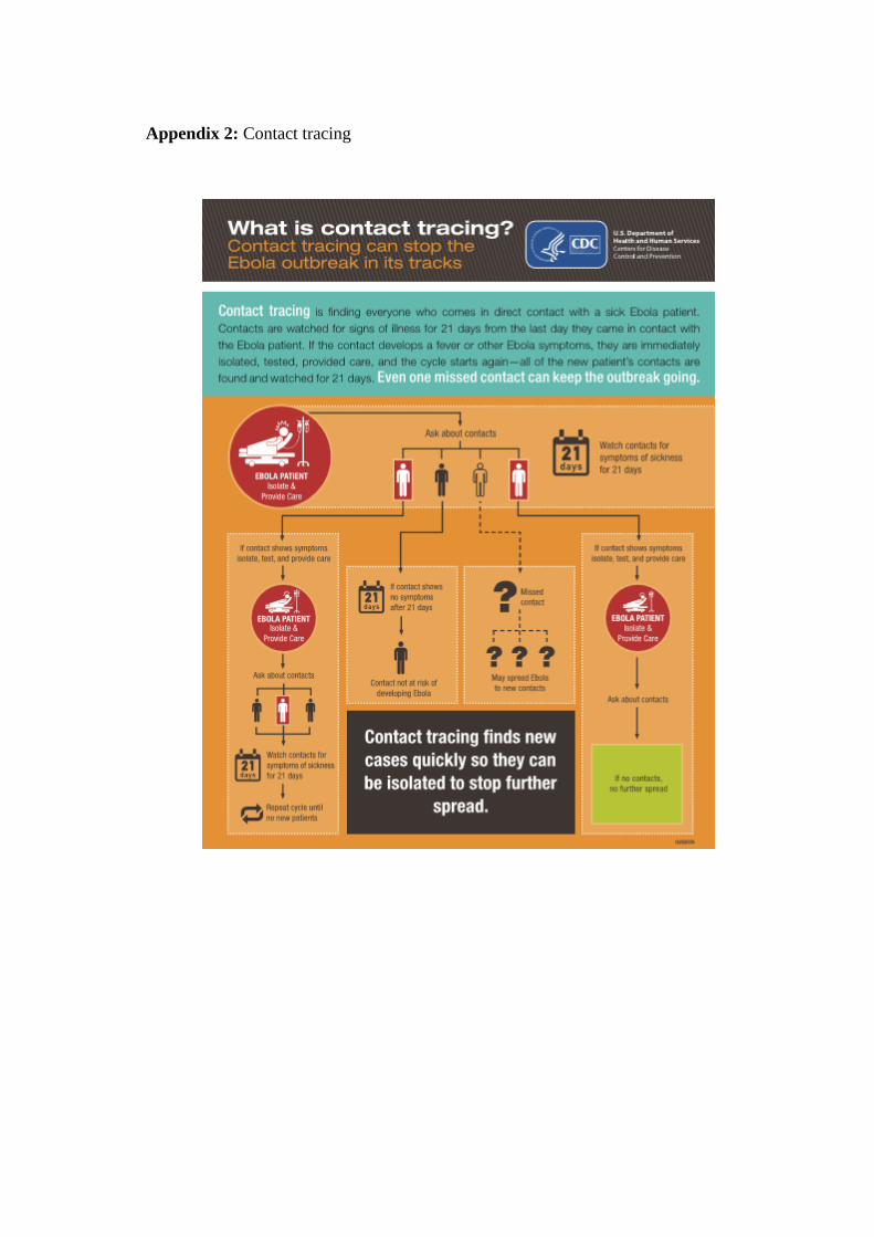
Appendix 2: Contact tracing

Appendix 3: Case Pauline
Article from MebMD Health News
“Nurse With Ebola, Pauline Cafferkey, Back in Hospital
Peter Russell | February 23, 2016
Scottish nurse Pauline Cafferkey has been transferred to a specialist hospital in London for the third time
since contracting Ebola in 2014.
The transfer came hours after the 40-year-old from South Lanarkshire was admitted to the Queen
Elizabeth University Hospital in Glasgow for routine monitoring at the Infectious Diseases Unit.
RAF Flight
She was put on an RAF Hercules aircraft which is flying her to London for transfer to the Royal Free
Hospital in London.
Ms Cafferkey has been treated in the hospital specialist isolation unit twice during 2015 after contracting
Ebola in Sierra Leone in 2014.
The Royal Free has issued a statement, saying: "We can confirm that Pauline Cafferkey is being
transferred to the Royal Free Hospital due to a late complication from her previous infection by the Ebola
virus. She will now be treated by the hospital's infectious diseases team under nationally agreed
guidelines.
"The Ebola virus can only be transmitted by direct contact with the blood or bodily fluids of an infected
person while they are symptomatic so the risk to the general public remains low and the NHS has well
established and practised infection control procedures in place."
Earlier, NHS Greater Glasgow and Clyde had described Ms Cafferkey's condition as 'stable'.
Ms Cafferkey was first admitted to the high level isolation unit (HLIU) at the Royal Free Hospital in
December 2014 after contracting the disease while working as part of a British team at the Kerry Town
treatment centre for Ebola patients in Sierra Leone. She was discharged in January this year after making
a recovery.
Meningitis

She was re-admitted to the Royal Free HLIU on the 8th October 2015 after the virus triggered a case of
viral meningitis.
Bodily tissues can harbour Ebola for some time after a patient appears to have recovered.
At one stage after her re-admission, Ms Cafferkey's condition was described as 'critical', but she was well
enough to be discharged from the isolation unit on 12th November last year.
It is not yet known what has prompted her re-admission today.
The Royal Free Hospital in London has an isolation unit and infection control protocols with specially
trained staff ready to deal with Ebola cases.
'A More Complex Disease Than Imagined'
Dr Derek Gatherer, lecturer in the Division of Biomedical and Life Sciences at Lancaster University,
comments in a statement: "It is now becoming clear that Ebola is a far more complex disease than we
previously imagined. The very large number of cases in West Africa since the end of 2013, have
displayed a wide range of rare but extremely unpleasant consequences for those who survive their first
battle with the disease.
"The meningitis that Ms Cafferkey suffered from at the end of last year is one of the most serious
complications of all, as it can be life-threatening. She was unlucky enough to be one of only a handful of
patients in whom it has been seen."
Dr Gatherer says the other main, but rare, complication seen in patients is eye inflammation which can
lead to blindness, while joint aches, headaches and general tiredness can last for months after recovery
from Ebola.
"Up to half of Ebola survivors will have some or several of this milder range of symptoms," says Dr
Gatherer. "Although not life-threatening, these can be problematic, especially in societies like west Africa
where a large number of people make their living from agricultural labour."
SOURCES:
Royal Free London NHS Foundation Trust. NHS Greater Glasgow and Clyde. Science Media Centre.
Reviewed on February 23, 2016”
Available at http://www.medscape.com/viewarticle/859308 accessed 19 May 2016.

Appendix 4: Ebola Info graphic with the outbreak events timeline

Appendix 5: Cleaning and disinfection in the ETC
Source: WHO
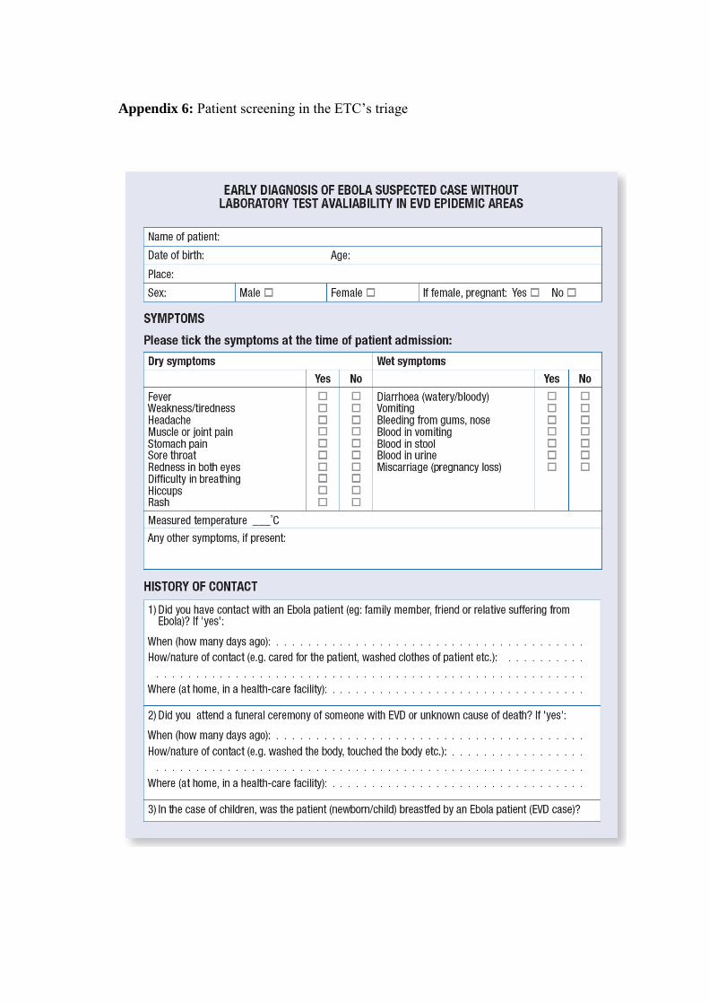
Appendix 6: Patient screening in the ETC‟s triage

Source: WHO

Appendix 7: Algorithm for admitting a patient in the ETC
Source: WHO
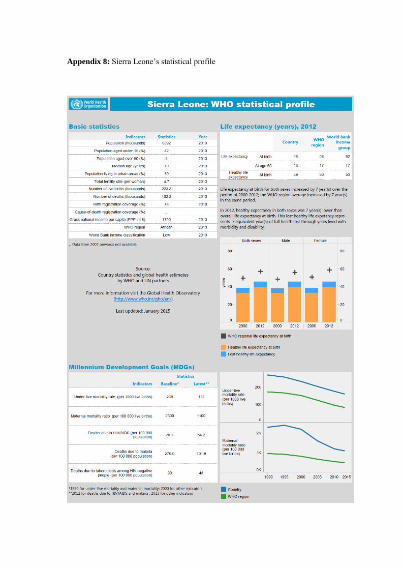
Appendix 8: Sierra Leone‟s statistical profile


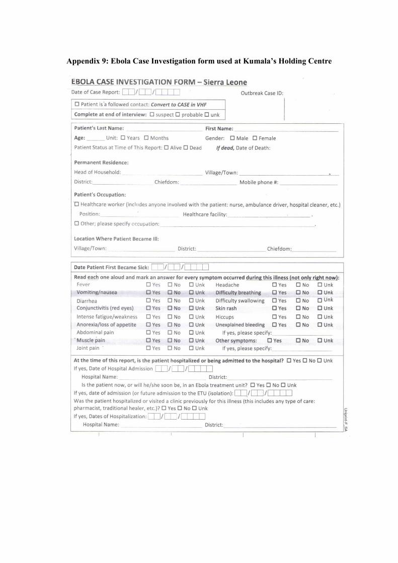
Appendix 9: Ebola Case Investigation form used at Kumala’s Holding Centre


Appendix 10: Table of results
Symptoms of the 10 survivors on admission to the HC, 8 and 10 months after
SURVIVOR 1 SURVIVOR 2
SYMPTOM ON
ADMISSION 8M AFTER 10M AFTER
ON
ADMISSION 8M AFTER 10M AFTER
FEVER X
VOMITING X
DIARRHOEA
FATIQUE
WEAKNESS X X
WEIGHT LOSS X X
APPETITE LOSS X X
ADBOMINAL PAIN X X
JOINT PAIN X
MUSCLE PAIN X
BACK PAIN X
CHEST PAIN X
HEADACHE X X X X
DIF. BREATHING
DIF. SWALLOWING
HICCUPS
SKIN RASH
EYE PROBLEMS X X
HEARING PROBLEMS
SLEEPING PROBLEMS
MOOD DISTURBANCE
BLEEDING
SURVIVOR 3 SURVIVOR 4
SYMPTOM ON
ADMISSION 8M AFTER 10M AFTER
ON ADMISSION
8M AFTER 10M AFTER
FEVER X X
NO DATA
VOMITING
DIARRHOEA
FATIQUE X
WEAKNESS X X X
WEIGHT LOSS X X X
APPETITE LOSS X
ADBOMINAL PAIN X X X

JOINT PAIN X X X
MUSCLE PAIN X
BACK PAIN X
CHEST PAIN X
HEADACHE X X
DIF. BREATHING X X
DIF. SWALLOWING X
HICCUPS
SKIN RASH
EYE PROBLEMS X X X X
HEARING PROBLEMS
SLEEPING PROBLEMS
MOOD DISTURBANCE
BLEEDING
SURVIVOR 5 SURVIVOR 6
SYMPTOM ON
ADMISSION 8M AFTER 10M AFTER
ON
ADMISSION 8M AFTER 10M AFTER
FEVER X
NO DATA NO
SYMPTOMS
X
VOMITING
DIARRHOEA X X
FATIQUE X
WEAKNESS X X X
WEIGHT LOSS X X
APPETITE LOSS
ADBOMINAL PAIN X
JOINT PAIN X X X
MUSCLE PAIN X X
BACK PAIN X
CHEST PAIN
HEADACHE X X
DIF. BREATHING X X
DIF. SWALLOWING X
HICCUPS
SKIN RASH
EYE PROBLEMS X X X
HEARING PROBLEMS
SLEEPING PROBLEMS
MOOD DISTURBANCE X
BLEEDING X

SURVIVOR 7 SURVIVOR 8
SYMPTOM ON
ADMISSION 8M AFTER 10M AFTER
ON
ADMISSION 8M AFTER 10M AFTER
FEVER X
NO
SYMPTOMS
X
NO DATA
VOMITING
DIARRHOEA
FATIQUE X X
WEAKNESS X X
WEIGHT LOSS X X
APPETITE LOSS
ADBOMINAL PAIN
JOINT PAIN X X
MUSCLE PAIN X
BACK PAIN X
CHEST PAIN X
HEADACHE X X
DIF. BREATHING
DIF. SWALLOWING
HICCUPS
SKIN RASH X
EYE PROBLEMS X
HEARING PROBLEMS
SLEEPING PROBLEMS
MOOD DISTURBANCE
BLEEDING
SURVIVOR 9 SURVIVOR 10
SYMPTOM ON
ADMISSION 8M AFTER 10M AFTER
ON
ADMISSION 8M AFTER 10M AFTER
FEVER
VOMITING X
DIARRHOEA X
FATIQUE X X X
WEAKNESS X X X X X
WEIGHT LOSS X X
APPETITE LOSS X
ADBOMINAL PAIN X X X X
JOINT PAIN X X X X
MUSCLE PAIN
BACK PAIN X X
CHEST PAIN X X
HEADACHE X X X X

DIF. BREATHING
DIF. SWALLOWING
HICCUPS
SKIN RASH
EYE PROBLEMS
HEARING PROBLEMS X
SLEEPING PROBLEMS X
MOOD DISTURBANCE X
BLEEDING X