newcastle university eprintseprint.ncl.ac.uk/.../197259/01b1e17a-264b-44f0-91e6-e8942a9e5663.pdf ·...
TRANSCRIPT
Newcastle University ePrints
Boczonadi V, Horvath R.
Mitochondria: Impaired mitochondrial translation in human disease.
International Journal of Biochemistry & Cell Biology 2014, 48, 77-84.
Copyright:
Copyright © 2014 The Authors. Published by Elsevier Ltd.
This is an open-access article distributed under the terms of the Creative Commons Attribution-
NonCommercial-No Derivative Works License, which permits non-commercial use, distribution, and
reproduction in any medium, provided the original author and source are credited.
DOI link to article:
http://dx.doi.org/10.1016/j.biocel.2013.12.011
Date deposited: 11th February 2014
This work is licensed under a Creative Commons Attribution-NonCommercial-No Derivative Works
License
ePrints – Newcastle University ePrints
http://eprint.ncl.ac.uk

O
M
VI
a
ARR1AA
KMMHTC
mno
Cf
1h
The International Journal of Biochemistry & Cell Biology 48 (2014) 77– 84
Contents lists available at ScienceDirect
The International Journal of Biochemistry& Cell Biology
journa l h om epage: www.elsev ier .com/ locate /b ioce l
rganelles in focus
itochondria: Impaired mitochondrial translation in human disease�
eronika Boczonadi, Rita Horvath ∗
nstitute of Genetic Medicine, Wellcome Trust Centre for Mitochondrial Research, Newcastle University, Newcastle upon Tyne, UK
r t i c l e i n f o
rticle history:eceived 28 September 2013eceived in revised form3 November 2013ccepted 26 December 2013vailable online 8 January 2014
a b s t r a c t
Defects of the mitochondrial protein synthesis cause a subgroup of mitochondrial diseases, which areusually associated with decreased activities of multiple respiratory chain (RC) enzymes. The clinical pre-sentations of these disorders are often disabling, progressive or fatal, affecting the brain, liver, skeletalmuscle, heart and other organs. Currently there are no effective cures for these disorders and treatment isat best symptomatic. The diagnosis in patients with multiple respiratory chain complex defects is particu-larly difficult because of the massive number of nuclear genes potentially involved in intra-mitochondrial
eywords:itochondrial respiratory chainitochondrial translationuman mitochondrial diseaseissue specific presentation
protein synthesis. Many of these genes are not yet linked to human disease. Whole exome sequencingrapidly changed the diagnosis of these patients by identifying the primary defect in DNA, and preventingthe need for invasive and complex biochemical testing. Better understanding of the mitochondrial proteinsynthesis apparatus will help us to explore disease mechanisms and will provide clues for developingnovel therapies.
ytosolic translation
Organelle facts
• Mitochondrial protein synthesis requires several mitochon-drial and nuclear-encoded factors for optimal translation.
• The clinical presentation of diseases due to defective mito-chondrial protein synthesis is very variable and tissuespecific presentations are common.
• The reasons behind the tissue specificity are largelyunknown.
• Besides mitochondrial tRNA mutations and mtDNA dele-tions or depletion, autosomal recessive mutations havebeen reported in genes encoding ribosomal proteins, ribo-some assembly proteins, mitochondrial aminoacyl-tRNAsynthetases, tRNA modifying enzymes and initiation, elon-gation and termination factors of translation.
• Frequent and clinically recognisable genetic causes ofhuman diseases due to impaired mitochondrial translationare caused by mutations in mitochondrial tRNA synthetaseand tRNA modifying genes.
• The potential interaction between cytosolic and mitochon-drial translation requires further investigations.
� This is an open-access article distributed under the terms of the Creative Com-ons Attribution-NonCommercial-No Derivative Works License, which permits
on-commercial use, distribution, and reproduction in any medium, provided theriginal author and source are credited.∗ Corresponding author at: Institute of Genetic Medicine, Newcastle University,entral Parkway, Newcastle upon Tyne NE1 3BZ, UK. Tel.: +44 191 2418855;
ax: +44 191 2418666.E-mail address: [email protected] (R. Horvath).
357-2725/$ – see front matter © 2014 The Authors. Published by Elsevier Ltd. All rights ttp://dx.doi.org/10.1016/j.biocel.2013.12.011
© 2014 The Authors. Published by Elsevier Ltd. All rights reserved.
1. Introduction
Mitochondrial diseases affect at least 1 in 5000 of the popu-lation and produce diverse clinical phenotypes often presentedas multi-systemic disorders (DiMauro et al., 2013; Ylikallio andSuomalainen, 2012; Vafai and Mootha, 2012). In addition tothe nucleus, human cells also harbour DNA in the mitochon-dria (mtDNA), which is essential for cell viability (Tuppen et al.,2010). This small (16.5 kb) genome is found in multiple copies inmitochondria, the subcellular organelles that often constitute morethan 20% of the total cell volume. OXPHOS (oxidative phosphor-ylation) is responsible for the production of ATP by generatinga proton gradient across the inner membrane of the mitochon-dria which is used by the mammalian cells (Greaves et al., 2012).The mitochondrial OXPHOS system comprises around 150 differentproteins out of which only 13 polypeptide subunits are encoded bythe mtDNA. In addition, the mtDNA encodes the small and largerRNAs, and 22 distinct mitochondrial tRNAs that are necessary forthe translation of only the mitochondrial-encoded proteins (Smitset al., 2010; Rötig, 2011; Chrzanowska-Lightowlers et al., 2011). Thenuclear-encoded subunits of the respiratory chain (RC) complexesas well as proteins that are inevitable for normal mitochondrialprotein synthesis (such as OXPHOS assembly, mtDNA metabolismand maintenance, mitochondrial cofactor biosynthesis, mitoribo-somal subunits and assembly factors, regulators of mitochondrialexpression and translation, etc.) are encoded by the nuclear genome
(nDNA) and synthetised in the cytosol before transported into theorganelle (Vafai and Mootha, 2012). The mitochondrial ribosomalproteins assemble with mitochondrial ribosomes 12S rRNA and 16SrRNA to form the mitochondrial ribosome (Pietromonaco et al.,reserved.
78 V. Boczonadi, R. Horvath / The International Journal of Biochemistry & Cell Biology 48 (2014) 77– 84

urnal
1tt
2
tatt2
2
mm2s2eisabmtfde
2
macmwpfGdotAa(
FapbngmLaTsfpos
V. Boczonadi, R. Horvath / The International Jo
991). Lately it was also reported that import of 5S rRNA is alsoransported to the mitochondria being an essential component ofhe mitochondrial ribosomes (Smirnov et al., 2011).
. Organelle function
The components responsible for the proper mitochondrialranslation are different from their cytosolic counterparts and theyre more related to those of bacteria however the mechanisms ofhe translation follows the same major steps: initiation, elongation,ermination and recycling of the ribosome (Christian and Spremulli,012) (Fig. 1).
.1. Initiation
The process of mitochondrial translation starts with the for-ation of the initiation complex. The separation of the twoitochondrial ribosomal subunits (28S and 39S) (Kuzmenko et al.,
013) allow this complex to be formed which consists of the 28Subunit, mRNA and fMET-tRNA and IF2/3mt (Koc and Spremulli,002; Christian and Spremulli, 2009). This is followed by thentrance of the mRNA into the IF3mt:28S subunit complex. IF3mt
s thought to support the correct position of mRNA to bind themall subunit at the peptidyl (P) site of the mitoribosome. When theppropriate start codon is present, the formylmethionin-tRNA canind to the first codon with the help of IF2mt. The association of theitoribosome stimulates the release of the initiation factors and
he elongation on the 55S ribosome commence. MTFMT is criticalor efficient human mitochondrial translation and reveal a humanisorder of Met-tRNA(Met) formylation (Tucker et al., 2011; Neevet al., 2013).
.2. Elongation
To coordinate accurate and specific codon anti-codon pairing,itochondrial elongation factor (EF-Tumt-GTP) and an amino-
cylated tRNA arrives to the A-site of the mitoribosome. Uponorrect codon-anticodon pairing the EF-Tumt-GDP leave theitoribosome and the aminoacyl-tRNA moves into the P sitehere peptide bond formation is catalysed extending the growingolypeptide chain. EF-Tsmt plays a role as a nucleotide exchangeactor and converts EF-Tumt to an active form (EF-Tumt-GTP). TheTP bound EFG1mt catalyses the translocation of the ribosomeuring the A and P site tRNAs move to the P and exit (E) sitesf the mitoribosome. This elongation step repeats itself until
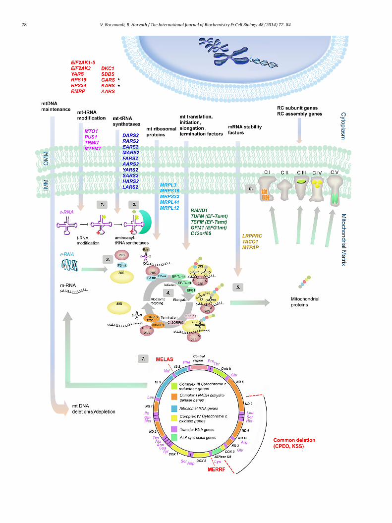
he stop codon (UAA, UAG, AGA or AGG) is encountered in the-site. Mutation of the mitochondrial elongation factors typicallyssociated with encephalopathy and other organ involvementliver, heart). Clinical symptoms are present in early infancy andig. 1. Schematic overview of human genes involved in mitochondrial protein synthesis dnd correctly replicated and transcribed. Mutations within nDNA-encoded genes responroteins also have to be imported into the mitochondria for accurate mitochondrial translased on their role in the translational machinery. The first group of genes (highlighteuclear genes (GARS, KARS (marked with black stars)) are transported to the mitochondrenes involved in mt-tRNA modification are: MTO1, PUS1, TRMU and MTFMT (highlighteditochondrial ARSs have been found to cause translational deficiencies in humans. Thes
ARS2 (highlighted in dark blue) (2). Nuclear genes encoding for ribosomal proteins and innd MRPL44 (light blue) (3). Genes represented in green are responsible for the mitochoSFM, GFM1, C12orf65. Nuclear genes such as translational activators and mRNA stability fynthesis (5). For the formation of the respiratory chain (RC) complexes both nuclear and mactors need to be synthetised, transported to the mitochondrial matrix and assembled iroteins are represented within each complex (CI: ND1, ND2, ND3, ND 4L, ND4, ND5, ND6;f the mitochondrial genome (7) showing the 13 proteins, 2 r-RNA, 22 t-RNA regions anhown in the diagram. Associated clinical phenotypes with the gene mutations mentione
of Biochemistry & Cell Biology 48 (2014) 77– 84 79
the affected children die early (Valente et al., 2007; Smeitink et al.,2006; Coenen et al., 2004; Smits et al., 2011a,b).
2.3. Termination
The mitochondrial release factor (mtRF1a) recognises the stopcodon and binds to the mitoribosome, induces hydrolysis of thepeptidyl-tRNA bond in the A-site releasing the mature protein fromthe site. Other termination release factors such as mtRF1, C12orf65and ICT1 are also thought to play an essential role in the termination(Richter et al., 2010). As a last step mitochondrial recycling factors(mtRRF1 and mtRRF2) translocate to the A-site to induce the releaseof the mRNA (Chrzanowska-Lightowlers et al., 2011). Up to dateonly the C12orf65 has been identified as a disease causing gene.Affected patients develop optic atrophy and ophthalmoplegia withLeigh syndrome (Antonicka et al., 2010).
2.4. Regulatory mechanisms
The expression of mitochondrial proteins is regulated by theirown translational activators that bind mitochondrial mRNAs usu-ally to their 5‘-untranslated regions, and each mitochondrial mRNAhas its own translational activator(s), which has been first shownin yeast (Herrmann et al., 2013). Recent studies showed that thesetranslational activators can be part of a feedback control loopswhich only permit translation if the downstream assembly ofnascent translation products can occur (Herrmann et al., 2013).Recently mutations in nuclear-encoded translational activators ofmitochondrial proteins such as TACO1 were also implicated inhuman disease (Weraarpachai et al., 2009). A regulatory role ofaminoacyl-tRNA synthetases has been suggested in both cytosolicand mitochondrial translation (Yao and Fox, 2013) and other fac-tors, such as MTERF3 has been implicated to coordinate crosstalkbetween transcription and translation for the regulation of mam-malian mtDNA (Wredenberg et al., 2013).
3. Cell Physiology
Impaired mitochondrial translation usually results in severecombined respiratory chain dysfunction through deficient func-tion of all mtDNA-encoded proteins however some nuclear geneshave been shown to alter the translation of single mitochondrial-encoded proteins. The defective mitochondrial proteins lead todeficient ATP production, and cellular energy deficit.
Human cells contain 17 cytoplasmic ARS polypeptides, includ-ing the bifunctional glutamyl-prolyl-tRNA synthetase (EPRS), and
18 mitochondrial ARS2 enzymes. Three ARS genes encode proteinswith dual localisation, present both in the cytoplasm and mitochon-dria (GARS, KARS, QARS), and the transport of the mitochondrialisoforms is ensured by a mitochondrial targeting signal.efects. Prior to mitochondrial protein synthesis the mtDNA needs to be maintainedsible for these functions lead to mtDNA deletion(s) and depletion. Several other
ation processes. These nuclear encoded genes are categorised into different groupsd in red) are the genes that are involved in cytosolic translation. Some of theseia and also function as mitochondrial aminoacyl t-RNA synthetase (ARS). Nuclear
in purple) (the functions of the genes are shown and labelled as (1). Up to date 10e genes are DARS2, RARS2, EARS2, MARS2, FARS2, AARS2, YARS2, SARS2, HARS2 andvolved in impaired mitochondrial translation are MRPL3, MRPS16, MRPS22, MRPL12ndrial translation steps: initiation, elongation and termination (4): RMND1, TUFM,actors (LRPPRC, TACO1 and MTPAP) also involved in impaired mitochondrial protein
itochondrial DNA are required. nDNA-encoded RC subunit genes and RC assemblynto functional enzyme complexes with the 13 mDNA-encoded proteins. These 13
CIII: CYTB; CIV: COX1, COX2, COX3; CV: ATPase6, ATPase8) (6). Schematic drawingd the control region. Mutations for MELAS, MERRF and for common deletions ared above are detailed in Table 1.

80 V. Boczonadi, R. Horvath / The International Journal of Biochemistry & Cell Biology 48 (2014) 77– 84
Table 1Nuclear DNA mutations involved in impaired mitochondrial translation and associated diseases in human. Sources: OMIM (Online Mendelian Inheritance in Man).
Nuclear genes involved in impaired cytosolic translation
Gene Protein Clinical presentation Age of onset OMIM References
EIF2AK1-5 eIF2B subunits �–� Vanishing white matter;childhood ataxia withcentral nervous systemhypomyelination (chronicprogressive, an episodicencephalopathy)
Childhood to adultage
604032 Leegwater et al.(2001); van der Knaapet al. (2002)
EIF2AK3 eIF2 � kinase PERK Wolcott–Rallisonsyndrome (diabetesmellitus, epiphysealdysplasia, kidney and liverdysfunction, mentalretardation, centralhypothyroidism anddysfunction of the exocrinepancreas)
Neonatal or earlychildhood
604032 Delépine et al. (2000)
GARS YARSKARS AARS
Glycyl-tRNA synthetasetyrosyl-tRNA synthetaselysyl-tRNA synthetasealanyl-tRNA synthetase.
Dominant intermediateCharcot–Marie–Tooth typeC (slowly progressivemixeddemyelinating-axonalneuropathy) or hereditarymotor neuropathy
Childhood to adultage
600287 603623 601421 601065 Antonellis et al. (2003)Jordanova et al. (2006)Rossor et al. (review)(2013)
RPS19 RPS24 Ribosomal protein S19ribosomal protein S24
Diamond–Blackfananaemia (abnormalities ofthe thumb, short stature,ventricular septal defects,kidney hypoplasia andcongenital glaucoma)
From birth 603474 602412 Draptchinskaia et al.(1999); Gazda et al.(2006)
RMRP Mitochondrial RNA proc.endoribonuclease
Cartilage-hair hypoplasia Neonatal, infantile 157660 Ridanpää et al. (2001)
DKC1 Dyskerin X-linked dyskeratosiscongenita (ectodermalabnormalities, bonemarrow failure andsusceptibility to cancer)
From birth 300126 Heiss et al. (1998)
SBDS Shwachman–Bodian–Diamondsyndrome protein
Shwachman–Diamondsyndrome (exocrinepancreatic insufficiency,bone marrow dysfunction,skeletal abnormalities andshort stature)
From birth 607444 Boocock et al. (2003)
Nuclear genes involved in impaired mitochondrial translation – tRNA-modifying enzymes
Gene Protein Clinical presentation Age of onset OMIM References
PUS1 Pseudouridine synthase Myopathy, lactic acidosisand sideroblastic anaemia(MLASA1)
Early childhood 608109 Bykhovskaya et al.(2004)
TRMU tRNA 5-methylaminome-thyl-2-thiouridylatemethyl-transferase
Reversible infantile liverfailure
Infantile 613070 Zeharia et al. (2009);Gaignard et al. (2013)Schara et al. (2011);
MTO1 Mitoch. translationoptimisation 1 homolog
Hypertrophiccardiomyopathy and lacticacidosis
Infantile 614702 Ghezzi et al. (2012)
MTFMT Mitoch. methionyl-tRNAformyltransferase
Leigh syndrome Early childhood 611766 Tucker et al. (2011)Neeve et al. (2013)
Nuclear genes involved in impaired mitochondrial translation – ribosomal proteins
Gene Protein Clinical presentation Age onset OMIM References
MRPL3 Mitochondrial ribosomalprotein L3
Hypertrophiccardiomyopathy andpsychomotor retardation
Infantile 614582 Galmiche et al. (2011)
MRPS16 Mitochondrial ribosomalprotein S16
Corpus callosum agenesia,hypothonia and fatalneonatal lactic acidosis
Neonatal 610498 Miller et al. (2004)
MRPS22 Mitochondrial ribosomalprotein S22
Cornelia de Lange-likesyndrome oedema,cardiomyopathy andtubulopathy
Neonatal 611719 Saada et al. (2007);Smits et al. (2011a,b)
MRPL12 Mitochondrial ribosomalprotein L12
Growth retardation andneurological deterioration
Neonatal 602375 Serre et al. (2013)

V. Boczonadi, R. Horvath / The International Journal of Biochemistry & Cell Biology 48 (2014) 77– 84 81
Table 1 (Continued)
Nuclear genes involved in impaired mitochondrial translation – ribosomal proteins
Gene Protein Clinical presentation Age of onset OMIM References
MRPL44 Mitochondrial ribosomalprotein L44
Hypertrophiccardiomyopathy
Neonatal 611849 Carroll et al. (2013)
Nuclear genes involved in impaired mitochondrial translation – aminoacyl-tRNA synthetases
Gene Protein Clinical presentation Age onset OMIM References
DARS2 Aspartyl-tRNA sythetase 2 Leukoencephalopathy withbrainstem and spinal cordinvolvement (LBSL)
Childhood oradulthood
610956 Scheper et al. (2007)
RARS2 Arginyl-tRNA synthetase 2 Pontocerebellar hypoplasiatype 6 (PCHD-6)
Neonatal 611523 Edvardson et al. (2007)
EARS2 Glutamyl-tRNA synthetase 2 Leukoencephalopathy withthalamus and brainsteminvolvement and highlactate (LTBL)
Infantile 612799 Steenweg et al. (2012)
MARS2 Methionyl-tRNA synthetase2
Autosomal recessivespastic ataxia withleukoencephalopathy
Juvenile oradulthood
609728 Bayat et al. (2012)
FARS2 Phenylalanyl-tRNAsynthetase 2
Alpers syndrome,encephalopathy, epilepsy,lactic acidosis
Neonatal andinfantile
611592 Elo et al. (2012);Shamseldin et al.(2012)
AARS2 Alanyl-tRNA synthetase 2 Hypertrophiccardiomyopathy
Infantile 614096 Götz et al. (2011)
YARS2 Tyrosyl-tRNA synthetase 2 Myopathy, lactic acidosis,and sideroblastic anaemia(MLASA2)
Infantile 613561 Riley et al. (2010)
SARS2 Seryl-tRNA synthetase 2 HUPRA syndrome(hyperuricemia,pulmonary hypertension,renal failure in infancy andalkalosis)
Infantile (4months)
613845 Belostotsky et al.(2011)
HARS2 Histidyl-tRNA synthetase 2 Perrault syndrome(sensorineural deafness,ovarian dysgenesis)
Juvenile oradulthood
600783 Pierce et al. (2011)
LARS2 Leucyl-tRNA synthetase 2 Perrault syndrome(sensorineural deafness,ovarian dysgenesis)
Juvenile 604544 Pierce et al. (2013)
Nuclear genes involved in impaired mitochondrial translation – initiation, elongation and termination factors
Gene Protein Clinical presentation Age onset OMIM References
RMND1 RMND Deafness, myopathy, renalinvolvement and a severebiochemical defect
Neonatal 614917 Garcia-Diaz et al.(2012): Janer et al.(2012)
TUFM Elongation factor Tu,mitochondrial (EF-TUmt)
Lactic acidosis,leukoencephalopathy andpolymicrogyria
Neonatal 610678 Valente et al. (2007)
TSFM Elongation factor Ts,mitochondrial (EF-Tsmt)
Encephalomyopathy,hypertrophiccardiomyopathy
Neonatal 610505 Smeitink et al. (2006)
GFM1 Elongation factor G 1,mitochondrial (EFG1mt)
Encephalopathy with orwithout liver involvement
Neonatal 609060 Coenen et al. (2004);Smits et al. (2011a);Valente et al. (2007)
C12orf65 Chromosome 12 openreading frame 65
Leigh syndrome, opticatrophy, ophthalmoplegia
Infantile 613559 Antonicka et al. (2010)
Nuclear genes involved in impaired mitochondrial transaltion – translation activators and mRNA stability factors
Gene Protein Clinical presentation Age onset OMIM References
LRPPRC Leucine-rich PPR-motifcontaining protein
Leigh syndromeFrench–Canadian variant(LSFC)
Infantile 220111 Mootha et al. (2003)
TACO1 Translational activator of Late-onset Leigh syndrome Juvenile 612958 Weraarpachai et al.
ataxia
e(sa
cytochrome c oxidase 1MTPAP Mitochondrial poly-A
polymeraseProgressive spastic
with optic atrophy
Beside disorders due to impaired mitochondrial translation, sev-
ral human disorders are caused by altered cytosolic translationYao and Fox, 2013). Interestingly these diseases also lead to tissuepecific clinical presentations mainly affecting brain, spinal cordnd peripheral neurons, illustrated by clinical presentations such as(2009)Juvenile (earlychildhood)
613672 Crosby et al. (2010)
Charcot–Marie–Tooth disease (CMT), distal hereditary motor neu-
ropathies (dHMN) or leukoencephalopathy with vanishing whitematter (VWM). Further implications of altered translation are high-lighted by variable and complex clinical presentations, includingdiseases of eye, cartilage, skin, hair and even cancer (Table 1).
8 urnal
4
vtLrttnriLmdrtmbweai
4
mmmetaHie2rrpr
4d
mostdwc
tge
4
lach(
2 V. Boczonadi, R. Horvath / The International Jo
. Organelle pathology
As it was predicted, mutations in the nuclear genes coding forarious components of the translation machinery could give riseo a wide spectrum of diseases and phenotypes (Chrzanowska-ightowlers et al., 2011) (Table 1). Mitochondrial protein synthesisequires several nuclear-encoded factors, such as ribosomal pro-eins, ribosome assembly proteins, aminoacyl-tRNA synthetases,RNA modifying enzymes and initiation, elongation and termi-ation factors of translation (Rötig, 2011) (Fig. 1). Autosomalecessive mutations have been reported in several of these factorsn association with variable clinical presentations (Chrzanowska-ightowlers et al., 2011). Here we note, that disorders ofitochondrial protein synthesis usually result in combined RC
eficiencies and associated with abnormal mitochondria (raggeded fibres, COX negative fibres) on histology. However a defect ofranslation activation factors or post-transcriptional regulators of
ammalian mtDNA expression may cause impairment in the sta-ility of certain mitochondrial transcripts, as reported in patientsith TACO1 and LRPPRC deficiency, respectively (Weraarpachai
t al., 2009; Harmel et al., 2013). Because these defects appear toffect only a single OXPHOS enzyme (COX), these patients showsolated COX deficiency.
.1. Defective mitochondrial translation due to mtDNA mutations
Frequent causes of impaired mitochondrial translation aretDNA rearrangements (e.g. Kearns–Sayre syndrome) that affectitochondrial tRNA and/or rRNA genes or single mt-tRNA pointutations. About half of the mtDNA mutations causing dis-
ases in humans occur in tRNA genes (MELAS, MERRF, etc.) andhe heterogeneous clinical manifestations usually reflect vari-ble heteroplasmy (Tuppen et al., 2010; Greaves et al., 2012).omoplasmic tRNA mutations with variable penetrance and clin-
cal presentations also occur and suggest the role of genetic orpigenetic modifiers in mitochondrial translation (Taylor et al.,003). Although most of these conditions are progressive and fatal,eversible infantile cytochrome c oxidase deficiency myopathy (oreversible infantile respiratory chain deficiency), due to a homo-lasmic mt-tRNAGlu mutation stands out by showing spontaneousecovery (Horvath et al., 2009).
.2. Defective mitochondrial translation due to nuclear geneefects
The currently defined disorders caused by nuclear defects ofitochondrial protein synthesis are usually early-onset, severe,
ften fatal diseases (Table 1) with extremely variable clinical pre-entations, and the reason behind is still unclear. Patients withranslation elongation factor or mitochondrial ribosomal proteinefects had an early age of onset and a severe multisystem diseaseith symptoms already present at birth or even prenatal in a few
ases (Table 1).The extreme variability and relative strict tissue specificity of
he diseases caused by mutations in mitochondrial tRNA synthetaseenes illustrate the importance of understanding the factors influ-ncing mitochondrial translation in different tissues.
.3. Neurological presentations
Some genes are selectively important in specific neuronal popu-ations, as exemplified by leukoencephalopathy with brainstem
nd spinal cord involvement (LBSL) due to mutations in the mito-hondrial aspartyl-tRNA synthetase 2 (DARS2), or pontocerebellarypoplasia caused by argynyl tRNA synthetase 2 (RARS2) defectScheper et al., 2007; Edvardson et al., 2007). Mutations in theof Biochemistry & Cell Biology 48 (2014) 77– 84
glutamyl-tRNA synthetase (EARS2) cause early onset severe neuro-logical disease (leukoencephalopathy involving the thalamus andbrainstem with high lactate, LTBL) (Steenweg et al., 2012; Ghezziet al., 2012). MTFMT deficiency leads to (relatively mild) Leigh syn-drome with or without optic atrophy (Tucker et al., 2011; Neeveet al., 2013).
4.4. Cardiac presentations
Recently autosomal recessive mutations were reported in theAARS2 and MTO1 genes in patients with infantile hypertrophiccardiomyopathy. MTO1 encodes the enzyme that catalyzes the5-carboxymethylamino-methylation of the wobble position inmt-tRNAGlu, mt-tRNAGln and mt-tRNALys (Ghezzi et al., 2012).Patents with clinically indistinguishable clinical presentation offatal infantile hypertrophic cardiomyopathy had mutations in themitochondrial alanyl-tRNA synthetase 2 (AARS2) gene (Götz et al.,2011).
4.5. Hepatic presentations
Autosomal recessive mutations in the tRNA 5-methylaminomethyl-2-thiouridylate methyltransferase (TRMU),which is responsible for 2-thiouridylation of the mt-tRNAGlu,mt-tRNAGln and mt-tRNALys cause a severe but reversible infantilehepatopathy (Zeharia et al., 2009; Schara et al., 2011; Sasarmanet al., 2011). Infants with reversible hepatopathy develop symp-toms between 2 and 4 months of age, but if they survive thisphase of liver failure, they recover and develop normally (Scharaet al., 2011). The disease course and age of manifestation in TRMUdeficiency shows remarkable similarities to reversible infantilemyopathy and recent studies suggest that infantile cysteine con-centrations may be important for the reversibility of both of thesediseases (Boczonadi et al., 2013).
4.6. Haematological presentations
As a further complication of mt-tRNA synthetase dysfunction,the involvement of blood cells has been implicated by mutationsin PUS1, resulting in mitochondrial myopathy, lactic acidosis andsideroblastic anaemia (MLASA) (Bykhovskaya et al., 2004). Mito-chondrial tyrosyl-tRNA synthetase 2 (YARS2) mutations have beenalso identified in families with MLASA, very similar, but earlieronset compared to the phenotype caused by deficiency of a mt-tRNA modifying enzyme PUS1 (Chrzanowska-Lightowlers et al.,2011).
4.7. Other presentations
Other characteristic, rare diseases are HyperUricemia, Pul-monary hypertension, Renal failure and Alkalosis (HUPRA)syndrome, which is caused by mutations in the mitochondrialseryl-tRNA synthetase 2 (SARS2) (Belostotsky et al., 2011) andPerrault syndrome, characterised by ovarian dysgenesis and sen-sorineural hearing loss due to mutations in the mitochondrialhistidyl-tRNA synthetase 2 (HARS2) (Pierce et al., 2011) and leucyl-tRNA synthetase 2 (LARS2) (Pierce et al., 2013).
5. Future outlook
Despite the rapid advances in technologies and the growingnumber of human disease genes and studies on mechanisms of
mammalian mitochondrial translation, its regulation still remainslargely unexplored. The variety and intriguing tissue and cell-typespecific clinical presentations in both mitochondrial and cytosolictranslation, and the dual function of some ARS enzymes suggest
urnal
stDitgdpdectcaisdd
fi(ftmtdIgoct2fatsim
R
A
A
B
B
B
B
B
C
C
C
V. Boczonadi, R. Horvath / The International Jo
ubstantial interaction and overlap between these two protein syn-hesis pathways, which have not been extensively studied to date.ue to the abundant proteins and factors required for maintain-
ng accurate mitochondrial translation, it is a challenge to identifyhe genetic defect in all cases. However rapid development ofenetic technologies (next generation sequencing) resulted in aynamic improvement in genetic diagnosis. Although there is ahenotypic diversity in patients with mitochondrial translationeficiencies, we observed some emerging clinical subgroups (Kempt al., 2011), which recently turned out to be associated with spe-ific genetic defects. In patients with neurological presentationRNA synthetases or tRNA modifying factors are the most likelyause of disease. AARS2 and MTO1 mutations are preferentiallyssociated with cardiomyopathy, mutations in TRMU present withnfantile, reversible liver failure, YARS2 and PUS1 mutations lead toideroblastic anaemia and myopathy, and RMND1 deficiency causeeafness, myopathy, renal involvement and a severe biochemicalefect (Table 1).
Defining the exact pathomechanisms will suggest new avenuesor treatment in these disorders, as it has been recently stud-ed in reversible COX deficiency myopathy and TRMU deficiencyBoczonadi et al., 2013). Downregulation of TRMU that is requiredor 2-thiouriylation in cells from patient with RIRCD led to a reduc-ion in levels of mt-tRNAGlu thiolation resulting in a defect of
itochondrial protein synthesis. Cysteine is essential for normalhiolation and supplementation of L-cysteine improved mitochon-rial gene translation not only in TRMU but also in RIRCD cells.
nteresting experimental progresses are being pursued towardsene therapeutic approaches for mitochondrial translational dis-rders. Engineered human mitochondrial tRNAs and mRNAs –ontaining RP import sequence – can be efficiently imported intohe mitochondria where they restore translation (Wang et al.,012). The use of exosomes the body’s own vehicle mechanismor delivering protein and genetic biomarkers are also promisingnd new avenues are being identified. Recently it has been shownhat mRNAs for most tRNA synthetases can be detected in exo-omes (Wang et al., 2013). The detection of mutations in factorsnvolved in mitochondrial translation widens our understanding of
itochondrial disease and highlights basic molecular mechanisms.
eferences
ntonellis A, Ellsworth RE, Sambuughin N, Puls I, Abel A, Lee-Lin SQ. Glycyl tRNAsynthetase mutations in Charcot–Marie–Tooth disease type 2D and distal spinalmuscular atrophy type V. Am J Hum Genet 2003;72:1293–9.
ntonicka H, Ostergaard E, Sasarman F, Weraarpachai W, Wibrand F, Pedersen AM,et al. Mutations in C12orf65 in patients with encephalomyopathy and a mito-chondrial translation defect. Am J Hum Genet 2010;87:115–22.
ayat V, Thiffault I, Jaiswal M, Tétreault M, Donti T, Sasarman F, et al. Mutationsin the mitochondrial methionyl-tRNA synthetase cause a neurodegenera-tive phenotype in flies and a recessive ataxia (ARSAL) in humans. PLoS Biol2012;10(3):e1001288.
elostotsky R, Ben-Shalom E, Rinat C, Becker-Cohen R, Feinstein S, Zeligson S, et al.Mutations in the mitochondrial seryl-tRNA synthetase cause hyperuricemia,pulmonary hypertension, renal failure in infancy and alkalosis, HUPRA syn-drome. Am J Hum Genet 2011;88(2):193–200.
oczonadi V, Smith PM, Pyle A, Gomez-Duran A, Schara U, Tulinus M, et al. Altered2-thiouridylation impairs mitochondrial translation in reversible infantile respi-ratory chain deficiency. Hum Mol Genet 2013 June [Epub ahead of print].
oocock GR, Morrison JA, Popovic M, Richards N, Ellis L, Durie PR, et al. Muta-tions in SBDS are associated with Shwachman–Diamond syndrome. Nat Genet2003;33:97–101.
ykhovskaya Y, Casas K, Mengesha E, Inbal A, Fischel-Ghodsian N. Missense muta-tion in pseudouridine synthase 1 (PUS1) causes mitochondrial myopathy andsideroblastic anemia (MLASA). Am J Hum Genet 2004;74(6):1303–8.
arroll CJ, Isohanni P, Pöyhönen R, Euro L, Richter U, Brilhante V, et al. Whole-exome sequencing identifies a mutation in the mitochondrial ribosome proteinMRPL44 to underlie mitochondrial infantile cardiomyopathy. J Med Genet
2013;50(3):151–9.hristian BE, Spremulli LL. Evidence for an active role of IF3mt in the initiation oftranslation in mammalian mitochondria. Biochemistry 2009;48(15):3269–78.
hristian BE, Spremulli LL. Mechanism of protein biosynthesis in mammalianmitochondria. Biochim Biophys Acta 2012;1819(9–10):1035–54.
of Biochemistry & Cell Biology 48 (2014) 77– 84 83
Chrzanowska-Lightowlers ZM, Horvath R, Lightowlers RN. 175th ENMC Inter-national Workshop: Mitochondrial protein synthesis in health and disease.Neuromuscul Disord 2011;21:142–7.
Coenen MJ, Antonicka H, Ugalde C, Sasarman F, Rossi R, Heister JG, et al. Mutantmitochondrial elongation factor G1 and combined oxidative phosphorylationdeficiency. N Engl J Med 2004;351:2080–6.
Crosby AH, Patel H, Chioza BA, Proukakis C, Gurtz K, Patton MA, et al. Defectivemitochondrial mRNA maturation is associated with spastic ataxia. Am J HumGenet 2010;87:655–60.
Delépine M, Nicolino M, Barrett T, Golamaully M, Lathrop GM, Julier C. eIF2AK3,encoding translation initiation factor 2-� kinase 3, is mutated in patients withWolcott–Rallison syndrome. Nat Genet 2000;25:406–9.
DiMauro S, Schon EA, Carelli V, Hirano M. The clinical maze of mitochondrial neu-rology. Nat Rev Neurol 2013(88):429–44.
Draptchinskaia N, Gustavsson P, Andersson B, Pettersson M, Willig TN, Dianzani I,et al. The gene encoding ribosomal protein S19 is mutated in Diamond–Blackfananaemia. Nat Genet 1999;21:169–75.
Edvardson S, Shaag A, Kolesnikova O, Gomori JM, Tarassov I, Einbinder T, et al. Dele-terious mutation in the mitochondrial arginyl-transfer RNA synthetase gene isassociated with pontocerebellar hypoplasia. Am J Hum Genet 2007;81:857–62.
Elo JM, Yadavalli SS, Euro L, Isohanni P, Götz A, Carroll CJ, et al. Mitochon-drial phenylalanyl-tRNA synthetase mutations underlie fatal infantile Alpersencephalopathy. Hum Mol Genet 2012;21(20):4521–9.
Gaignard P, Gonzales E, Ackermann O, Labrune P, Correia I, Therond P, et al. Mito-chondrial infantile liver disease due to TRMU gene mutations: three new cases.JIMD Rep 2013;11:117–23.
Galmiche L, Serre V, Beinat M, Assouline Z, Lebre AS, Chretien D, et al. Rötig Exomesequencing identifies MRPL3 mutation in mitochondrial cardiomyopathy. HumMutat 2011;32:1225–31.
Garcia-Diaz B, Barros MH, Sanna-Cherchi S, Emmanuele V, Akman HO, Ferreiro-Barros CC, et al. Infantile encephaloneuromyopathy and defective mitochondrialtranslation are due to a homozygous RMND1 mutation. Am J Hum Genet2012;91(4):729–36.
Gazda HT, Kho AT, Sanoudou D, Zaucha JM, Kohane IS, Sieff CA, et al. Defectiveribosomal protein gene expression alters transcription, translation, apo-ptosis, and oncogenic pathways in Diamond–Blackfan anemia. Stem Cells2006;24:2034–44.
Ghezzi D, Baruffini E, Haack TB, Invernizzi F, Melchionda L, Dallabona C,et al. Mutations of the mitochondrial-tRNA modifier MTO1 cause hyper-trophic cardiomyopathy and lactic acidosis. Am J Hum Genet 2012;90:1079–87.
Götz A, Tyynismaa H, Euro L, Ellonen P, Hyötyläinen T, Ojala T, et al. Exomesequencing identifies mitochondrial alanyl-tRNA synthetase mutations in infan-tile mitochondrial cardiomyopathy. Am J Hum Genet 2011;88:635–42.
Greaves LC, Reeve AK, Taylor RW, Turnbull DM. Mitochondrial DNA and disease. JPathol 2012;226:274–86.
Harmel J, Ruzzenente B, Terzioglu M, Spåhr H, Falkenberg M, LarssonNG. The leucine-rich pentatricopeptide repeat-containing protein (LRPPRC)does not activate transcription in mammalian mitochondria. J Biol Chem2013;288(22):15510–9.
Heiss NS, Knight SW, Vulliamy TJ, Klauck SM, Wiemann S, Mason PJ, et al. X-linkeddyskeratosis congenita is caused by mutations in a highly conserved gene withputative nucleolar functions. Nat Genet 1998;19:32–8.
Herrmann JM, Woellhaf MW, Bonnefoy N. Control of protein synthesis in yeastmitochondria: the concept of translational activators. Biochim Biophys Acta2013;1833(2):286–94.
Horvath R, Kemp JP, Tuppen HA, Hudson G, Oldfors A, Marie SK, et al. Molecularbasis of infantile reversible cytochrome c oxidase deficiency myopathy. Brain2009;132:3165–74.
Janer A, Antonicka H, Lalonde E, Nishimura T, Sasarman F, Brown GK, et al. An RMND1Mutation causes encephalopathy associated with multiple oxidative phosphor-ylation complex deficiencies and a mitochondrial translation defect. Am J HumGenet 2012;91(4):737–43.
Jordanova A, Irobi J, Thomas FP, Van Dijck P, Meerschaert K, Dewil M, et al. Disruptedfunction and axonal distribution of mutant tyrosyl-tRNA synthetase in dominantintermediate Charcot–Marie–Tooth neuropathy. Nat Genet 2006;38:197–202.
Kemp JP, Smith PM, Pyle A, Neeve VC, Tuppen HA, Schara U, et al. Nuclear factorsinvolved in mitochondrial translation cause a subgroup of combined respiratorychain deficiency. Brain 2011;134:183–95.
Koc EC, Spremulli LL. Identification of mammalian mitochondrial translational ini-tiation factor 3 and examination of its role in initiation complex formation withnatural mRNAs. J Biol Chem 2002;38:35541–9.
Kuzmenko A, Atkinson GC, Levitskii S, Zenkin N, Tenson T, Hauryliuk V, et al. Mito-chondrial translation initiation machinery: conservation and diversification.Biochimie 2013, S0300-9084(13) 00247-2.
Leegwater PA, Vermeulen G, Könst AA, Naidu S, Mulders J, Visser A, et al. Subunitsof the translation initiation factor eIF2B are mutated in leukoencephaly withvanishing white matter. Nat Genet 2001;29:383–8.
Miller C, Saada A, Shaul N, Shabtai N, Ben-Shalom E, Shaag A, et al. Defective mito-chondrial translation caused by a ribosomal protein (MRPS16) mutation. AnnNeurol 2004;56:734–8.
Mootha VK, Lepage P, Miller K, Bunkenborg J, Reich M, Hjerrild M, et al. Identifica-tion of a gene causing human cytochrome c oxidase deficiency by integrativegenomics. Proc Natl Acad Sci USA 2003;100:605–10.
Neeve VC, Pyle A, Boczonadi V, Gomez-Duran A, Griffin H, Santibanez-Koref M, et al.Clinical and functional characterisation of the combined respiratory chain defect
8 urnal
P
P
P
R
R
R
R
R
S
S
S
S
S
S
S
S
4 V. Boczonadi, R. Horvath / The International Jo
in two sisters due to autosomal recessive mutations in MTFMT. Mitochondrion2013, S1567-7249(13)00041-X.
ierce SB, Chisholm KM, Lynch ED, Lee MK, Walsh T, Opitz JM, et al. Mutationsin mitochondrial histidyl tRNA synthetase HARS2 cause ovarian dysgenesisand sensorineural hearing loss of Perrault syndrome. Proc Natl Acad Sci USA2011;108(16):6543–8.
ierce SB, Gersak K, Michaelson-Cohen R, Walsh T, Lee MK, Malach D, et al.Mutations in LARS2, encoding mitochondrial leucyl-tRNA synthetase, lead topremature ovarian failure and hearing loss in Perrault syndrome. Am J HumGenet 2013;92(4):614–22.
ietromonaco S, Denslow N, O‘Brien TW. Proteins of mammalian mitochondrialribosomes. Biochimie 1991;73:827–36.
ichter R, Rorbach J, Pajak A, Smith PM, Wessels HJ, Huynen MA, et al. A functionalpeptidyl-tRNA hydrolase ICT1, has been recruited into the human mitochondrialribosome. EMBO J 2010;29(6):1116–25.
idanpää M, van Eenennaam H, Pelin K, Chadwick R, Johnson C, Yuan B, et al. Muta-tions in the RNA component of RNase MRP cause a pleiotropic human disease,cartilage-hair hypoplasia. Cell 2001;104:195–203.
iley LG, Cooper S, Hickey P, Rudinger-Thirion J, McKenzie M, Compton A, et al.Mutation of the mitochondrial tyrosyl-tRNA synthetase gene YARS2, causesmyopathy, lactic acidosis, and sideroblastic anemia—MLASA syndrome. Am JHum Genet 2010;87:52–9.
ossor AM, Polke JM, Houlden H, Reilly MM. Clinical implications of geneticadvances in Charcot–Marie–Tooth disease. Nat Rev Neurol 2013 September.,http://dx.doi.org/10.1038/nrneurol.2013.179.
ötig A. Human diseases with impaired mitochondrial protein synthesis. BiochimBiophys Acta 2011;1807:1198–205.
aada A, Shaag A, Arnon S, Dolfin T, Miller C, Fuchs-Telem D, et al. Antenatalmitochondrial disease caused by mitochondrial ribosomal protein (MRPS22)mutation. J Med Genet 2007;44:784–6.
asarman F, Antonicka H, Horvath R, Shoubridge EA. The 2-thiouridylase function ofthe human MTU1 (TRMU) enzyme is dispensable for mitochondrial translation.Hum Mol Genet 2011;20:4634–43.
chara U, von Kleist-Retzow JC, Lainka E, Gerner P, Pyle A, Smith PM, et al. Acuteliver failure with subsequent cirrhosis as the primary manifestation of TRMUmutations. J Inherit Metab Dis 2011;34(1):197–201.
cheper GC, van der Klok T, van Andel RJ, van Berkel CG, Sissler M, Smet J, et al.Mitochondrial aspartyl-tRNA synthetase deficiency causes leukoencephalopa-thy with brain stem and spinal cord involvement and lactate elevation. Nat Genet2007;39:534–9.
erre V, Rozanska A, Beinat M, Chretien D, Boddaert N, Munnich A, et al. Mutationsin mitochondrial ribosomal protein MRPL12 leads to growth retardation, neuro-logical deterioration and mitochondrial translation deficiency. Biochim BiophysActa 2013;1832(8):1304–12.
hamseldin HE, Alshammari M, Al-Sheddi T, Salih MA, Alkhalidi H, Kentab A, et al.Genomic analysis of mitochondrial diseases in a consanguineous populationreveals novel candidate disease genes. J Med Genet 2012;49:234–41.
meitink JA, Elpeleg O, Antonicka H, Diepstra H, Saada A, Smits P, et al. Distinct
clinical phenotypes associated with a mutation in the mitochondrial translationelongation factor EFTs. Am J Hum Genet 2006;79:869–77.mirnov A, Entelis N, Martin RP, Tarassov I. Biological significance of 5S rRNAimport into human mitochondria: role of ribosomal protein MRP-L18. GenesDev 2011;25:1289–305.
of Biochemistry & Cell Biology 48 (2014) 77– 84
Smits P, Smeitink J, van den Heuvel L. Mitochondrial translation and beyond: pro-cesses implicated in combined oxidative phosphorylation deficiencies. J BiomedBiotechnol 2010:737385.
Smits P, Antonicka H, van Hasselt PM, Weraarpachai W, Haller W, Schreurs M, et al.Mutation in subdomain G′ of mitochondrial elongation factor G1 is associatedwith combined OXPHOS deficiency in fibroblasts but not in muscle. Eur J HumGenet 2011a;19:275–9.
Smits P, Saada A, Wortmann SB, Heister AJ, Brink M, Pfundt R, et al. Mutation inmitochondrial ribosomal protein MRPS22 leads to Cornelia de Lange-like phe-notype, brain abnormalities and hypertrophic cardiomyopathy. Eur J Hum Genet2011b;19:394–9.
Steenweg ME, Ghezzi D, Haack T, Abbink TE, Martinelli D, van Berkel CG, et al.Leukoencephalopathy with thalamus and brainstem involvement and high lac-tate ‘LTBL’ caused by EARS2 mutations. Brain 2012;135:1387–94.
Taylor RW, Giordano C, Davidson MM, d’Amati G, Bain H, Hayes CM, et al. Ahomoplasmic mitochondrial transfer ribonucleic acid mutation as a causeof maternally inherited hypertrophic cardiomyopathy. J Am Coll Cardiol2003;41:1786–96.
Tucker EJ, Hershman SG, Köhrer C, Belcher-Timme CA, Patel J, Goldberger OA, et al.Mutations in MTFMT underlie a human disorder of formylation causing impairedmitochondrial translation. Cell Metab 2011;14:428–34.
Tuppen HA, Blakely EL, Turnbull DM, Taylor RW. Mitochondrial DNA mutations andhuman disease. Biochim Biophys Acta 2010;1797:113–28.
Vafai SB, Mootha VK. Mitochondrial disorders as windows into an ancient organelle.Nature 2012;491(7424):374–83.
Valente L, Tiranti V, Marsano RM, Malfatti E, Fernandez-Vizarra E, Donnini C,et al. Infantile encephalopathy and defective mitochondrial DNA translation inpatients with mutations of mitochondrial elongation factors EFG1 and EFTu. AmJ Hum Genet 2007;80:44–58.
van der Knaap MS, Leegwater PA, Könst AA, Visser A, Naidu S, Oudejans CB,et al. Mutations in each of the five subunits of translation initiation factoreIF2B can cause leukoencephalopathy with vanishing white matter. Ann Neurol2002;51:264–70.
Weraarpachai W, Antonicka H, Sasarman F, Seeger J, Schrank B, Kolesar JE, et al.Mutation in TACO1, encoding a translational activator of COX I, results incytochrome c oxidase deficiency and late-onset Leigh syndrome. Nat Genet2009;41:833–7.
Wredenberg A, Lagouge M, Bratic A, Metodiev MD, Spåhr H, Mourier A, et al. MTERF3regulates mitochondrial ribosome biogenesis in invertebrates and mammals.PLoS Genet 2013;9(1):e1003178.
Yao P, Fox PL. Aminoacyl-tRNA synthetases in medicine and disease. EMBO Mol Med2013(3):332–43.
Ylikallio E, Suomalainen A. Mechanisms of mitochondrial diseases. Ann Med2012;44:41–59.
Zeharia A, Shaag A, Pappo O, Mager-Heckel AM, Saada A, Beinat M, et al. Acuteinfantile liver failure due to mutations in the TRMU gene. Am J Hum Genet2009;85:401–7.
Wang F, Xu Z, Zhou J, Lo WS, Lau CF, Nangle LA, et al. Regulated capture
by exosomes of mRNAs for cytoplasmic tRNA synthetases. J Biol Chem2013;288(41):29223–8.Wang G, Shimada E, Zhang J, Hong JS, Smith GM, Teitell MA, et al. Correcting humanmitochondrial mutations with targeted RNA import. Proc Natl Acad Sci USA2012;109(13):4840–5.