nih public access adv biochem eng biotechnol · a sperm forming a single celled embryo, the zygote....
TRANSCRIPT
Totipotency, Pluripotency and Nuclear Reprogramming
Shoukhrat Mitalipov andDivision of Reproductive Sciences, Oregon National Primate Research Center, Oregon Health andScience University, 505 N.W. 185th Avenue, Beaverton, Oregon 97006, USA; Oregon Stem CellCenter, Oregon Health and Science University, 505 N.W. 185th Avenue, Beaverton, Oregon 97006,USA; and Department of Obstetrics and Gynecology, School of Medicine, Oregon Health andScience University, 505 N.W. 185th Avenue, Beaverton, Oregon 97006, USA, [email protected]
Don WolfDivision of Reproductive Sciences, Oregon National Primate Research Center, Oregon Health andScience University, 505 N.W. 185th Avenue, Beaverton, Oregon 97006, USA
AbstractMammalian development commences with the totipotent zygote which is capable of developing intoall the specialized cells that make up the adult animal. As development unfolds, cells of the earlyembryo proliferate and differentiate into the first two lineages, the pluripotent inner cell mass andthe trophectoderm. Pluripotent cells can be isolated, adapted and propagated indefinitely in vitro inan undifferentiated state as embryonic stem cells (ESCs). ESCs retain their ability to differentiateinto cells representing the three major germ layers: endoderm, mesoderm or ectoderm or any of the200+ cell types present in the adult body. Since many human diseases result from defects in a singlecell type, pluripotent human ESCs represent an unlimited source of any cell or tissue type forreplacement therapy thus providing a possible cure for many devastating conditions. Pluripotent cellsresembling ESCs can also be derived experimentally by the nuclear reprogramming of somatic cells.Reprogrammed somatic cells may have an even more important role in cell replacement therapiessince the patient’s own somatic cells can be used for reprogramming thereby eliminating immunebased rejection of transplanted cells. In this review, we summarize two major approaches toreprogramming: (1) somatic cell nuclear transfer and (2) direct reprogramming using geneticmanipulations.
KeywordsEmbryonic stem cells; iPS cells; Pluripotent; Somatic cell nuclear transfer; Totipotent
1 TotipotencyTotipotency is defined in Wikipedia as the ability of a single cell to divide and produce all thedifferentiated cells in an organism, including extraembryonic tissues. Totipotent cells formedduring sexual and asexual reproduction include spores and zygotes. In some organisms, cellscan dedifferentiate and regain totipotency. For example, a plant cutting or callus can be usedto grow an entire plant. Mammalian development commences when an oocyte is fertilized bya sperm forming a single celled embryo, the zygote. Consistent with the definition, the zygoteis totipotent, meaning that this single cell has the potential to develop into an embryo with allthe specialized cells that make up a living being, as well as into the placental support structurenecessary for fetal development. Thus, each totipotent cell is a self-contained entity that cangive rise to the whole organism. This is said to be true for the zygote and for early embryonicblastomeres up to at least the 4-cell stage embryo (see Fig. 1). Experimentally, totipotency canbe demonstrated by the isolation of a single blastomere from a preimplantation embryo and
NIH Public AccessAuthor ManuscriptAdv Biochem Eng Biotechnol. Author manuscript; available in PMC 2009 September 26.
Published in final edited form as:Adv Biochem Eng Biotechnol. 2009 ; 114: 185–199. doi:10.1007/10_2008_45.
NIH
-PA Author Manuscript
NIH
-PA Author Manuscript
NIH
-PA Author Manuscript

subsequently monitoring its ability to support a term birth following transfer into a suitablerecipient. This approach was pioneered in rats and has been realized in several mammalianspecies including nonhuman primates [1–4]. In the latter case, we confirmed the ability ofisolated blastomeres from 2- and 4-cell stage, IVF produced embryos of the rhesus monkey tosupport term pregnancies and to produce live animals [5]. As embryo development progressesto the 8-cell stage and beyond depending on the species, the individual blastomeres thatcomprise the embryo gradually lose their totipotency. It is generally believed that thisrestriction in developmental potential indicates irreversible differentiation and specializationof early embryonic cells into the first two lineages, the inner cell mass (ICM) that includescells that will give rise to the fetus and the trophectoderm (TE), and an outer layer of cells thatis destined to an extraembryonic fate (Fig. 1).
A complication in assessing the state of potency of blastomeres isolated from more advancedstages of development is insufficient cytoplasmic volume. Thus, although the blastomeres mayin fact be totipotent, embryonic development of relatively small isolated blastomeres arrestsat or near the time of blastulation. Recall that the zygote and early blastomeres undergo severalunusual mitotic or cleavage divisions that are not accompanied by a corresponding growth ofcytoplasm, that is, there is no change in embryo size despite the presence of more cells orblastomeres and each individual blastomere becomes smaller. The embryonic genome at theseearly stages is transcriptionally quiescent and development is regulated by maternally inheritedfactors present at the time of fertilization in the oocyte [6]. The transition in developmentalregulation with activation of the embryonic genome and a complete loss of dependence onoocyte factors occurs before the blastocyst stage in a species-specific manner. Additionally,by the late morula or early blastocyst stage the embryo ceases cleavage divisions and resumesnormal mitotic divisions with concomitant increases in cell volume during the S-phase. Thelikelihood that early blastomeres retain totipotency for a major part of preimplantationdevelopment but experimentally we cannot prove it is directly supported by the fact that theaddition of oocyte cytoplasm to a blastomere of the 8- to 16-cell stage embryo can restore, orperhaps more appropriately allow expression of, its full developmental potential. Thisapproach, embryonic cell nuclear transfer, has been employed in the monkey to demonstratethe totipotency of 8- to 16-cell stage blastomeres whereby reconstructed embryos whentransferred to a recipient resulted in a term birth [7].
It is also known that conglomerates of embryonic cells at a later stage of development candevelop into an organism. An experimental manipulation that supports this concept involvesblastocyst splitting. Cutting the embryo into halves with an approximately equal distributionof TE and ICM cells can lead to the production of viable infants [5,8]. Obviously, embryosplitting that creates demi embryos with highly distorted ratios of ICM to TE cells isinconsistent with the production of live births.
2 PluripotencyThe Wikipedia definition in the broad sense means “having more than one potential outcome.”In cell biology, the definition of pluripotency has come to refer to a stem cell that has thepotential to differentiate into any of the three germ layers: endoderm, mesoderm or ectoderm.Pluripotent stem cells can give rise to any fetal or adult cell type. However, a single cell or aconglomerate of pluripotent cells cannot develop into a fetal or adult animal because they lackthe potential to organize into an embryo. In contrast, many progenitor cells that are capable ofdifferentiating into a limited number of cell fates are described as multipotent. Somatic stemcells such as neural, bone marrow-derived, or hematopoietic cells would fit into this lattercategory.
Mitalipov and Wolf Page 2
Adv Biochem Eng Biotechnol. Author manuscript; available in PMC 2009 September 26.
NIH
-PA Author Manuscript
NIH
-PA Author Manuscript
NIH
-PA Author Manuscript

At least some of the embryo’s ICM cells are pluripotent, meaning that they can form virtuallyevery somatic and germ cell type in the body. These ICM cells are self sustained and theirpluripotency is maintained by endogenously expressed factors. In vivo, pluripotent cells withinthe ICM exist transiently; as the developmental program unfolds they differentiate into cellsof the next embryonic or fetal stage. However, they can be isolated, adapted and propagatedin vitro in an undifferentiated state as embryonic stem cells (ESCs) [9,10]. ESCs were firstderived in 1981 from the ICM of the inbred mouse by Martin [10] and Evans and Kaufman[9]. In 1998, ESCs were successfully isolated from surplus, IVF-produced human embryos[11].
ESCs express specific markers or characteristics similar but not identical to the transientpluripotent cells of an embryo. This includes stage specific embryonic antigens, enzymaticactivities such as alkaline phosphatase and telomerase, and “stemness” genes that are rapidlydown-regulated upon differentiation, including OCT4 and NANOG. Under specific conditions,ESCs can proliferate indefinitely in an undifferentiated state, suggesting that the transcriptionalactivity and epigenetic regulators capable of supporting pluripotency can be maintained in vitroin ESCs. However, when released from the influence of these culture conditions or followingtheir introduction back into a host embryo, ESCs retain their ability to differentiate into anycell-type, just like ICM cells. Alternatively, they can differentiate in vivo in teratomas intocells representing the three major germ layers: endoderm, mesoderm and ectoderm or they canbe directed to differentiate in vitro into any of the 200+ cell types present in the adult body.Since many human diseases result from defects in a single cell type, pluripotent human ESCsmay become an unlimited source of any cell or tissue type for replacement therapy thusproviding a possible cure for many devastating diseases.
Parenthetically, one of the challenges before clinical transplantation studies involving hESCscan begin concerns the immune response anticipated after transplantation [12,13]. HumanESCs are routinely derived from IVF embryos and transplantation of such cells into geneticallyunrelated patients will incite an immune response and result in rejection. Histocompatibilityis one of major unsolved problems in transplant medicine. Rejection of unmatched transplantedtissues is provoked by alloantigens present on graft tissues by the recipient’s immune system.The alloantigens or antigenic proteins on the surface of transplant tissues that mostly causeimmune rejection are the blood group antigens (ABO) and the major histocompatibilitycomplex (MHC) proteins, also designated in humans as human leukocyte antigens (HLA).Matching donor and recipient HLA types is important to reduce a cytotoxic T-cell response inthe recipient, and subsequently improve the chances of survival of the transplant. However,tissue or organ transplantation from one individual to another is a daunting task due to theexistence of two classes of HLA molecules (Class I, and II), each encoded by multiple genesand most importantly, each of these genes represented by multiple alleles. For example, thereare 22 different alleles identified so far for the class I HLA-A gene and 42 alleles for HLA-B.Thus, due to HLA polymorphism, the chances of finding a donor–recipient match based onjust a few HLA genes (HLA-A, -B, and -DR) could be one in several million [14]. Therefore,the need for developing approaches for deriving histocompatible pluripotent cells is commonlyrecognized.
3 Nuclear ReprogrammingHochedlinger and Jaenisch define nuclear reprogramming as the reversal of the differentiationstate of a mature cell to one that is characteristic of the undifferentiated embryonic state [15].Let us first look at the forward process of development and differentiation. It is now generallyrecognized that genetic material is usually not lost during development and differentiation.Consequently, the process of differentiation must reflect the expression at each stage of aunique cohort of specific genes, its transcriptome, and it now appears that such differential
Mitalipov and Wolf Page 3
Adv Biochem Eng Biotechnol. Author manuscript; available in PMC 2009 September 26.
NIH
-PA Author Manuscript
NIH
-PA Author Manuscript
NIH
-PA Author Manuscript

expression is determined or regulated by reversible epigenetic changes gradually imposed onthe genome during development.
Epigenetic mechanisms that have been implicated in the regulation of differential gene activityinclude modifications to the histones (such as acetylation, methylation, phosphorylation,ubiquitination, and ADP-ribosylation) and methylation of DNA at CpG dinucleotides (seereviews by [16,17]). These specific epigenetic modifications regulate expression or silencingof genes at the level of transcription, mediated by the level of packaging DNA into chromatin.For example, acetylation of histones H3 and H4 and methylation of H3 at the lysine 4 position(H3 Lys-4) unfolds and loosens up the DNA template and makes it accessible to transcriptionfactors. Thus, these epigenetic mechanisms are generally associated with active genetranscription. Conversely, methylation of H3 Lys-9 and H3 Lys-27 induce DNA compactionand subsequently gene silencing.
In this review, we will summarize two major approaches to nuclear reprogramming or reversingthe developmental process: (1) somatic cell nuclear transfer (SCNT) and (2) directreprogramming using genetic manipulations. It should be noted that interest in both of thesestrategies derives, in large measure, from the potential production and use of histocompatiblehuman ESCs in regenerative medicine.
4 Epigenetic Reprogramming by SCNTThe concept of reprogramming of a patient’s somatic cells into pluripotent ESCs was conceivedbased on two independent breakthroughs in the field of developmental biology in the late1990s: success with cloning of animals by SCNT [18,19] and derivation of human ESCs[11]. SCNT, or cloning, dates back to 1962 when John Gurdon first demonstrated that somaticcells from Xenopus laevis could be reprogrammed back into an early embryonic state by factorspresent in an egg cytoplasm and support development of an adult frog [20]. Thus, it becameclear that the cytoplasm of the oocyte has the ability to reprogram gene expression and that asingle somatic cell nucleus has the capacity to yield a whole new organism [21].
Research in SCNT involving other vertebrates including mammals continued for severaldecades and culminated in groundbreaking announcements, first in 1996 [18] and then in 1997[19] that sheep could be produced by SCNT using fetal and adult somatic cells. Thisaccomplishment was quickly reproduced in other mammals including mice [22], cattle [23,24], pigs [25], goats [26], rabbits [27], cats [28], mules [29], horses [30], rats [31], and dogs[32].
As mentioned above, ESCs were first derived in 1981 in the mouse [9,10]. Exploiting the abilityof mouse ESCs to contribute to germ-line chimeras and homologous recombination technologyfor the creation of knock-out mice and mammalian gene function analysis revolutionized thefield of experimental biology [33]. To date, an estimated 10,000 mutated mice have beengenerated worldwide using the gene targeting technique. In recognizing their enormouscontribution to the advances in every field of biology and medicine, the 2007 Nobel Prize inPhysiology and Medicine was awarded to three scientists who pioneered the derivation ofmouse ESCs and gene targeting [34].
The establishment of mouse ESCs has instigated similar studies in other mammals. Workingwith nonhuman primates, James Thomson of the Wisconsin National Primate Research Centerreported in 1995 the successful isolation of ESCs from rhesus macaque, in vivo flushedblastocysts [35]. Unlike mouse ESCs, monkey ESCs grew as flat colonies and expressedslightly different surface markers than did mouse cells. Primate ESCs are relativelycumbersome to maintain and manipulate, requiring considerable technical expertise andattention confounded by a requirement for manual passaging and their slow growth rate.
Mitalipov and Wolf Page 4
Adv Biochem Eng Biotechnol. Author manuscript; available in PMC 2009 September 26.
NIH
-PA Author Manuscript
NIH
-PA Author Manuscript
NIH
-PA Author Manuscript

Nevertheless, these cells were indeed pluripotent and capable of differentiating into cell typesof all three germ layers. ESCs were also successfully isolated in other nonhuman primatespecies including marmosets and cynomolgus macaques [36,37]. In the rhesus macaque, anadditional 25 cell lines were produced from in vitro produced embryos at the Oregon NationalPrimate Research Center [38]. In 1998, following protocols and markers developed in themonkey, the isolation of ESC lines from surplus IVF-produced human embryos was reported[11]. Subsequently, approximately 65 human ESC lines were approved in the United Statesfor Federal research support in August of 2001; however only a few of those lines are currentlyavailable and under study (http://stem-cells.nih.gov/research/registry/).
Despite the remarkable strides that have been made to date with mouse and primate ESCs,success in other species has been limited. ESC-like cells have been described in several speciesincluding sheep [39], cattle [40], pigs [41], rabbits [42] and rats [43]. However, the pluripotencyof these cells and their ability to maintain an undifferentiated phenotype over long term cultureremains questionable.
The conceptual unification of SCNT and embryonic stem cell derivation technology suggestedthat it might be possible to produce preimplantation human embryos by SCNT and then deriveisogenic embryonic stem cells from the resulting SCNT embryos [44,45]. Human ESCsproduced by this approach called “therapeutic cloning” would subsequently be differentiatedinto therapeutically useful cells and transplanted back into a patient suffering from adegenerative disease. The proof of the concept was first demonstrated in the mouse in 2000with the isolation of pluripotent ESCs from adult somatic cell nuclei [46]. These SCNT-derivedESCs expressed canonic pluripotent markers and were able to differentiate readily into varioussomatic cell types in vitro or in vivo in teratomas and chimeras. Despite multiple abnormalitiesobserved in cloned offspring, mouse ESCs derived by SCNT were transcriptionallyindistinguishable from their counterparts derived from fertilized embryos [47,48], consistentwith the notion that ESCs derived from reprogrammed somatic cells have an identicaltherapeutic potential with “wild type” IVF-derived ESCs. This exciting scientific advanceindicated that it may soon be possible to provide patients with pluripotent cells tailored for agiven therapeutic purpose.
However, despite this remarkable progress, the feasibility of therapeutic cloning in primatesremained questionable. Early attempts demonstrated that human and nonhuman primate SCNTembryos were unable to develop efficiently into blastocysts and typically arrested at earlycleavage stages [49,50]. This indicated an inability of primate SCNT embryos to activateembryonic genes and sustain the developmental program, possibly due to lacking or incompletenuclear reprogramming. These challenges along with retraction of two high profile papers thatcontained fabricated data on human SCNT [51] significantly dampened scientific enthusiasm.The ability to derive primate ESCs by SCNT until recently was uncertain.
We initially reported incomplete nuclear remodeling following standard SCNT in the monkey,including nuclear envelope breakdown (NEBD) and premature chromosome condensation(PCC), and correlated this observation with a decline in maturation promoting factor (MPF)activity [52]. Although, a direct link between NEBD, PCC and successful reprogramming wasnot clear, we presumed that remodeling could be particularly beneficial for efficient nuclearreprogramming by allowing access of reprogramming factors to the somatic cell’s chromatin.We introduced several modifications to SCNT protocols that prevented MPF decline andinduced robust NEBD and PCC. Importantly, these modifications resulted in improved SCNTembryo development and significantly increased blastocyst rates, suggesting that MPF activityis essential for efficient nuclear reprogramming. The modified protocols allowed routineproduction of SCNT blastocysts from various donor somatic cells providing the foundation forrapid advances in the derivation of ESCs. More recently, we succeeded in the derivation of
Mitalipov and Wolf Page 5
Adv Biochem Eng Biotechnol. Author manuscript; available in PMC 2009 September 26.
NIH
-PA Author Manuscript
NIH
-PA Author Manuscript
NIH
-PA Author Manuscript
two ESC lines from rhesus macaque SCNT blastocysts using adult male skin fibroblasts asnuclear donors (Fig. 2) [53]. DNA analysis confirmed that nuclear DNA was identical to donorsomatic cells and that mitochondrial DNA originated from oocytes. Both cell lines exhibitednormal ESC morphology, expressed key stemness markers, were transcriptionally similar tocontrol ESCs and differentiated into multiple cell types in vitro and in vivo. These resultsrepresent a significant advancement in understanding the role of nuclear remodeling events inreprogramming following SCNT and demonstrate the first successful reprogramming of adultprimate somatic cells into pluripotent ESCs. Currently, we are focused on furtherimprovements in reprogramming by SCNT and efficient derivation of ESCs in the nonhumanprimate model. In our initial report, the efficiency of this approach was quite low, requiringapproximately 150 oocytes to produce a single ESC line [53]. However, based on our currentSCNT outcomes yielding nearly threefold higher blastocyst development and ESC derivationrates over our previously reported efficiency, as few as ten or less monkey oocytes are requiredto produce one ESC line (Mitalipov, unpublished results). These results suggest that systematicoptimization of SCNT approaches to define critical reprogramming factors will likely succeedin the efficient generation of patient-specific ESCs for therapeutic applications.
Our recent data also strongly support the notion that oocyte-induced reprogramming of primatesomatic cells results in complete erasure of somatic memory and the resetting of a new ESC-specific epigenetic state. Imprinted gene expression, methylation, telomere length and X-inactivation analyses of SCNT-derived primate ESCs were consistent with accurate andextensive epigenetic reprogramming of somatic cells by oocyte-specific factors ((Mitalipov,unpublished results).
A variation on the SCNT theme that has received recent attention is called altered nucleartransfer (ANT). Reprogramming by oocyte-specific factors after SCNT employs endogenousepigenetic pathways/programs. Thus SCNT provides a paradigm for identification of naturalepigenetic factors in an egg that accompany nuclear reprogramming and promotes utilizationof these factors for direct reprogramming. However, utilization of an SCNT approach forreprogramming of human somatic cells into pluripotent ESCs poses ethical concerns since itinvolves the creation and subsequent destruction of preimplantation stage embryos withpotential for full-term development. Thus, ANT proposes the creation of pluripotent stem cellsby preemptive alterations prior to SCNT insuring that no totipotent embryo is involved [54,55]. These alterations should preclude the integrated organic unity and developmental potentialthat are the defining characteristics of a living organism, while still allowing the formation ofthe ICM cell lineage from which pluripotent stem cells can be derived. ANT proposes to alterthe nucleus of a somatic cell and/or the cytoplasm of an enucleated oocyte prior to SCNT thatwould prevent formation of a totipotent zygote. However, residual oocyte factors should becapable of reprogramming an introduced nucleus with subsequent development to a stage thatwould support pluripotent stem cell isolation in the absence of a trophectodermal lineage.
Mature metaphase II (MII) oocytes are one of the largest cells produced by the human body.They contain key maternally inherited transcriptional and epigenetic factors essential for“natural” reprogramming of highly specialized gametic genomes into totipotent and pluripotentcells. Therefore, it is not surprising that oocyte-specific factors are able to reprogramtransplanted somatic nuclei, although with less efficiency than that which occurs in the embryoafter fertilization. As indicated above, maternally inherited factors in the oocyte convert atranscriptionally-quiescent embryonic genome into an active one during early embryonicdevelopment and following embryonic genome activation, control of the developmentalprogram is gradually shifted to embryonic factors. Among these maternal transcription factors,whose functions have been well defined, are Oct4 and Sox2, which are both essential forformation of the ICM in mouse preimplantation embryos. Cdx2 plays a similar role in thedevelopment of TE. In early cleavage-stage mouse embryos these transcription factors are
Mitalipov and Wolf Page 6
Adv Biochem Eng Biotechnol. Author manuscript; available in PMC 2009 September 26.
NIH
-PA Author Manuscript
NIH
-PA Author Manuscript
NIH
-PA Author Manuscript

expressed in all blastomeres. At the blastocyst stage, Oct4 and Sox2 are detected exclusivelyin the ICM cells, while Cdx2 is confined to the TE [56]. The role and expression pattern ofthese factors is poorly studied in other species including primates. However, we have showna similar expression profile for OCT4 in monkey preimplantation embryos [57]. Thehomeodomain protein, Nanog, is also detected exclusively in the ICM of mouse embryos andcooperates with Oct4 and Sox2 to control a set of target genes that have important functionsin maintaining pluripotency and ICM formation. Currently, little is known about maternalepigenetic factors that induce histone modifications and DNA methylation. Recent worksuggests that expression of Nanog in embryos may be regulated by the histone argininemethyltransferase Carm1 [58]. Interestingly, overexpression of Carm1 upregulates both Nanogand Sox2 and was able to direct development of individual blastomeres into an ICM fate.
As stated above, Cdx2 is one of the earliest known transcription factors that is essential forformation and function of the TE lineage [56]. Cdx2-deficient mouse embryos fail to maintaina blastocoel and cannot form the TE, but nonetheless, development of the pluripotent lineageof the ICM is relatively unaffected [59,60]. Additional evidence for a key role of Cdx2 comesfrom Tead4 knockout embryos which are devoid of both the TE lineage and Cdx2 expression[61,62]. Interestingly, Cdx2-deficient ICMs can generate functional ESCs. Recent evidencealso suggests that somatic cells lacking Cdx2 can be used for SCNT, resulting in formation ofthe single ICM lineage suitable for isolation of ESC lines [63]. This demonstrates that inhibitionof TE specific factors during SCNT can significantly alter the developmental program andprevent formation of a totipotent embryo without compromising reprogramming to thepluripotent state, thus providing a scientific basis for the ANT concept.
On the other hand, Cdx2-deficient nuclei in this study were complemented by maternal factorsincluding Cdx2 before the onset of embryonic genome activation. Therefore, SCNT embryoswere not obviously abnormal until the maternal-to-embryonic transition point. To solve thisethical dilemma, maternal Cdx2 transcripts must be inactivated as well. Moreover, it remainsto be determined whether this approach will work in other species including primates.
5 Direct ReprogrammingPossibly one of the greatest developments in the stem cell research field in the past 2 years isthe discovery that introduction and ectopic expression of several genes can induce pluripotencyin somatic cells. A research group led by Shinya Yamanaka of Kyoto University found thatmurine somatic cells transduced with retroviral vectors carrying only four transcription factors,namely Oct4, Sox2, c-Myc and Klf4 can revert their epigenetic state to become ESC-like[64]. These cells termed induced pluripotent stem (iPS) cells were similar in their propertiesto ESCs in terms of marker expression, transcriptional activity and the ability to differentiateinto a variety of cell types in chimeras. The relative simplicity with which iPS cells can begenerated compared with SCNT makes this technique an attractive approach for studying theprinciples of nuclear reprogramming and also to evaluate their potential for clinicalapplications. Indeed, mouse iPS cells were quickly developed in several laboratories and haverecently been used to successfully treat sickle cell anemia in mice [65].
In November of 2007, two independent groups led by Shinya Yamanaka and James Thomsonreported that using a similar transduction approach they were able to generate iPS cells fromhuman somatic cells [66,67]. These human ESC-like cells also expressed markers of ESCs andwere capable of differentiating into cell types of all three germ layers. Yamanaka’s group usedthe same quartet of four factors that worked in the mouse, while the Thomson lab demonstratedthat a slightly new combination, OCT4, SOX2, NANOG and LIN28 can also generate humaniPS cells. It is interesting to note, that the efficiency of reprogramming was lower with adultsomatic cells than with cells of fetal or embryonic origin. Moreover, some adult somatic cell-
Mitalipov and Wolf Page 7
Adv Biochem Eng Biotechnol. Author manuscript; available in PMC 2009 September 26.
NIH
-PA Author Manuscript
NIH
-PA Author Manuscript
NIH
-PA Author Manuscript

derived iPS cells did not contribute to all cell types following differentiation in teratomas. Itis likely that there could be additional factors that may enhance production of iPS cells fromadult somatic cells. Indeed, a recent report suggests that hTERT and SV40 large T can enhancethe reprogramming efficiency of Yamanaka’s factors on human adult somatic cells [68].
Recent reports also suggest that the kinetics of reprogramming significantly differs betweeniPS and SCNT approaches. Direct reprogramming of somatic cells to iPS cells appears to bea much slower process with activation of the endogenous Oct4 or Nanog in the mouse observedon day 16 post-transduction [69]. In contrast, Oct4 expression in mouse SCNT embryos canbe detected after the 4-cell stage or on day 2 after SCNT [70].
The direct genetic manipulation of somatic cells into iPS cells carries an advantage over SCNTsince it does not produce totipotent cells and does not require human eggs. From a bioethicalviewpoint this approach would resolve concerns about producing and destroying humanembryos. However, this approach currently has serious limitations as a source of cells forregenerative medicine. Reprogramming using c-Myc results in tumor development inapproximately 20% of chimeric mice derived by injection of iPS cells [71]. Recent findingssuggest that c-Myc is not absolutely necessary for iPS cell induction, although it appears thatreprogramming efficiency is much lower when the oncogene is omitted [72]. Another concernis that introduction of multiple copies of transgenes may cause insertional mutations and disruptthe function of many endogenous genes. Continuous overexpression of transgenes is alsoproblematic due to the possibility of incomplete silencing of these transgenes duringdifferentiation. The residual incidence of even a few pluripotent cells in transplanted tissuesmay cause tumors. Although the retroviral-delivered genes are silenced in most iPS cells, thereis the likelihood of reactivation of these transgenes in differentiated cells and the possibilityof spontaneous reversion of transplanted cells back to the pluripotent state, leading to the riskfor malignant progression.
To avoid these pitfalls each patient-specific iPS cell line must be rigorously tested in animalmodels before therapeutic applications. These concerns suggest that further advances in thederivation of iPS cells without gene transfer will be required to overcome these problems. Inthe near future, novel reprogramming approaches that involve transient gene delivery systemor small molecules may prove to be a safer way of generating iPS cells suitable for clinicalapplications. It will be necessary to carry out a detailed analysis of iPS cells to understand fullythe mechanisms of reprogramming and their role in regenerative medicine. It is also essentialto continue to study SCNT-induced reprogramming and to compare carefully the properties ofiPS cell lines to those derived by SCNT.
References1. Nicholas J, Hall B. Experiments on developing rats: II. The development of isolated blastomeres and
fused eggs. J Exp Zool 1942;90:441–459.2. Johnson WH, et al. Production of four identical calves by the separation of blastomeres from an in
vitro derived four-cell embryo. Vet Rec 1995;137(1):15–16. [PubMed: 7483226]3. Willadsen SM, Polge C. Attempts to produce monozygotic quadruplets in cattle by blastomere
separation. Vet Rec 1981;108(10):211–213. [PubMed: 6939169]4. Tarkowski AK. Experiments on the development of isolated blastomers of mouse eggs. Nature
1959;184:1286–1287. [PubMed: 13836947]5. Mitalipov SM, et al. Monozygotic twinning in rhesus monkeys by manipulation of in vitro-derived
embryos. Biol Reprod 2002;66(5):1449–1455. [PubMed: 11967209]6. Minami N, Suzuki T, Tsukamoto S. Zygotic gene activation and maternal factors in mammals. J Reprod
Dev 2007;53(4):707–715. [PubMed: 17827882]
Mitalipov and Wolf Page 8
Adv Biochem Eng Biotechnol. Author manuscript; available in PMC 2009 September 26.
NIH
-PA Author Manuscript
NIH
-PA Author Manuscript
NIH
-PA Author Manuscript

7. Mitalipov SM, et al. Rhesus monkey embryos produced by nuclear transfer from embryonicblastomeres or somatic cells. Biol Reprod 2002;66(5):1367–1373. [PubMed: 11967199]
8. Ozil JP. Production of identical twins by bisection of blastocysts in the cow. J Reprod Fertil 1983;69(2):463–468. [PubMed: 6631814]
9. Evans MJ, Kaufman MH. Establishment in culture of pluripotential cells from mouse embryos. Nature1981;292:154–156. [PubMed: 7242681]
10. Martin GR. Isolation of a pluripotent cell line from early mouse embryos cultured in mediumconditioned by terato-carcinoma stem cells. Proc Natl Acad Sci USA 1981;78:7634–7638. [PubMed:6950406]
11. Thomson JA, et al. Embryonic stem cell lines derived from human blastocysts. Science 1998;282(5391):1145–1147. [PubMed: 9804556]
12. Ginis I, Rao MS. Toward cell replacement therapy: promises and caveats. Exp Neurol 2003;184(1):61–77. [PubMed: 14637081]
13. Dawson L, et al. Safety issues in cell-based intervention trials. Fertil Steril 2003;80(5):1077–1085.[PubMed: 14607552]
14. Taylor CJ, et al. Banking on human embryonic stem cells: estimating the number of donor cell linesneeded for HLA matching. Lancet 2005;366(9502):2019–2025. [PubMed: 16338451]
15. Hochedlinger K, Jaenisch R. Nuclear reprogramming and pluripotency. Nature 2006;441(7097):1061–1067. [PubMed: 16810240]
16. Gan Q, et al. Concise review: epigenetic mechanisms contribute to pluripotency and cell lineagedetermination of embryonic stem cells. Stem Cells 2007;25(1):2–9. [PubMed: 17023513]
17. Jenuwein T, Allis CD. Translating the histone code. Science 2001;293(5532):1074–1080. [PubMed:11498575]
18. Campbell KH, et al. Sheep cloned by nuclear transfer from a cultured cell line. Nature 1996;380(6569):64–66. [PubMed: 8598906]
19. Wilmut I, et al. Viable offspring derived from fetal and adult mammalian cells. Nature (London)1997;385(6619):810–813. [PubMed: 9039911]
20. Gurdon JB. The developmental capacity of nuclei taken from intestinal epithelium cells of feedingtadpoles. J Embryol Exp Morph 1962;10:622–640. [PubMed: 13951335]
21. Pomerantz J, Blau HM. Nuclear reprogramming: a key to stem cell function in regenerative medicine.Nat Cell Biol 2004;6(9):810–816. [PubMed: 15340448]
22. Wakayama T, et al. Full-term development of mice from enucleated oocytes injected with cumuluscell nuclei. Nature 1998;394(6691):369–374. [PubMed: 9690471]
23. Kato Y, et al. Eight calves cloned from somatic cells of a single adult. Science 1998;282(5396):2095–2098. [PubMed: 9851933]
24. Cibelli JB, et al. Cloned transgenic calves produced from nonquiescent fetal fibroblasts. Science1998;280(5367):1256–1258. [PubMed: 9596577]
25. Polejaeva IA, et al. Cloned pigs produced by nuclear transfer from adult somatic cells. Nature(London) 2000;407(6800):86–90. [PubMed: 10993078]
26. Baguisi A, et al. Production of goats by somatic cell nuclear transfer. Nat Biotechnol 1999;17(5):456–461. [PubMed: 10331804]
27. Chesne P, et al. Cloned rabbits produced by nuclear transfer from adult somatic cells. Nat Biotechnol2002;20(4):366–369. [PubMed: 11923842]
28. Shin T, et al. A cat cloned by nuclear transplantation. Nature 2002;415(6874):859. [PubMed:11859353]
29. Woods GL, et al. A mule cloned from fetal cells by nuclear transfer. Science 2003;301(5636):1063.[PubMed: 12775846]
30. Galli C, et al. Pregnancy: a cloned horse born to its dam twin. Nature 2003;424(6949):635. [PubMed:12904778]
31. Zhou Q, et al. Generation of fertile cloned rats by regulating oocyte activation. Science 2003;302(5648):1179. [PubMed: 14512506]
32. Lee BC, et al. Dogs cloned from adult somatic cells. Nature 2005;436(7051):641. [PubMed:16079832]
Mitalipov and Wolf Page 9
Adv Biochem Eng Biotechnol. Author manuscript; available in PMC 2009 September 26.
NIH
-PA Author Manuscript
NIH
-PA Author Manuscript
NIH
-PA Author Manuscript

33. Capecchi MR. Altering the genome by homologous recombination. Science 1989;244(4910):1288–1292. [PubMed: 2660260]
34. Mak TW. Gene targeting in embryonic stem cells scores a knockout in Stockholm. Cell 2007;131(6):1027–1031. [PubMed: 18083089]
35. Thomson JA, et al. Isolation of a primate embryonic stem cell line. Proc Natl Acad Sci USA 1995;92(17):7844–7848. [PubMed: 7544005]
36. Thomson JA, et al. Pluripotent cell lines derived from common marmoset (Callithrix jacchus)blastocysts. Biol Reprod 1996;55(2):254–259. [PubMed: 8828827]
37. Suemori H, et al. Establishment of embryonic stem cell lines from cynomolgus monkey blastocystsproduced by IVF or ICSI. Dev Dyn 2001;222(2):273–279. [PubMed: 11668604]
38. Mitalipov S, et al. Isolation and characterization of novel rhesus monkey embryonic stem cell lines.Stem Cells 2006;24(10):2177–2186. [PubMed: 16741224]
39. Handyside AH, et al. Towards the isolation of embryonal stem cells from the sheep. Rouxs Arch DevBiol 1987;196:185–190.
40. Evans MJ, et al. Derivation and preliminary characterization of pluripotent cell lines from porcineand bovine blastocysts. Theriogenology 1990;33:125–128.
41. Notarianni E, et al. Maintenance and differentiation in culture of pluripotential embryonic cell linesfrom pig blastocysts. J Reprod Fertil 1990;41:51–56.
42. Giles JR, et al. Pluripotency of cultured rabbit inner cell mass cells detected by isozyme analysis andeye pigmentation of fetuses following injection into blastocysts or morulae. Mol Reprod Dev 1993;36(2):130–138. [PubMed: 8257563]
43. Iannaccone PM, et al. Pluripotent embryonic stem cells from the rat are capable of producing chimeras.Dev Biol 1994;163(1):288–292. [PubMed: 8174785]
44. Gurdon JB, Colman A. The future of cloning. Nature 1999;402(6763):743–746. [PubMed: 10617195]45. Lanza RP, Cibelli JB, West MD. Human therapeutic cloning. Nat Med 1999;5(9):975–977. [PubMed:
10470061]46. Munsie MJ, et al. Isolation of pluripotent embryonic stem cells from reprogrammed adult mouse
somatic cell nuclei. Curr Biol 2000;10(16):989–992. [PubMed: 10985386]47. Brambrink T, et al. ES cells derived from cloned and fertilized blastocysts are transcriptionally and
functionally indistinguishable. Proc Natl Acad Sci USA 2006;103:933–938. [PubMed: 16418286]48. Wakayama S, et al. Equivalency of nuclear transfer-derived embryonic stem cells to those derived
from fertilized mouse blastocysts. Stem Cells 2006;24(9):2023–2033. [PubMed: 16690779]49. Stojkovic M, et al. Derivation of a human blastocyst after heterologous nuclear transfer to donated
oocytes. Reprod Biomed Online 2005;11(2):226–231. [PubMed: 16168222]50. Mitalipov SM, et al. Rhesus monkey embryos produced by nuclear transfer from embryonic
blastomeres or somatic cells. Biol Reprod 2002;66(5):1367–1373. [PubMed: 11967199]51. Kennedy D. Editorial retraction. Science 2006;311:336. [PubMed: 16424324]52. Mitalipov SM, et al. Reprogramming following somatic cell nuclear transfer in primates is dependent
upon nuclear remodeling. Hum Reprod 2007;22(8):2232–2242. [PubMed: 17562675]53. Byrne JA, et al. Producing primate embryonic stem cells by somatic cell nuclear transfer. Nature
2007;450(7169):497–502. [PubMed: 18004281]54. Condic ML. Alternative sources of pluripotent stem cells: altered nuclear transfer. Cell Prolif
2008;41:7–19. [PubMed: 18181941]55. Hurlbut WB. Altered nuclear transfer: a way forward for embryonic stem cell research. Stem Cell
Rev 2005;1(4):293–300. [PubMed: 17142870]56. Niwa H, et al. Interaction between Oct3/4 and Cdx2 determines trophectoderm differentiation. Cell
2005;123(5):917–929. [PubMed: 16325584]57. Mitalipov SM, et al. Oct-4 expression in pluripotent cells of the rhesus monkey. Biol Reprod 2003;69
(6):1785–1792. [PubMed: 12890723]58. Torres-Padilla ME, et al. Histone arginine methylation regulates pluripotency in the early mouse
embryo. Nature 2007;445(7124):214–218. [PubMed: 17215844]59. Chawengsaksophak K, et al. Cdx2 is essential for axial elongation in mouse development. Proc Natl
Acad Sci USA 2004;101(20):7641–7645. [PubMed: 15136723]
Mitalipov and Wolf Page 10
Adv Biochem Eng Biotechnol. Author manuscript; available in PMC 2009 September 26.
NIH
-PA Author Manuscript
NIH
-PA Author Manuscript
NIH
-PA Author Manuscript
60. Strumpf D, et al. Cdx2 is required for correct cell fate specification and differentiation oftrophectoderm in the mouse blastocyst. Development 2005;132(9):2093–2102. [PubMed: 15788452]
61. Nishioka N, et al. Tead4 is required for specification of trophectoderm in pre-implantation mouseembryos. Mech Dev 2008;125:270–283. [PubMed: 18083014]
62. Yagi R, et al. Transcription factor TEAD4 specifies the trophectoderm lineage at the beginning ofmammalian development. Development 2007;134(21):3827–3836. [PubMed: 17913785]
63. Meissner A, Jaenisch R. Generation of nuclear transfer-derived pluripotent ES cells from clonedCdx2-deficient blastocysts. Nature 2006;439(7073):212–215. [PubMed: 16227971]
64. Takahashi K, Yamanaka S. Induction of pluripotent stem cells from mouse embryonic and adultfibroblast cultures by defined factors. Cell 2006;126(4):663–676. [PubMed: 16904174]
65. Hanna J, et al. Treatment of sickle cell anemia mouse model with iPS cells generated from autologousskin. Science 2007;318(5858):1920–1923. [PubMed: 18063756]
66. Takahashi K, et al. Induction of pluripotent stem cells from adult human fibroblasts by defined factors.Cell 2007;131(5):861–872. [PubMed: 18035408]
67. Yu J, et al. Induced pluripotent stem cell lines derived from human somatic cells. Science2007;318:1917–1920. [PubMed: 18029452]
68. Park IH, et al. Reprogramming of human somatic cells to pluripotency with defined factors. Nature2008;451(7175):141–146. [PubMed: 18157115]
69. Brambrink T, et al. Sequential expression of pluripotency markers during direct reprogramming ofmouse somatic cells. Cell Stem Cell 2008;2(2):151–159. [PubMed: 18371436]
70. Boiani M, et al. Oct4 distribution and level in mouse clones: consequences for pluripotency. GenesDev 2002;16(10):1209–1219. [PubMed: 12023300]
71. Okita K, Ichisaka T, Yamanaka S. Generation of germline-competent induced pluripotent stem cells.Nature 2007;448(7151):313–317. [PubMed: 17554338]
72. Nakagawa M, et al. Generation of induced pluripotent stem cells without Myc from mouse and humanfibroblasts. Nat Biotechnol 2008;26(1):101–106. [PubMed: 18059259]
Mitalipov and Wolf Page 11
Adv Biochem Eng Biotechnol. Author manuscript; available in PMC 2009 September 26.
NIH
-PA Author Manuscript
NIH
-PA Author Manuscript
NIH
-PA Author Manuscript

Fig. 1.Development and reprogramming. Ontogeny begins from a single cell, the zygote. The zygoteand each blastomere of the early embryo are totipotent with the potential to develop into thewhole organism. As development unfolds, the developmental potential of individualblastomeres gradually declines resulting subsequently in pluripotent, multipotent, unipotentand terminally differentiated somatic cells. However, developmental potential of somatic cellscan be reinstated to the totipotent stage by SCNT or to the pluripotent state by directreprogramming
Mitalipov and Wolf Page 12
Adv Biochem Eng Biotechnol. Author manuscript; available in PMC 2009 September 26.
NIH
-PA Author Manuscript
NIH
-PA Author Manuscript
NIH
-PA Author Manuscript
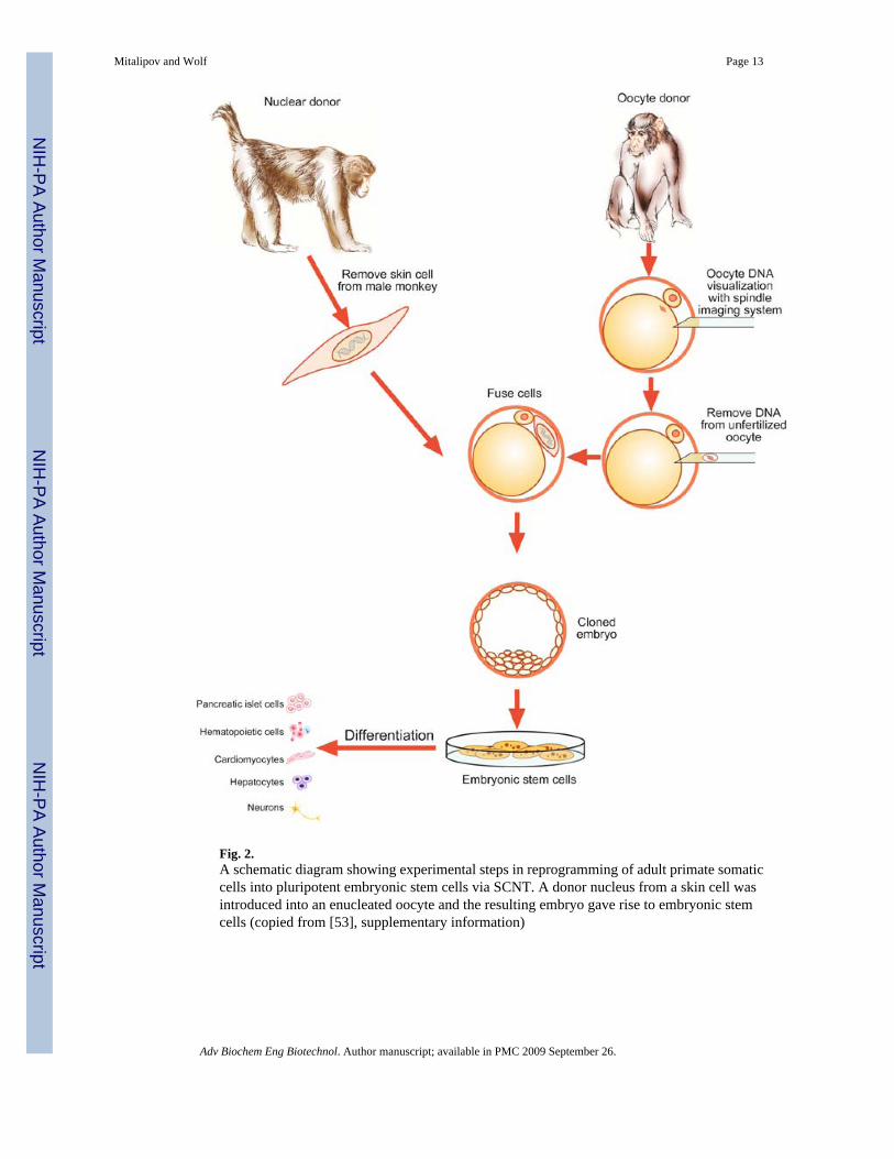
Fig. 2.A schematic diagram showing experimental steps in reprogramming of adult primate somaticcells into pluripotent embryonic stem cells via SCNT. A donor nucleus from a skin cell wasintroduced into an enucleated oocyte and the resulting embryo gave rise to embryonic stemcells (copied from [53], supplementary information)
Mitalipov and Wolf Page 13
Adv Biochem Eng Biotechnol. Author manuscript; available in PMC 2009 September 26.
NIH
-PA Author Manuscript
NIH
-PA Author Manuscript
NIH
-PA Author Manuscript