nobel prize for chemistry 2014 awarded to trio for ... nobel prize for chemistry... · nobel prize...
TRANSCRIPT
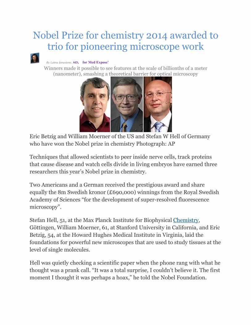
Nobel Prize for chemistry 2014 awarded to trio for pioneering microscope work
By Laima Jonusiene, MD, for Med Expose’
Winners made it possible to see features at the scale of billionths of a meter (nanometer), smashing a theoretical barrier for optical microscopy
Eric Betzig and William Moerner of the US and Stefan W Hell of Germany
who have won the Nobel prize in chemistry Photograph: AP
Techniques that allowed scientists to peer inside nerve cells, track proteins
that cause disease and watch cells divide in living embryos have earned three
researchers this year’s Nobel prize in chemistry.
Two Americans and a German received the prestigious award and share
equally the 8m Swedish kronor (£690,000) winnings from the Royal Swedish
Academy of Sciences “for the development of super-resolved fluorescence
microscopy”.
Stefan Hell, 51, at the Max Planck Institute for Biophysical Chemistry,
Göttingen, William Moerner, 61, at Stanford University in California, and Eric
Betzig, 54, at the Howard Hughes Medical Institute in Virginia, laid the
foundations for powerful new microscopes that are used to study tissues at the
level of single molecules.
Hell was quietly checking a scientific paper when the phone rang with what he
thought was a prank call. “It was a total surprise, I couldn’t believe it. The first
moment I thought it was perhaps a hoax,” he told the Nobel Foundation.
In a series of breakthroughs, the scientists overcame what many regarded as a
fundamental barrier to improving the resolution of optical microscopes and
showed that it was possible to see features at the scale of billionths of a metre.
Now there is theoretically no structure too small to be studied.
“Their groundbreaking work has brought optical microscopy into the nano-
dimension,” the Nobel jury said. “Today, nanoscopy is used worldwide and
new knowledge of the greatest benefit to mankind is produced on a daily
basis.”
Neurons in the brain of a living zebrafish embryo, imaged using nanoscopy.
For comparison, portions of the video show what one would see with ‘adaptive
optics (AO) and deconvolution’ (techniques for removing image distortions)
turned on, and off. Video: Eric Betzig/Janelia Research Campus/HHMI
For more than a century, the level of detail that could be seen through optical
microscopes was thought to hit an immovable limit at around a fifth of a
micrometre. The apparent barrier emerged from work by a microscopist called
Ernst Abbe who in 1873 showed that optical microscopes could never see
features smaller than roughly half the wavelength of visible light.
The Nobel prizewinners refused to believe the orthodoxy. After a decade spent
trying to sidestep Abbe’s theoretical limit, Stefan Hell demonstrated his
breakthrough. In 2000, he revealed the first stimulated emission depletion
(STED) microscope images of an E coli bacterium. The resolution was
unprecedented. Before taking the pictures, Hell prepared the bug by tagging it
with fluorescent proteins that glowed when light was shone on them. The
microscope flashed laser light onto the samples in a way that allowed only a
tiny region to glow at a time. By combining hundreds of snapshots of the
glowing sample taken from slightly different positions, he built up a picture
with a resolution far below the 200 nanometre limit.
Hell’s ideas met with substantial scepticism from the scientific community.
“People believed ‘this barrier has been around since 1873 and the resolution is
what it is and doing something about it is kind of crazy, not very realistic’,” he
said.
“But it was my view that so much physics had happened in the 20th century. I
felt there must be something, some kind of phenomenon that leads you
beyond the barrier.”
“I’ve always enjoyed doing challenging things and challenging common
wisdom,” he added.
Moerner and Betzig took a similar approach. Their own breakthrough came
when they discovered fluorescent proteins that could be turned on and off at
will. With these in hand, they developed single-molecule microscopy. The
technique uses weak pulses of laser light to make only a fraction of the
fluorescent tags light up. The microscope takes a picture of these glowing tags,
then fires another shot of laser light. This time, a different fraction of tags light
up, and another picture is taken. By repeating the process hundreds of times
and then superimposing the images, the scientists created pictures with higher
resolution than Abbe ever thought possible.
Moerner said that on hearing the news that he’d won a Nobel, “I was
incredibly excited and thrilled and of course your heart races.” Betzig said he
felt an equal measure of happiness and fear. “The fear is your life being
changed. I really like my life the way it is now!”
“It’s ironic in a way because, trained as a physicist, as a young man I would
look down on chemists,” he added.
“It’s no exaggeration to say that super-resolution fluorescence microscopy has
revolutionised imaging, so this year’s Nobel Prize in Chemistry is very well
deserved. The resolution in microscopy had been limited to 200 nanometres –
about the size of the smallest bacteria – for several hundred years,” said
Stefanie Reichelt, head of light microscopy at the Cancer Research UK
Cambridge Research Institute.
“The new imaging developments suddenly gave us a more than tenfold
increase in resolution and we can now see individual components of cells in
great detail. But these are much more than just pretty images – at this
resolution, we can begin to understand much more clearly what is happening
in important biological processes.”
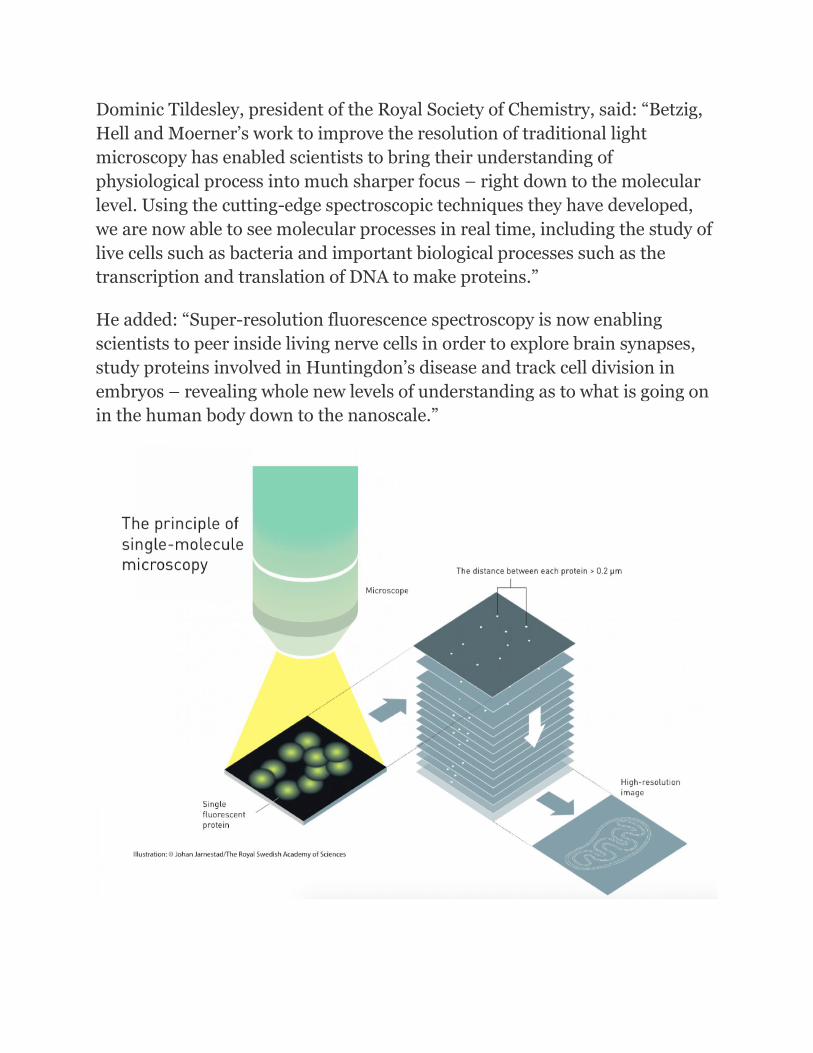
Dominic Tildesley, president of the Royal Society of Chemistry, said: “Betzig,
Hell and Moerner’s work to improve the resolution of traditional light
microscopy has enabled scientists to bring their understanding of
physiological process into much sharper focus – right down to the molecular
level. Using the cutting-edge spectroscopic techniques they have developed,
we are now able to see molecular processes in real time, including the study of
live cells such as bacteria and important biological processes such as the
transcription and translation of DNA to make proteins.”
He added: “Super-resolution fluorescence spectroscopy is now enabling
scientists to peer inside living nerve cells in order to explore brain synapses,
study proteins involved in Huntingdon’s disease and track cell division in
embryos – revealing whole new levels of understanding as to what is going on
in the human body down to the nanoscale.”
Nobel Prize in Chemistry 2014 –
Superesolution Microscopy 0
BY CIARAN MURPHY-ROYAL ON OCT 21, 2014CHEMISTRY, SCIENCE, SCIENCE NEWS, TOPICAL
Alfred Nobel
As some of you may have heard, this year’s Nobel Prizes have been awarded to many great and deserving
people. This highly prestigious award, started by the dynamite tycoon Alfred Nobel in 1895 (following his
death), comes only after having contributed some brilliant work in your field, be it Literature, Peace, Physics,
Chemistry or even Physiology & Medicine. I was compelled to write a piece about this as both the chemistry
and the Physiology & Medicine prizes are quite close to home. I worked on a part of the brain that was
awarded the prize in Physiology & Medicine and have a great number of friends who use the type of
microscope which was awarded the prize in chemistry. If you are wondering how and when you are most
likely to win a Nobel Prize, you can take a read of an earlier piece I wrote on the subject.
In this post I would like to highlight and try to explain the brilliant work carried out by a small group of
scientists which literally changed the way we look at the world. I am indeed talking about the Nobel Prize in
chemistry which was awarded equally to two physicists and a chemist; Stefan Hell, Eric Betzig and William
Moerner. This prize was awarded specifically for the development of superesolved fluorescence microscopy.
In short, these guys invented new ways of looking at very small things under the microscope in such detail that
was believed to be impossible to achieve. Sounds pretty impressive, eh?
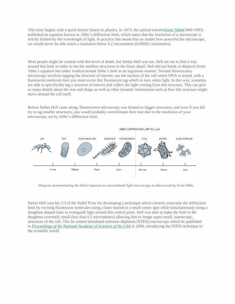
This story begins with a quick history lesson in physics. In 1873, the optical scientistErnst Abbe(1840-1905)
published an equation known as Abbe’s diffraction limit, which states that the resolution of a microscope is
strictly limited by the wavelength of light. In practice this meant that no matter how powerful the microscope,
we would never be able reach a resolution below 0.2 micrometers (0.00002 centimeters).
Most people might be content with this level of detail, but Stefan Hell was not. Hell set out to find a way
around this limit in order to see the smallest structures in the finest detail. Hell did not break or disprove Ernst
Abbe’s equation but rather worked around Abbe’s limit in an ingenious manner. Normal fluorescence
microscopy involves tagging the structure of interest, say the nucleus of the cell where DNA is stored, with a
fluorescent molecule then you must excite this fluorescent tag which in turn, emits light. In this way, scientists
are able to specifically tag a structure of interest and collect the light coming from this structure. This can give
us many details about the size and shape as well as other dynamic information such as how this structure might
move around the cell itself.
Before Stefan Hell came along, fluorescence microscopy was limited to bigger structures, and even if you did
try to tag smaller structures, you would probably overestimate their size due to the resolution of your
microscope, set by Abbe’s diffraction limit.
Diagram demonstrating the limits imposed on conventional light microscopy as discovered by Ernst Abbe.
Stefan Hell won his 1/3 of the Nobel Prize for developing a technique which cleverly overcame the diffraction
limit by exciting fluorescent molecules using a laser trained to a small centre spot while simultaneously using a
doughnut shaped laser to extinguish light around this central point. Hell was able to make the hole in the
doughnut extremely small (less than 0.2 micrometers) allowing him to image super-small, nanoscopic,
structures of the cell. This he coined stimulated emission depletion (STED) microscopy which he published
in Proceedings of the National Academy of Sciences of the USA in 2000, introducing the STED technique to
the scientific world.
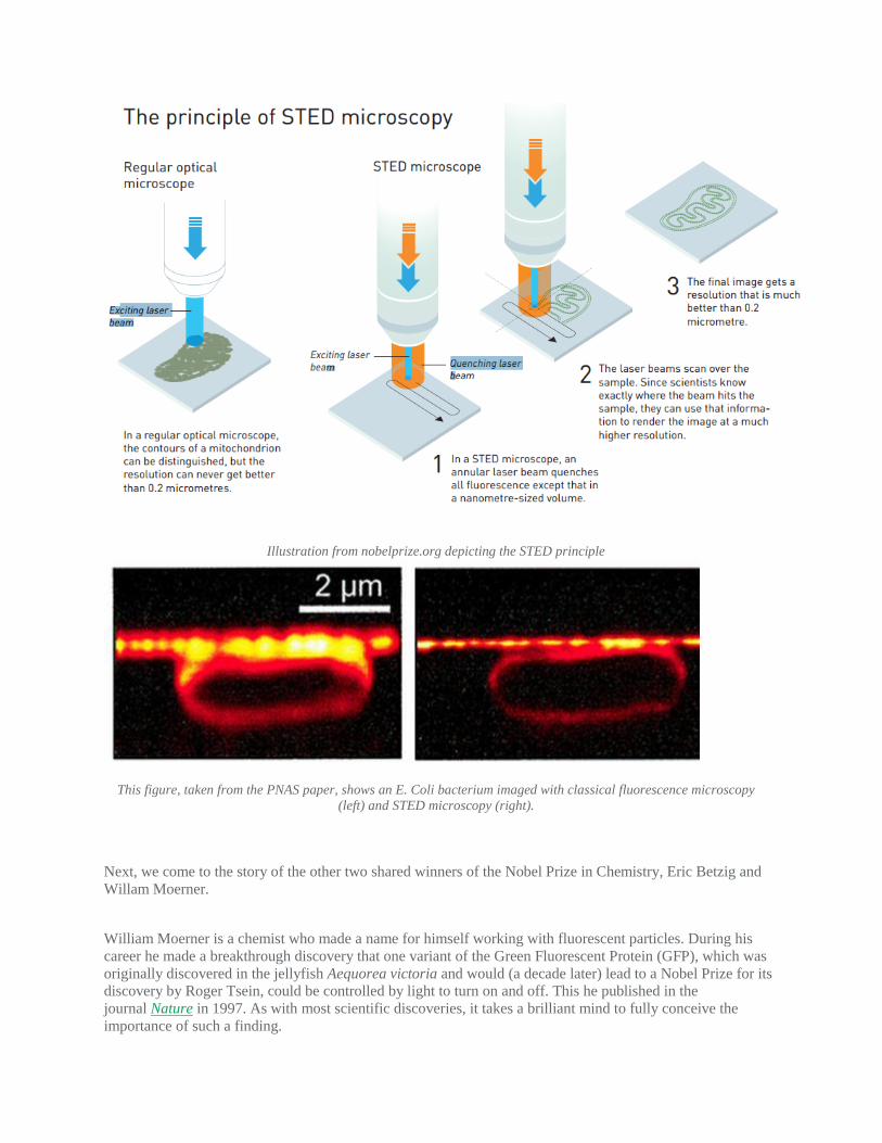
Illustration from nobelprize.org depicting the STED principle
This figure, taken from the PNAS paper, shows an E. Coli bacterium imaged with classical fluorescence microscopy
(left) and STED microscopy (right).
Next, we come to the story of the other two shared winners of the Nobel Prize in Chemistry, Eric Betzig and
Willam Moerner.
William Moerner is a chemist who made a name for himself working with fluorescent particles. During his
career he made a breakthrough discovery that one variant of the Green Fluorescent Protein (GFP), which was
originally discovered in the jellyfish Aequorea victoria and would (a decade later) lead to a Nobel Prize for its
discovery by Roger Tsein, could be controlled by light to turn on and off. This he published in the
journal Nature in 1997. As with most scientific discoveries, it takes a brilliant mind to fully conceive the
importance of such a finding.
Eric Betzig turned out to be one of the brilliant people to put Moerner’s controllable fluorescent particles to
work in a novel way which would allow him to join the elite club of Nobel Prize winners. Building on
Moerner’s findings, Betzig published a paper demonstrating a method to circumvent Abbe’s diffraction limit,
using low amounts of light in order to excite only a fraction of the fluorescent molecules which tag the
structure of interest. This allowed the precise calculation of the position of each individual fluorescent particle
before the light died out. This process was repeated over and over allowing the reconstruction of a
superesolution image of the structure being studied. Betzig and his colleagues published this technique called
photoactivated localisation (PALM) microscopy in Science in 2006 and brought this technique to the attention
of the scientific community at large.
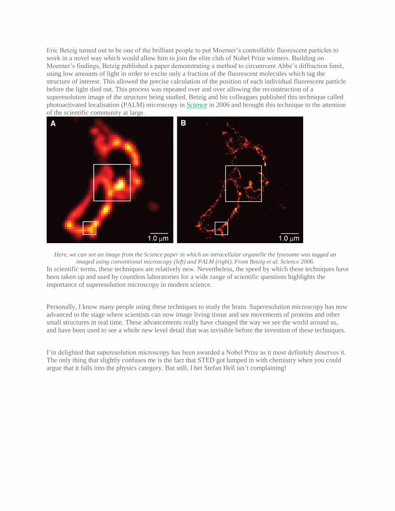
Here, we can see an image from the Science paper in which an intracellular organelle the lysosome was tagged an
imaged using conventional microscopy (left) and PALM (right). From Betzig et al. Science 2006.
In scientific terms, these techniques are relatively new. Nevertheless, the speed by which these techniques have
been taken up and used by countless laboratories for a wide range of scientific questions highlights the
importance of superesolution microscopy in modern science.
Personally, I know many people using these techniques to study the brain. Superesolution microscopy has now
advanced to the stage where scientists can now image living tissue and see movements of proteins and other
small structures in real time. These advancements really have changed the way we see the world around us,
and have been used to see a whole new level detail that was invisible before the invention of these techniques.
I’m delighted that superesolution microscopy has been awarded a Nobel Prize as it most definitely deserves it.
The only thing that slightly confuses me is the fact that STED got lumped in with chemistry when you could
argue that it falls into the physics category. But still, I bet Stefan Hell isn’t complaining!

Royal Rife had an idea but he could not validate it and his attackers stopped
progress
Royal Raymond Rife, June 1931 -Popular Science Magazine
Royal Raymond Rife (May 16, 1888 – August 5, 1971) was an American inventor and early exponent of high-magnification time-lapse cine-micrography.[1][2] In the 1930s, he claimed that by using a specially designed optical microscope, he could observe microbes which were too small to visualize with previously existing technology.[3] Rife also reported that a 'beam ray' device of his invention could weaken or destroy the pathogens by energetically exciting destructive resonances in their constituent chemicals.[4] Rife's claims could not be independently replicated,[5] and were discredited by independent researchers during the 1950s. Rife blamed the scientific rejection of his claims on a conspiracy involving the American Medical Association (AMA), the Department of Public Health, and other elements of "organized medicine", which had "brainwashed and intimidated" his colleagues.[6]
Interest in Rife's claims was revived in some alternative medical circles by the 1987 book The Cancer Cure That Worked, which claimed that Rife had succeeded in curing cancer, but that his work was suppressed by a powerful conspiracy headed by the AMA.[5] After this book's publication, a variety of devices bearing Rife's name were marketed as cures for diverse diseases such as cancer and AIDS. An analysis by Electronics Australia found that a typical 'Rife device' consisted of a nine-volt battery, wiring, a switch, a timer and two short lengths of copper tubing, which delivered an "almost undetectable" current unlikely to penetrate the skin.[7] Several marketers of other 'Rife devices' have been convicted for health fraud, and in some cases cancer patients who used these devices as a replacement for medical therapy have died.[8] Rife devices are currently classified as a subset of radionics devices, which are generally viewed as pseudomedicine by mainstream experts.[5]
BACILLI REVEALED BY NEW MICROSCOPE; Dr. Rife's Apparatus,
Magnifying 17,000 Times, Shows Germs Never Before Seen.
PERMISSIONS
Special to The New York Times. ();
November 22, 1931,
, Section , Page 19, Column , words
Life and work
Little reliable published information exists describing Rife's life. In 1929, he was granted a patent for a high-intensity microscope lamp.[9] On November 20, 1931, forty-four doctors attended a dinner advertised as "The End To All Diseases" at the Pasadena estate of Milbank Johnson, honoring Arthur I. Kendall of Northwestern Medical School and Rife, the developer of the 'Rife microscope'.[citation needed] Moving microorganisms from prepared, diseased human tissue[citation needed] were reportedly seen, still-photographed and also filmed with motion-picture equipment.[10]
In a 1932 report in Science, Mayo Clinic physician Edward C. Rose now wrote that in addition to other small particles viewable with the standard lab microscope, small turquoise bodies termed 'eberthella typhi' not visible with the standard lab microscopes were seen in filtrate using a Rife microscope. Rosenow attributed their detection to "the ingenious methods employed rather than excessively high magnification".[11] Subsequently, details of one of Rife's microscopes, as well as obtained micrographs, were included in the 1944 Annual Report of the Board of Regents of the Smithsonian Institution.[12]
Rife claimed to have documented a "Mortal Oscillatory Rate" for various pathogenic organisms, and to be able to destroy the organisms by vibrating them at this particular rate. According to the San Diego Evening Tribune in 1938, Rife stopped short of claiming that he could cure cancer, but did argue that he could "devitalize disease organisms" in living tissue, "with certain exceptions".[4]
Rife's microscope, techniques and claimed results have been consistently denied and discredited by the medical community, who've concluded that his results were simply not possible to obtain,
observing the known laws of physics. An obituary in the Daily Californian described his death at the age of 83 on August 5, 1971, stating that he died penniless and embittered by the failure of his devices to garner scientific acceptance.[6]
Modern revival, marketing, and health fraud
Interest in Rife was revived in the 1980s by author Barry Lynes, who wrote a book about Rife entitled The Cancer Cure That Worked. The book claimed that Rife's 'beam ray' device could cure cancer, but that all mention of his discoveries was suppressed in the 1930s by a wide-ranging conspiracy headed by the American Medical Association. The American Cancer Society described Lynes' claims as implausible, noting that the book was written "in a style typical of conspiratorial theorists" and defied any independent verification.[5]
In response to this renewed interest, devices bearing Rife's name began to be produced and marketed in the 1980s. Such 'Rife devices' have figured prominently in several cases of health fraud in the U.S., typically centered around the uselessness of the devices and the grandiose claims with which they are marketed. In a 1996 case, the marketers of a 'Rife device' claiming to cure numerous diseases including cancer and AIDS were convicted of felony health fraud.[13] The sentencing judge described them as "target[ing] the most vulnerable people, including those suffering from terminal disease" and providing false hope.[14] In 2002 John Bryon Krueger, who operated the Royal Rife Research Society, was sentenced to 12 years in prison for his role in a murder and also received a concurrent 30-month sentence for illegally selling Rife devices. In 2009 a U.S. court convicted James Folsom of 26 felony counts for sale of the Rife devices sold as 'NatureTronics', 'AstroPulse', 'BioSolutions', 'Energy Wellness', and 'Global Wellness'.[15]
Several deaths have resulted from the use of Rife machines in place of standard medical treatment. In one case, a U.S. court found that the marketer of a Rife device had violated the law and that, as a result of her actions, a cancer patient had ceased chemotherapy and died.[16] In Australia, the use of Rife machines has been blamed for the deaths of cancer patients who might have been cured with conventional therapy.[7]
In 1994, the American Cancer Society reported that Rife machines were being sold in a "pyramid-like, multilevel marketing scheme". A key component in the marketing of Rife devices has been the claim, initially put forward by Rife himself, that the devices were being suppressed by an establishment conspiracy against cancer "cures".[5] Although 'Rife devices' are not registered by the U.S. Food and Drug Administration and have been linked to deaths among cancer sufferers, the Seattle Times reported that over 300 people attended the 2006 Rife International Health Conference in Seattle, where dozens of unregistered devices were sold.[8]
References
1. "Local Man Bares Wonders of Germ Life: Making Moving Pictures of Microbe Drama". San Diego Union. November 3, 1929.
2. H. H. Dunn (June 1931). "Movie New Eye of Microscope in War on Germs". Popular Science 118 (6): 27, 141. ISSN 0161-7370.
3. "BACILLI REVEALED BY NEW MICROSCOPE; Dr. Rife's Apparatus, Magnifying 17,000 Times, Shows Germs Never Before Seen.". The New York Times. November 22, 1931. p. 19.
4. ^ to:a b Jones, Newell (1938-05-06). "Dread Disease Germs Destroyed By Rays, Claim Of S. D. Scientist: Cancer Blow Seen After 18-year Toil by Rife". San Diego Evening Tribune. p. 1.
5. ^ to:a b c d e "Questionable methods of cancer management: electronic devices" (PDF). CA Cancer J Clin 44 (2): 115–27. 1994.doi:10.3322/canjclin.44.2.115. PMID 8124604.
6. ^ to:a b Del Hood (August 11, 1971). "Scientific Genius Dies: Saw Work Discredited". Daily Californian.
7. ^ to:a b Hills, Ben (2000-12-30). "Cheating Death". Sydney Morning Herald. Retrieved 2009-01-11.
8. ^ to:a b Willmsen, Christine; Michael J. Berens (2007-12-21). "Pair indicted on fraud charges in medical-device probe". Seattle Times. Retrieved 2008-04-24.
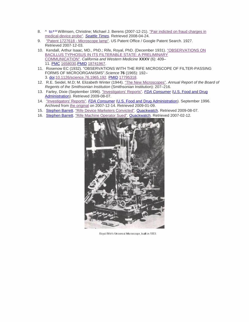
9. "Patent 1727618 - Microscope lamp". US Patent Office / Google Patent Search. 1927. Retrieved 2007-12-03.
10. Kendall, Arthur Isaac, MD., PhD.; Rife, Royal, PhD. (December 1931). "OBSERVATIONS ON BACILLUS TYPHOSUS IN ITS FILTERABLE STATE: A PRELIMINARY COMMUNICATION". California and Western Medicine XXXV (6): 409–11. PMC 1658030.PMID 18741967.
11. Rosenow EC (1932). "OBSERVATIONS WITH THE RIFE MICROSCOPE OF FILTER-PASSING FORMS OF MICROORGANISMS".Science 76 (1965): 192–3. doi:10.1126/science.76.1965.192. PMID 17795318.
12. R.E. Seidel, M.D; M. Elizabeth Winter (1944). "The New Microscopes". Annual Report of the Board of Regents of the Smithsonian Institution (Smithsonian Institution): 207–216.
13. Farley, Dixie (September 1996). "Investigators' Reports". FDA Consumer (U.S. Food and Drug Administration). Retrieved 2009-08-07.
14. "Investigators' Reports". FDA Consumer (U.S. Food and Drug Administration). September 1996. Archived from the original on 2007-12-14. Retrieved 2009-01-09.
15. Stephen Barrett. "Rife Device Marketers Convicted". Quackwatch. Retrieved 2009-08-07.
16. Stephen Barrett. "Rife Machine Operator Sued". Quackwatch. Retrieved 2007-02-12.
Microscope lamp US 1727618 A ABSTRACT
CLAIMS
DESCRIPTION (OCR text may contain errors)
5613b 10, 1929. R R R 1,727,618
MICROSCOPE LAMP Filed Aug. 2, I927 J 5 TOR.
\ I BY ATTORNEY Patented Sept. 10, 1929.
UNITED STATES BOY a. RIFE, or SAN DIEGO, CALIFORNIA. I
' IMIGROSCOPE LA r.
Application filed. August 2, 1927. Serial No. 210,099.
My invention relates to microscope lamps and the objects of my invention are: first, to provide a lamp of
this class which is positiond directly below the stage of the microo scope; second, to provide a device of
this class which fits into the mirror yoke of the microscope; third, to provide a device of this class in which
the intensity of light may be easily controlled; fourth, to provide a delovice of this class in which the lamp
is of ample intensity for the most minute or microscopic studies; fifth, to provide a device of this class
which is attached to the microscope and is not an accessory thereto; sixth, to pro- 15'vide a device of this
class which provides superior quality of fiat and uniform light which is excellent for microscopic and
microphotographic work; seventh, to provide a de vice of this class which is well ventilated to prevent
excessive heat; eighth, to provide a device of this class in which the light emitted therefrom does not
fluctuate and therefore reduces to a minimum the strain on the operators eyes; and ninth, to provide a
device 251 of this class which is simple of construction, easy to install on any conventional micro scope,
neat in appearance, durable, efficient in its action, and which will not readily deteriorate or get out of
order.
With these and other objects in view as will appear hereinafter, my invention consists of certain novel
features of construction, combination and arrangement of parts and portions as will be hereinafter
described in detail and particularly set forth in the appended claims, reference being had to the
accompanying drawings and to the characters of reference thereon which form a part of this application,
in which:
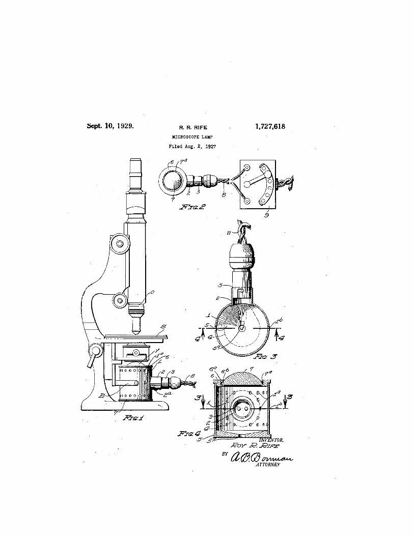
Figure 1 is a side elevational view of my microscope lamp shown inconnection with a conventional
microscope; Fig. 2 is a top or plan view of my microscope lamp shown in connection with a rheostat
means for varying the intensity of the light of the lamp;
Fig. 3 is an enlarged sectional view of my microscope lamp through 33 of Fig. 4,
with certain parts shown in plan to facilitate the illustration, and with the light bulb 60.. therein shown by
dotted lines, and Fig. 4:
is sectional elevational view thereof through 4 -4 of Fig. 3 with the light bulb therein shown by dotted lines.
Similar characters 1 of reference refer to similar parts and portions throughout the several Views of the
drawings.
The lamp housing 1, lamp socket support 2, lamp socket 3, incandescent lamp 4:, reflector 5, lens
support 6, lens 7, cord 8, and the rheostat 9, constitute the principal parts and portions of my
microscopelamp;
My lamp is positioned below the stage S and at the side thereof opposite the objective 0, and is mounted
in its preferred form, on the conventional mirror yoke B of the ,microscope, in place of the usual mirror as
will be described later.- I
The housing 1, is cylindrical, is open at its ends and is provided with a plurality of per-, forations 1 in the
walls thereof. Extending from an opening in the side wall of the housing 1, is a lamp socket support 2.- Its
inner end is flanged and is soldered or otherwisesecured to the housing 1. The support 2, is provided with
a clip means 2 for friction ally engaging the lamp socket 3 which is positioned therein. The lamp socket 3,
is similar to the conventional automobile lamp socket and may be adjustably positioned in the lamp
socket support 2, An incandescent lamp 4 is removably secured in the lamp socket 3. Positioned over the
lower open end of the housing 4. is a reflector 5, which is preferably metallic and which is provided with a
reflecting surface on its upper. side. An opening 5 is provided in the reflector 5, which is centered therein
and which, with the perforations 1 in the, housing 1, permits thoroughcirculation of air around the lamp 9O
4.. Thehole5, also permits thelight emitting, portion ofthe lamp 4 to be more easily centered on the axial
line of the microscope lamp.- Positioned over the upper open end of the housing 1, is "a lens support ,6,-
which is provided with a large central openingfi therein. The loweredge of the opening 6, has i an inwardly
extending flange 6", on which rests the lens 7. The lens 7 is plane; A
convex and is frostedon its plane and inner side. The lens 7, is held in position by means of plastic
material 7 The cord 8, which! furnishes electricity to the incandescent lamp 4, is connected with the
rheostat 9, which varies the strength of current and thereby regulates the intensity of the light of the lamp
4. As shown in Fig. 1 of the drawings the microscope lampv is mounted under the stage of the
microscope in place of the microscope mirror. For thispurpose, the housing 1, is provided with two
oppositely disposed openings in the side walls thereof in which extend j ec-tions of the mirror yoke B.
It is obvious from the construction as illustrated in the drawings and included in the foregoing
specification, that there is provided a microscope lamp as aimed at and set forth in the objects of my
invention, andalthough I have shown and described a particular construction, combination and
arrangement of parts and portions, I do not wish to be limited to this particular construction, combination
and arrangement, but
pro-
desire to include in the scope of my invention the construction, combination and arrangement.
substantially as set forth in the appended claimsp I Having thus described my invention what I claim as
new and desire to secure y etters Patent is: i
1. In a microscope lamp of the class described, thecombination with a microscope having a stationary
base and a pivotal ob-. ective, of a cylindrical'housing pivotally mounted between said base and said
objective in connection with and in alignment with said objective and movable therewith, a lens positioned
over the one end of said housing, a reflector'positioned over the other end of said housing, andan
incandescent lamp extending into said housing from the side thereof at a right angle to its axis, between
said lens and said reflector.
2 In a microscope lamp of the class described, the combination with a microscope hav ng a stationary
base and a pivotal obect1ve,of a'cylindrical housing pivotally mounted between said base and said
objectlve m connection with and in alignment with sald objective and movable therewith, a lens posltioned
over the one end of said housmg, a reflector positioned over the other end of said housing, an
incandescent lamp extending into said housing from the side thereof at a right angle to its normally
verticalaxis, and means to facilitate the positioning of the light emitting portion of said lamp on the
axialline of said housing.
3. In a means of the class described, the combination with a microscope having a stage and an objective
at one side thereof, of a support carried by and shifta-ble with said stage, and a lamp mounted on said
supat the opposite side of said stage, of a lamp,
mounted on said support and directed toward said stage.
, In testimony whereof, I have hereunto set my hand at San Diego, California, this 16th day of July, 1927.
ROY R. RIFE.
REFERENCED BY
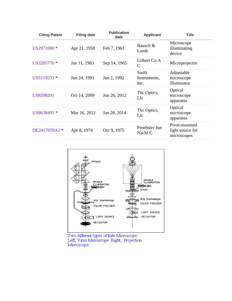
Citing Patent Filing date Publication
date Applicant Title
US2971080 * Apr 21, 1958 Feb 7, 1961 Bausch &
Lomb
Microscope
illuminating
device
US3205770 * Jan 11, 1963 Sep 14, 1965 Gilbert Co A
C Microprojector
US5119233 * Jun 24, 1991 Jun 2, 1992
Swift
Instruments,
Inc.
Adjustable
microscope
illuminator
US8208201 Oct 14, 2009 Jun 26, 2012 Tbc Optics,
Llc
Optical
microscope
apparatus
US8638495 * Mar 16, 2012 Jan 28, 2014 Tbc Optics,
Llc
Optical
microscope
apparatus
DE2417059A1 * Apr 8, 1974 Oct 9, 1975 Proebster Jun
Nachf C
Pivot-mounted
light source for
microscopes