non amplified nucleic acid probes signal ... - · pdf fileanalysis non-amplified nucleic acid...
TRANSCRIPT
MOLECULAR DIAGNOSTIC TECHNIQUES
◄
NON-AMPLIFIED NUCLEIC ACID PROBES ................................................................................................. 2 SIGNAL AMPLIFICATION TECHNIQUES ................................................................................................... 2
bDNA ................................................................................................................................................... 2 Hybrid Capture Assays ......................................................................................................................... 3
TARGET AMPLIFICATION TECHNIQUES.................................................................................................. 4 PCR ...................................................................................................................................................... 4 RT-PCR ................................................................................................................................................ 6 Nested PCR .......................................................................................................................................... 7 Multiplex PCR ...................................................................................................................................... 7 Competitive PCR .................................................................................................................................. 7 Transcription amplification Methods (NASBA and TMA) .................................................................. 7 Strand Displacement Amplification ................................................................................................... 10
PROBE AMPLIFCATION TECHNIQUES ................................................................................................... 12 Ligase chain reaction .......................................................................................................................... 12 Cleavase-Invader Assays .................................................................................................................... 13 Cycling probe technology ................................................................................................................... 14
AUTOMATION ......................................................................................................................................... 15 PRODUCT DETECTION AND ANALYSIS ................................................................................................... 16
Gel Analysis ....................................................................................................................................... 16 Probing on a gel .................................................................................................................................. 16 Single-strand conformation polymorphism (SSCP) ........................................................................... 16 Restriction fragment length polymorphism (RFLP) ........................................................................... 17 Colorimetic Microtitre Plate Systems (CMP) ..................................................................................... 17 Slot, dot and spot blot assays .............................................................................................................. 17 Reverse blot assays ............................................................................................................................. 17 Hybridisation protection assays .......................................................................................................... 17 Real-time PCR .................................................................................................................................... 18
Fluorescent dye ............................................................................................................................... 18 Melting curve analysis .................................................................................................................... 18 Taqman assay or 5’ nuclease PCR assay ........................................................................................ 19 Fluorescent real time energy transfer (FRET) ................................................................................ 20 Molecular beacons .......................................................................................................................... 21 Quantitation by real-time PCR ....................................................................................................... 21
Direct sequencing ............................................................................................................................... 22 DNA microarrays ............................................................................................................................... 23
See article
Some terms:
Annealing = process of binding two complementary strands of DNA
Amplicon = A PCR product that is a copy synthesised from the original DNA or RNA target
region
Avidin = a protein found in egg white with high affinity for biotin
Biotinylated primer = an oligonucleotide that has a biotin molecule attached at the 5’ end –
this is used to label amplified DNA via avidin
cDNA = complimentary DNA, the product of reverse transcription of RNA
Hybridisation = the process of forming a double stranded DNA molecule e.g. between probe
and target sequence
Probe = a small piece of DNA that is used to isolate complementary target from the reaction
mixture and are labelled with a reporter molecule, e.g. fluorophore
rTth = thermostable DNA polymerase original isolated from Thermus theophilus - optimal
activity 70-80 degrees and survives the denaturing steps of PCR. Also has reverse
transcription properties in the presence of manganese
Taq polymerase = thermostable DNA polymerase derived from Thermus aquaticus
Transcription = process of making RNA from DNA
UNG = uracil N-glycosylase – an enzyme that removes uracil residues from DNA. Cytidine
spintaeously de-methylate to form uracil and UNG recognises this as inappropriate for DNA
and removes them. This is the active ingredient in AmpErase.
Hy-probe assays = used on LightCycler. Employs FRET for detection during cycles
TaqMan = PCR assays similar Hy-probe but only uses single probe and there is no melt-curve
analysis
NON-AMPLIFIED NUCLEIC ACID PROBES Probes are segments of DNA or RNA which are labelled with a reporter molecule (radio-isotopes,
enzymes or chemiluminescent). The probes bind to complementary nucleic acid sequences with high
specificity. Can vary in size from 15-1000s of nucleotides, but usually are <50 nucelotides.
Probes can be used to detect pathogens in clinical samples, or for identification after isolation by
culture.
The format of the probe can be one of three:
Liquid phase. Most common method – hybridisation protection assay. Brand is GenProbe.
Uses a single-stranded DNA probe labelled with acridinium ester. After incubation with the
probe, the mixture is subject to alkaline hydrolysis. The probe is protected from hydrolysis if
it is bound. Upon addition of peroxides, the intact acridinium ester then emits light. Good
because don’t have to remove unbound probe. Can be completed in several hours.
Solid phase. Here the target nucleic acids are bound to nylon or nitrocellulose, and the probe
is in solution incubated with the solid phase. A washing step then occurs before adding a
substrate to detect the bound probes by radioactivity, fluorescence, luminescence or colour
development. Complex and time consuming.
In situ hybridisation. Here the nucleic acid remain in tissue or cells, fixed on a microscope
slide. Can be less sensitive than other methods.
Generally need large number of organisms for probes, due to the lack of amplification (poor analytical
sensitivity).
Most popular application is for culture confirmation for mycobacteria and dimorphic fungi.
SIGNAL AMPLIFICATION TECHNIQUES Here the probe or target does not increase in concentration, but the number of labelled molecules
attached to the target is increased. Generally the analytical sensitivity is lower than target amplification,
but this is improving, and some (bDNA) are close to those for target amplification.
Advantages
Allows quantitation, since signal is directly proportional to amount of target in the original
sample
Reduced false positives due to cross contamination
Not affected by enzyme inhibitors, simplifying extraction techniques
Larger probes are used, reducing false negatives due to sequence heterogeneity
Can measure RNA directly without needing to make cDNA
bDNA
bDNA is a solid phase sandwich hybridisation assay.
Here the branched DNA molecule contains 15 identical branches, each of which can bind three labelled
probes.
Here the target nucleic acid is captured onto the solid phase by a combination of capture probes
attached to the microwell plate which bind to target-specific capture probes in the solution, which in
turn bind to the target nucleic acid.

A second set of target-specific probes join the target nucleic acid to ‘pre-amplifier’ molecules: each of
these ‘pre-amplifier’ molecules can bind up to eight bDNA molecules. Each bDNA molecule has 15
branches, each of which then can bind up to 3 enzyme-labelled probes.
The enzyme is ALP, and the substrate is dioxetane which emits light, measured in a luminometer.
Third generation bDNA assays use iso-C and iso-G in the pre-amplifier and labelled probes. These
bind with each other, but not naturally-occuring bases. This reduces non-specific background signal
due to non-specific binding, and as a result improves the detection limit. Limit is as low as 50
copies/ml for HIV. Also used for quantitation of HCV and HBV. Commercially available from Bayer,
and automation has been developed for incubation, washing, reading and data processing.
Picture in Murray p236 is better.
Hybrid Capture Assays
In hybrid capture the target DNA is hybridised with an RNA probe. The hybids are then captured on
the solid phase by antihybrid antibodies. In turn, multiple ALP-conjugated antihybrid antibodies bind
to each of the immobilised hybrids, and a chemiluminescent substrate is added, and the signal
measured in a luminometer. The intensity of the emitted light is proportional to the amount of target
DNA in the specimen. Has been developed for HBV, HPV and CMV (Digene). Also developed for
HIV, C. trachomatis and N. gonorrhoeae.

TARGET AMPLIFICATION TECHNIQUES Here copies of the target nucleic acid are synthesised using two oligonucleotide primers that bind to
either end of the target sequence, one on the + strand, the other on the – strand. Each synthesised target
sequence acts as a template for further amplification. Thus, target amplifaction techniques are prone to
contamination with product molecules,, leading to false positives.
Contamination can be reduced through special laboratory design, practices and work flow.
PCR
PCR basically consists of target DNA, two oligonucleotide primers, heat stable DNA polymerase, an
equimolar mixture of deoxyribonucleotide triphosphates (dNTPs; dATP, dCTP, dGTP and dTTP),
MgCl2, KCl and a Tris-HCl buffer.
The length of the target is generally <100 to several hundred bases.
The PCR cycles consist of a pre-heating step to dissociate the two strands of DNA, then cooling to
permit annealing of the primers to the target. During the subsequent extension phase, DNA polymerase

then extends the primers towards each other. These primer extension products are then dissociated from
the target by heating, and can then form as template for subsequent rounds of primer annealing and
extension.
Each round theoretically produces a doubling in template. So after n cycles, the target has been
amplified 2n fold. Thus 20 cycles produces just over 10
6 copies, while 30 produces just over 10
9. In
practice non-ideal reaction conditions and inhibition means that amplification is not 100% efficient. To
take this into account, the formula for amplification can be expressed as (1+e)n where e is the
efficiency (0 ≤ e ≤ 1).
The process is carried out in a programmable thermal cycler that controls the temperature and the
length of time in each step.

RT-PCR
Reverse transcriptase PCR is used for amplification of RNA targets. As it was first described, heat-
labile reverse transcriptase was used in the first step, to produce cDNA (complementary DNA) from
the RNA target. Then the cDNA is amplified using thermostable DNA polymerase. However, since the
RT was thermolabile, the dissociation steps had to be carried out at a lower temperature than traditional
PCR, thus making the amplification inefficient and prone to false positives due to non-specific
annealing. However, thermostable DNA polymerase from Thermus thermophilus was subsequently
discovered: it can function efficiently as both RT and DNA polymerase. Using this enzyme makes the
assay more specific and efficient. Commerical kits using this enzyme from Roche include those for
HCV and HIV quantitation. (? RNaseH removes the orginial RNA template from the hybrid product
produced by the RT-PCR step ? – see sheet) See CMN review
Nested PCR
Nested PCR increases both sensitivity and specificity. Here after standard amplification for, typicially,
15-30 cycles, the products are subjected to further cycles of amplification using a second set of primers
that anneal to targets internal to the first set of primers. Increased sensitivity due to high copy numbers,
and increased specificity due to annealing of the second set of primers to sequences found only in the
first round products.
Disadvantages is high rate of contamination that occur when transferring the first round products to the
tube for the second round. Can avoid by sepearating using wax, or by using single-tube protocols.
Realistically, though don’t usually need nested PCR to achieve high sensivity and specificity.
Multiplex PCR
Here the one reaction mixture has multiple primer pairs targeting different sequences. Need to carefully
select the primers such that annealing temperatures are similar, and that the primers lack
complemetarity. More complicated. Generally, sensitivity diminishes with each extra primer pair that is
added. See article.
Competitive PCR
Although there is a linear relationship between the quantity of input template and the amount of
product in PCR, the actual final yield may vary unpredictably due to variations in efficiency. This may
vary due to sample preparation, inhibitors, purification procedures and thermal cycler performance.
Thus simple quantitation of the product and use of external standard reference curves are not reliable.
This has been developed to accurately quantitate DNA or RNA in a sample. It is felt to be the most
robust quantitation method. Here as well as template from the clinical sample (target template), a
second template (competitor template) of known concentration, with the same primer binding
sequences, but different intervening sequence (but of similar size and base composition), is added to
the mixture. The subsequent efficiency for amplification of both the target template and the competitor
template are the same, since they are occurring in the same tube with the same primers. The yield of the
PCR is represented by the formula:
Y = I (1 + e)n
Where Y is the quantity of the PCR product, I is the quantity of the template at the beginning of the
reaction, e is the efficiency of the reaction and n is the number of cycles. Since Y and I for the
competitor template (Yc and Ic) and Y for the target template (Yt) is known, the yield used to determine I
for the target template (It) can be calculated:
c
ctt
Y
IYI
Competitive PCR can be done for both standard PCR and RT-PCR. It is the method used for
quantitative assays for CMV, HIV-1 and HCV by Roche.
Transcription amplification Methods (NASBA and TMA)
These are both isothermal RNA amplification methods modelled after retroviral replication. In both
methods, cDNA is made from the RNA template using reverse transcriptase, but then RNA copies are
synthesised from the cDNA using RNA polymerase.
NASBA (nucleic-acid sequence-based amplification) uses avian myeloblastosis virus RT, RNase H and
T7 bacteriophage RNA polymerase.
TMA (transcription-mediated amplification) uses an RT enzyme with endogenous RNase H activity
and T7 bacteriophage RNA polymerase.
When cDNA is produced from template, the DNA primer used has sequence complementary to the
target only at the 3’ end. At the 5’ end there is the T7 RNA polymerase promoter sequence.
In the first step, reverse transcriptase extends this DNA primer in the 3’ direction, transcribing the
RNA sequence to cDNA.
In the next step, the original RNA sequence from the sample that is now hybridised to cDNA is
degraded by RNase H. This is necessary to allow the second primer to bind to the cDNA.
Next the second primer (an primer) binds to the cDNA, and is extended by DNA polymerase activity of
the reverse transcriptase. This extension continues to where the original primer is, and transcribes the
T7 RNA polymerase promoter sequence also. Thus dsDNA has been produced, with the RNA
promoter at one end.
In the next step, RNA polymerase recognises this promoter sequence and uses the + strand of the
dsDNA to make multiple copies of ss – sense RNA.
Next thse ss – sense RNA sequences are copied to cDNA by reverse transcriptase, then degraded by
RNase H and the cDNA is turned into dsDNA, and thus the cycles continue.

See picture in Murray p238.
For TMA, the single-stranded RNA products are detected by a type of hybridisation protection assay
(TMA is marketed by GenProbe who market the hybridisation protection probes). In TMA, the
aciridium esters can have either fast or slow chemiluminescent kinetics, so that can analyse signals
from two hybridisation reactions simultaneously.
For NASBA (marketed by bioMérieux) detection is with probes labelled with tris(2,2’
bispyridine)ruthenium which are detected by electrochemiluminesence. NASBA has also been used
with molecular beacon technology. Quantitation can be achieved using competitor RNA molecules
(three of different concentrations are used in the NASBA HIV-1 kit).
Strengths of transcription based amplification systems include:
No requirement for thermal cycler

Rapid kinetics
Single stranded product that does not require denaturation prior to detection
Contamination risk lower due to single tube assays and a labile RNA product
Limitations of transcription based amplification include:
Poor performance with DNA targets
Complex multienzyme systems that may be less stable.
TMA has been used for Mycobacterium tuberculosis, C. trachomatis, N. gonorrhoeae, HCV and HIV-
1.
NASBA has been developed for HIV-1 and CMV detection and quantitation. Kits are also available for
in-house uses.
Strand Displacement Amplification
This is another isothermal (52.5ºC) technique. Marketed as ProbeTec ET by Becton Dickinson, and
used for M. tuberculosis, C. trachomatis and N. gonorrhoeae as well as HIV quantitation. Here
amplification is performed by SDA, and detection is real-time using fluorescence resonance energy
transfer (FRET).
There are two phases in SDA: target generation and exponential target amplification. The process
involves both strands of dsDNA simultaneously. dUTP, dATP and dGTP are used, but the CTP is
thioated.
In the target generation phase, the dsDNA target is denatured, and each strand hybridises to two
primers, B1 (bumper primer) and S1 (contains a restriction site for BsoB1) for the + sense strand, and B2
and S2 for the – sense strand. Subsequent extension of both primers by DNA polymerase occurs, and
the extension of B1 thus displaces S1, while B2 displaces S2. The displacement of S1 and S2 means that
the opposite strand primers (B2-S2 and B1-S1 respectively) are free to bind to them, and further action of
DNA polymerase yields dsDNA, one side of which has thiodCTP instead of dCTP.
In the exponential target amplification phase, the BsoB1 restriction enzyme recognises the sequence in
the S1 site and S2 site and nicks them, but only on the strands that have normal dCTP, not thiodCTP.
Once nicked, DNA polymerase extends from the nick downstream, displacing the downstream side of
the nick, thus recreating the double stranded species, one side of which has normal dCTP and the other
side of which has thiodCTP, and thus is protected from nicking.

Detection
This involves fluorescent real time energy transfer (FRET). The single stranded products that are
displaced in the exponential phase can bind to detector probes for real-time detection. The detector
probes are single stranded DNA molecules containing fluorescein and rhodamine labels. The region
between the two labels forms a stem-loop structure. The loop contains the recognition sequence for the
BsoB1 enzyme. One side of the stem contains the fluorescein, and the other side contains the
rhodamine and the proximate location of the rhodamine quenches the fluorescein.
The probe binds to the target sequence is extended by DNA polymerase but at the same time is
displaced by the amplification primer. This then causes it to bind to the opposite strand and is extended
in the other direction, which leads to linearization of the loop of the probe. The BsoB1 site on the probe
is then made of normal dCTP (not thiodCTP) and it is thus cleaved by the restriction endonuclease,

separating the two ends of the probe and removing the quenching effect of the rhodamine producing
fluorescence.
This detection method differs from the Taqman method (5’ nuclease chemistry) in that restriction
endonuclease rather than exonuclease activity of DNA polymerase are used to separate the quencher
and fluorophore, and that the process is isothermal.
SDA has a reported sensitivity of as few as 10-50 copies. If you use it to detect a multicopy gene, you
can theoretically detect one organism.
Can be adapted to quantitation by adding an RT step (RT-SDA).
Advantage of SDA:
Isothermal (after initial target denaturation) so don’t need expensive thermal cyclers
Can be done in a single tube.
Disadvantages of SDA:
Low temperatures can result in non-specific primer hybdridisation thus amplyifying non-
target sequence and decreasing the sensitivity of the technique. Can be partially resolved by
using organic solvents that increase the stringency of primer binding or by using newer more
thermostable polymerases capable of strand displacement.
PROBE AMPLIFCATION TECHNIQUES Here the amplification products contain sequence from the initial probes, not from the target.
Ligase chain reaction
Here two sets of probes against targets that are located close to one another (but with as small gap
between them) are annealed to the target DNA, and the gap or nick is joined by DNA ligase. Only
bound probes can be ligated. These ligated probes can then serve as template for more pairs of probes
to be ligated.
It is the ligated probes which are detected, since either end has a different ‘hapten’ – a capture hapten
and detection hapten. During detection, the capture hapten is recognised by antibodies on a solid phase
(or microparticle), then a wash step occurs, and the detector haptens on the other end of the ligated
probes is recognised by a conjugated antibody. A substrate is then added to complete the detection.
This has been marketed by Abbot as LCx. Marketed for detection of C. trachomatis and N.
gonorrhoeae and M. tuberculosis (? All withdrawn?). The LCx analyser, automatically chemically
inactivates the product after detection to help reduced cross-contamination.

Cleavase-Invader Assays
Cleavase is an enzyme of the DNA polymerase family that recognises and cleaves particular DNA
structures – 5’ single-stranded flaps of a branched base-paired duplex. It recognises them without
regard to the actual sequence they contain.
In the assay, two overlapping probes are used, an ‘invader’ oligo and a ‘probe’ oligo. The positioning
of the invader oligo prevents the 5’ end of the probe oligo from annealing – only the 3’ end of the
probe oligo anneals. This structure formed by the probe oligo and the target sequence is thus a 5’
single-stranded flap of a branched base-paired duplex – recognised by cleavase which cleaves the
probe oligo, thus releaseing the 5’ end. The generation of this free 5’ end of the oligo means that target
sequence must be present (cleavase will only cleave it when it is bound as described).

A thermostable cleavase is used, and the reaction is run at a high temperature, at which there is
equilibration of primer binding, allowing exchanging of binding – in other words, once the probe oligo
is cleaved, the bound 3’ end may dissociate, and an uncleaved probe oligo can bind, and it to cleaved.
Cleavage products can be detected by various methods including FRET probes.
Avantages of Invader assays include:
Ability to detect point mutations by designing invader probes that overlap only by a single
base pair – the one of interest. This will lead to differential cleavage of the probe oligo,
depending on whether the mutation is present or absent.
The amount of target sequence is not amplified, so cross-contamination is less of a problem.
Cleavase can also be used in genotyping in cleavage fragment length polymorphisms (CFLPs)
analogous to RFLPs, since heating and rapid cooling of DNA leads to hairpin structures that act as sites
for cleavase. Has been used for HCV.
Cycling probe technology
Here a chimeric DNA-RNA-DNA probe labelled with fluoroscein at the 5’ end and biotin at the 3’ end
is used. RNase H digests the middle RNA part of the probe, but only when it is bound to the target
DNA. It is carried out at an annealing temperature, allowing the free DNA ends to then dissociate and
another chimeric probe to bind to the target.
Detection involves adding a mouse anti-fluorescein antibody-gold conjugate then running on a
nitrocellulose membrane with strepavidin on the test line and anti-mouse IgG antibody on the control
line. If the target gene is not present, the uncut probe, bound to the mouse anti-fluorescein-gold
conjugate is captured by streptavidin to form a test line. However, if the target gene is present, the
probe is cut, so only the 5’ end of the probe binds to the streptavidin on the control line, so no line is
formed. The control line mops up excess anti-fluorescein antibody-gold conjugate, and a line should
form here to display proper sample flow.
Has been developed for detection of mecA in S. aureus and VanA and VanB in VRE.
The picture below shows detection in a microtitre well, not membrane-based detection.

AUTOMATION The three main steps of target and probe amplification assays are processing, amplification and
detection. Amplification and detection are easy to automate, processing less so.
COBAS Amplicor by Roche automates amplification and detection. It has a 48 sample capacity, and
internal controls are usually used to detect inhibitors. Furthermore, UNG is used to prevent carryover

contamination (see QA). Available for TB, HIV, HCV detection and quantitation, CMV, Chalmydia,
Gonorrhoea, M. avium and M. intracellulare.
Following amplification cycles, a denaturation solution is added to denature the nucleic acid to single
stranded product. Then, magnetic particles coated with oligonucleotide probes specific for the target
sequence (and some specific for the internal control) are added. The amplicons are biotin labelled
(because the primers were biotin labelled?), and after a washing step, an avidin-HPO conjugate is
added, then after another wash, H2O2 and TMB substrate is added, and the colour change measured.
Abbot LCx automates the detection of LCR products.
Bayer bDNA system 340 platform automates incubation, washing, reading and data prcessing steps.
NASBA assays can have separate systems for automation of sample preparation and detection systems.
Manufacturers are working on fully automated systems.
PRODUCT DETECTION AND ANALYSIS Gel Analysis
Visualisation on agarose gels after electrophoresis and ethidium bromide staining was the earliest
detection method.
Probing on a gel
Specific probes can be used to determine the specificity of bands on agarose gels, after transferring
(southern blotting) them to nitrocellulose or nylon membranes. This increased both sensitivitiy and
specificity. Radionucleotide probes were detected by placing the membrane in proximity with X-ray
films, and the hybrids thus visualised as dark bands. Enzyme-labelled probes can be visualised on the
membrane as light-producing (chemiluminescent substrate) or coloured (chromogenic substrate) bands.
Alternatively, the PCR products can be labelled in the amplification stage, eg: with digoxigenin-dUTP,
and an enzyme labelled anti-digoxigenin-antibody can be used for detection.
Single-strand conformation polymorphism (SSCP)
Information about the base composition of bands on a gel can be obtained by performing single-strand
confirmation polymorphism (SSCP) or restriction fragment length polymorphism (RFLP) analysis.
In SSCP the PCR product is denatured, then subject to electrophoresis on a gel. Variations in the
physical conformation of the single-stranded fragments due to variations in the base composition will
lead to differential gel migration. Has been used to detect mutations in M. tuberculosis that lead to
rifampicin resistance.
Restriction fragment length polymorphism (RFLP)
In RFLP restriction endonucleases cleave the fragments at specific recognition sites, and the resulting
fragments are separated on a gel. The resulting banding pattern provides information about the
sequence. Can be combined with hybridisation probes.
Colorimetic Microtitre Plate Systems (CMP)
Similar to an enzyme immunoassay. Here specific oligonucleotide probes against the amplified DNA
are on the solid phase of a microtitre plate. Bound product is then detected (after washing) by an
enzyme conjugate and (after a second wash) by an appropriate substrate. The nature of the detector
conjugate can vary:
One method involves using biotinylated primers in the amplification stage. The subsequent
biotin-containing PCR product is hybridised to the capture probe on the solid phase, and
detected using a streptavidin-enzyme conjugate and chromogenic substrate.
Another method involves substituting digoxigenin-dUTP in the amplification stage for dTTP,
thus labelling the PCR product with digoxigenin, which is detected using enzyme-conjugated
antidigoxidenin antibodies.
Another method is to use enzyme-conjugated antibodies against dsDNA.
Slot, dot and spot blot assays
Here, the amplification product is transferred to a membrane for probing, but without prior gel
electrophoresis, so that no information about the size of the product is obtained. For example in dot blot
assays, the PCR product is heated to denature the DNA to single stranded, then rapidly cooled to keep
it in the single-stranded state. Next it is ‘spotted’ using a pipette (1µl) onto a nylon membrane, then
exposed to UV light which fixes the DNA to the membrane. Then it is incubated with the hybridisation
probes.
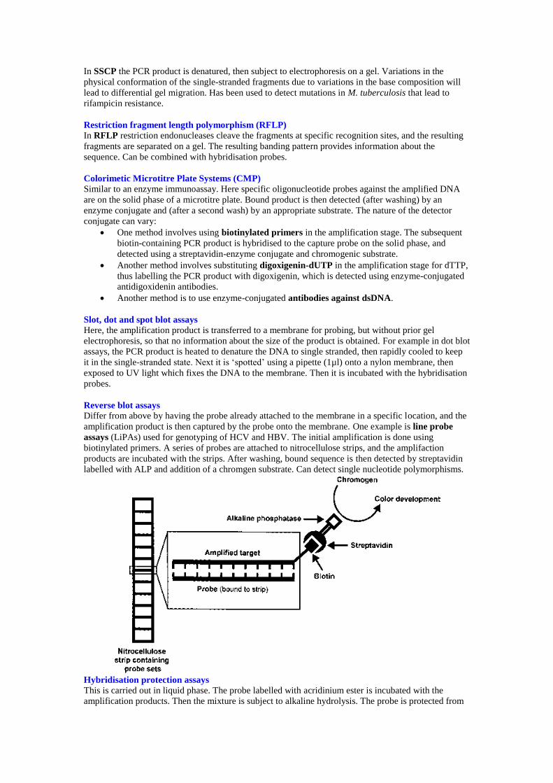
Reverse blot assays
Differ from above by having the probe already attached to the membrane in a specific location, and the
amplification product is then captured by the probe onto the membrane. One example is line probe
assays (LiPAs) used for genotyping of HCV and HBV. The initial amplification is done using
biotinylated primers. A series of probes are attached to nitrocellulose strips, and the amplifaction
products are incubated with the strips. After washing, bound sequence is then detected by streptavidin
labelled with ALP and addition of a chromgen substrate. Can detect single nucleotide polymorphisms.
Hybridisation protection assays
This is carried out in liquid phase. The probe labelled with acridinium ester is incubated with the
amplification products. Then the mixture is subject to alkaline hydrolysis. The probe is protected from

hydrolysis if it is bound. Upon addition of peroxides, the intact acridinium ester then emits light. Good
because don’t have to remove unbound probe. Same principle can be used for non-amplified probing.
Real-time PCR
Also known as homogeneous or kinetic PCR. It refers to the amplification and detection steps
occurring simultaneously in the same tube. Specialised cyclers that have precision optics that monitor
fluorescence emission from sample wells are required (light-cycler or ABI Prism).
Advantages:
Integrated system
Constant monitoring
Rapid cycling
Low contamination
Potential quantification
Fluorescent dye
In the simplest format, a fluorescent dye that binds preferentially to dsDNA is added to the mixture
(one such dye is SYBR Green I). It binds non-specifically to any dsDNA, and it emits much more
fluorescence when bound to dsDNA than when unbound. As the amount of dsDNA in the mixture
increases with subsequent cycles, the amount of fluorescence also increases. However, the dye binds to
all dsDNA including primer dimers and other non-specific dsDNA.
Melting curve analysis
This is used to increase the specificity – the specific amplified product will have a characteristic peak at
its predicted melting temperature – the temperature at which half the strands become dissociated (Tm)
whereas non-specific dsDNA will give different Tms or give broader peaks.
Melt curves are affected by:
Sequence variation
Sequence length
C+G content
Strand complimentarity
High-resolution melt curve analysis (HRMC) – dsDNA binding dyes, requires very precise
instrumentation but able to detect a single mutation. Useful for identifying SNPs e.g. for spa typing of
MRSA. Curves depend on initial concentration in the sample, so can be quantified.

Fluorescent hybdridisation probes can also be added to the reaction mixture to increase specificity.
They are labelled with just fluorescent dyes or with combinations of fluorescent and quencher dyes.
There are a number of different techniques:
Taqman assay or 5’ nuclease PCR assay
Taq polymerase has 5’-to-3’ exonuclease activity. Dual-labelled non-extendable probes with a reporter
at one end and a quencher at the other end are used. As amplification proceeds, the transcription
machinery encounters the probe and the exonuclease cleaves the reporter from the probe, releasing it
from the proximity of the quencher, producing an increase in fluorescence.

Fluorescent real time energy transfer (FRET)
This is another method involving real-time detection with fluorescent-labelled probes. Here two
probes are used that anneal close to each other. One is labelled with a donor dye that emits fluorescence
that is transmitted to the second, labelled with a reporter dye, that is excited from the fluorescence of
the nearby donor, and emits its own fluorescence at a longer wavelength. This fluorescence is detected.
The second probe is also phosphorylated at the 3’ terminus to prevent extension by Taq polymerase.
This method is used by the Roche LightCycler

Molecular beacons
These are another method for real-time detection and quantification. Here, hairpin shaped
oligonucleotides are labelled at one end with a fluorophore (reporter dye) and at the other end by a
quencher. The loop of the hairpin contains sequence complentary to the amplified target. In the hairpin
configuration, reporter and quencher are approximated, so no fluorescence in detectable. However,
when the probe encounters the amplified target sequence, the stem separates and it binds to the
sequence (the probe-target configuration is more stable than the hairpin configuration since the
complementary sequence is longer). As a result, the reporter and quencher are no longer approximated,
and fluorescence is detected.
Quantitation by real-time PCR
Quantitation in real-time PCR is done by observing at which cycle number the amount of fluorescence
passes a given threshold (CT). The CT for a set of standards is determined for the assay: the graph of the

log of the initial concentration versus the CT for a set of standards forms a straight line – and this can be
used to determine the initial concentration in an unknown sample. The standards can be internal or
external.
Direct sequencing
Here a sequencing reaction is performed on the amplified products, where as well as normal dNTPs,
fluorescent-labelled dye-terminator nucleotides (di-deoxynucleotides – ddNTPs) are included in the
reaction mix, and only one primer rather than a pair (forward or reverse). Each of the four nucleotides
is labelled with a different dye. When the translation machinery adds one of the dye-terminator
nucleotides to the growing chain of the amplified product, translation is terminated. The result is
amplified product of different lengths, each ending with one of four dye-labelled nucleotide. Ordering
the products according to length, then observing the particular dye-labelled nucleotide attached to each
product according to its length reveals the sequence of the target DNA. Traditionally the ordering has
been done on polyacrylamide gel electrophoresis (PAGE) and the reading by laser scanning. However,
capillary electrophoresis has reduced the labour-intensiveness of sequencing. A second reaction with
the opposite primer may also be done to confirm sequence integrity.

DNA microarrays
See article.
Also known as DNA chips. Here hundreds or thousands of oligonucleotides are attached to a solid
support in a precise pattern. The labelled amplification product is hybridised to different parts of the
probes, and hybridisation signals are mapped to various positions within the array. Full sequencing of
the amplificication product can actually be achieved if the number of probes is large enough.
Chips can be manufactured using similar techniques for manufacture of semiconductors where light
can be used to direct the position of synthesis of short oligonucleotides in a silica wafer so that on a
15mm chip, thousands of probes can be established.
After incubation with the labelled amplified product, a scanning laser confocal microscope
automatically detects the pattern of surface fluorescence on the chip, and it is analysed by computer
software.
Can have control spots with deliberate nucleotide mismatches to look for non-specific binding. Can
also use competitor technology (see article).
MALDI-TOF (Matrix-Assisted Laser Desorption Ionization Time of Flight Mass Spectrometry)
Based on peptide spectra derived from ‘soft’ ionized bacteria. Usually uses a laser and mass
spectrometer that compares spectra to known signatures on a database. Can provide rapid results and is
relatively inexpensive. Some discrepant results and requires at least 4 or more colonies to get results.
Can have difficulties in identifying some common organisms such as S. aureus and S. pneumoniae.
In a recent real-time trial, 95% of 1660 isolates were correctly identified at least to genus level. Can be
used directly from colonies and get results in a couple of hours and overall cost savings once initial
capital outlay.