non-contiguous finished genome sequence of staphylococcus ... · medical biotechnology lab....
TRANSCRIPT

Standards in Genomic Sciences (2014) 9:1118-1127 DOI:10.4056/sigs.5491045
The Genomic Standards Consortium
Non-contiguous finished genome sequence of Staphylococcus capitis CR01 (pulsetype NRCS-A)
H. Lemriss1,2, Martins Simões P2,3, S. Lemriss1, M. Butin3, A. Ibrahimi4, S. El Kabbaj1, JP Rasigade2,3, F. Laurent2,3,5
1Department of Biosecurity PCL3, Laboratory of Research and Medical Analysis of the Fra-ternal of Gendarmerie Royale, Rabat, Morocco.
2International Centre for Research in Infectious diseases, INSERM U1111, University of Lyon, Lyon, France.
3Department of Clinical Microbiology, Northern Hospital Group, Hospices Civils de Lyon, Lyon, France.
4Medical Biotechnology lab. (MedBiotech), Medical and Pharmacy School, University Mo-hammed V Souissi, Rabat, Morocco.
5National Reference Center for Staphylococci, Hospices Civils de Lyon, Lyon, France.
Correspondence:
Keywords:
Staphylococcus capitis (NCRS-A), draft-genome, methicillin resistance, late-onset sepsis
Staphylococcus capitis is a coagulase-negative staphylococci (CoNS) commonly found in the human microflora. Recently, a clonal population of Staphylococcus capitis (denominated NRCS-A) was found to be a major cause of late-onset sepsis (LOS) in several neonatal inten-sive care units in France. Here, we report the complete genome sequence and annotation of the prototype Staphylococcus capitis NCRS-A strain CR01. The 2,504,472 bp long genome (1 chromosome and no plasmids) exhibits a G+C content of 32.81%, and contains 2,468 pro-tein-coding and 59 tRNA genes and 4 rRNA genes.
Abbreviations: EMBL- European Molecular Biology Laboratory, NCBI- National Center for Biotechnology Information (Bethesda, MD, USA), RDP- Ribosomal Database Project (East Lansing, MI, USA)
IntroductionA frequent cause of low-weight newborns mortali-ty and morbidity in Neonatal Intensive Care Units (NICUs) are late-onset sepsis (LOS), that are de-fined as sepsis occurring after 3 days of age. The most frequently encountered pathogens are coag-ulase-negative staphylococci (CoNS) and within those Staphylococcus epidermidis has been shown to be the most prevalent [1,2]. However, a few studies have reported the emergence of Staphylo-coccus capitis as a main CoNS- and LOS- causative pathogen in NICU settings [2-4]. A study in French NICUs [2] has demonstrated the spread of a single clonal population of methicillin-resistant S. capitis (pulsotype NRCS-A) associated to reduced suscep-tibility to vancomycin, the first line of antibiotics used in cases of LOS. Moreover, this clone has also been recently identified in NICUs in Belgium, United Kingdom and Australia, which suggests a worldwide distribution. In contrast, in adult bac-
teremia, S. capitis are rarely found and when de-tected, it presents a bigger diversity in terms of genotypes as well as antimicrobial susceptibility profiles than neonates bacteremia. In order to elucidate the molecular mechanisms behind the wide spreading of the S. capitis NRCS-A clone in NICUs throughout the world, we se-quenced a prototype strain (CR01).
Classification and information A strain belonging to the clonal population of Staphylococcus capitis NCRS-A pulsetype (Table 1) was isolated from the blood culture of a preterm infant with LOS, hospitalized in the NICU of the Northern Hospital Group Center (Hospices Civils de Lyon, Lyon, France) and suffering of LOS. Species identification of the bacterial isolates and antimicrobial susceptibility testing (AST) were performed, respectively, using Vitek MS

Lemriss et al.
http://standardsingenomics.org 1119
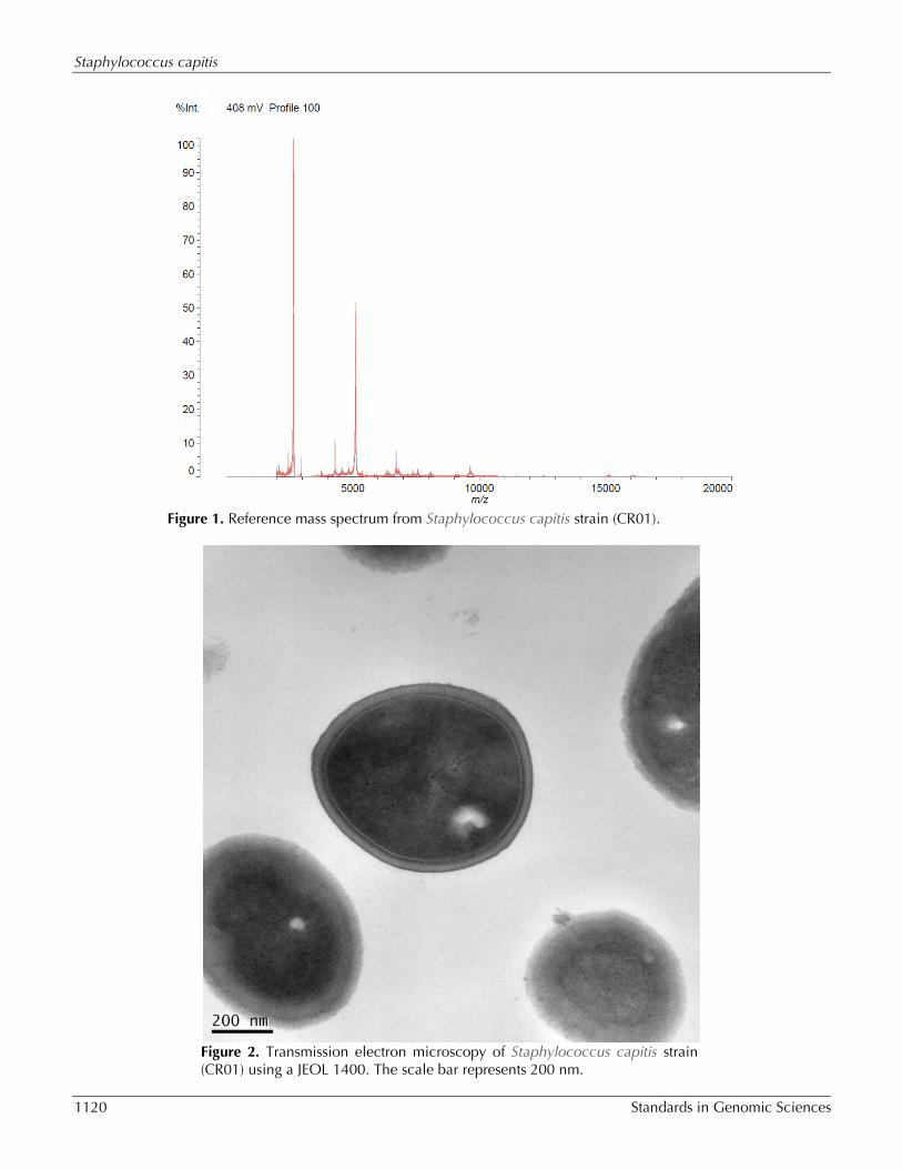
(bioMérieux, Marcy l’Etoile), 16S rDNA sequenc-ing, the automated BD Phoenix system (Becton Dickinson, Sparks, MD) and with Shimadzu-MALDI-TOF MS system (Shimadzu Corporation), as implemented on [21]. The strain was identified as being aStaphylococcus capitis by VITEK MS with 99.9% and at 93.7% by the MALDI-TOF MS, using the Shimadzu Launchpad software program and the SARAMIS database application (AnagnosTec GmbH) for au-tomatic measurement and identification (Figure 1). Based on the information provided by the manufacture, when the score is ≥70%, identifica-tion is considered of high confidence.
The antimicrobial susceptibility test (AST) results were analyzed according to the recommendations of the French Microbiology Society [22]. The S. capitis bacteremia was considered positive based on a single positive blood culture [2,23]. The S. capitis NCRS-A isolate CR01, as all isolates from this clone, is resistant to penicillin, methicillin, gentamicin, rifampicin, hetero-resistant to vancomycin and sensitive to fusidic acid and fluoroquinolones. Table 1, Figure 2 and Figure 3 show detailed in-formation concerning general features of Staphy-lococcus capitis strain (CR01) and position within the genus Staphylococcus.
Table 1. Classification and general features of Staphylococcus capitis strain CR01, pulsetype-NRCS-A ac-cording the MIGS recommendation [5].
MIGS ID Property Term Evidence codea Current classification
Domain Bacteria Phylum Firmicutes Class Bacilli Order Bacillales Family Staphylococcaceae Genus Staphylococcus Species Staphylococcus capitis Strain CR01, pulsetype-NRCS-A
TAS [6] TAS [7,8] TAS [9,10] TAS [11,12] TAS [13,14] TAS [11,15,16] TAS [17] TAS [2,18]
Gram stain Positive TAS [19] Cell shape Coccoid TAS [17] Motility Non-motile TAS [19] Sporulation Non-sporuating TAS [19] Temperature range Mesophilic IDA Optimum temperature 37°C TAS [17] Carbon source Carbohydrates, (glucose, sacharose,
fructose, manitol, mannose) TAS [17]
Energy source Chemoorganotropic TAS [17] Terminal electron recep-
tor O2 TAS [17]
MIGS-6 Habitat Skin of humans TAS [17] MIGS-6.3 Salinity Physiological TAS [17] MIGS-22 Oxygen Facultative anaerobes TAS [17] MIGS-15 Biotic relationship Free-living TAS [17] MIGS-14 Pathogenicity Opportunistic pathogen (Nosocom-
ial bacteremia in premature neo-nates)
TAS [2]
MIGS-4 Geographic location NICU Lyon, France TAS [2] MIGS-5 Sample collection time 2007 IDA MIGS-4.1 MIGS-4.2
Latitude – Longitude 45° 45' 35" N 4° 50' 32" E IDA
MIGS-4.3 Depth Not applicable IDA MIGS-4.4 Altitude 162 m IDA
a) Evidence codes - IDA: Inferred from Direct Assay; TAS: Traceable Author Statement (i.e., a direct re-port exists in the literature); NAS: Non-traceable Author Statement (i.e., not directly observed for the liv-ing, isolated sample, but based on a generally accepted property for the species, or anecdotal evidence). These evidence codes are from the Gene Ontology project [20].
Staphylococcus capitis
1120 Standards in Genomic Sciences
Figure 1. Reference mass spectrum from Staphylococcus capitis strain (CR01).
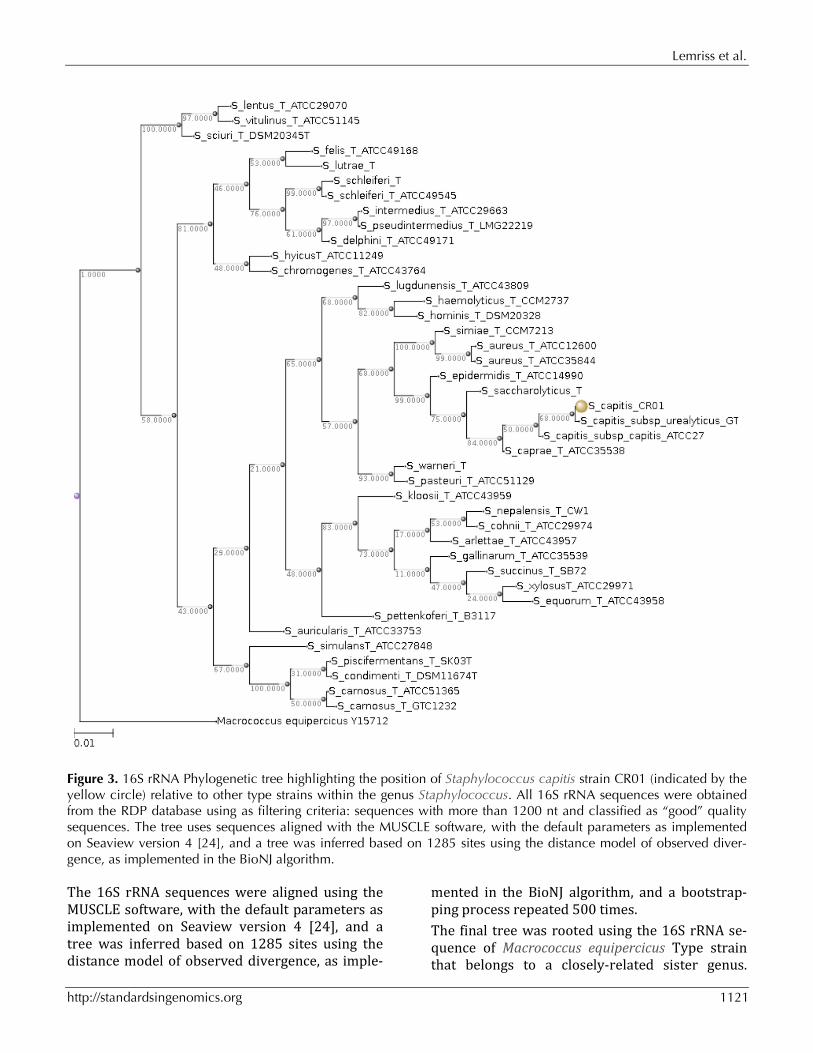
Figure 2. Transmission electron microscopy of Staphylococcus capitis strain (CR01) using a JEOL 1400. The scale bar represents 200 nm.
Lemriss et al.
http://standardsingenomics.org 1121
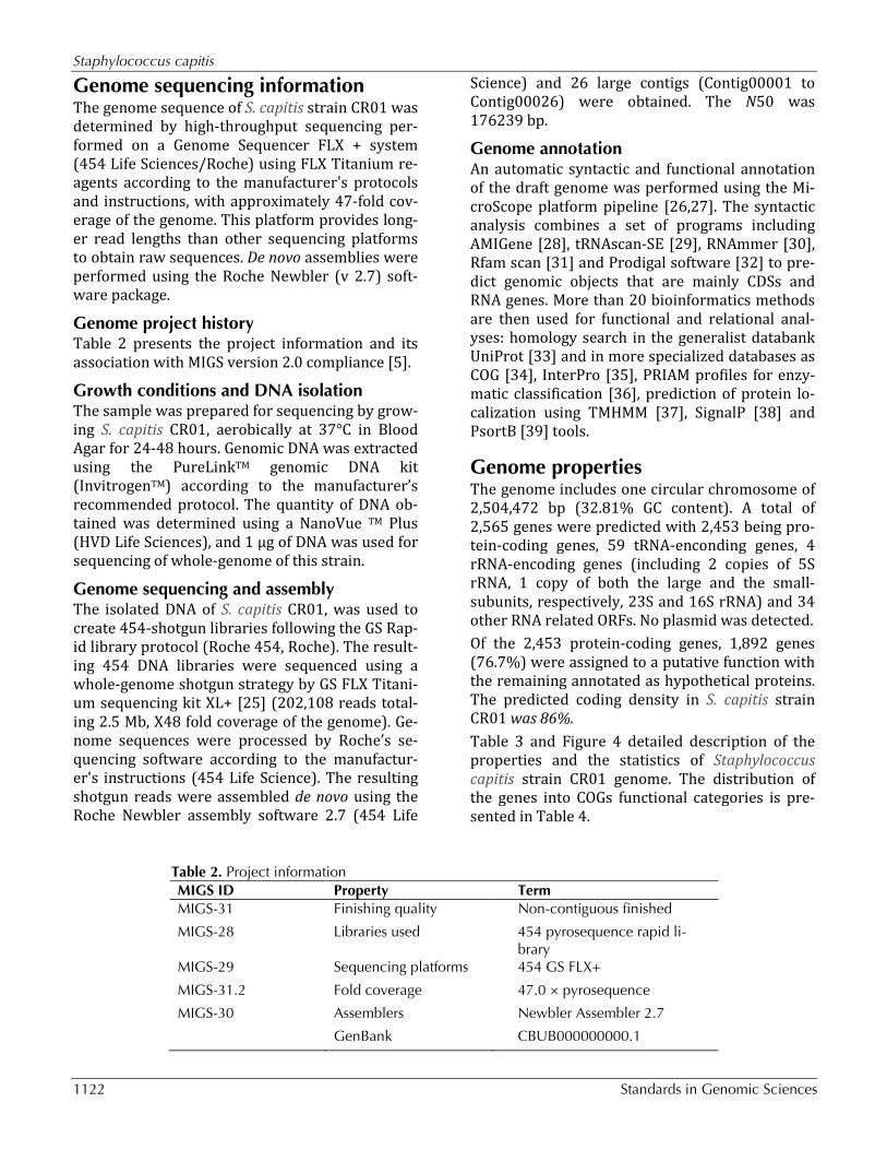
Figure 3. 16S rRNA Phylogenetic tree highlighting the position of Staphylococcus capitis strain CR01 (indicated by the yellow circle) relative to other type strains within the genus Staphylococcus. All 16S rRNA sequences were obtained from the RDP database using as filtering criteria: sequences with more than 1200 nt and classified as “good” quality sequences. The tree uses sequences aligned with the MUSCLE software, with the default parameters as implemented on Seaview version 4 [24], and a tree was inferred based on 1285 sites using the distance model of observed diver-gence, as implemented in the BioNJ algorithm.
The 16S rRNA sequences were aligned using the MUSCLE software, with the default parameters as implemented on Seaview version 4 [24], and a tree was inferred based on 1285 sites using the distance model of observed divergence, as imple-
mented in the BioNJ algorithm, and a bootstrap-ping process repeated 500 times. The final tree was rooted using the 16S rRNA se-quence of Macrococcus equipercicus Type strain that belongs to a closely-related sister genus.
Staphylococcus capitis
1122 Standards in Genomic Sciences
Genome sequencing information The genome sequence of S. capitis strain CR01 was determined by high-throughput sequencing per-formed on a Genome Sequencer FLX + system (454 Life Sciences/Roche) using FLX Titanium re-agents according to the manufacturer's protocols and instructions, with approximately 47-fold cov-erage of the genome. This platform provides long-er read lengths than other sequencing platforms to obtain raw sequences. De novo assemblies were performed using the Roche Newbler (v 2.7) soft-ware package.
Genome project history Table 2 presents the project information and its association with MIGS version 2.0 compliance [5].
Growth conditions and DNA isolation The sample was prepared for sequencing by grow-ing S. capitis CR01, aerobically at 37°C in Blood Agar for 24-48 hours. Genomic DNA was extracted using the PureLinkTM genomic DNA kit (InvitrogenTM) according to the manufacturer’s recommended protocol. The quantity of DNA ob-tained was determined using a NanoVue TM Plus (HVD Life Sciences), and 1 µg of DNA was used for sequencing of whole-genome of this strain.
Genome sequencing and assembly The isolated DNA of S. capitis CR01, was used to create 454-shotgun libraries following the GS Rap-id library protocol (Roche 454, Roche). The result-ing 454 DNA libraries were sequenced using a whole-genome shotgun strategy by GS FLX Titani-um sequencing kit XL+ [25] (202,108 reads total-ing 2.5 Mb, X48 fold coverage of the genome). Ge-nome sequences were processed by Roche’s se-quencing software according to the manufactur-er's instructions (454 Life Science). The resulting shotgun reads were assembled de novo using the Roche Newbler assembly software 2.7 (454 Life
Science) and 26 large contigs (Contig00001 to Contig00026) were obtained. The N50 was 176239 bp.
Genome annotation An automatic syntactic and functional annotation of the draft genome was performed using the Mi-croScope platform pipeline [26,27]. The syntactic analysis combines a set of programs including AMIGene [28], tRNAscan-SE [29], RNAmmer [30], Rfam scan [31] and Prodigal software [32] to pre-dict genomic objects that are mainly CDSs and RNA genes. More than 20 bioinformatics methods are then used for functional and relational anal-yses: homology search in the generalist databank UniProt [33] and in more specialized databases as COG [34], InterPro [35], PRIAM profiles for enzy-matic classification [36], prediction of protein lo-calization using TMHMM [37], SignalP [38] and PsortB [39] tools.
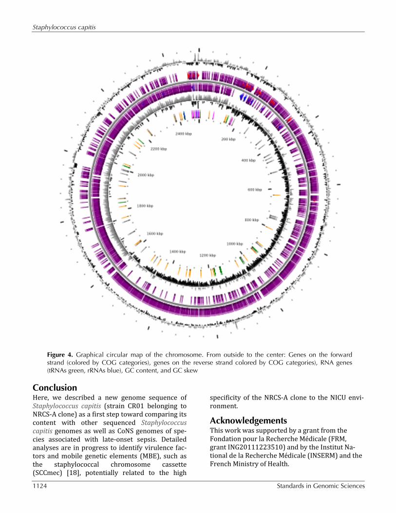
Genome properties The genome includes one circular chromosome of 2,504,472 bp (32.81% GC content). A total of 2,565 genes were predicted with 2,453 being pro-tein-coding genes, 59 tRNA-enconding genes, 4 rRNA-encoding genes (including 2 copies of 5S rRNA, 1 copy of both the large and the small-subunits, respectively, 23S and 16S rRNA) and 34 other RNA related ORFs. No plasmid was detected. Of the 2,453 protein-coding genes, 1,892 genes (76.7%) were assigned to a putative function with the remaining annotated as hypothetical proteins. The predicted coding density in S. capitis strain CR01 was 86%. Table 3 and Figure 4 detailed description of the properties and the statistics of Staphylococcus capitis strain CR01 genome. The distribution of the genes into COGs functional categories is pre-sented in Table 4.
Table 2. Project information MIGS ID Property Term MIGS-31 Finishing quality Non-contiguous finished
MIGS-28 Libraries used 454 pyrosequence rapid li-brary
MIGS-29 Sequencing platforms 454 GS FLX+
MIGS-31.2 Fold coverage 47.0 × pyrosequence
MIGS-30 Assemblers Newbler Assembler 2.7
GenBank CBUB000000000.1
Lemriss et al.
http://standardsingenomics.org 1123
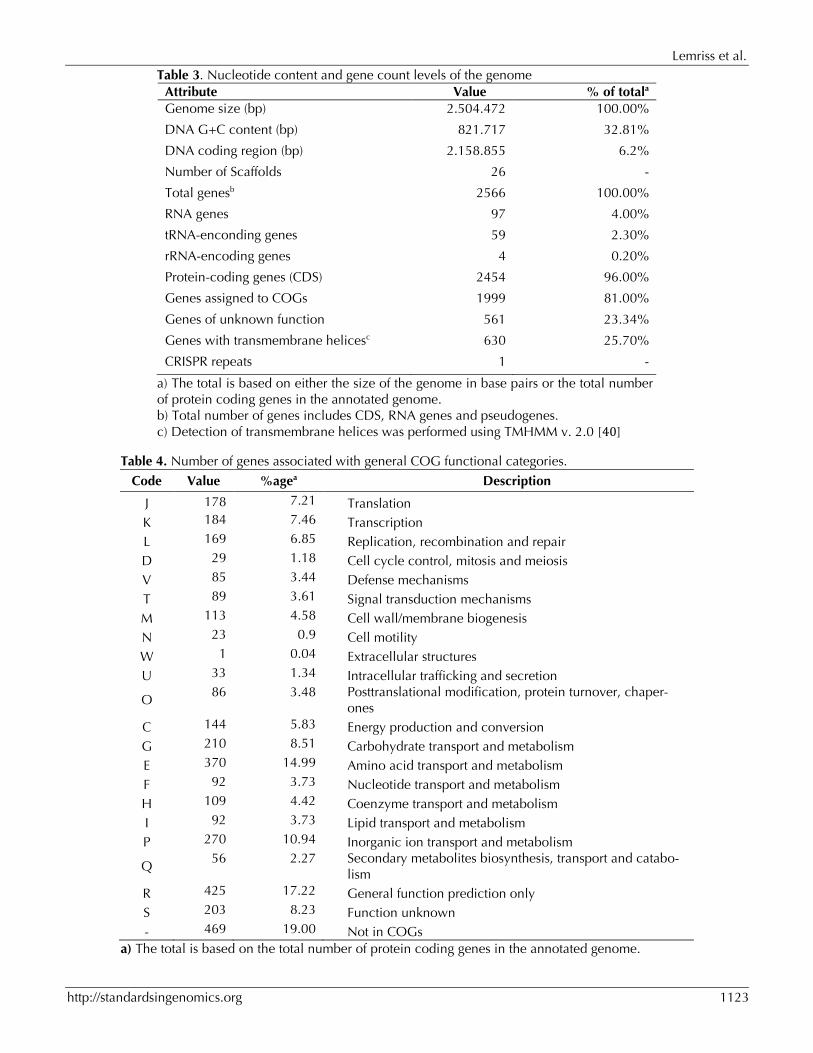
Table 3. Nucleotide content and gene count levels of the genome Attribute Value % of totala Genome size (bp) 2.504.472 100.00%
DNA G+C content (bp) 821.717 32.81%
DNA coding region (bp) 2.158.855 6.2%
Number of Scaffolds 26 -
Total genesb 2566 100.00%
RNA genes 97 4.00%
tRNA-enconding genes 59 2.30%
rRNA-encoding genes 4 0.20%
Protein-coding genes (CDS) 2454 96.00%
Genes assigned to COGs 1999 81.00%
Genes of unknown function 561 23.34%
Genes with transmembrane helicesc 630 25.70%
CRISPR repeats 1 -
a) The total is based on either the size of the genome in base pairs or the total number of protein coding genes in the annotated genome. b) Total number of genes includes CDS, RNA genes and pseudogenes. c) Detection of transmembrane helices was performed using TMHMM v. 2.0 [40]
Table 4. Number of genes associated with general COG functional categories.
Code Value %agea Description
J 178 7.21 Translation K 184 7.46 Transcription L 169 6.85 Replication, recombination and repair D 29 1.18 Cell cycle control, mitosis and meiosis V 85 3.44 Defense mechanisms T 89 3.61 Signal transduction mechanisms M 113 4.58 Cell wall/membrane biogenesis N 23 0.9 Cell motility W 1 0.04 Extracellular structures U 33 1.34 Intracellular trafficking and secretion
O 86 3.48 Posttranslational modification, protein turnover, chaper-
ones C 144 5.83 Energy production and conversion G 210 8.51 Carbohydrate transport and metabolism E 370 14.99 Amino acid transport and metabolism F 92 3.73 Nucleotide transport and metabolism H 109 4.42 Coenzyme transport and metabolism I 92 3.73 Lipid transport and metabolism P 270 10.94 Inorganic ion transport and metabolism
Q 56 2.27 Secondary metabolites biosynthesis, transport and catabo-
lism R 425 17.22 General function prediction only S 203 8.23 Function unknown - 469 19.00 Not in COGs
a) The total is based on the total number of protein coding genes in the annotated genome.
Staphylococcus capitis
1124 Standards in Genomic Sciences
Figure 4. Graphical circular map of the chromosome. From outside to the center: Genes on the forward strand (colored by COG categories), genes on the reverse strand colored by COG categories), RNA genes (tRNAs green, rRNAs blue), GC content, and GC skew
ConclusionHere, we described a new genome sequence of Staphylococcus capitis (strain CR01 belonging to NRCS-A clone) as a first step toward comparing its content with other sequenced Staphylococcus capitis genomes as well as CoNS genomes of spe-cies associated with late-onset sepsis. Detailed analyses are in progress to identify virulence fac-tors and mobile genetic elements (MBE), such as the staphylococcal chromosome cassette (SCCmec) [18], potentially related to the high
specificity of the NRCS-A clone to the NICU envi-ronment.
Acknowledgements This work was supported by a grant from the Fondation pour la Recherche Médicale (FRM, grant ING20111223510) and by the Institut Na-tional de la Recherche Médicale (INSERM) and the French Ministry of Health.
Lemriss et al.
http://standardsingenomics.org 1125
References1. Klingenberg C, Rønnestad A, Anderson AS, Abra-
hamsen TG, Zorman J, Villaruz A, Flægstad T, Ot-to M. Sollid, Ericson J. Persistent strains of coagu-lase-negative staphylococci in a neonatal inten-sive care unit: virulence factors and invasiveness. Clin Microbiol Infect 2007; 13:1100-1111. Pub-Med http://dx.doi.org/10.1111/j.1469-0691.2007.01818.x
2. Rasigade JP, Raulin O, Picaud JC, Tellini C, Bes M, Grando J, Ben Saïd M, Claris O, Etienne J, Tigaud S, Laurent F. Methicillin-ResistantStaphylococcus capitis with Reduced Vancomycin Susceptibility Causes Late-Onset Sepsis in Intensive Care Neonates. PLoS ONE 2012; 7:e31548. PubMed http://dx.doi.org/10.1371/journal.pone.0031548
3. Ng PC, Chow VC, Lee CH, Ling JM, Wong HL, Chang RCY. Persistent Staphylococcus capitis septicemia in a preterm infant. Pediatr Infect Dis J 2006; 25:652-654. PubMed http://dx.doi.org/ 10.1097/01.inf.0000225785.32137.d3
4. de Silva GDI, Kantzanou M, Justice A, Massey RC, Wilkinson AR, Day NPJ, Peacock SJ. The ica Operon and Biofilm Production in Coagulase-Negative Staphylococci Associated with Carriage and Disease in a Neonatal Intensive Care Unit. J Clin Microbiol 2002; 40:382-388. PubMed http://dx.doi.org/10.1128/JCM.40.02.382-388.2002
5. Field D, Garrity G, Gray T, Morrison N, Selengut J, Sterk P, Tatusova T, Thomson N, Allen MJ, Angiuoli et al. The minimum information about a genome sequence (MIGS) specification. Nat Biotechnol 2008; 26:541-547. PubMed http://dx.doi.org/10.1038/nbt1360
6. Woese CR, Kandler O, Wheelis ML. Towards a nat-ural system of organisms: proposal for the do-mains Archaea, Bacteria, and Eukarya. Proc Natl Acad Sci USA 1990; 87:4576-4579. PubMed http://dx.doi.org/10.1073/pnas.87.12.4576
7. Gibbons NE, Murray RGE. Proposals Concerning the Higher Taxa of Bacteria. Int J Syst Bacteriol 1978; 28:1-6; http://dx.doi.org/10.1099/00207713-28-1-1.
8. Murray RGE. The Higher Taxa, or, a Place for Everything...? In: Holt JG (ed), Bergey's Manual of Systematic Bacteriology, First Edition, Volume 1, The Williams and Wilkins Co., Baltimore, 1984, p. 31-34.
9. List of new names and new combinations previ- ously effectively, but not validly, published. List
no. 132. Int J Syst Evol Microbiol 2010; 60:469-472. http://dx.doi.org/10.1099/ijs.0.022855-0
10. Ludwig W, Schleifer KH, Whitman WB. Class I. Bacilli class nov. In: De Vos P, Garrity G, Jones D, Krieg NR, Ludwig W, Rainey FA, Schleifer KH, Whitman WB (eds), Bergey's Manual of Systemat- ic Bacteriology, Second Edition, Volume 3, Springer-Verlag, New York, 2009, p. 19-20.
11. Skerman VBD, McGowan V, Sneath PHA. Ap- proved Lists of Bacterial Names. Int J Syst Bacteriol 1980; 30:225-420. http://dx.doi.org/10.1099/00207713-30-1-225
12. Prévot AR. In: Hauderoy P, Ehringer G, Guillot G, Magrou. J., Prévot AR, Rosset D, Urbain A (eds), Dictionnaire des Bactéries Pathogènes, Second Edition, Masson et Cie, Paris, 1953, p. 1-692.
13. List Editor. List of new names and new combina- tions previously effectively, but not validly, pub- lished. List no. 132. Int J Syst Evol Microbiol 2010; 60:469-472. http://dx.doi.org/10.1099/ijs.0.022855-0
14. Schleifer KH, Bell JA. Family VIII.Staphylococcaceae fam. nov. In: De Vos P, Garrity G, Jones D, Krieg NR, Ludwig W, Rainey FA, Schleifer KH, Whitman WB (eds), Bergey's Manual of Systematic Bacteriology, Second Edi- tion, Volume 3, Springer-Verlag, New York, 2009, p. 392.
15. Rosenbach FJ. In: Bergmann JF (ed), Microorganismen bei den Wund-Infections- Krankheiten des Menschen., Wiesbaden, 1884, p. 1-122.
16. Judicial Commission. Opinion 17. Conservation of the Generic name Staphylococcus Rosenbach, Designation of Staphylococcus aureus Rosenbach as the Nomenclatural Type of the Genus Staphy- lococcus Rosenbach, and Designation of the Neotype culture of Staphylococcus aureus Rosenbach. Int Bull Bacteriol Nomencl Taxon 1958; 8:153-154.
17. Kloos WE, Musselwhite MS. Distribution and Per-sistence of Staphylococcus and Micrococcus Spe-cies and Other Aerobic Bacteria on Human Skin. Appl Microbiol 1975; 30:381-385. PubMed
18. Martins Simões P, Rasigade JP, Lemriss H, Butin M, Ginevra C, Lemriss S, Goering RV, Ibrahimi A, Picaud JC, Vandenesch FEL, et al. Characteriza-tion of a novel staphylococcal chromosome cas-sette (SCCmec) within a composite SCC island in neonatal sepsis-associated Staphylococcus capitis pulsotype NRCS-A. Antimicrob Agents
Staphylococcus capitis
1126 Standards in Genomic Sciences
Chemother 2013; 57:6354. PubMed http://dx.doi.org/10.1128/AAC.01576-13
19. Schleifer KH, Bell JA. Family VIII.Staphylococcaceae fam. nov. In: De Vos P, Garrity G, Jones D, Krieg NR, Ludwig W, Rainey FA, Schleifer KH, Whitman WB (eds), Bergey's Manual of Systematic Bacteriology, Second Edi- tion, Volume 3, Springer-Verlag, New York, 2009, p. 392.
20. Ashburner M, Ball CA, Blake JA, Botstein D, But-ler H, Cherry JM, Davis AP, Dolinski K, Dwight SS, Eppig JT, et al. Gene ontology: tool for the unification of biology. The Gene Ontology Con-sortium. Nat Genet 2000; 25:25-29. PubMed http://dx.doi.org/10.1038/75556
21. Cherkaoui A, Hibbs J, Emonet S, Tangomo M, Girard M, Francois P, Schrenzel J. Comparison of two matrix-assisted laser desorption ionization-time of flight mass spectrometry methods with conventional phenotypic identification for routine identification of bacteria to the species level. J Clin Microbiol 2010; 48:1169-1175. PubMed http://dx.doi.org/10.1128/JCM.01881-09
22. French Society for Microbiology. Recommandations du Comite de l'Antibiogramme de la Société Française de Microbiologie. 2009. Available at: wwwsfmasofr/doc/downloadphp?doc=DiU8C&fic =casfm_2009pdf. Accessed 22 February 2009.
23. Hall KK, Lyman JA. Updated review of blood cul-ture contamination. Clin Microbiol Rev 2006; 19:788-802. PubMed http://dx.doi.org/10.1128/CMR.00062-05
24. Gouy M, Guindon S, Gascuel O. SeaView Ver-sion 4: A Multiplatform Graphical User Interface for Sequence Alignment and Phylogenetic Tree Building. Mol Biol Evol 2010; 27:221-224. Pub-Med http://dx.doi.org/10.1093/molbev/msp259
25. Fleischmann RD, Adams MD, White O, Clayton RA, Kirkness EF, Kerlavage AR, Bult CJ, Tomb JF, Dougherty BA, Merrick JM, et al. Whole-genome random sequencing and assembly ofHaemophilus influenzae Rd. Science 1995; 269:496-512. Pub-Med http://dx.doi.org/10.1126/science.7542800
26. Vallenet D, Belda E, Calteau A, Cruveiller S, Engelen S, Lajus A, Le Fèvre F, Longin C, Mornico D, Roche D, et al. MicroScope—an integrated microbial resource for the curation and compara-tive analysis of genomic and metabolic data. Nu-cleic Acids Res 2013; 41:D636-D647. PubMed http://dx.doi.org/10.1093/nar/gks1194
27. Vallenet D, Labarre L, Rouy Z, Barbe V, Bocs S, Cruveiller S, Lajus A, Pascal G, Scarpelli C, Médigue C. MaGe: a microbial genome annota-tion system supported by synteny results. Nucleic Acids Res 2006; 34:53-65. PubMed http://dx.doi.org/10.1093/nar/gkj406
28. Bocs S, Cruveiller S, Vallenet D, Nuel G, Médigue C. AMIGene: Annotation of MIcrobial Genes. Nucleic Acids Res 2003; 31:3723-3726. PubMed http://dx.doi.org/10.1093/nar/gkg590
29. Lowe TM, Eddy SR. tRNAscan-SE: a program for improved detection of transfer RNA genes in ge-nomic sequence. Nucleic Acids Res 1997; 25:955-964. PubMed http://dx.doi.org/10.1093/nar/25.5.0955
30. Lagesen K, Hallin P, Rødland EA, Staerfeldt HH, Rognes T, Ussery DW. RNAmmer: consistent and rapid annotation of ribosomal RNA genes. Nucle-ic Acids Res 2007; 35:3100-3108. PubMed http://dx.doi.org/10.1093/nar/gkm160
31. Gardner PP, Daub J, Tate JG, Nawrocki EP, Kolbe DL, Lindgreen S, Wilkinson AC, Finn RD, Grif-fiths-Jones S, Eddy SR, Bateman A. Rfam: updates to the RNA families database. Nucleic Acids Res 2009; 37:D136-D140. PubMed http://dx.doi.org/10.1093/nar/gkn766
32. Hyatt D, Chen GL, Locascio PF, Land ML, Lar-imer FW, Hauser LJ. Prodigal: prokaryotic gene recognition and translation initiation site identifi-cation. BMC Bioinformatics 2010; 11:119. Pub-Med http://dx.doi.org/10.1186/1471-2105-11-119
33. UnitProt Consortium. The Universal Protein Re-source (UniProt) 2009. Nucleic Acids Res 2009; 37:D169-D174. PubMed http://dx.doi.org/10.1093/nar/gkn664
34. Tatusov RL, Fedorova ND, Jackson JD, Jacobs AR, Kiryutin B, Koonin EV, Krylov DM, Mazumder R, Mekhedov SL, Nikolskaya AN, et al. The COG database: an updated version includes eukary-otes. BMC Bioinformatics 2003; 4:41. PubMed http://dx.doi.org/10.1186/1471-2105-4-41
35. Hunter S, Apweiler R, Attwood TK, Bairoch A, Bateman A, Binns D, Bork P, Das U, Daugherty L, Duquenne L, et al. InterPro: the integrative pro-tein signature database. Nucleic Acids Res 2009; 37:D211-D215. PubMed http://dx.doi.org/10.1093/nar/gkn785
36. Claudel-Renard C, Chevalet C, Faraut T, Kahn D. Enzyme-specific profiles for genome annotation: PRIAM. Nucleic Acids Res 2003; 31:6633-6639. PubMed http://dx.doi.org/10.1093/nar/gkg847
Lemriss et al.
http://standardsingenomics.org 1127
37. Sonnhammer EL, von Heijne G, Krogh A. A hid-den Markov model for predicting transmembrane helices in protein sequences. Proc Int Conf Intell Syst Mol Biol 1998; 6:175-182. PubMed
38. Bendtsen JD, Nielsen H, von Heijne G, Brunak S. Improved prediction of signal peptides: SignalP 3.0. J Mol Biol 2004; 340:783-795. PubMed http://dx.doi.org/10.1016/j.jmb.2004.05.028
39. Gardy JL, Laird MR, Chen F, Rey S, Walsh CJ, Es-ter M, Brinkman FS. PSORTb v.2.0: expanded prediction of bacterial protein subcellular locali-zation and insights gained from comparative pro-
teome analysis. Bioinformatics 2005; 21:617-623. PubMed http://dx.doi.org/10.1093/bioinformatics/bti057
40. Moller S, Croning MDR, Apweiler R. Evaluation of methods for the prediction of membrane span-ning regions. Bioinformatics 2001; 17:646-653. PubMed http://dx.doi.org/10.1093/bioinformatics/17.7.646