nonhealing cellulitis in a 54-year-old man with diabetes
TRANSCRIPT

I M BOARD REVIEW DAVID L. LONGWORTH, MD, JAMES K. STOLLER, MD, EDITORS
C R E D I !
STEVEN D. MAWHORTER, M D D e p a r t m e n t o f I n f e c t i o u s D isease , C l e v e l a n d C l i n i c ; r e s e a r c h i n t e r e s t s in c l i n i c a l i n f e c t i o u s d isease , t r a v e l m e d i c i n e , a n d t h e i m m u n o l o g y o f i n f e c t i o n s
Nonhealing cellulitis in a 54'yeai>old man with diabetes mellitus
54-YEAR'OLD MAN with type 1 (insulin-dependent) diabetes mellitus presents
to the emergency department with a 1-week history of fever with temperatures as high as 103°F (39.4°C) and progressive erythema, warmth, swelling, and tenderness of his left medial thigh. He says he has not had any trau-ma to this area. He is admitted to the hospital and receives cl indamycin 900 mg intra-venously every 8 hours. Though his white blood cell count declines from 22.0 to 12.0 x 10⁹/L, he continues to have significant pain and cutaneous signs consistent with cellulitis.
Init ial eva lua t ion Blood cultures are negative. A duplex ultra-sound scan is negative for deep vein thrombo-sis. Plain radiographs of the left thigh show no gas, foreign body, or evidence of osteomyelitis. Magnetic resonance imaging (MRI) reveals diffuse changes in soft tissue and muscle with-out the fascial enhancement that is consistent with diffuse infiltrative edema or inflammato-ry disease (FIGURE 1 ) . N O discrete abscess is noted.
T h e patient is transferred to a tertiary care center for further management.
Physical examination. O n admission, the patient's temperature is 99.9°F {31.TC), and his blood pressure is 142/84 mm Hg. His left thigh is slightly red and warm, with extremely tender "woody" induration. T h e balance of his examination is normal, with no evidence of cutaneous anesthesia, nerve entrapment, or neuropathy. Computed tomography ( C T ) performed 24 hours after his MRI scan now shows ring-enhancing lesions in the deep muscles of the medial left thigh (FIGURE 1 ) . Aspiration and culture of the lesions is per-formed.
WHAT IS THE DIAGNOSIS?
1 W h a t is the most likely diagnosis in this case?
• Improper antibiotic therapy of cellulitis • Diabetic myonecrosis • Pyomyositis • Traumatic hematoma
Improper antibiotic therapy of cellulitis needs to be considered but is unlikely in this patient. The specific bacterial etiology of cel-lulitis is often not known. A leading edge cul-ture (not done in this case) is instructive only 30% of the time at best. (A leading edge cul-ture is performed by inserting a needle in the subcutaneous tissue and aspirating the inflamed interface between normal and cel-lulitic tissue.) Published data indicate that clindamycin usually covers the most likely organisms found in typical cellulitis—staphy-lococci, streptococci, and rare anaerobes.
Aside from diabetes mellitus, this patient has no obvious immunocompromising condi-tions that might limit antibiotic effectiveness. However, in a diabetic patient, a wider range of organisms may be responsible for unrespon-sive cellulitis. For example, if the cellulitis is severe, we need to consider synergistic necro-tizing cellulitis, also called synergistic non-clostridial myonecrosis, in which Gram-nega-tive organisms often contribute as part of a mixed aerobic and anaerobic infection. A combination of a beta-lactam and a beta-lac-tamase inhibitor, such as piperacillin-tazobac-tam or ticarcillin-clavulanate or a carbapen-em, provides appropriate broader coverage.
Necrotizing fasciitis, which is very simi-lar to the synergistic cellulitis-myonecrosis
If cellulitis does not respond to antibiotics in a diabetic patient, look for another diagnosis
C L E V E L A N D C L I N I C J O U R N A L O F M E D I C I N E V O L U M E 6 7 • N U M B E R 1 J A N U A R Y 2 0 0 0 13 on December 1, 2021. For personal use only. All other uses require permission.www.ccjm.orgDownloaded from
IM BOARD REVIEW
syndromes mentioned above, is another possi-bility. It can be caused by group A Streptococcus pyogenes, Clostridium species, or mixed aerobic and anaerobic bacteria. It typi-cally proceeds rapidly through extreme pain to cutaneous anesthesia with bullae formation, not seen in this case.1 It should be emphasized, however, that surgical consultation with full-thickness biopsy or exploration is appropriate when patients have signs and symptoms of toxemia or the diagnosis is uncertain. Gas pro-duction in tissues is too unreliable a sign to use in ruling in or ruling out severe myocutaneous infection.
Diabetic myonecrosis, an u n c o m m o n complication of diabetes mellitus, was first described in 19652 and is believed to result from atherosclerotic and diabetic microangio-pathic vascular infarction of skeletal muscle. It typically involves the thigh muscles and pre-sents with severe pain over days to weeks, evolving into a well-defined mass-like lesion. Cultures are negative and treatment is sup-portive, usually wi thout ant ibiot ics .3
Unfortunately, lesions recur or new ones arise in 42% of cases.4
This pat ient had no o ther diabet ic microangiopathic organ involvement and had positive aspirate cultures, making diabetic myonecrosis less likely.
Traumatic hematomas can be difficult to differentiate from abscesses. Hema tomas appear nearly identical to abscesses on CT, and become visible early on both C T and MRI. In addition, hematomas can become secondarily infected. Since the immune sys-tem mounts an acute inflammatory reaction tha t assists in resolving even sterile hematomas, one may even observe fever and local signs similar to those of infection.
This patient did not use anticoagulants or have a history of trauma, making this possibil-ity unlikely.
Pyomyositis—a pyogenic infection of the skeletal muscle—is due to Staphylococcus aureus in more than 90% of cases.5 '6 It may arise by deeper extension from cellulitis, or by direct hematogenous seeding of the muscle. This condition typically has a subacute pre-sentation with fever, persistent severe pain, tenderness, swelling, and "woody" induration. In contrast, myositis with rhabdomyolysis has
Pyomyositis has a subacute presentation
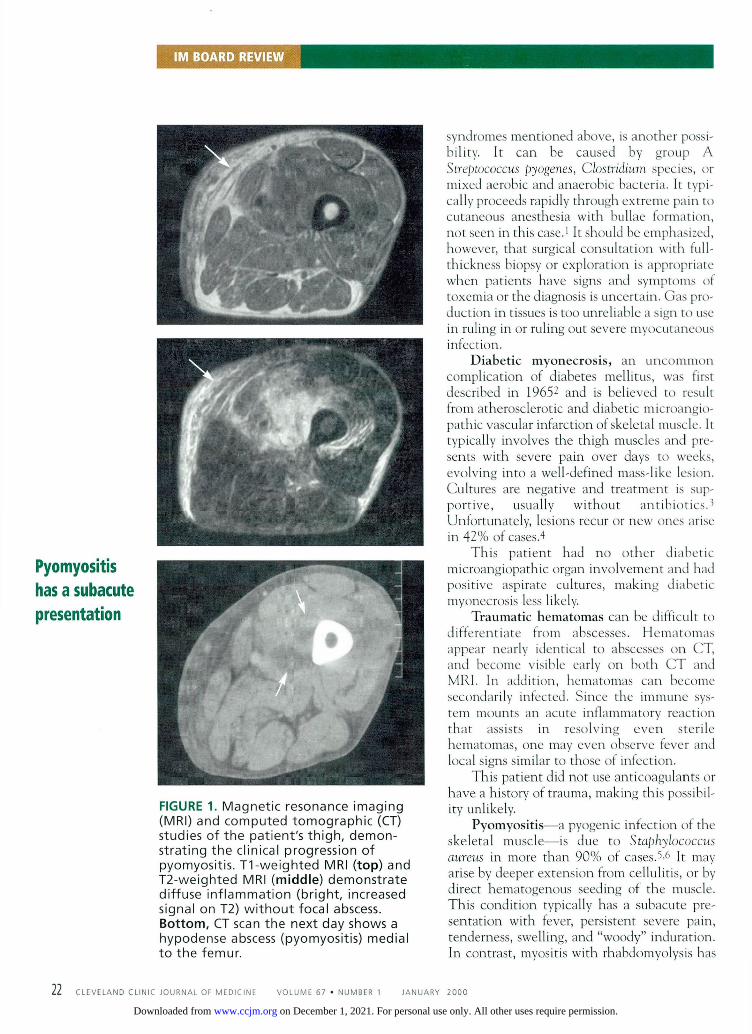
FIGURE 1. Magnetic resonance imaging (MRI) and computed tomographic (CT) studies of the patient's thigh, demon-strating the clinical progression of pyomyositis. T1-weighted MRI ( top ) and T2-weighted MRI ( m i d d l e ) demonstrate diffuse inf lammation (bright, increased signal on T2) w i thou t focal abscess. B o t t o m , CT scan the next day shows a hypodense abscess (pyomyositis) medial to the femur.
2 2 C L E V E L A N D C L I N I C J O U R N A L OF M E D I C I N E V O L U M E 6 7 • N U M B E R 1 J A N U A R Y 2 0 0 0
on December 1, 2021. For personal use only. All other uses require permission.www.ccjm.orgDownloaded from

a more acute onset and muscle swelling with "bogginess." The most commonly infected muscles are those of the thigh. Pyomyositis is uncommon hut has an increased incidence in diabetic patients.5
Though classically described in patients from tropical climates who are otherwise nor-mal, pyomyositis has been noted in immuno-compromised patients in temperate climates. Various theories attempt to explain a geo-graphic distribution of this disease, though limited access to timely medical care, more common in tropical locations, is plausible.
This case fulfills all the classic features of pyomyositis in a diabetic patient in a temper-ate climate.
• WHAT IS THE EVALUATION?
2 Evaluation of nonhealing cellulitis in a patient with diabetes should include all except which one of the following?
• Blood cultures • Muscle enzyme determinations • Ultrasonography, CT, MRI, or all three • Aspiration and culture • Electromyelographic testing
Blood cultures are positive in 5% to 29% of diabetic patients with pyomyositis, though it is of ten unclear whether positive results rep-resent an original bacteremia or seeding from the infected muscle.5 Conversely, autopsy studies found spontaneous muscle abscesses in fewer than 1% of patients who died of staphy-lococcal septicemia.5
Muscle enzyme determinations (ie, serum concentrations of creatine phosphoki-nase, aspartate, aminotransferase, lactate dehydrogenase, and aldolase) are usually nor-mal in pyomyositis,6 in spite of extensive asso-ciated myonecrosis. T h e primary utility of these tests may lie in helping to exclude rhab-domyolysis and necrotizing fasciitis, in which they are more commonly elevated. Given the severity of these conditions, it is reasonable to check these enzymes.
Radiographic imaging has several roles in evaluating unresponsive cellulitis. For exam-ple, ultrasonography can help document deep venous thrombosis, which can occasionally
mimic cellulitis and does not respond to antibiotics. Ultrasonography can also demon-strate local fluid collections. However, C T and MRI provide much more detail and give important information about the extent of disease and the involvement of adjacent struc-tures, such as possible bony involvement in a case of pyomyositis due to contiguous osteomyelitis.3 In addition, an imaging test can pinpoint the abscess, which would facili-tate the drainage procedure.
A word of caution: Since severe group A streptococcal fasciitis does not produce gas, lack of gas on radiographic studies should no t delay an aggressive evaluation in cases in which necrotizing fasciitis is sus-pected. 1
As in any abscess-associated infection, definitive treatment involves antibiotics and adequate drainage of the abscess. In our case, aspiration and culture also confirmed the presence of oxacillin-sensitive S aureus in the involved muscle, providing useful data to guide antibiotic therapy. This culture result was obtained even though antibiotic therapy had already been started, demonstrating the benefit of draining abscesses therapeutically and diagnostically, even in patients on active therapy.
Electromyelographic testing can be very helpful in diagnosing some forms of inflam-matory myositis but does not have a role in this patient.
1 WHAT CAUSES PYOMYOSITIS?
3 Pyomyositis may develop as a result of all except which one of the following?
• Immunocompromised state • Hematogenous spread during transient
bacteremia • Local spread from a contiguous bone or
soft-tissue infection • Prior muscle-infection by parasites in
tropical pyomyositis
T h e etiology of pyomyositis is likely multifac-torial.
Immunocompromise. In its nontropical form, pyomyositis tends to occur in immuno-compromised hosts. Pyomyositis can be seen
Gas is an unreliable sign of severe myocutaneous infection
C L E V E L A N D C L I N I C J O U R N A L OF M E D I C I N E V O L U M E 6 7 • N U M B E R 1 J A N U A R Y 2 0 0 0 13
on December 1, 2021. For personal use only. All other uses require permission.www.ccjm.orgDownloaded from

IM BOARD REVIEW
Abscesses require antibiotics and drainage
in the profound immunocompromised setting of AIDS, or the limited immune changes of diabetes.
Hematogenous spread. Patients with dia-betes mellitus have an increased prevalence of nasal and cutaneous colonization with S aureus. They also have an increased incidence of S aureus infections, including internal seed-ing with bacteremia.7
However, as men t ioned above, most patients with pyomyositis do not have posi-tive blood cultures: most studies have found a rate of only approximately 5%, though this may vary. Hence, if bacteremic muscle seed-ing occurs, it likely happens during episodes of transient bacteremia not detected on blood cultures. A n autopsy series of patients who died of S aureus bacteremia noted only 1% had muscle abscesses, emphasizing the rarity of occult pyomyositis.5 In experiments in rabbits wi th S aureus bacteremia, pyomyositis developed only after antecedent muscle trauma.
Local spread. In some cases, muscle abscess may develop from contiguous osteomyelitis or other soft-tissue infections. Occasionally, deep pyomyositis results from lymphatic spread of overlying cellulitis. C T and MRI studies may help establish or rule out these conditions.
Parasitic infect ion. In t ropical pyomyositis, some experts have suggested tha t vigorous physical activity or prior mus-cle involvement from more prevalent para-sites or viral diseases explains the increased rate of disease in otherwise-normal hosts. However, a case-control study8 found no increased rate of infection associated with malnutri t ion, myositis, or antecedent infec-tion (bacterial, parasitic, or viral). Tropical pyomyositis may develop because of limited access to ant ib io t ics , a l lowing minor S
aureus infections ro progress to deeper mus-cle involvement. Of the possibilities given, this is the least likely in our pat ient .
• SUMMARY: PURSUING ALTERNATIVE DIAGNOSES WHEN TREATMENT FAILS
Our case demonstrates the importance of con-tinuing to pursue an alternative diagnosis vig-orously when cellulitis does not respond to appropriate empiric antibiotic therapy in a patient with diabetes mellitus. It also points out the need for repetitive imaging studies to identify pyomyositis as it moves from the ini-tial invasive stage to the purulent stage when the diagnosis is more easily made.
Definitive treatment, including drainage, is necessary to avoid the development of the late or septic stage. Woody induration with persistent pain in an ill patient without tox-emia may be a particularly useful clue and should alert the clinician to the possibility of pyomyositis. E3
• REFERENCES
1. File TM, Tan JS, Dispersio JR. Diagnosing and treating the "flesh-eating bacteria syndrome." Cleve Clinic J Med 1998; 65:241-249.
2. Angervall L, Stener R. Tumoriform focal muscular degen-eration in two diabetic patients. Diabetologia 1965; 1:39-42.
3. Scully RE, Mark EJ, McNeely WF, Ebeling SH, Phillips LD (editors). Weekly dinicopathological exercises. Case 29-1997. N Engl J Med 1997; 337:839-845. Bodner RA, Younger DS, Rosokljia G. Diabetic muscle infarction. Muscle Nerve 1994; 17:949-950.
5. Walling DM, Kaeling WG Jr. Pyomyositis in patients with diabetes mellitus. Rev Infect Dis 1991; 13:797-802.
6. Dunkerley GR, Older J. Onwochei B, Pazienza J. Pyomyositis. Am Fam Phys 1996; 54:565-569. Breen JD, Karchmer AW. Staphylococcus aureus infections in diabetic patients Infect Dis Clin North Am 1995; 9:11-24. Eason R, OsbourneJ, Ansford T, Stallman N, Forsyth JRL. Tropical pyomyositis in the Solomon Islands: clinical and aetiological features. Trans R SocTrop Med Hyg 1989; 83:275-278.
4 .
7.
Ì J ' J J ^
Î J ^ U J - J 1
- f É I j f
Category I CME Credit. Test your knowledge
of clinical topics.
IN THIS ISSUE PAGE 71
2 4 C L E V E L A N D C L I N I C J O U R N A L OF M E D I C I N E V O L U M E 6 7 • N U M B E R 1 J A N U A R Y 2 0 0 0
on December 1, 2021. For personal use only. All other uses require permission.www.ccjm.orgDownloaded from

BRIEF S U M M A R Y - A R T H R O T E C ® (d ic lofenac s o d i u m a n d misoprosto l ) B e f o r e p r e s c r i b i n g , p l e a s e c o n s u l t c o m p l e t e p r e s c r i b i n g i n f o r m a t i o n . ®
C O N T R A I N D I C A T I O N S A N D W A R N I N G S ARTHROTEC®, because of the abor t i fac ient p roper ty of the misopros to l componen t , is contra-ind icated in w o m e n w h o are pregnant . (See PRECAUTIONS.) Reports, p r imar i l y f r o m Brazil, of congen i ta l anomal ies and reports of fetal death subsequent to misuse of m isopros to l alone, as an abor t i fac ient , have been received. Patients mus t be advised of the abor t i fac ient p roper ty and w a r n e d not to g ive the d r u g to others. ARTHROTEC shou ld not be used in w o m e n of ch i ldbear-ing potent ia l unless the pat ient requires nonstero ida l an t i - i n f lammatory drug (NSAID) therapy and is at h igh risk of deve lop ing gastr ic or duodena l u lcerat ion or fo r deve lop ing compl ica t ions f r o m gastr ic or duodena l ulcers associated w i t h the use of the NSAID. (See WARNINGS.) In such patients, ARTHROTEC m a y be prescr ibed if the pat ient: • has had a negat ive s e r u m pregnancy test w i t h i n t w o weeks pr ior to beg inn ing therapy. • is capable of c o m p l y i n g w i t h ef fect ive cont racept ive measures. • has received bo th oral and wr i t t en warn ings of the hazards of misoprosto l , the risk of possi-
b le cont racept ion fa i lure, and the danger to other w o m e n of ch i ldbear ing potent ia l shou ld the drug be taken by mistake.
• w i l l beg in ARTHROTEC on ly on the second or th i rd day of the next no rma l mens t rua l per iod.
I N D I C A T I O N S A N D U S A G E ARTHROTEC® is indicated fo r t rea tment of the s igns and s y m p t o m s o f osteoar thr i t is (OA) or rheu-ma to id arthr i t is (RA) in pat ients at h igh risk of deve lop ing NSAID- induced gastr ic and duodena l ulcers and the i r compl ica t ions . See WARNINGS—Gastrointestinal effects f o r a list of fac tors that may increase the risk of NSAID- induced gastr ic and duodena l ulcers and the i r compl ica t ions . C O N T R A I N D I C A T I O N S
See b o x e d CONTRAINDICATIONS AND WARNINGS related to misoprosto l . ARTHROTEC is contra-ind ica ted in pat ients w i t h hypersens i t i v i t y o r al lergic react ion to d ic lofenac, m isopros to l , o ther pros-tag land ins , o r aspi r in /NSAIDs. Severe, rarely fatal, anaphy lac to id react ions t o d ic lo fenac Na have been repor ted. W A R N I N G S Regarding diclofenac:
G a s t r o i n t e s t i n a l ( G i l e f f e c t s — r i s k o f G l u l c e r a t i o n , b l e e d i n g a n d p e r f o r a t i o n : Ser ious Gl toxic i ty , such as i n f l ammat ion , b leeding, u lcerat ion and per forat ion of the s tomach, smal l intest ine or large intes-t ine, can occur at any t i m e , w i t h or w i t hou t wa rn i ng s y m p t o m s , in pat ients t reated w i t h NSAIDs. Remain alert for u lcerat ion and bleeding, even in the absence of p rev ious Gl tract s y m p t o m s . In fo rm pat ients about the s igns and/or s y m p t o m s and the steps to take if t hey occur. On ly 1/5 pat ients w h o deve lop a ser ious upper Gl adverse event on NSAID therapy is symptomat i c . It has been demon-st ra ted that upper Gl ulcers, g ross b leeding, or per forat ion, caused by NSAIDs, appears to occur in app rox ima te l y 1% of pat ients t reated for 3 - 6 months , and in 2 - 4 % of pat ients treated for one year, w i t h an increasing l i ke l ihood of deve lop ing a ser ious Gl event du r ing chronic therapy. However , even shor t - te rm therapy has risk. Prescribe NSAIDs w i t h ex t reme caut ion in those w i t h a pr ior h is tory of ulcer disease or Gl b leeding. Most spon taneous repor ts of fatal Gl events are in e lder ly or debi l -i tated pat ients and there fore special care shou ld be taken in t reat ing th is popu la t ion . T o m i n i m i z e t h e p o t e n t i a l r i sk f o r a n a d v e r s e e v e n t , t h e l o w e s t e f f e c t i v e d o s e s h o u l d b e u s e d f o r t h e s h o r t e s t p o s s i b l e d u r a t i o n , For very high-r isk pat ients, a l ternate therapies that d o not invo lve NSAIDs s h o u l d be cons idered. Studies have s h o w n that pat ients w i th a h istory of pept ic ulcer disease and/or Gl b leed ing, and w h o use NSAIDs, have a greater than 10-fold risk for deve lop ing a Gl bleed than pat ients w i th neither of these risk factors, In add i t ion to a past h is to ry of ulcer disease, pharmaco-e p i d e m i o l o g y ! studies have ident i f ied several o ther cond i t ions or co- therapies that may increase the risk for Gl bleeding, such as: t rea tment w i t h oral cor t icostero ids, t rea tmen t w i th ant icoagulants , longer dura t ion of NSAID therapy, o lder age, smok ing , a lcoho l ism, p o o r general heal th and H. pylori posi t ive status.
H e p a t i c e f f e c t s : Elevat ions of l iver tests may occur. Border l ine e levat ions {<3x upper l im i t o f norma l , or ULIM) or greater e levat ions of t ransaminases occur red in abou t 15% o f d ic lo fenac- t reated pat ients. In c l in ical t r ia ls, mean ing fu l e levat ions ( > 3 x U L N ) of AST (SGOT) occurred in abou t 2% of pat ients. Mean ing fu l e levat ions of ALT and/or AST occurred in about 4%, and marked e levat ions ( > 8 x U L N ) in abou t 1%, of pat ients t reated 2 - 6 mon ths . In an open- label tr ial, e levated ALT or AST w a s observed m o r e o f ten in pat ients rece iv ing d ic lofenac than other NSAIDs. Postmarket ing, rare cases of severe hepat ic react ions, i nc lud ing l iver necrosis, jaundice, and fu lm inan t fatal hepat i t is w i t h and w i t h o u t jaund ice have been repor ted ; some underwen t l iver t ransplantat ion. Severe hepat ic react ions can occur at any t i m e w i t h o u t a p r o d r o m e of d i s t i ngu ish ing symp toms . Transaminases s h o u l d therefore be per iod ica l ly measured beg inn ing w i t h i n 4 to 8 weeks after in i t ia t ing t reatment . The m isopros to l c o m p o n e n t does not appear to exacerbate these diclofenac effects. If abnormal liver tests persist or worsen , if c l in ical s igns and/or s y m p t o m s consistent w i t h l iver disease develop, or if sys temic man-i festat ions occur, d iscont inue ARTHROTEC t reatment immediate ly . In fo rm patients of the wa rn i ng s igns and s y m p t o m s of hepato tox ic i ty (eg, nausea, fa t igue, lethargy, prur i tus , jaundice, r igh t upper quad-rant tenderness, and " f lu - l i ke" symp toms ) , and the appropr ia te act ion pat ients shou ld take if these signs and s y m p t o m s appear. A n a p h y l a c t o i d r e a c t i o n s : Anaphy lac to id react ions may occur in pat ients w i t hou t k n o w n pr ior expo-sure t o ARTHROTEC or its c o m p o n e n t s . ARTHROTEC shou ld not be g iven to pat ients w i t h the aspir in t r iad. The t r i ad typical ly occurs in asthmat ic pat ients w h o exper ience rh in i t is w i t h or w i t h o u t nasal polyps, or w h o exhibit severe, potentially fatal b ronchospasm after tak ing asp i r in o r o ther NSAIDs. Emergency help shou ld be sought in cases whe re an anaphy lac to id react ion occurs. A d v a n c e d r e n a l d i s e a s e : In pat ients w i t h advanced kidney disease, t rea tment w i t h ARTHROTEC is not r ecommended . P R E C A U T I O N S
G e n e r a l : ARTHROTEC cannot be used to subst i tu te for cor t icostero ids or to treat fo r cor t icos tero id insuf f ic iency. ARTHROTEC's an t i - i n f l ammato ry act iv i ty may d im in i sh the ut i l i ty o f th is d iagnost ic s ign. Renal effects: Use caut ion w h e n in i t ia t ing ARTHROTEC in dehydra ted pat ients. Rehydrate pat ients f i rst . A l so use caut ion in pat ients w i t h k idney disease (see WARNINGS—Advanced renal disease). Long- te rm admin is t ra t ion of d ic lofenac has resul ted in renal pap i l la ry necrosis and other renal medu l la ry changes. Pat ients w i t h impa i red renal func t ion , heart fa i lure, or l iver dys func t ion , those tak ing d iuret ics and ACE inh ib i tors , and the elderly are at greatest r isk of renal decompensa t ion . D iscont inuat ion of NSAID the rapy is usual ly f o l l owed by recovery to the pre t reatment state. Diclo-fenac metabo l i tes are e l im ina ted pr imar i l y by the k idneys and pat ients w i t h s ign i f icant ly impa i red renal func t ion should be m o r e c losely mon i to red . Hematologic effects: Anem ia may occur ; it may be due to f l u id retent ion, Gl b l o o d loss, or an incomple te ly descr ibed effect upon ery thropoies is . Pat ients shou ld have their hemog lob in or hematocr i t checked if they exh ib i t s igns or s y m p t o m s of anemia. ARTHROTEC may inter fere to s o m e extent w i t h platelet func t ion and vascular responses to b leed ing. Patients w i t h coagu la t i on d isorders o r rece iv ing ant icoagu lants should be carefully mon -i tored. Aseptic meningitis: Asept ic men ing i t i s w i t h fever and coma has been observed on rare occa-sions in pat ients on d ic lo fenac. Fluid retention and edema: Fluid retent ion and edema may occur. Use w i t h caut ion in pat ients w i t h a h is tory of cardiac decompensa t ion , hyper tens ion, or o ther con-d i t ions pred ispos ing to f l u i d retent ion. Preexisting asthma: Do not use in pat ients w i t h aspir in-sen-si t ive as thma because of the risk of severe fatal b ronchospasm. Use w i t h caut ion in pat ients w i t h preex is t ing asthma. Porphyria: Use in pat ients w i t h hepat ic porphyr ia shou ld be avo ided. L a b o r a t o r y t e s t s : Patients s h o u l d have the i r CBC and a chemis t ry prof i le checked per iodical ly . If cl in-ical s igns and s y m p t o m s consistent w i t h l iver or renal disease develop, systemic mani fes ta t ions occur (eg, eos inoph i l ia , rash, etc) o r if abno rma l l iver tests persist or worsen, discontinue ARTHROTEC. D r u g i n t e r a c t i o n s : ARTHROTEC m a y increase the serum levels of d igox in , methot rexate , l i th ium and phenobarb i ta l ; pat ients s h o u l d be mon i t o red for toxic i ty . ARTHROTEC may increase cyc lospor ine nephro tox ic i ty , exacerbate Gl b leed ing in pat ients on war far in , and inh ib i t the act iv i ty o f ant ihyper-tens ives and d iuret ics. Use caut ion in admin is te r ing ARTHROTEC w i t h any of these agents, part icu-lar ly if renal func t ion is impa i red . Asp i r i n may d im in i sh the therapeut ic effect of d ic lofenac and coadmin is t ra t i on is not r e c o m m e n d e d . Dic lofenac Na may alter a d iabet ic pat ient 's response to insu l in or oral hypog lycemic agents. An tac ids con ta in ing magnes ium m a y exacerbate d iarrhea and shou ld not be coadmin is te red w i t h ARTHROTEC.
A n i m a l t o x i c o l o g y : A reversib le increase in the n u m b e r of n o r m a l surface gastr ic epithelial cells occur red in t h e dog, rat, a n d mouse du r ing long- te rm tox i co logy studies w i th misopros to l . An appar-ent response of the f ema le mouse to m isopros to l in long- te rm studies at 100-1000 x the h u m a n dose w a s hyperostos is , m a i n l y of the medu l la of s ternebrae. These effects have not been seen in h u m a n studies. C a r c i n o g e n e s i s , m u t a g e n e s i s , i m p a i r m e n t o f f e r t i l i t y : A n i m a l studies t o evaluate the potent ia l fo r carc inogenes is and effects on fer t i l i ty have been pe r fo rmed w i t h each componen t of ARTHROTEC given alone. ARTHROTEC itself w a s not genotox ic in the AMES test, the Chinese hamster ovary cell (CHO/HGPRT) f o r w a r d mu ta t i on test, the rat l ymphocy te c h r o m o s o m e aberrat ion test or the mouse micronuc leus test. In a 24 -mo rat carc inogen ic i ty study, oral misoprostol at doses up to 2 4 x the rec-o m m e n d e d m a x i m u m h u m a n dose of 0.6 m g / m 2 / d a y was not tumor igen ic . In a 21 -mo mouse car-c inogen ic i ty study, oral m isop ros to l at doses up t o 8 0 x the recommended m a x i m u m h u m a n dose was not tumor igen ic . M isopros to l , w h e n admin is te red to male and fema le b reed ing rats in an oral dose- range of 1 - 1 0 0 x the r e c o m m e n d e d m a x i m u m h u m a n dose p roduced dose-re lated pre- and
pos t - imp lan ta t ion losses and a s igni f icant decrease in the number of l ive pups born at the h ighest dose. These f i nd ings suggest the possib i l i ty o f a general adverse effect on fer t i l i ty in males and females. In a 24-mo rat carc inogenic i ty study, oral d ic lofenac Na w a s not t umor i gen i c at Q.08x the recommended m a x i m u m human dose of 148 mg/m 2 /day . In a 24 -mo m o u s e carc inogen ic i t y study, oral d ic lo fenac Na at doses up to 0 .006x the r e c o m m e n d e d m a x i m u m h u m a n dose in males and 0.02x the r e c o m m e n d e d m a x i m u m human dose in females w a s not tumor igen ic . Dic lofenac Na at oral doses up to 0 .16x the recommended m a x i m u m h u m a n dose w a s f o u n d t o have no effect on fer t i l i ty and reproduc t ive per formance of male and female rats. P r e g n a n c y : Pregnancy category X: See boxed CONTRAINDICATIONS AND WARNINGS regard ing misopros to l . ARTHROTEC is contra ind icated in pregnancy. Non-teratogenic effects: M isopros to l may endanger pregnancy (may cause miscarr iage) and thereby cause harm to the fe tus when admin is te red to a p regnant w o m a n . M isopros to l p roduces uter ine cont ract ions, uter ine bleeding, and expu ls ion of the products of concept ion . M iscar r iages caused by m isopros to l m a y be incomplete. In s tudies in w o m e n undergo ing elect ive t e rm ina t i on of p regnancy du r ing the f irst t r imester , misopros to l caused part ial or comp le te expu ls ion of t h e p roduc ts of con-cept ion in 11% of t h e subjects and increased uter ine b leed ing in 41%. Reports, p r i m a r i l y f r o m Brazil, of congeni ta l anoma l ies and repor ts of feta l death subsequent to misuse of m isop ros to l a lone, as an abor t i fac ient , have been received (see CONTRAINDICATIONS AND WARNINGS). If a w o m a n is or becomes pregnant wh i le tak ing th is d rug , the drug s h o u l d be d iscon t inued and the pat ient appr ised of the potent ia l hazard to the fetus. The dic lofenac Na c o m p o n e n t of ARTHROTEC, like o ther NSAIDs w h i c h are pros tag land in- inh ib i t ing drugs, may affect the fetal card iovascu lar sys tem caus ing premature c losure o f the ductus arter iosus. NSAIDs may also inh ib i t u ter ine cont rac t ions. Teratogenic effects: A n i m a l tests have revealed no ev idence of te ra togen ic potent ia l for ARTHROTEC, misopros to l , or d ic lo fenac.
N u r s i n g m o t h e r s : Because of the potent ia l for ser ious adverse react ions in nurs ing infants, ARTH-ROTEC is not r e c o m m e n d e d for use by nurs ing mothers . P e d i a t r i c u s e : Safety and efficacy have not been establ ished.
G e r i a t r i c u s e : In c l in ical tr ial patients >65 years of age, no overal l d i f ferences w e r e observed between eff icacy, adverse events or PK prof i les of o lder and younge r pat ients. However, the e lder ly are l ikely to to lerate adverse events less we l l than younge r pat ients. A D V E R S E R E A C T I O N S
A d v e r s e r e a c t i o n s a s s o c i a t e d w i t h A R T H R O T E C G a s t r o i n t e s t i n a l : In c l in ical trials, the mos t f requent ly repor ted adverse events were Gl d isorders: abdomina l pain (21%), diarrhea (19%), dyspeps ia (14%), nausea (11%), and f la tu lence (9%). ARTH-ROTEC can cause m o r e Gl s y m p t o m s than dic lofenac alone. These events led t o d iscon t inua t ion of therapy in 9% of pat ients . Diarrhea and abdomina l pain deve loped early in the course of therapy, and w e r e usual ly se l f - l im i ted (resolved after 2 to 7 days). Rare instances of p ro found d iar rhea leading to severe dehydra t ion have been repor ted in pat ients receiv ing misopros to l . Pat ients w i t h an under-ly ing cond i t i on such as i n f l ammato ry bowe l disease, or those in w h o m dehydra t ion , w e r e it to occur, w o u l d be dangerous , shou ld be mon i t o red careful ly. The inc idence of d iar rhea can be m in im ized by admin is te r ing ARTHROTEC w i t h f o o d and by avo id ing coadmin is t ra t i on w i t h magnes ium-con -ta in ing antacids. G y n e c o l o g i c a l : Postmenopausal vag ina l b leed ing may occur (see be low) and s h o u l d be evaluated to rule out gyneco log ic pathology. Other : Adverse exper iences repor ted occasional ly or rarely w i t h ARTHROTEC, d ic lo fenac or other NSAIDs, or m isopros to l are: Body as a whole: Asthenia, death, fa t igue, fever, in fect ion, malaise, sepsis. Cardiovascular system: A r r h y t h m i a , atr ia l f i b r i l l a t i on , CHF, hype r t ens i on , h y p o t e n s i o n , increased CPK and/or LDH, Ml, palp i tat ions, phlebi t is , PVCs, syncope, tachycard ia , vascul i t is . Central and peripheral nervous system: Coma, convu ls ions , d ip lop ia , d rows iness , hyperesthes ia , hyper to-nia, hypoesthesia, men ing i t i s , migra ine , neuralg ia, paresthesia, t r emor , ver t igo . Digestive: Anorex ia , dry mou th , dysphagia , enterit is, esophageal u lcerat ion, gast roesophageal ref lux, Gl b leed ing, Gl neo-p lasm ben ign, g lossi t is , hematemesis, hemor rho ids , intest inal per fora t ion, pept ic ulcer, s tomat i t i s and ulcerat ive s tomat i t i s , tenesmus, Female reproductive disorders: Breast pain, dysmeno r rhea , in termenst rua l b leed ing, leukorrhea, menst rua l d isorder, menor rhag ia , vag ina l hemor rhage . Hemic and lymphatic system: Agranulocytos is , aplast ic anemia , coagu la t ion t ime increased, ecchymos is , eos inoph i l ia , epistaxis, hemoly t ic anemia, leukocytosis, leukopenia, l ymphadenopa thy , melena, pul-m o n a r y embo l i sm , purpura , pancytopenia , rectal b leeding, t h rombocy them ia , t h rombocy topen ia . Hypersensitivity: A n g i o e d e m a , la ryngea l /pharyngea l edema, ur t icar ia. Liver and biliary system: A b n o r m a l hepat ic function, hepatitis, jaundice, liver fa i lure, pancreat i t is . Mate reproductive disor-ders: Impotence, per ineal pain. Metabolic and nutritional: A lka l ine phosphatase increased, dehy-dra t ion , hyponat remia , hyperg lycemia, hypog lycemia , BUN increased, hyperu r i cemia , per iorb i ta l edema, porphyr ia , gou t , hypercholestero lemia, we igh t changes. Musculoskeletal system: Ar thra l -gia, mya lg ia . Psychiatric: Anxiety, asthenia, concent ra t ion impa i red , confus ion , depress ion, disor i-entat ion, d ream abnormal i t ies, ha l luc inat ions, i r r i tabi l i ty , mala ise, nervousness, parano ia , psychot ic react ion, somno lence . Respiratory system: As thma , cough ing , dyspnea, hyperven t i l a t ion , pneumo-nia, respi ratory depress ion. Skin and appendages: Acne, alopecia, e ry thema m u l t i f o r m e , eczema, exfo l ia t ive dermat i t i s , pemph igo id react ion, photosensi t iv i ty , p ru r i tus ani, sk in u lcera t ion , Stevens-J o h n s o n synd rome , sweat ing increased, tox ic ep iderma l necrolys is. Special senses: Hear ing impai r -ment , taste loss, tas te perversion. Urinary system: Cysti t is, dysur ia , hematur ia , in ters t i t ia l nephr i t is , m ic tu r i t ion f requency, noctur ia, nephrot ic synd rome, o l igur ia /po lyur ia , papi l lary necrosis, prote in-uria, renal fa i lure, u r ina ry tract in fect ion. Vision: Amb l yop ia , b lu r red v is ion, con junc t iv i t i s , g laucoma, ir i t is, lac r imat ion abno rma l , night b l indness, v is ion abnorma l . O V E R D O S A G E
The tox ic dose of ARTHROTEC has not been de te rmined . However , s igns of ove rdosage f r o m the c o m p o n e n t s of the p roduc t may inc lude: diclofenac— Gl comp la in t s , con fus ion , d rows iness or general hypoton ia ; misoprostol— sedat ion, t remor , convu ls ions, dyspnea, a b d o m i n a l pa in, d iarrhea, fever, palp i tat ions, hypotens ion, or bradycardia. Overdosage s y m p t o m s shou ld be t rea ted w i t h sup-por t ive therapy. In case of acute overdosage, gastr ic lavage is r ecommended . Induced d iures is may be benef ic ial . The use of oral act ivated charcoal may help to reduce absorp t ion . D O S A G E A N D A D M I N I S T R A T I O N
ARTHROTEC is admin is te red as ARTHROTEC 50 (50 m g dic lofenac Na /200 meg misopros to l ) or as ARTHROTEC 75 (75 m g diclofenac Na /200 meg misoprosto l ) . O A : T h e r e c o m m e n d e d dosage for m a x i m a l Gl mucosa l protect ion is ARTHROTEC 50 t id. H A : T h e r e c o m m e n d e d dosage is ARTHROTEC 50 t id or qid. For OA and RA pat ients w h o exper ience in to lerance, ARTHROTEC 75 b id or ARTHROTEC 50 b id can be used, but are less ef fect ive in prevent ing ulcers. ARTHROTEC f ixed dose c o m b i n a t i o n is not appropr ia te for patients who w o u l d not receive the appropr ia te dose o f bo th ingred ients . Doses of the c o m p o n e n t s del ivered w i t h these reg imens are as fo l lows :
RA regimen
OA regimen
Diclofenac Na (mg/day)
Misoprostol (meg/day)
ARTHROTEC 50 q id - 200 800
t i d t id 150 600
b id bid 100 400
ARTHROTEC 75 b id b id 150 400
S P E C I A L D O S I N G C O N S I D E R A T I O N S : ARTHROTEC conta ins m isopros to l , w h i c h p r o v i d e s pro tec t ion against gastr ic and duodena l ulcers. For gastr ic ulcer prevent ion, the 200 meg q i d and t i d reg imens are therapeut ica l ly equivalent , but more protect ive than the b id reg imen. For d u o d e n a l ulcer pre-ven t ion , the q id reg imen is more protect ive than the t i d or bid reg imens . However , the q id reg imen is less we l l to le ra ted than the t i d reg imen because of usual ly se l f - l imi ted d ia r rhea re lated to the misopros to l dose (see ADVERSE REACTIONS—Gastrointestinal), and the b id r eg imen m a y be better to lera ted than t i d in some patients. Dosages may be ind iv idual ized us ing the separate products (misopros to l and d ic lofenac), after w h i c h the pat ient may be c h a n g e d to the appropr ia te ARTHRO-TEC dose. If c l in ica l ly indicated, m isopros to l co- therapy w i t h ARTHROTEC, or use of t h e ind iv idua l c o m p o n e n t s t o op t im ize the misopros to l dose and/or f requency of admin is t ra t ion , m a y be appro-priate. The tota l dose of misoprosto l shou ld not exceed 800 meg/day, and no m o r e t h a n 200 meg of m isopros to l shou ld be admin is tered at any one t ime. Doses of d ic lofenac h igher than 150 mg/day in OA or h igher than 225 mg/day in RA are not recommended .
12/30/97 - P97AR13195V
Packaged by G.D. Searle & Co. Mfd. by Searle, Morpeth, England For G.D. Searle Inter-American Co. Chicago IL 60680 USA
SEARLE
Address medical inquiries to: G.D. Searle & Co. Healthcare Information Services 5200 Old Orchard Road Skokie IL 60077
Printed in USA
© 1999 Searle, Box 5110, Chicago, IL 60680-5110 December 1999*ARl8564T*Printed in USA
on December 1, 2021. For personal use only. All other uses require permission.www.ccjm.orgDownloaded from