noninvasive ventilation in acute heart failure
TRANSCRIPT
EMERGENCY MEDICINE (FRANK PEACOCK, SECTION EDITOR)
Noninvasive Ventilation in Acute Heart Failure
Josep Masip1,2
Published online: 30 May 2019# Springer Science+Business Media, LLC, part of Springer Nature 2019
AbstractPurpose of Review To assess the role of noninvasive ventilation (NIV) in acute heart failure (AHF).Recent Findings NIV rapidly improves the respiratory distress and reduces the need for intubation and even mortality in patientswith acute cardiogenic pulmonary edema (ACPE). Therefore, NIV is indicated as first line therapy in ACPE. NIV may also beconsidered in some cases of cardiogenic shock after stabilization. CPAP is an easier and cheaper technique that is recommendedas first-line therapy, particularly in pre-hospital or low-equipped areas. Noninvasive pressure support ventilation is equallyeffective in these scenarios, and may be preferable in patients with mild fatigue or significant hypercapnia, including those withassociated chronic obstructive pulmonary disease (COPD). High flow nasal cannula is an alternative for patients who needprolonged ventilation or those who show poor tolerance to these techniques.Summary NIV should be used as a first-line therapy in all patients with ACPE and should be considered in stable cardiogenicshock and AHF associated to COPD.
Keywords Noninvasive ventilation . Acute heart failure . CPAP . Pressure support . High-flow nasal cannula
Introduction
Acute respiratory failure (ARF) is a frequent complication inclinical practice, and it is usually managed with conventionaloxygen therapy (COT), mainly high-flow “Venturi”masks, orlow-flow reservoir masks and thin nasal cannulas. However,ARF is not often fully compensated with COT and requiresgreater respiratory support. Noninvasive ventilation (NIV), atechnique that emerged in the 1980s, consisting of the appli-cation of positive intrathoracic pressure to conscious patientsthrough different interfaces, has shown to be useful in thissetting by reducing the need for EI and invasive mechanicalventilation (IMV) and decreasing some of its associated risks,mainly ventilator-associated pneumonia [1]. Since its intro-duction, NIV has been extended to different areas of the hos-pital, the pre-hospital setting and even care at home, while
IMV has remained limited to critical units or the operatingtheater. NIV is currently used to treat ARF in different acutescenarios [2••] (Table 1), having COPD exacerbation andacute cardiogenic pulmonary edema (ACPE) as the strongestindications.
Acute Respiratory Failure in AHF Syndromes
Although nearly 90% of acute heart failure (AHF) patientscomplain of dyspnea [3] and most of them show some degreeof lung congestion [4], less than half present ARF with hyp-oxemia [5]. Among the different AHF scenarios [4, 6, 7],significant ARF is essentially seen in ACPE, in cardiogenicshock (CS), and in cases of AHF associated with other lungalterations.
Acute Cardiogenic Pulmonary Edema Acute cardiogenic pul-monary edema is produced by a rapid increase in pulmonarycapillary hydrostatic pressure with fluid filtration that exceedsthe lymphatic interstitial drainage capacity [8]. ARF occurswhen an excess of interstitial and alveoli flooding results ina significant reduction of gas exchange and a concomitantshunt effect. Therefore, the key findings are the combinationof ARF (hypoxemia and/or hypercapnia) with significant
This article is part of the Topical Collection on Emergency Medicine
* Josep [email protected]
1 Intensive Care Department, Consorci Sanitari Integral, University ofBarcelona, Jacint Verdaguer 90, ES-08970 Sant JoanDespí, Barcelona, Spain
2 Cardiology Department, Hospital Sanitas CIMA, Barcelona, Spain
Current Heart Failure Reports (2019) 16:89–97https://doi.org/10.1007/s11897-019-00429-y
respiratory distress (tachypnea and increased work of breath-ing). Criteria for the diagnosis have been recently published[9••] (Table 2) [10, 11]. Patients frequently present hyperten-sion on admission. Hypertensive ACPE more frequently havepreserved left ventricular (LV) ejection fraction (EF), hyper-capnia, and better prognosis than those with lower blood pres-sure (BP) [12]. Some of them may have a very rapid
presentation, “flash ACPE,” without previous accumulationof fluids and mostly diastolic LV dysfunction [13].
Cardiogenic Shock Patients have significant ARF for severalreasons. There are pulmonary edema and ventilation-perfusion inequality due to an increase in pulmonary deadspace due to a fall of lung perfusion. In addition, the circula-tory failure reduces the central venous oxygen content (SvO2)secondary to a greater tissue extraction.
Other Scenarios Patients with AHF may also have COPD,pneumonia, asthma, large pleural effusion, pulmonary embo-lism, or atelectasis, which may precipitate or aggravate ARF.In cases with isolated right ventricular (RV) failure, ARF ismainly seen in cases of acute pulmonary thromboembolism ordecompensated chronic pulmonary hypertension.
Effect of NIV in AHF
Positive airway pressure increases oxygenation and decreasesthe work of breathing [14]. Ventilatory support additionallyimproves alveolar ventilation with further decreases in thework of breathing and carbon dioxide level [15]. NIV hasshown to produce faster improvement of the ARF, shorteningthe critical phase, decreasing the risk of endotracheal intuba-tion, and, potentially, reducing mortality in high-risk patients.However, in patients with isolated RV failure, positive pres-sure should be avoided because it increases RV afterload,impairing RV function [16].
Modalities of NIV
The main modalities in AHF are continuous positive airwaypressure (CPAP), noninvasive pressure support ventilation(NIPSV), and, more recently, high-flow nasal cannula (HFNC).
CPAP CPAP is the simplest technique and consists of the appli-cation of a high flow in the mask exceeding the breathing de-mand of the patients, leading to a continuous positive pressure
Table 2 Diagnostic criteria for acute pulmonary edema
Clinical criteria (all of them)
• Acute respiratory distressa
• Physical examinationb
• Orthopnea
• Respiratory failurec
Diagnostic Confirmation (at least two of the following)
• Clear signs of pulmonary congestion on chest radiography or CT scan
• Multiple B-lines on lung ultrasoundd
• Elevated pulmonary capillary pressure on catheterization
• Increased total lung water on pulse contour and thermodilutionanalysis system
• Signs of elevated filling pressures on echocardiographye
• Significant elevation of natriuretic peptidesf
RR respiratory rate, CT computer tomographya Respiratory distress: acute increase in the work of breathing (assessed bysingle inspection), significant tachypnea (RR > 25breaths/min)1 , may bewith the use of accessory muscles or abdominal paradoxb Crackles +/– wheezes over the lungs, third heart sound2
c Oxygen saturation on room air by pulse-oximetry (SpO2) < 90%.Arterial blood gases may also show PaO2 < 60 mmHg, PaCO2 >45 mmHg, or PaO2/FiO2 < 300mHgd ≥ 3 B-lines in 2 chest zones on each hemithorax (references 7,8)e E/E′ > 15. Other parameters of elevated left atrial pressure may also beconsideredf Nt-ProBNP >900 (or >1800 in older than 75)1 RR may be lower, and orthopnea may be absent in obtunded patients2 Patients with low systolic blood pressure (i.e., < 90 mmHg) may beconsidered to have cardiogenic shock rather than ACPE3 In “flash pulmonary edema,” BNP may be lower
(Taken from reference 9 in the text)
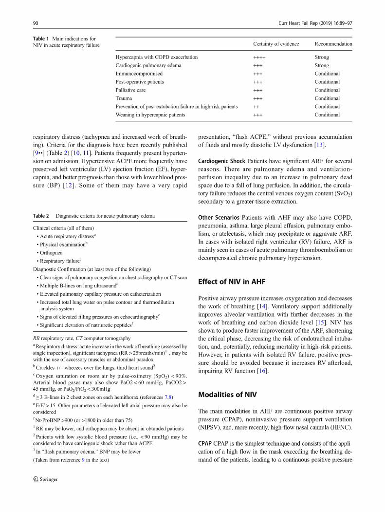
Table 1 Main indications forNIV in acute respiratory failure Certainty of evidence Recommendation
Hypercapnia with COPD exacerbation ++++ Strong
Cardiogenic pulmonary edema +++ Strong
Immunocompromised +++ Conditional
Post-operative patients +++ Conditional
Palliative care +++ Conditional
Trauma +++ Conditional
Prevention of post-extubation failure in high-risk patients ++ Conditional
Weaning in hypercapnic patients +++ Conditional
90 Curr Heart Fail Rep (2019) 16:89–97
into the lungs. It can be applied without the aid of a ventilator, byusing a source of air or oxygen and a mask equipped with PEEPvalve, or with the Boussignac system [17].
NIPSV This modality requires a ventilator. It is programmedwithtwo levels of pressure: expiratory pressure (EPAP) or positiveend-expiratory pressure (PEEP), and inspiratory pressure(IPAP), which is obtained with pressure support. It is also callednoninvasive intermittent positive pressure ventilation (NIPPV),or sometimes bilevel or BiPAP. This method requires some ex-perience for setting the ventilator to the changing needs of thepatient. Adequate synchrony is essential. The respiratory rate isnot pre-set and depends exclusively on the patient.
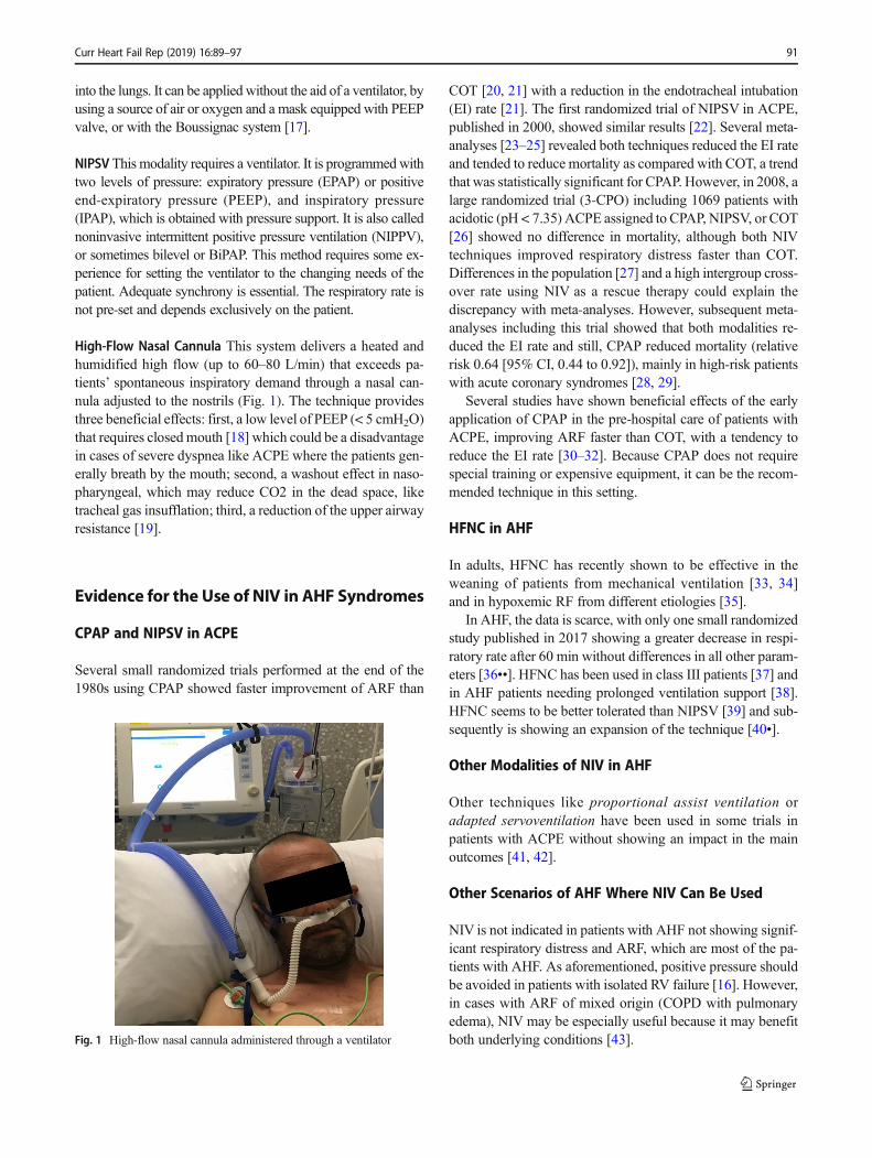
High-Flow Nasal Cannula This system delivers a heated andhumidified high flow (up to 60–80 L/min) that exceeds pa-tients’ spontaneous inspiratory demand through a nasal can-nula adjusted to the nostrils (Fig. 1). The technique providesthree beneficial effects: first, a low level of PEEP (< 5 cmH2O)that requires closedmouth [18] which could be a disadvantagein cases of severe dyspnea like ACPE where the patients gen-erally breath by the mouth; second, a washout effect in naso-pharyngeal, which may reduce CO2 in the dead space, liketracheal gas insufflation; third, a reduction of the upper airwayresistance [19].
Evidence for the Use of NIV in AHF Syndromes
CPAP and NIPSV in ACPE
Several small randomized trials performed at the end of the1980s using CPAP showed faster improvement of ARF than
COT [20, 21] with a reduction in the endotracheal intubation(EI) rate [21]. The first randomized trial of NIPSV in ACPE,published in 2000, showed similar results [22]. Several meta-analyses [23–25] revealed both techniques reduced the EI rateand tended to reduce mortality as compared with COT, a trendthat was statistically significant for CPAP. However, in 2008, alarge randomized trial (3-CPO) including 1069 patients withacidotic (pH < 7.35) ACPE assigned to CPAP, NIPSV, or COT[26] showed no difference in mortality, although both NIVtechniques improved respiratory distress faster than COT.Differences in the population [27] and a high intergroup cross-over rate using NIV as a rescue therapy could explain thediscrepancy with meta-analyses. However, subsequent meta-analyses including this trial showed that both modalities re-duced the EI rate and still, CPAP reduced mortality (relativerisk 0.64 [95% CI, 0.44 to 0.92]), mainly in high-risk patientswith acute coronary syndromes [28, 29].
Several studies have shown beneficial effects of the earlyapplication of CPAP in the pre-hospital care of patients withACPE, improving ARF faster than COT, with a tendency toreduce the EI rate [30–32]. Because CPAP does not requirespecial training or expensive equipment, it can be the recom-mended technique in this setting.
HFNC in AHF
In adults, HFNC has recently shown to be effective in theweaning of patients from mechanical ventilation [33, 34]and in hypoxemic RF from different etiologies [35].
In AHF, the data is scarce, with only one small randomizedstudy published in 2017 showing a greater decrease in respi-ratory rate after 60 min without differences in all other param-eters [36••]. HFNC has been used in class III patients [37] andin AHF patients needing prolonged ventilation support [38].HFNC seems to be better tolerated than NIPSV [39] and sub-sequently is showing an expansion of the technique [40•].
Other Modalities of NIV in AHF
Other techniques like proportional assist ventilation oradapted servoventilation have been used in some trials inpatients with ACPE without showing an impact in the mainoutcomes [41, 42].
Other Scenarios of AHF Where NIV Can Be Used
NIV is not indicated in patients with AHF not showing signif-icant respiratory distress and ARF, which are most of the pa-tients with AHF. As aforementioned, positive pressure shouldbe avoided in patients with isolated RV failure [16]. However,in cases with ARF of mixed origin (COPD with pulmonaryedema), NIV may be especially useful because it may benefitboth underlying conditions [43].Fig. 1 High-flow nasal cannula administered through a ventilator
Curr Heart Fail Rep (2019) 16:89–97 91
NIV and Myocardial Infarction
Two trials in the 1990s suggested that NIPSV could increasethe risk of acute myocardial infarction (AMI) [44, 45].However, no other trial has reproduced these results, includingrandomized studies specifically designed to assess this issue[46–48]. One randomized trial analyzed the effect of CPAP inpatients with AMI showing advantages over COT [49]. Inaddition, in 3-CPO, NIV was safely used in patients withAMI, who accounted for nearly 50% of the population en-rolled, with no differences in the incidence of AMI betweengroups [26].
NIV in Cardiogenic Shock
There are no studies analyzing NIV in CS. However, in theinternational registry “Cardshock study” [50], NIV was usedin nearly 13% of the patients, after correction of hypotension,avoiding EI in the majority [51••]. Therefore, although theindication of NIV remains limited in hypotensive patients, itmay be cautiously considered in selected CS patients.
CPAP or NIPSV
Although theoretically NIPSV should be superior to CPAPbecause it provides inspiratory help for breathing, no trialsor meta-analyses have demonstrated a clear advantage ofone technique over the other for important outcomes but pa-tients treated with NIPSV have shown faster improvement inseveral physiological variables [45, 52, 53]. In several caseseries of patients with ACPE, NIPSV was most clearly effec-tive in those with hypercapnia [22, 54]. In addition, in patientswithARF from different etiologies, CPAP has beenmost oftenused in hypoxemic patients, while NIPSV may be more effec-tive in those with hypercapnia. Consequently, although eithertechnique can be used as a first-line treatment in ACPE, itseems reasonable to prefer NIPSV in patients with severehypercapnia, including those with COPD.
Recommendations for NIV in AHF
NIV has shown an expansion in the last decades, particularlyin ACPE [55•]; however, there is a wide variation amongcenters, from nearly 0 to almost 100% [56]. ACPE is currentlythe second most frequent indication for NIV [57]. Data from2430 patients who required ventilatory support in theADHERE registry supported the use of NIV to avoid EI[15]. The latest ESC guidelines gave NIV a class IIa recom-mendation with level of evidence B [58, 59] in patients withAHF and respiratory distress (respiratory rate > 25 breaths/min, SpO2 < 90%). The NICE guidelines in AHF recom-mended NIV in patients with ACPE with severe dyspnea
and acidemia [60]. Finally, recent guidelines from ERC/ATSrecommended NIV, either bilevel NIV or CPAP, for patientswith ARF due to ACPE and suggested it in the pre-hospitalsetting [2••].
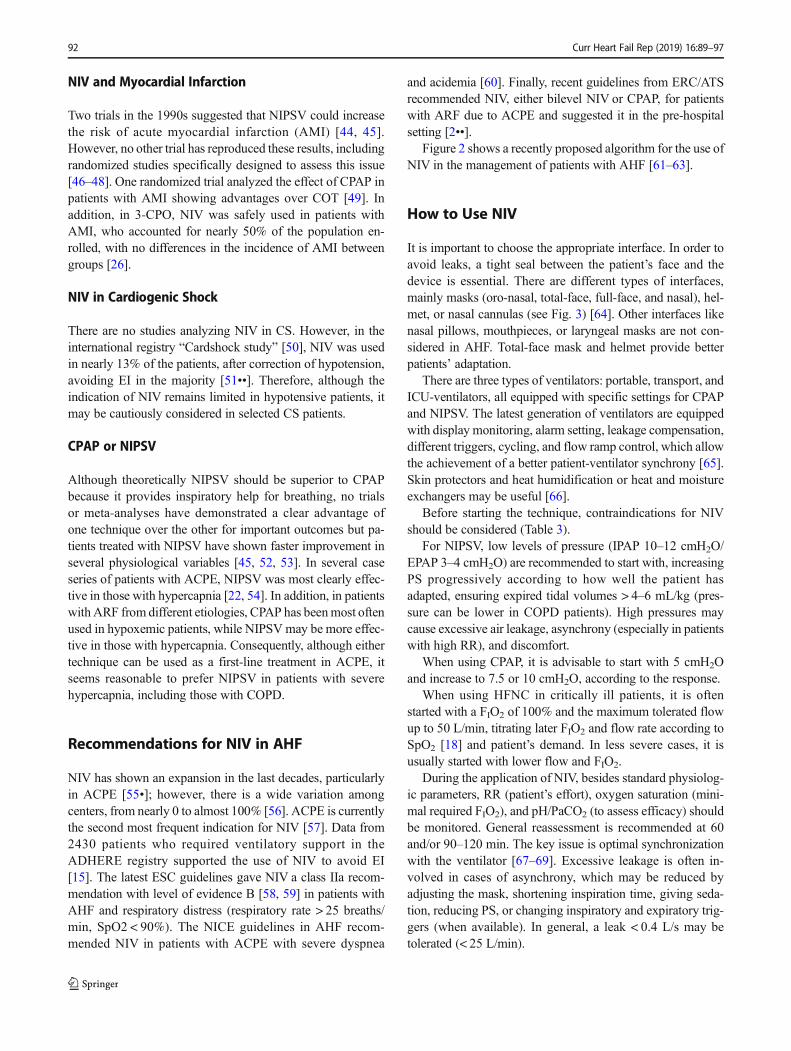
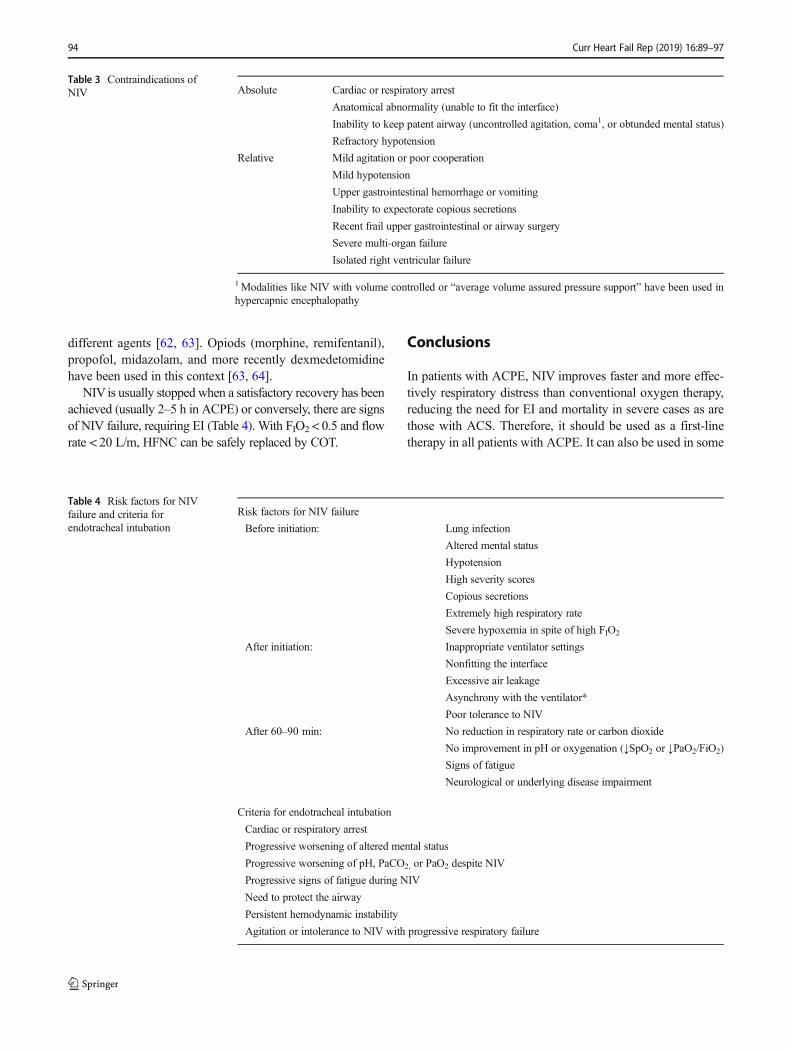
Figure 2 shows a recently proposed algorithm for the use ofNIV in the management of patients with AHF [61–63].
How to Use NIV
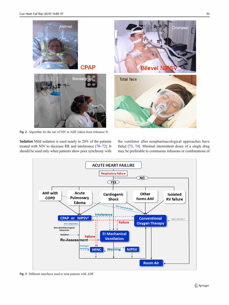
It is important to choose the appropriate interface. In order toavoid leaks, a tight seal between the patient’s face and thedevice is essential. There are different types of interfaces,mainly masks (oro-nasal, total-face, full-face, and nasal), hel-met, or nasal cannulas (see Fig. 3) [64]. Other interfaces likenasal pillows, mouthpieces, or laryngeal masks are not con-sidered in AHF. Total-face mask and helmet provide betterpatients’ adaptation.
There are three types of ventilators: portable, transport, andICU-ventilators, all equipped with specific settings for CPAPand NIPSV. The latest generation of ventilators are equippedwith display monitoring, alarm setting, leakage compensation,different triggers, cycling, and flow ramp control, which allowthe achievement of a better patient-ventilator synchrony [65].Skin protectors and heat humidification or heat and moistureexchangers may be useful [66].
Before starting the technique, contraindications for NIVshould be considered (Table 3).
For NIPSV, low levels of pressure (IPAP 10–12 cmH2O/EPAP 3–4 cmH2O) are recommended to start with, increasingPS progressively according to how well the patient hasadapted, ensuring expired tidal volumes > 4–6 mL/kg (pres-sure can be lower in COPD patients). High pressures maycause excessive air leakage, asynchrony (especially in patientswith high RR), and discomfort.
When using CPAP, it is advisable to start with 5 cmH2Oand increase to 7.5 or 10 cmH2O, according to the response.
When using HFNC in critically ill patients, it is oftenstarted with a FIO2 of 100% and the maximum tolerated flowup to 50 L/min, titrating later FIO2 and flow rate according toSpO2 [18] and patient’s demand. In less severe cases, it isusually started with lower flow and FIO2.
During the application of NIV, besides standard physiolog-ic parameters, RR (patient’s effort), oxygen saturation (mini-mal required FIO2), and pH/PaCO2 (to assess efficacy) shouldbe monitored. General reassessment is recommended at 60and/or 90–120 min. The key issue is optimal synchronizationwith the ventilator [67–69]. Excessive leakage is often in-volved in cases of asynchrony, which may be reduced byadjusting the mask, shortening inspiration time, giving seda-tion, reducing PS, or changing inspiratory and expiratory trig-gers (when available). In general, a leak < 0.4 L/s may betolerated (< 25 L/min).
92 Curr Heart Fail Rep (2019) 16:89–97
Sedation Mild sedation is used nearly in 20% of the patientstreated with NIV to decrease RR and intolerance [70–72]. Itshould be used only when patients show poor synchrony with
the ventilator after nonpharmacological approaches havefailed [73, 74]. Minimal intermittent doses of a single drugmay be preferable to continuous infusions or combinations of
Fig. 3 Different interfaces used to treat patients with AHF
Fig. 2 Algorithm for the use of NIV in AHF (taken from reference 9)
Curr Heart Fail Rep (2019) 16:89–97 93
different agents [62, 63]. Opiods (morphine, remifentanil),propofol, midazolam, and more recently dexmedetomidinehave been used in this context [63, 64].
NIV is usually stopped when a satisfactory recovery has beenachieved (usually 2–5 h in ACPE) or conversely, there are signsof NIV failure, requiring EI (Table 4). With FIO2 < 0.5 and flowrate < 20 L/m, HFNC can be safely replaced by COT.
Conclusions
In patients with ACPE, NIV improves faster and more effec-tively respiratory distress than conventional oxygen therapy,reducing the need for EI and mortality in severe cases as arethose with ACS. Therefore, it should be used as a first-linetherapy in all patients with ACPE. It can also be used in some
Table 4 Risk factors for NIVfailure and criteria forendotracheal intubation
Risk factors for NIV failure
Before initiation: Lung infection
Altered mental status
Hypotension
High severity scores
Copious secretions
Extremely high respiratory rate
Severe hypoxemia in spite of high FIO2
After initiation: Inappropriate ventilator settings
Nonfitting the interface
Excessive air leakage
Asynchrony with the ventilator*
Poor tolerance to NIV
After 60–90 min: No reduction in respiratory rate or carbon dioxide
No improvement in pH or oxygenation (↓SpO2 or ↓PaO2/FiO2)
Signs of fatigue
Neurological or underlying disease impairment
Criteria for endotracheal intubation
Cardiac or respiratory arrest
Progressive worsening of altered mental status
Progressive worsening of pH, PaCO2, or PaO2 despite NIV
Progressive signs of fatigue during NIV
Need to protect the airway
Persistent hemodynamic instability
Agitation or intolerance to NIV with progressive respiratory failure
Table 3 Contraindications ofNIV Absolute Cardiac or respiratory arrest
Anatomical abnormality (unable to fit the interface)
Inability to keep patent airway (uncontrolled agitation, coma1, or obtunded mental status)
Refractory hypotension
Relative Mild agitation or poor cooperation
Mild hypotension
Upper gastrointestinal hemorrhage or vomiting
Inability to expectorate copious secretions
Recent frail upper gastrointestinal or airway surgery
Severe multi-organ failure
Isolated right ventricular failure
1Modalities like NIV with volume controlled or “average volume assured pressure support” have been used inhypercapnic encephalopathy
94 Curr Heart Fail Rep (2019) 16:89–97
patients with cardiogenic shock without refractory hypoten-sion and in patients with AHF associated with lung diseaseshowing ARF. CPAP is cheaper and easy to use, and it ismainly indicated in low-equipped areas, whereas NIPSV ispreferred in cases with significant hypercapnia, although re-quires some expertise and adequate setting. HFNC may beconsidered in patients with ARF who can keep the mouthclosed and require prolonged ventilation or not tolerating oth-er forms of NIPSV.
Compliance with Ethical Standards
Conflict of Interest The author declares that he has no conflicts ofinterest.
Human and Animal Rights and Informed Consent This article does notcontain any studies with human or animal subjects performed by any ofthe authors.
References
Papers of particular interest, published recently, have beenhighlighted as:• Of importance•• Of major importance
1. Girou E, Brun-Buisson C, Taillé S, Lemaire F, Brochard L. Seculartrends in nosocomial infections and mortality associated with non-invasive ventilation in patients with exacerbation of COPD andpulmonary edema. JAMA. 2003;290:2985–91.
2.•• Rochwerg B, Brochard L, Elliott MW, Hess D, Hill NS, Nava S,et al. Official ERS/ATS clinical practice guidelines: noninvasiveventilation for acute respiratory failure. Eur Respir J. 2017;50:1602426. https://doi.org/10.1183/13993003.02426-2016 Analyzesthe evidence of the usefulness of NIV in the most frequentdifferent acute respiratory failure scenarios and providesdifferent levels of recommendation.
3. Mebazaa A, Pang PS, TavaresM, Collins SP, StorrowAB, Laribi S,et al. The impact of early standard therapy on dyspnoea in patientswith acute heart failure: the URGENT-dyspnoea study. Eur Heart J.2010;31:832–41.
4. Dickstein K, Cohen-Solal A, Filippatos G, McMurray JJ,Ponikowski P, Poole-Wilson PA, et al. ESC guidelines for the di-agnosis and treatment of acute and chronic heart failure 2008: thetask force for the diagnosis and treatment of acute and chronic heartfailure 2008 of the European Society of Cardiology. Eur Heart J.2008;29:2388–442.
5. Park JJ, Choi DJ, Yoon CH, Oh IY, Lee JH, Ahn S, et al. Theprognostic value of arterial blood gas analysis in high-risk acuteheart failure patients: an analysis of the Korean Heart Failure(KorHF) registry. Eur J Heart Fail. 2015;17:601–11.
6. Nieminen MS, Böhm M, Cowie MR, Drexler H, Filippatos GS,Jondeau G, et al. Executive summary of the guidelines on the diag-nosis and treatment of acute heart failure. The task force on acuteheart failure of the European Society of Cardiology endorsed by theEuropean Society of Intensive Care Medicine (ESICM). Eur HeartJ. 2005;26:384–416.
7. Chioncel O, Mebazaa A, Harjola V-P, Coats AJ, Piepoli MF,Crespo-Leiro MG, et al. Clinical phenotypes and outcome of
patients hospitalized for acute heart failure: the ESC Heart FailureLong-Term Registry. Eur J Heart Fail. 2017;19:1242–54.
8. Ware LB, Matthay MA. Clinical practice. Acute pulmonary edema.N Engl J Med. 2005;353:2788–96.
9.•• Masip J, Peacock WF, Price S, Cullen L, Martin-Sanchez FJ,Seferovic P, et al. Indications and practical approach to noninvasiveventilation in acute heart failure. Eur Heart J. 2018;39:17–25 Thisis a position paper from two AHF working groups of the ESC.The review describes the main AHF phenotypes showing ARFand analyzes the evidence of NIV on each one of them.Indications and practical recommendations are also provided.
10. Volpicelli G, Elbarbary M, Blaivas M, Lichtenstein DA, Mathis G,Kirkpatrick AW, et al. International evidence-based recommenda-tions for point-of-care lung ultrasound. Intensive Care Med.2012;38:577–91.
11. Pivetta E, Goffi A, Lupia E, Tizzani M, Porrino G, Ferreri E, et al.Lung ultrasound-implemented diagnosis of acute decompensatedheart failure in the ED: a SIMEU multicenter study. Chest.2015;148:202–10.
12. Figueras J, Bañeras J, Peña-Gil C,Masip J, Barrabés JA, RodriguezPalomares J, et al. Acute arterial hypertension in acute pulmonaryedema. Mostly a trigger or an associated phenomenon? Can JCardiol. 2016;32:1214–20.
13. Kramer K, Kirkman P, Kitzman D, Little WC. Flash pulmonaryedema: association with hypertension and reoccurrence despite cor-onary revascularization. Am Heart J. 2000;140:451–5.
14. Tobin MJ. Advances in mechanical ventilation. N Engl J Med.2001;344:1986–96.
15. Tallman TA, Peacock WF, Emerman CL, Lopatin M, Blicker JZ,Weber J, et al. Noninvasive ventilation outcomes in 2,430 acutedecompensated heart failure patients: an ADHERE registry analy-sis. Acad Emerg Med. 2008;15:355–62.
16. Harjola VP,MebazaaA,Čelutkienė J, Bettex D, Bueno H, ChioncelO, et al. Contemporary management of acute right ventricular fail-ure: a statement from the heart failure association and the workinggroup on pulmonary circulation and right ventricular function of theEuropean Society of Cardiology. Eur J Heart Fail. 2016;18:226–41.
17. Moritz F, Benichou J, Vanheste M, Richard JC, Line S, Hellot MF,et al. Boussignac continuous positive airway pressure device inemergency care of acute cardiogenic pulmonary oedema: a random-ized pilot study. Eur J Emerg Med. 2003;10:204–8.
18. Luo J-c, Lu M-s, Zhao Z-h, Jiang W, Xu B, Weng L, et al. Positiveend-expiratory pressure effect of 3 high-flow nasal cannula devices.Respir Care. 2017;62:888–95.
19. Lee JH, Rehder KJ, Williford L, Cheifetz IM, Turner DA. Use ofhigh flow nasal cannula in critically ill infants, children, and adults:a critical review of the literature. Intensive CareMed. 2013;39:247–57.
20. Räsänen J, Heikklä J, Downs J, Nikki P, Väisänen I, Viitanen A.Continuous positive airway pressure by face mask in acute cardio-genic pulmonary edema. Am J Cardiol. 1985;55:296–300.
21. Bersten AD, Holt AW, Vedig AE, Skowronski GA, Baggoley CJ.Treatment of severe cardiogenic pulmonary edema with continuouspositive airway pressure delivered by face mask. N Engl J Med.1991;325:1825–30.
22. Masip J, Betbesé AJ, Páez J, Vecilla F, Cañizares R, Padró J, et al.Non-invasive pressure support ventilation versus conventional ox-ygen therapy in acute cardiogenic pulmonary oedema: a random-ized trial. Lancet. 2000;356:2126–32.
23. Peter JV, Moran JL, Phillips-Hughes J, Graham P, Bersten AD.Effect of non-invasive positive pressure ventilation (NIPPV) onmortality in patients with acute cardiogenic pulmonary oedema: ameta-analysis. Lancet. 2006;367:1155–63.
24. Masip J, Roque M, Sánchez B, Fernández R, Subirana M, ExpósitoJ. Noninvasive ventilation in acute cardiogenic pulmonary edema.Systematic review and meta-analysis. JAMA. 2005;294:3124–30.
Curr Heart Fail Rep (2019) 16:89–97 95
25. Winck J, Azevedo LF, Costa-Pereira A, Antonelli M, Wyatt JC.Efficacy and safety of non-invasive ventilation in the treatment ofacute cardiogenic pulmonary edema: a systematic review andmeta-analysis. Crit Care. 2006;10:R69.
26. GrayA, Goodacre S, NewbyDE,MassonM, Sampson F, Nicholl J,et al. Noninvasive ventilation in acute cardiogenic pulmonary ede-ma. N Engl J Med. 2008;359:142–51.
27. Masip J,Mebazaa A, Filippatos G. Noninvasive ventilation in acutecardiogenic pulmonary edema. N Engl J Med. 2008;359:2068–9.
28. Weng CL, Zhao YT, Liu QH, Fu CJ, Sun F, Ma YL, et al. Meta-analysis: noninvasive ventilation in acute cardiogenic pulmonaryedema. Ann Intern Med. 2010;152:590–600.
29. Masip J, Páez J, Merino M, Parejo S, Vecilla F, Riera C, et al. Riskfactors for intubation as a guide for noninvasive ventilation in pa-tients with severe acute cardiogenic pulmonary edema. IntensiveCare Med. 2003;29:1921–8.
30. Ducros L, Logeart D, Vicaut E, Henry P, Plaisance P, Collet JP, et al.CPAP for acute cardiogenic pulmonary oedema from out-of-hospital to cardiac intensive care unit: a randomised multicentrestudy. Intensive Care Med. 2011;37:1501–9.
31. Plaisance P, Pirracchio R, Berton C, Vicaut E, Payen D. A random-ized study of out-of-hospital continuous positive airway pressurefor acute cardiogenic pulmonary oedema: physiological and clinicaleffects. Eur Heart J. 2007;28:2895–901.
32. Foti G, Sangalli F, Berra L, Sironi S, Cazzaniga M, Rossi GP, et al.Is helmet CPAP first line pre-hospital treatment of presumed severeacute pulmonary edema? Intensive Care Med. 2009;35:656–62.
33. Hernández G, Vaquero C, Colinas L, Cuena R, González P, CanabalA, et al. Effect of Postextubation high-flow nasal cannula vs non-invasive ventilation on reintubation and Postextubation respiratoryfailure in high-risk patients: a randomized clinical trial. JAMA.2016;316:1565–74.
34. Hernández G, Vaquero C, González P, Subira C, Frutos-Vivar F,Rialp G, et al. Effect of postextubation high-flow nasal cannula vsconventional oxygen therapy on reintubation in low-risk patients: arandomized clinical trial. JAMA. 2016;315:1354–61.
35. Frat JP, Thille AW, Mercat A, Girault C, Ragot S, Perbet S, et al.High-flow oxygen through nasal cannula in acute hypoxemic respi-ratory failure. N Engl J Med. 2015;372:2185–96.
36.•• Makdee O, Monsomboon A, Surabenjawong U, Praphruetkit N,Chaisirin W, Chakorn T, et al. High-flow nasal cannula versus con-ventional oxygen therapy in emergency department patients withcardiogenic pulmonary edema: a randomized controlled trial. AnnEmerg Med. 2017;70:465–72 This is the first trail analyzing theeffect of HFNC in ACPE compared with conventional oxygentherapy. No significant differences in the outcomes were found,but the study showed a decrease in respiratory rate in the firsthour with HFNC.
37. Roca O, Pérez-Terán P, Masclans JR, Pérez L, Galve E, EvangelistaA, et al. Patients with New York Heart Association class III heartfailure may benefit with high flow nasal cannula supportive therapy.High flow nasal cannula in heart failure. J Crit Care. 2013;28:741–6.
38. Carratalá JM, Llorens P, Brouzet B, Albert AR, Fernández-CañadasJM, Carbajosa J, et al. High-flow therapy via nasal cannula in acuteheart failure. Rev Esp Cardiol. 2011;64:723–35.
39. Frat JP, Brugiere B, Ragot S, Chatellier D, Veinstein A, Goudet V,et al. Sequential application of oxygen therapy via high-flow nasalcannula and noninvasive ventilation in acute respiratory failure: anobservational pilot study. Respir Care. 2015;60:170–8.
40.• Corley A, Rickard CM, Aitken LM, Johnston A, Barnett A, FraserJF, et al. High-flow nasal cannulae for respiratory support in adultintensive care patients. Cochrane Database Syst Rev. 2017;5:CD010172 Analyzes the studies assessing the effect of HFNCin acute respiratory failure and concludes that there is no
enough evidence of the superiority of HFNC compared withconventional oxygen or classical forms of NIV.
41. Rusterholtz T, Bollaert PE, Feissel M, Romano-Girard F, HarlayML, Zaehringer M, et al. Continuous positive airway pressure vs.proportional assist ventilation for noninvasive ventilation in acutecardiogenic pulmonary edema. Intensive Care Med. 2008;34:840–6.
42. Nakano S, Kasai T, Tanno J, Sugi K, Sekine Y, Muramatsu T, et al.The effect of adaptive servo-ventilation on dyspnoea, haemody-namic parameters and plasma catecholamine concentrations inacute cardiogenic pulmonary oedema. Eur Heart J AcuteCardiovasc Care. 2015;4:305–15.
43. Cabrini L, Landoni G, Oriani A, Plumari VP, Nobile L, Greco M,et al. Noninvasive ventilation and survival in acute care settings: acomprehensive systematic review and metaanalysis of randomizedcontrolled trials. Crit Care Med. 2015;43:880–8.
44. Mehta S, Jay GD, Woolard RH, Hipona RA, Connolly EM, CiminiDM, et al. Randomized, prospective trial of bilevel versus continu-ous positive airway pressure in acute pulmonary edema. Crit CareMed. 1997;25:620–8.
45. Sharon A, Shpirer I, Kaluski E, Moshkovitz Y, Milovanov O, PolakR, et al. High-dose intravenous isosorbide-dinitrate is safer andbetter than Bi-PAP ventilation combined with conventional treat-ment for severe pulmonary edema. J Am Coll Cardiol. 2000;36:832–7.
46. Bellone A, Monari A, Cortellaro F, Vettorello M, Arlati S, Coen D.Myocardial infarction rate in acute pulmonary edema: noninvasivepressure support ventilation versus continuous positive airway pres-sure. Crit Care Med. 2004;32:1860–5.
47. Yamamoto T, Takeda S, Sato N, Akutsu K, Mase H, Nakazato K,et al. Noninvasive ventilation in pulmonary edema complicatingacute myocardial infarction. Circ J. 2012;76:2586–91.
48. Ferrari G, Olliveri F, De Filippi G, et al. Noninvasive positive air-way pressure and risk of myocardial infarction in acute cardiogenicpulmonary edema: continuous positive airway pressure vs nonin-vasive positive pressure ventilation. Chest. 2007;132:1804–9.
49. Takeda S, Nejima J, Takano T, Nakanishi K, Takayama M,Sakamoto A, et al. Effect of nasal continuous positive airway pres-sure on pulmonary edema complicating acute myocardial infarc-tion. Jpn Circ J. 1998;62:553–38.
50. Harjola VP, Lassus J, Sionis A, Køber L, Tarvasmäki T, Spinar J,et al. CardShock study investigators; GREAT network. Clinicalpicture and risk prediction of short-term mortality in cardiogenicshock. Eur J Heart Fail. 2015;17:501–9.
51.•• Hongisto M, Lassus J, Tarvasmaki T, Sionis A, Tolppanen H,Lindholm MG, et al. Use of noninvasive and invasive mechanicalventilation in cardiogenic shock: a prospective multicenter study.Int J Cardiol. 2016;230:191–7. This is the first and the only pub-lished article that addresses this issue.
52. Park M, Lorenzi-Filho G, Feltrim MI, Viecili PR, Sangean MC,Volpe M, et al. Oxygen therapy, continuous positive airway pres-sure, or noninvasive bilevel positive pressure ventilation in thetreatment of acute cardiogenic pulmonary edema. Arq BrasCardiol. 2001;76:221–30.
53. Liesching T, Nelson DL, Cormier KL, Sucov A, Short K,Warburton R, et al. Randomized trial of bilevel versus continuouspositive airway pressure for acute pulmonary edema. J EmergMed.2014;46:130–40.
54. Nava S, Carbone G, DiBattista N, Bellone A, Baiardi P, CosentiniR, et al. Noninvasive ventilation in cardiogenic pulmonary edema: amulticenter randomized trial. Am J Respir Crit Care Med.2003;168:1432–7.
55.• Demoule A, Chevret S, Carlucci A, Kouatchet A, Jaber S, MezianiF, et al. Changing use of noninvasive ventilation in critically illpatients: trends over 15 years in francophone countries. Intensive
96 Curr Heart Fail Rep (2019) 16:89–97
Care Med. 2016;42:82–92. This one of the largest and recentregistries analyzing the use of NIV in the last 15 years.
56. Kulkarni VT, Kim N, Dai Y, Dharmarajan K, Safavi KC, Bikdeli B,et al. Hospital variation in noninvasive positive pressure ventilationfor acute decompensated heart failure. Circ Heart Fail. 2014;7:427–33.
57. Burns KE, Sinuff T, Adhikari NK, Meade MO, Heels-Ansdell D,Martin CM. Bilevel noninvasive positive pressure ventilation foracute respiratory failure: survey of Ontario practice. Crit Care Med.2005;33:1477–83.
58. Ponikowski P, Voors AA, Anker SD, Bueno H, Cleland JG, CoatsAJ, et al. 2016 ESC guidelines for the diagnosis and treatment ofacute and chronic heart failure. Eur Heart J. 2016;37:2129–200.
59. McMurray JJ, Adamopoulos S, Anker SD, Auricchio A, BöhmM,Dickstein K, et al. ESC guidelines for the diagnosis and treatment ofacute and chronic heart failure 2012: the task force for the diagnosisand treatment of acute and chronic heart failure 2012 of theEuropean Society of Cardiology. Developed in collaboration withthe Heart Failure Association (HFA) of the ESC. Eur Heart J.2012;33:1787–847.
60. National Clinical Guideline Centre (UK). Acute heart failure. diag-nosing andmanaging acute heart failure in adults. London: NationalInstitute for Health and Care Excellence (UK); 2014.
61. Mebazaa A, Yilmaz MB, Levy P, Ponikowski P, Peacock WF,Laribi S, et al. Recommendations on pre-hospital & early hospitalmanagement of acute heart failure: a consensus paper from theHeart Failure Association of the European Society of Cardiology,the European Society of Emergency Medicine and the Society ofAcademic EmergencyMedicine. Eur J Heart Fail. 2015;17:544–58.
62. Mebazaa A, Yilmaz MB, Levy P, Ponikowski P, Peacock WF,Laribi S, et al. Recommendations on pre-hospital & early hospitalmanagement of acute heart failure: a consensus paper from the heartfailure Association of the European Society of cardiology, theEuropean Society of Emergency Medicine and the Society ofAcademic Emergency Medicine - Short version. Eur Heart J.2015;36:1958–66.
63. Mueller C, Christ M, CowieM, Cullen L,Maisel AS,Masip J, et al.European Society of Cardiology-acute cardiovascular care associa-tion position paper on acute heart failure: a call for interdisciplinarycare. Eur Heart J Acute Cardiovasc Care. 2015.
64. Hauaji F, Vilella LM, Gonçalves C, Oliveira LC. The Total facemask is more comfortable than the oronasal mask in noninvasiveventilation but is not associated with improved outcome.Respiration. 2011;82:426–30.
65. Carteaux G, Lyazidi A, Cordoba-Izquierdo A, Vignaux L, Jolliet P,Thille AW, et al. Patient-ventilator asynchrony during noninvasiveventilation: a bench and clinical study. Chest. 2012;142:367–76.
66. Lellouche F, L’Her F, Abroug F, Deye N, Rodriguez PO, Rabbat A,et al. Impact of the humidification device on intubation rate duringnoninvasive ventilation with ICU ventilators: results of a multicen-ter randomized controlled trial. Intensive Care Med. 2014;40:211–9.
67. Hess DR. Patent-ventilator interaction during noninvasive ventila-tion. Respir Care. 2011;56:153–65.
68. Thille A, Rodriguez P, Cabello B, Lellouche F, Brochard L. Patient-ventilator asynchrony during assisted mechanical ventilation.Intensive Care Med. 2006;32:1515–22.
69. Vignaux L, Vargas F, Roeseler J, Tassaux D, Thille AW,Kossowsky MP, et al. Patient-ventilator asynchrony during non-invasive ventilation for acute respiratory failure: a multicenterstudy. Intensive Care Med. 2009;35:840–6.
70. Devlin JW, Nava S, Fong JJ, Bahhady I, Hill NH. Survey of seda-tion practices during noninvasive positive-pressure ventilation totreat acute respiratory failure. Crit Care Med. 2007;35:2298–302.
71. Matsumoto T, Tomii K, Tachikawa R, Otsuka K, Nagata K, OtsukaK, et al. Role of sedation for agitated patients undergoing noninva-sive ventilation: clinical practice in a tertiary referral hospital. BMCPulm Med. 2015;15:71.
72. Muriel A, Peñuelas O, Frutos-Vivar F, Arroliga AC, Abraira V,Thille AW, et al. Impact of sedation and analgesia during noninva-sive positive pressure ventilation on outcome: a marginal structuralmodel causal analysis. Intensive Care Med. 2015;41:1586–600.
73. Conti G, Hill NS, Nava S. Is sedation safe and beneficial in patientsreceiving NIV? No. Intensive Care Med. 2015;41:1692–5.
74. Hilbert G, Navalesi P, Girault C. Is sedation safe and beneficial inpatients receiving NIV? Yes. Intensive Care Med. 2015;41:1688–91.
Publisher’s Note Springer Nature remains neutral with regard tojurisdictional claims in published maps and institutional affiliations.
Curr Heart Fail Rep (2019) 16:89–97 97