north american snake envenomation: diagnosis,...
TRANSCRIPT

North American snake envenomation:diagnosis, treatment, and management
Barry S. Gold, MD, FACPa,*, Robert A. Barish, MD,FACP, FACEPb, Richard C. Dart, MD, PhD, FACEPc
aDivision of Emergency Medicine, Department of Surgery, University of Maryland School
of Medicine, 419 West Redwood Street, Suite 280, Baltimore, MD 21201, USAbOffice of the Dean, University of Maryland School of Medicine, 655 West Baltimore Street,
14-011, Baltimore, MD 21201-1559, USAcRocky Mountain Poison and Drug Center, Denver Health and Hospitals Authority, 1001
777 Bannock Street, Denver, CO 80204-4507, USA
Few animals have captured the human imagination more than the snake.Beginning with the Garden of Eden, world cultures have cast the snake insymbolic roles. It has been seen as a symbol of life, love, health, virtue,wealth, procreation, wisdom, and immortality. Alternatively, it has repre-sented death, hate, disease, sin, and poverty. Humans can become obsessedwith snakes—we fear them, worship them, and exploit them—but perhaps itis our fear of being bitten that lies at the core of our obsession. This ob-session leads to one of the most multifaceted problems confronted byphysicians—the management and treatment of individuals bitten by venom-ous snakes. Snake venom poisoning is a complex medical emergency thathas local effects at the bite site and can affect every major organ systemprimarily or secondarily.
Epidemiology
Approximately 15% of the 3000 species of snakes found worldwide areconsidered dangerous to humans [1]. The commonly encountered venomousspecies of snakes are members of several families. The largest family is theViperidae, which includes the African, European, and Middle-Easternvipers. Within this family is the subfamily of Crotalinae (pit vipers),comprising the rattlesnakes, cottonmouths, and copperheads of North
* Corresponding author.
E-mail address: [email protected] (B.S. Gold).
Emerg Med Clin N Am 22 (2004) 423–443
0733-8627/04/$ - see front matter � 2004 Elsevier Inc. All rights reserved.
doi:10.1016/j.emc.2004.01.007

424 B.S. Gold et al / Emerg Med Clin N Am 22 (2004) 423–443
America, the fer-de-lances of Central and South America, and the Asiaticpit vipers. The other major family is the Elapidae, consisting of cobras,mambas, kraits, coral snakes, and all venomous snakes of Australia. Lessfrequently encountered are the Hydrophidae (true sea snakes) and theLacticaudidae (sea kraits) families and members of the Colubridae, in-cluding the boomslang, bird snake, and keelback [2].
Approximately 45,000 snakebites occur annually in the United States,8000 of which are from venomous snakes [3]. Envenomation can causemorbidity of significant extent and duration [4] and accounts for five to sixdeaths per year [5]. Deaths typically occur in children, the elderly, and inthose for whom treatment is not given, is postponed, or is administered ininsufficient quantities. Deaths are also seen among members of fundamen-talist religious sects who handle venomous snakes during religious rites [6].
The victim profile has changed little since the late 1950s, when Parrishconducted his snake venom poisoning survey of the United States [3]. Themajority of victims remain men between the ages of 17 and 27 years [7].Alcohol intoxication is a contributing factor in many envenomations [7].More than 95% of the bites are on extremities, and most occur betweenApril and October, the peak months being July and August. These monthscoincide with the times when native snakes are active and humans are moreprone to be bitten during outdoor activities.
Parrish found that North Carolina, Arkansas, Texas, Georgia, WestVirginia, Mississippi, Louisiana, and Oklahoma had the highest bite ratesper 100,000 population. In the decades since his study there has been a shiftin prevalence toward the southwestern United States. This change isattributed to migration of the population westward along with the near-decimation of rattlesnake populations in the East and Northeast. Othertrends are a lower percentage of bites associated with agriculture anda higher percentage of bites from captive native and nonnative snakes [6].
Venomous snakes in the United States
Only 25 of the more than 120 species of snakes indigenous to the UnitedStates are venomous [8]. The majority of these snakes belong to thesubfamily Crotalinae (pit vipers: rattlesnakes, cottonmouths, and copper-heads [Fig. 1]). The coral snake (Elapidae) is the only other native venomoussnake. At least one species of indigenous poisonous snake has beenidentified in every state except Alaska, Maine, and Hawaii [3]. Exoticvenomous snakes found in zoos, schools, snake farms, and amateur andprofessional collections account for an increasing number of bites [9].
Pit vipers (Crotalids)
Pit vipers are named for the heat-sensitive foramen (pit) located betweeneach eye and nostril (Fig. 2), which senses the location and presence of

425B.S. Gold et al / Emerg Med Clin N Am 22 (2004) 423–443
warm-blooded prey or predators and guides the direction of the strike. Pitvipers have a triangular head, owing to the venom glands in both temporalregions. The pupils are elliptic. These snakes have two curved, canalizedfangs, each surrounded by a membranous sheath. The fangs are long andmoveable and retract posteriorly when the mouth is closed. At least threepairs of replacement fangs lie behind each functional fang. These fangs arein various stages of development and ready to move forward to replace shedor broken fangs. Fangs are replaced through the life cycle of thesnake; consequently, no fangless or defanged snake remains harmlessindefinitely [6]. Based on their ability to sense the size of their prey usingtheir pit, venomous snakes can regulate the amount of venom theyexcrete. The amount of venom discharged in defensive bites is less controlled[1].
Rattlesnakes (Crotalus and Sistrurus)
Sixteen species of rattlesnakes have been identified, making up twogenera—Crotalus and Sistrurus. The genus Crotalus includes the largerspecies; Sistrurus includes the smaller pygmy rattlesnakes and massasaugas.The cardinal characteristic of the rattlesnake is the tail rattle, which isformed by a group of interlocking keratin rings that vibrate against eachother, producing the characteristic buzzing sound when the snake isaroused. It is speculated that the rattle functions as a warning device topredators [10]. Throughout history, the myth has persisted that rattlesnakesalways ‘‘rattle’’ before striking. In fact, many strikes occur without warning.Juvenile rattlesnakes might lack functional rattle segments and are oftensoundless.
Rattlesnakes are native to every state with the exception of Alaska,Hawaii, Maine, and the District of Columbia. Most bites result from theeastern diamondback rattlesnake (C adamanteus), the western diamondbackrattlesnake (C atrox), the prairie and Pacific rattlesnakes (C viridis), thetimber rattlesnake (C horridus), and the pygmy rattlesnake (S miliarius). Theeastern and western diamondback rattlesnakes account for the mostfatalities [6].
Moccasins (Agkistrodon)
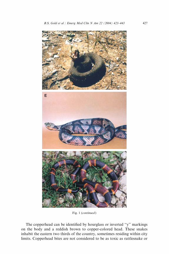
There are two species of moccasins—the cottonmouth (A piscivorus) andthe copperhead (A contortrix). Native to the southeastern and south centralsections of the country, the cottonmouth is a semiaquatic snake that can beidentified by dark olive to black coloring. It derives its descriptive namefrom the pale white color of the oral mucosa, which is highly visible whenthe mouth is fully opened. The cottonmouth is considered to be pugnaciousand is capable of biting while submerged.

426 B.S. Gold et al / Emerg Med Clin N Am 22 (2004) 423–443
Fig. 1. Venomous snakes of North America. (A) Eastern diamondback rattlesnake (Crotalus
adamanteus). (B) Western diamondback rattlesnake (C atrox). (C) Timber rattlesnake (C
horridus). (D) Cottonmouth (Agkistrodon piscivorus). (E) Copperhead (A contortix). (F) Eastern
coral snake (Micrurus fulvius fulvius). (From Gold BS, Dart RC, Barish RA. Bites of venomous
snakes. N Engl J Med 2002;347(5):347–58; with permission; Panels A, B, and F courtesy of
Kristen Wiley, Kentucky Reptile Zoo, Slade, Kentucky.)
427B.S. Gold et al / Emerg Med Clin N Am 22 (2004) 423–443
The copperhead can be identified by hourglass or inverted ‘‘y’’ markingson the body and a reddish brown to copper-colored head. These snakesinhabit the eastern two thirds of the country, sometimes residing within citylimits. Copperhead bites are not considered to be as toxic as rattlesnake or
Fig. 1 (continued )

428 B.S. Gold et al / Emerg Med Clin N Am 22 (2004) 423–443
cottonmouth bites and rarely require treatment; however, severe envenom-ations left untreated in children and the elderly can result in death.
Coral snakes (Micruroides and Micrurus)
The coral snakes are members of the Elapidae family, with two genera ofcoral snake native to the United States, Micruroides and Micrurus. TheArizona or Sonoran coral snake (Micruroides euryxanthus), a small snakewith an average length of 15 to 20 inches, is found in Arizona and NewMexico. The eastern coral snake (Micrurus fulvius fulvius) and the Texascoral snake (Micrurus fulvius tenere) are larger, with average lengths of 20 to
Venomous snakes Nonvenomous snakes
Crotalus atroxWestern diamondbackrattlesnake
Triangle-shaped head
Elliptical pupil
Heat-sensing pit
Retractablefangs
Venomgland
Venomduct
Analplate
Skull of a member offamily Viperidae
Rattle(rattlesnakes)
Single row ofsubcaudal plates
Double row ofsubcaudal plates
Analplate
Elaphe guttataCorn snake
Rounded head
Round pupil
No heat-sensing pit
No fangs
Skull of a member offamily Colubridae
Fig. 2. Comparison of venomous snakes (pit vipers) and nonvenomous snakes in the United
States. (From Gold BS, Dart RC, Barish RA. Bites of venomous snakes. N Engl J Med
2002;347(5):347–58; with permission.)

429B.S. Gold et al / Emerg Med Clin N Am 22 (2004) 423–443
45 inches. They are native from North Carolina southward to Florida andthrough the Gulf states to Texas.
Although various color phases such as black and albino have beenidentified, coral snakes in the United States can be identified by charac-teristic broad red and black bands separated by yellow (or cream) bands ontheir bodies. A number of harmless snakes can mimic the coral snake, whichhas given rise to the rhyme ‘‘red on yellow will kill a fellow; red on blackvenom lack.’’ This rhyme applies only to coral snakes native to the UnitedStates. Coral snakes in the United States possess black snouts and roundpupils but lack facial pits. Their fangs are short and fixed, and they injecttheir venom by a succession of chewing movements. These snakes are shy,secretive, and nocturnal. They are not aggressive and account for only 20 to25 bites per year [11].
Nonnative venomous snakes (exotic species)
Because of the increasing popularity of herpetoculture as a business anda hobby, the incidence of snakebites inflicted by nonnative venomousspecies in the United States is increasing. Between 1977 and 1995 Mintonreported that he consulted on 54 cases of bites from nonnative venomoussnakes involving at least 29 species [12]. The most common species was thecobra, which constituted 40% of the group [12]. The popularity of the cobraseems to arise from its perceived reputation as the ultimate deadly snake.They are popular with zoos and amateur snake keepers and are readilyavailable in the animal trade.
Cobras are members of the Elapidae family. Themajority of cobras belongto the genus Naja, of which the monocellate cobra (Naja kaouthia) appearsto be the species that is most available in the United States. Other popularspecies include the Chinese cobra (N atra), the Indian cobra (N naja), andthe African Cape cobra (N nivea). Cobras have a pair of short, fixed fangsin the front of their mouth, which allows them to bite a victim and maintaina hold while injecting venom in a succession of chewing movements.
Other popular species of nonnative snake are the African vipers belongingto the family Viperidae. These snakes are in the genus Bitis and include thepuff adder (B arietans), the gaboon viper (B gabonica), and the rhinocerosviper (B nasicornis). These large, heavy snakes tend to have colorful markings,which make them popular in zoos and with amateur collectors. The brightlycolored, nonnative, arboreal vipers are also commonly maintained incaptivity.
Venomous or nonvenomous?
The essential components of a definitive diagnosis of snake venompoisoning are positive identification of the snake and clinical manifestations

430 B.S. Gold et al / Emerg Med Clin N Am 22 (2004) 423–443
of envenomation. In the assessment of a reported venomous snakebite, onemust distinguish the bite from one from a nonvenomous snake, bites fromother animals (eg, rat), and puncture wounds caused by inanimate objects.The victim or companion might be able to make a positive identification.The snake—alive, dead, or in parts—can be brought in with the victim. It isimportant to remember when handling recently killed or decapitated snakesthat the bite reflex remains active for several minutes, rendering the snakecapable of inflicting a bite [13]. Snake parts should not be handled directly; ifpossible they should be placed in a canvas bag or container. Expertassistance for establishing a positive identification is available by contactingherpetologists from zoos or aquaria. About 25% of all pit viper bitesand 50% of all coral snakebites in the United States do not result inenvenomation and are considered ‘‘dry’’ bites [14]. In the absence of positiveidentification, objective symptoms and signs of an envenomation becomethe focus of diagnosis.
Systemic symptoms and signs
The most common reaction to any snakebite is impending doom. Manypeople believe that bites from a venomous snake inevitably will result indeath. Consequently, victims might appear emotionally unstable andexhibit extreme lethargy and withdrawal. Fear might cause symptoms suchas nausea, vomiting, diarrhea, fainting, tachycardia, and cold, clammy skin.Thus, it is important to differentiate these autonomic reactions fromsystemic symptoms and signs resulting from the envenomation. Confusioncould lead to unwarranted treatment.
The primary local clinical findings after most pit viper bites emerge within30 to 60 minutes. Common characteristics of crotaline envenomationinclude the presence of one or more fang marks—including evidence ofpuncture wounds and scratches, pain, edema, erythema, or ecchymosis ofthe bite site and adjacent tissues. Localized burning pain and earlyprogressive edema around the bite site are common. Pain, which is usuallyevident within 5 minutes following envenomation, is probably the result ofedema, swelling, and the release of substance P. Pain is present in more than90% of pit viper envenomations, with the exception of bites from theMojaverattlesnake (Crotalus scutulatus), which might cause little or no pain.Edema, both proximal and distal to the bite site, appears within 10 minutes,is rarely delayed more than 30 minutes following the envenomation (butoccasionally does not appear for several hours), and is the result of smallvessel and capillary injury. Occasionally, bullae might be noted withinseveral hours of the envenomation and can be serous or hemorrhagic. Theremight be signs of lymphangitis with tender regional lymph nodes along withincreased temperature over the injured part. Ecchymosis might appear overthe bite site within 3 to 6 hours. Ecchymosis is most severe following bites by

431B.S. Gold et al / Emerg Med Clin N Am 22 (2004) 423–443
eastern and western diamondbacks and prairie, Pacific, and timber rattle-snakes. It is less severe after copperhead and cottonmouth bites. Systemicmanifestations usually include nausea, vomiting, perioral paresthesia,tingling of the fingertips and toes, fasciculations, lethargy, and weakness.Subjective complaints of a rubbery, minty, or metallic taste are frequentafter envenomation by some larger species of rattlesnake. More severesystemic effects include altered mental status, severe tachycardia, tachypnea,respiratory distress, and hypotension (systolic blood pressure\80 mmHg).Coagulopathy frequently occurs following bites by rattlesnakes and canresult in a consumptive coagulopathy manifested by hypofibrinogenemia,prolonged prothrombin time (PT), decreased or unmeasurable activatedpartial thromboplastin time (A-PTT) with a platelet count of less than20,000/mm3, or a combination of these signs. Pit viper venom alters capi-llary membrane permeability, resulting in loss of electrolytes, albumin, andred blood cells into the bite site, manifested clinically as edema anderythema. Altered red blood cell membrane permeability can cause hemo-lysis. Initially, hypoalbuminemia and hemoconcentration occur, followed bypooling of blood and fluids in the microvasculature, resulting in hypo-volemic shock and acidosis; however, this process can occur concomitantlyin other organs such as the lungs, myocardium, kidneys, peritoneum, and,rarely, central nervous system. Renal failure might be secondary to hypo-tension, hemolysis, consumptive coagulopathy, or the nephrotoxic effectsof the venom components themselves.
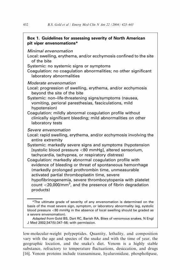
An essential element in the evaluation of snakebites by North Americanpit vipers is assessing the severity of the envenomation (Box 1). Multiplefactors influence the severity of any venomous snakebite: the species and sizeof the snake, the amount and toxicity of the venom injected, the location ofthe bite, first aid treatments performed, timing of definitive treatment,comorbid conditions, and the victim’s unique susceptibility to the venom [6].
Coral snake envenomations produce a paucity of local effects, but theremight be tremors, marked salivation, and altered sensorium, includingdrowsiness and euphoria. The neurologic signs are usually cranial nervepalsies manifested by ptosis, dysarthria, dysphagia, dyspnea, and seizures.The ultimate cause of death is paralysis of the respiratory musculature. Theonset of clinical manifestations might be delayed up to 12 hours followingthe envenomation [15]. Once signs and symptoms have appeared, preventionof neurotoxicity and reversal of changes that have already occurred mightnot be possible and might require airway management.
Pharmacology of venoms
Snake venoms are chemically complex mixtures of proteins, many ofwhich possess enzymatic properties. These enzymes contribute to thedeleterious effects of the venom, but the lethal components might be small
432 B.S. Gold et al / Emerg Med Clin N Am 22 (2004) 423–443
low-molecular-weight polypeptides. Quantity, lethality, and compositionvary with the age and species of the snake and with the time of year, thegeographic location, and the snake’s diet. Venom is a highly stablesubstance, refractory to temperature fluctuations, desiccation, and drugs[16]. Venom proteins include transaminase, hyaluronidase, phospholipase,
Box 1. Guidelines for assessing severity of North Americanpit viper envenomations*
Minimal envenomationLocal: swelling, erythema, and/or ecchymosis confined to the site
of the biteSystemic: no systemic signs or symptomsCoagulation: no coagulation abnormalities; no other significant
laboratory abnormalities
Moderate envenomationLocal: progression of swelling, erythema, and/or ecchymosis
beyond the site of the biteSystemic: non–life-threatening signs/symptoms (nausea,
vomiting, perioral paresthesias, fasciculations, mildhypotension)
Coagulation: mildly abnormal coagulation profile withoutclinically significant bleeding; mild abnormalities on otherlaboratory tests
Severe envenomationLocal: rapid swelling, erythema, and/or ecchymosis involving the
entire extremitySystemic: markedly severe signs and symptoms (hypotension
[systolic blood pressure\80 mmHg], altered sensorium,tachycardia, tachypnea, or respiratory distress)
Coagulation: markedly abnormal coagulation profile withevidence of bleeding or threat of spontaneous hemorrhage(markedly prolonged prothrombin time, unmeasurableactivated partial thromboplastin time, severehypofibrinogenemia, severe thrombocytopenia with plateletcount\20,000/mm3, and the presence of fibrin degradationproducts)
*The ultimate grade of severity of any envenomation is determined on thebasis of the most severe sign, symptom, or laboratory abnormality (eg, systolicblood pressure\80 mmHg in the absence of local swelling should be graded asa severe envenomation).
Adapted from Gold BS, Dart RC, Barish RA. Bites of venomous snakes. N EnglJ Med 2002;347(5):347–56; with permission.

433B.S. Gold et al / Emerg Med Clin N Am 22 (2004) 423–443
phosphodiesterase, and endonucleases and range in molecular weight from6000 to 100,000 daltons (Table 1) [17]. Electron microscopy shows that theseproteins damage capillary endothelium, resulting in blebs, dilation of theperinuclear space, and plasma membrane destruction [18]. Plasma anderythrocytes leak into the tissues, resulting in massive accumulation of fluidin intracellular spaces, which is manifested as edema and erythema orecchymoses. Plasma loss reduces the circulating blood volume and leads tohypovolemic shock, hemoconcentration, and lactic acidosis. The peptides ofsnake venom appear to bind to multiple receptor sites in the prey [19].
Crotaline venom components have the most deleterious effects on thecardiovascular, respiratory, hematologic, respiratory, and nervous systems.Consequently, attempting to label a venom as a ‘‘neurotoxin,’’ ‘‘hemo-toxin,’’ ‘‘cardiotoxin,’’ or ‘‘myotoxin’’ is misleading [20].
Treatment
Field and prehospital treatment
Following a bite from a venomous snake, the victim should be movedbeyond striking distance, placed at rest, reassured, kept warm, andtransported to the nearest medical facility as soon as possible. The injuredarea should be immobilized in a functional position below the level of the
Table 1
Enzymes in North American snake venoms
Crotalus Sistrurus Agkistrodon Micrurus
Proteolytic enzymes + + + 0
Arginine ester hydrolase + NK + NK
Thrombin-like enzyme + NK + NK
Collagenase + NK + NK
Hyaluronidase + NK + NK
Phospholipase A2(A) + NK + +
Phospholipase B ? NK NK NK
Phosphomonoesterase + NK + NK
Phosphodiesterase + + + NK
Acetylcholinesterase 0 0 0 NK
Rnase + NK NK NK
Dnase + NK NK NK
59-nucleotidase + NK + NK
NAD-nucleotidase 0 0 + NK
L-amino acid oxidase + + + NK
Lactate dehydrogenase NK NK NK NK
NK, not known.
From Russell FE. Snake venom poisoning. Great Neck (NY): Scholium International; 1983.
p. 179; with permission.
Modified from Gold BS, Dart RC, Barish RA. Bites of venomous snakes. N Engl J Med
2002;347(5):347–56.

434 B.S. Gold et al / Emerg Med Clin N Am 22 (2004) 423–443
heart. All rings, watches, and constrictive clothing should be removed. Nostimulants such as caffeine or alcohol should be administered. Signs andsymptoms of an envenomation might not develop immediately, whichmakes transportation to the nearest medical facility essential to ensureprompt and ongoing evaluation and necessary treatment if envenomationhas occurred. Previously recommended first aid measures involving the useof tourniquets, incision and suction, cryotherapy, and electric shock therapyare strongly discouraged [6,21]. Paramedical personnel should focustreatment on support of airway/breathing, circulation, administration ofoxygen, establishment of intravenous (IV) access on the contralateral side,and transportation of the victim to the nearest medical facility. Tourniquetsthat are not producing limb-threatening ischemia and constriction bandsthat have been placed as first aid should be left in place until the victim isevaluated in the hospital and, if necessary, antivenom infusion has beeninitiated.
Emergency department treatment
The principles of treatment of persons bitten by venomous snakes areaggressive supportive care and administration of antivenom. When airway,breathing, and circulation have been established, a rapid, detailed historyshould be obtained. Key points include the time of the bite, a generaldescription of the snake, first aid measures employed, comorbid medicalconditions, drug/food allergies, allergy to horse or sheep products, andhistory of snakebites and consequent therapy. The physical examinationshould be complete, with special attention paid to the cardiovascular,pulmonary, and neurologic systems. The bite site should be examined forfang/tooth marks and scratches. Baseline circumferential measurements atseveral points above and below the bite site should be documented. Thesemeasurements should be repeated and documented every 15 to 30 minutesthroughout treatment. Any advancing edge of swelling should be markedwith the time with an indelible marker. This mark serves as an index of localprogression and a guide for additional antivenom administration [20].Baseline laboratory studies should include complete blood count withplatelet count, coagulation profile (PT, A-PTT, fibrinogen), electrolytes,blood urea nitrogen, serum creatinine, and urinalysis. Additionally, testingsuch as creatinine kinase, blood typing with crossmatching, chest radiog-raphy, and electrocardiography might be indicated based on age, comorbidhistory, or severity of the envenomation [14]. Tetanus prophylaxis/immuni-zation should be administered based on the patient’s history. It should beemphasized that a bite might appear to be innocuous at first. An essentiallyunremarkable presenting physical and laboratory examination is nota reliable indicator of the severity of the envenomation. Because the onsetof signs and symptoms can be delayed, all crotaline snakebite patientsshould be observed in the emergency department for a minimum of 8 hours

435B.S. Gold et al / Emerg Med Clin N Am 22 (2004) 423–443
[21], similar to patients who have other diseases for which observationalmedicine is warranted. If no clinical or laboratory abnormalities areobserved during this time, the patient can be discharged. It is important torecognize that a mild envenomation syndrome at 1 hour could progress tosevere within several hours and lead to death without continuous obser-vation for progression and appropriate treatment. Therefore, monitoring inan intensive care unit (ICU) is recommended for all patients treated withantivenom. There have been no controlled trials to establish efficacy ofpretreatment with histamine H2 receptor blockers or corticosteroids in theacute phase of treatment. One controlled trial established the efficacy ofpretreatment with epinephrine [22]. Mild sedation with a benzodiazepine isrecommended for moderate to severe envenomations. Analgesia should bebased on the severity of the pain. Pain can usually be managed with opioidanalgesics. Aspirin and other nonsteroidal anti-inflammatory drugs, whichcan augment bleeding, should be avoided. Sedation and opioids shouldbe avoided in all victims of coral snake envenomations and in Mojaveand eastern diamondback rattlesnake poisonings because of their potentneurotoxic venom effects.
The same general protocol applies to coral snakebite victims. For a con-firmed coral snakebite, treatment should be initiated immediately. Once theneurotoxic effects of coral snake envenomation have become manifest, theyare difficult to reverse with antivenom and can last 3 to 6 days despitetreatment. If the snake is not recovered but coral snakebite is suspected, theindividual should be admitted for 24 to 48 hours of observation because thevenom effects can develop hours after envenomation and are not reversedeasily. Because of the potent neurotoxic component, ventilatory supportmight be required. These patients require frequent monitoring of oxygensaturation and baseline and serial pulmonary function, with special attentionpaid to peak flow and vital capacity.
Antivenoms
Two commercial antivenoms are available for the treatment of NorthAmerican pit viper envenomations. Since 1954 Wyeth Laboratories(Philadelphia) has produced Antivenin (Crotalidae) Polyvalent (ACP)(equine), which contributed to a marked decrease in mortality rate fromcrotaline snakebite—from 5% to 25% in the nineteenth century to less than0.5% for patients treated with antivenom in a health care facility [19]. InOctober 2000 a new antivenom for crotaline snakebite, Crotalidae Poly-valent Immune Fab (Ovine)/(CroFab�, Savage Laboratories, Melville,New York), was approved by the U.S. Food and Drug Administration.Characteristics of the two antivenoms are compared in Table 2. Currentsupplies ofWyeth antivenom for coral snakebite are expected to last at least 2years. Foreign companies that produce antivenoms for nonindigenous coralsnakes are investigating opportunities to import their products.

436 B.S. Gold et al / Emerg Med Clin N Am 22 (2004) 423–443
FabAV is an ovine immune serum produced by immunizing sheep withfour species of crotaline venom. The serum is then harvested and digestedwith papain to produce antibody fragments, Fab and Fc. The Fc portion iseliminated during purification, yielding two individual Fab fragments thatare combined to form FabAV. The new product was an average of 5.2 times(range 3.0–11.7) more potent than ACP when tested in animals using 14different crotaline venoms [23].
FabAV has been evaluated in two prospective trials [24,25]. The trialsused a clinical research tool, the snakebite severity score (SSS), to documentthe severity of envenomation more objectively than in previous work [26]. Inboth studies the mean SSS improved during the initial infusion of FabAVand continued through the 12-hour efficacy evaluation. The decrease in theseverity of illness, as represented by the SSS, was related to improvement ofthe components reflecting the coagulation, central nervous system, gastro-intestinal, and cardiovascular systems. Each of these score componentsimproved during the initial infusion and continued to improve throughoutthe evaluation period. Thus, venom-induced abnormalities in these organsystems were reversible. In contrast, the local injury (pain, swelling,ecchymosis) component of the score improved slightly, but the changewas not statistically significant. This observation might be explained by the
Table 2
Comparison of Antivenin (Crotalidae) Polyvalent and Crotalidae Polyvalent Immune Fab
(Ovine)
Antivenin (Crotalidae)
Polyvalent
Crotalidae Polyvalent Immune
Fab (Ovine)
Animal source Horse Sheep
Venoms used to
immunize animal
Crotalus adamanteus
(eastern diamondback)
C adamanteus (eastern
diamondback)
C atrox (western diamondback) C atrox (western diamondback)
C durissus terrificus
(tropical rattlesnake)
C scutulatus (Mojave)
Bothrops atrox (fer-de-lance) Agkistrodon piscivorus
(cottonmouth)
Immunoglobulin
(molecular weight)
IgG (150,000 daltons) Fab (50,000 daltons)
Purification method Ammonium sulfate
precipitation
Sodium sulfate precipitation and
affinity purification
Constituents
Total protein 2.1 g/vial \1.5 g/vial
Albumin Albumin 120 mg/vial
(6% w/w)
Albumin (\0.5% w/w)
Total antibody IgG (18.9% w/w) Fab (>85% w/w)
Fc (\3% w/w)
Color Yellow White
w/w, weight per weight.
From Gold BS, Dart, RC, Barish RA. Bites of venomous snakes. N Engl J Med
2002;347(5):347–58; with permission.

437B.S. Gold et al / Emerg Med Clin N Am 22 (2004) 423–443
fact that this injury involves local hemorrhage, cell swelling, and cell death,processes that are not quickly reversible.
An important and unexpected observation during clinical trials wasrecurrence after completion of FabAV treatment. Recurrence was defined asthe recrudescence of any venom effect after that abnormality had resolved.Edema at the envenomated site recurred at the bite site within 18 hours oftreatment, and recurrence of hypoprothrombinemia was found in onepatient during a 7-day follow-up visit.
A new dosing schedule was devised and tested in the second trial, whichcompared a single dose of FabAV to a loading dose followed by threemaintenance doses at 6, 12, and 18 hours after initial control was achieved.This study inadvertently became the first prospective evaluation of therecurrence phenomenon in the United States; however, recurrence has beendescribed following the use of antivenoms around the world [27]. Inaddition, other reliable data show the phenomenon occurred in the UnitedStates before the study.
Safety of antivenoms
Animal sera products can produce a wide range of adverse events rangingfrom cutaneous reactions to death from anaphylaxis. Reactions can occurduring infusion, such as anaphylactoid reactions or anaphylaxis, or theymight be delayed, as in serum sickness. Based on retrospective data, theincidence of acute reactions following administration of ACP ranges from23% to 56% [28–30]. The incidence of acute reactions to the FabAV inclinical trials was 14.3% (occurring in 6 of 42 patients) [31].
Retrospective studies by Jurkovich et al [30] and Downey et al [32]documented an incidence of serum sickness with ACP ranging from 18% to86%. In the only prospective study of ACP reactions, serum sicknessdeveloped in six of eight patients [33]. Following administration of FabAV,the rate of serum sickness was 16% (occurring in 6 of 38 patients); however,five of the reactions were associated with one early lot of the studyantivenom in which Fc had been retained inadvertently.
Clinical use of antivenoms
Indications for use of antivenom in the United States have not beenrigorously defined. After rattlesnake bites, the indications include pro-gressive venom effects such as worsening local injury (eg, pain, swelling,ecchymosis), a coagulation abnormality, or systemic effects (eg, hypoten-sion, altered mental status). Early administration of antivenom binds venomcomponents and thereby reverses some effects of envenomation such ashypotension and coagulopathy and prevents further progression of localmanifestations.
Because ACP is derived from horse serum, intended recipients should beskin tested for sensitivity before administration of the antivenom, as

438 B.S. Gold et al / Emerg Med Clin N Am 22 (2004) 423–443
recommended by the manufacturer; however, skin testing has no predictivevalue [34]. A negative skin test does not guarantee lack of hypersensitivity;conversely, a positive skin test does not predict the development of an acutereaction. Therefore, careful monitoring during subsequent antivenomadministration is vital. In a patient who has an envenomation that is con-sidered to be threatening to life or limb who has a positive reaction to thepreliminary skin test, pretreatment with H1 and H2 blockers followed byantivenom should be administered in a critical care setting equipped to treatanaphylaxis. If a reaction occurs, the infusion should be stopped immedi-ately (early reactions usually result from too rapid an infusion rate). Afteradministration of epinephrine, H1 and H2 blockers, and isotonic fluids, theantivenom should be further diluted and the infusion resumed at a slowerrate.
ACP is most effective when given within the first 4 hours after en-venomation and is less effective after 12 hours; however, it has reversedcoagulopathies after 24 hours. The amount of the initial dose should beguided by the severity and progression of local effects, systemic symptomsand signs, and results of coagulation studies. In general, patients who haveminimal rattlesnake envenomation require no antivenom, moderate casesusually require 10 to 15 vials (100–150 mL) initially, and severe cases at least15 vials (�150 mL). Patients who have profound circulatory collapse shouldreceive 20 vials (200 mL) initially. In contrast to rattlesnake bites, bites fromcottonmouths (water moccasins) usually require smaller doses of antivenom.Antivenom is unnecessary for most copperhead and pygmy rattlesnakebites, except in children, the elderly, and patients who have debilitatingconditions such as diabetes mellitus or coronary artery disease.
Reconstituted ACP antivenom should be diluted in 250 to 1000 mL ofnormal saline or 5% dextrose in water and given by IV drip, slowly, at 50 to75 mL/hour for the first 10 minutes. If no reaction occurs, the remainder canbe infused over 1 hour. Antivenom should never be injected into the fingeror toe. Administration of IV fluids should be minimized in children and theelderly unless shock or hypovolemia is present.
The need for additional antivenom doses should be guided by monitoringfor progression of local, systemic, or coagulopathic abnormalities. If localfindings, other signs, or laboratory test results progress, the initial dose ofantivenom is repeated every 1 to 2 hours.
When coral snake (Micrurus fulvius) envenomation is proven or stronglysuspected, five vials of coral snake antivenom should be administered imme-diately. If symptoms develop, an additional 10 to 15 vials might be necessaryand the patient should be monitored in an ICU for respiratory depression.
FabAV is administered according to the concepts of initial control andmaintenance therapy (Fig. 3). A large FabAV dose (four to six vials)is administered to achieve initial control, defined as reversal or markedattenuation of all venom effects: local injury, systemic effects, andcoagulopathy. In the majority of cases, 8 to 12 vials is sufficient to provide

439B.S. Gold et al / Emerg Med Clin N Am 22 (2004) 423–443
initial control; however, 22 vials were needed in one case [35]. After controlhas been established, an additional two vials are infused at 6, 12, and 18hours to prevent local recurrence. The regimen is based on a randomizedcomparative trial of two dosage schedules. In the arm of the trial that didnot include maintenance doses, 8 of 16 patients in the ‘‘as-needed’’ grouprequired treatment with additional (unplanned) antivenom doses forrecurrent venom effects. None of the scheduled patients received additionaldoses outside the protocol [25]; however, optimal dosing schedules forFabAV are still being reviewed.
A concern with lyophilized antivenoms involves reconstitution. When thedecision to treat has been made, any time lost to antivenom preparationrisks worsening of venom effects. A 1-hour delay needed to reconstitute vialsof ACP can be life-threatening in a patient who has a rapidly progressingenvenomation. Reconstitution time was documented during the clinicaltrials of FabAV. All vials were judged ready for infusion within 30 minutes[25]. In the clinical trials, the initial dose of FabAV was diluted to a volumeof 250 mL in a crystalloid fluid and the total dose was infused over 1 hour.After slow infusion for the first 10 minutes, the rate was increased tocomplete the infusion within 1 hour.
Follow-up care
The wound should be covered with a sterile dressing and the affectedextremity maintained in a functional position. After the third day, blebs,
Fig. 3. The clinical use of Crotalidae Polyvalent Immune Fab (Ovine). *Reconstitute each vial,
dilute the entire dose in a crystalloid fluid to a total volume of 250 mL, and administer over the
course of 1 hour. (Modified from Gold BS, Dart RC, Barish RA. Bites of venomous snakes.
N Engl J Med 2002;347(5):347–58; with permission)

440 B.S. Gold et al / Emerg Med Clin N Am 22 (2004) 423–443
vesicles, and necrotic tissue might require debridement. The goals of follow-up treatment are preservation of function, jointmobility, andmuscle strength.
Complications of envenomation and treatment
Wound infections are surprisingly rare following pit viper bites. There-fore, antibiotics are recommended only when there is clinical and micro-biologic evidence of wound infection. The choice of antibiotic should bebased on culture and sensitivity results.
Severe rattlesnake envenomations can be associated with increasedcompartment pressures. The local reaction to envenomation manifestingwith marked swelling, tenderness, tenseness, hypesthesia, and pain mightmimic true compartment syndrome. In cases of suspected compartmentsyndrome, clinical diagnosis requires objective evidence of compartmentpressure elevations greater than 30 mmHg measured with a Stryker hand-held digital monitor (Stryker Corporation, Kalamazoo, Michigan). Ifcompartment pressure is elevated, the authors recommend limb elevation inconjunction with the administration of mannitol, 1 to 2 gm/kg, given IVsimultaneously with an additional four to six vials of CroFab over 1 hour[36]. Compartment syndrome in patients who have rattlesnake envenoma-tion is thought to be caused by myonecrosis related to the action of thevenom components more so than to increased compartment pressures [37].The additional antivenom should effectively neutralize the venom compo-nents, thereby reducing compartment pressure. If these measures fail toreduce compartment pressure over 4 hours and the patient has circulatorycompromise, surgical intervention might be required [37–39]. Surgicalintervention primarily uses fasciotomy as a means of lowering elevatedcompartment pressure. Intense debate flourishes regarding the use offasciotomy because it does not prevent the progression of the envenomationsyndrome, treat coagulopathies, or obviate the need for additionalantivenom, yet it is still considered to be routine practice in some areas ofthe United States. Evidence regarding the efficacy of surgical fasciotomy issparse [37]. Fasciotomy can lengthen the course of treatment significantlyand is associated with nerve damage, disfiguring scars, contractures, andloss of limb function [36].
Serum sickness is a type III hypersensitivity reaction that might be seen 7to 21 days following treatment with a foreign equine or ovine serum. Serumsickness manifests as fever, rash, arthralgias, and lymphadenopathy. Itresponds well to a tapering course of prednisone, generally starting at a doseof 60 mg/day with a rapid taper over a 7- to 10-day period.
Assistance in managing venomous snakebites
A regional poison center should be contacted for assistance in managingpatients who have been bitten by a native or exotic venomous snake. The

441B.S. Gold et al / Emerg Med Clin N Am 22 (2004) 423–443
regional centers can be reached through the national hotline at 1-800-222-1222. The centers are staffed by individuals trained in all types of poisoningsand maintain a list of consulting physicians throughout the United Stateswho are experienced in management and treatment of venomous snakebite.
The clinical use of antivenoms remains complex. The dynamic and erraticcourse of the snake envenomation syndrome requires close patient moni-toring in an ICU along with prudent clinical decision making. Consultationwith a physician experienced in the use of antivenom is recommended.
Summary
Snake venom poisoning constitutes a medical emergency. It is a complextype of poisoning that not only affects the local bite site but can also involvemultiple organ systems. In the United States, poisonous snakes account forapproximately 8000 bites annually (almost all from pit vipers), resulting inabout five or six fatalities [3,5]. The majority of deaths occur in children, theelderly, and untreated or undertreated individuals. Diagnosis and treatmentare based on clinical signs and symptoms of envenomation along withidentification of the snake. First aid interventions should focus on transportof the victim to the nearest emergency department as soon as possible.Previously advocated measures such as tourniquet, incision and suction,cryotherapy, and electric shock should be avoided [6,21]. The efficacy oftreatment is enhanced by prompt administration of sufficient quantities ofthe appropriate neutralizing antivenom in conjunction with aggressivesupportive care in an ICU. Consultation with a physician experienced in themanagement of envenomated patients is strongly advised.
References
[1] Russell FE. When a snake strikes. Emerg Med 1990;22:21–43.
[2] Russell FE. Snakebite. In: Encyclopedia Britannica medical and health annual. Chicago:
Encyclopedia Britannica 1989. p. 443.
[3] Parrish HM. Incidence of treated snakebites in the United States. Public Health Rep 1966;
81:269–76.
[4] Spiller HA, Bosse GM. Prospective study of morbidity associated with snakebite
envenomation. J Toxicol Clin Toxicol 2003;41(2):125–30.
[5] Langley RL, MorrowWE. Deaths resulting from animal attacks in the United States. Wild
Env Med 1997;8:8–16.
[6] Gold BS, Wingert WA. Snake venom poisoning in the United States: a review of
therapeutic practice. South Med J 1994;87:579–89.
[7] Wingert WA, Chan L. Rattlesnake bites in southern California and rationale for
recommended treatment. West J Med 1988;148:37–44.
[8] Gold BS. Bites and stings. In: BeersMH, BerkowR, editors. TheMerckmanual of diagnosis
and therapy. 17th edition. New York, John Wiley & Sons, 1999, Merck & Co. p. 2644.
[9] Gold BS. Neostigmine for the treatment of neurotoxicity following envenomation by the
Asiatic cobra. Ann Emerg Med 1996;28:87–9.
442 B.S. Gold et al / Emerg Med Clin N Am 22 (2004) 423–443
[10] Klauber LM. Rattlesnakes: their habits, life histories, and influence on mankind. Berkeley
(CA): University of California Press; 1956.
[11] Russell FE, Dart RC. Toxic effects of animal toxins. In: Amdur MO, Doull J, Klaasen CD,
editors. Casarett and Doull’s toxicology: the basic science of poisons. 4th edition. New
York: MacMillan; 1990. p. 104–36.
[12] Minton SA Jr. Bites by non-native venomous snakes in the United States. Wild Env Med
1996;4:297–303.
[13] Gold BS, Barish RA. Venomous snakebites: current concepts in diagnosis, treatment and
management. Emerg Med Clin N Am 1992;10:249–67.
[14] Gold BS. Snake venom poisoning. In: Rakel R, editor. Conn’s current therapy, 2000.
Philadelphia: WB Saunders; 2000. p. 1139–41.
[15] Kitchens CS, VanMierop LHS. Envenomation by the eastern coral snake (Micrurus fulvius
fulvius): a study of 39 victims. JAMA 1987;258:1615–8.
[16] Russell FE, Eventor J. Lethality of crude and lyophilized Crotalus venom. Toxicon 1964;2:
81–4.
[17] Stocker KF. Composition of snake venoms. In: Stocker KW, editor. Medical use of snake
venom proteins. Boca Raton (FL): CRC Press; 1990. p. 33–56.
[18] Ownby C. Pathology of rattlesnake envenomation. In: Tu AT, editor. Rattlesnake venoms.
New York: Marcel Dekker; 1982. p. 163–209.
[19] Russell FE. Snake venompoisoning.GreatNeck (NY): Scholium International; 1983. p. 163.
[20] Russell FE. Snake venom poisoning in the US. Annu Rev Med 1980;31:247–59.
[21] Gold BS, Dart RC, Barish RA. Bites of venomous snakes. N Engl J Med 2002;347(5):
347–56.
[22] Premawardhena AP, de Silva CE, Fonseka MM, et al. Low dose subcutaneous adrenaline
to prevent acute adverse reactions to antivenom serum in people bitten by snakes:
randomized, placebo controlled trial. BMJ 1999;318(7190):1041–3.
[23] Consroe P, Egen NB, Russell RE, et al. Comparison of a new ovine antigen binding
fragment (Fab) for United States Crotalidae with the commercial antivenin for protection
against venom-induced lethality in mice. Am J Trop Med Hyg 1995;53:507–10.
[24] Dart RC, Seifert SA, Carroll L, et al. Affinity-purified, mixed monospecific crotalid
antivenom ovine Fab for the treatment of crotalid venom poisoning. Ann Emerg Med
1997;30:33–9.
[25] Dart RC, Seifert SA, Boyer LV, et al. A randomized multicenter trial of crotalinae
polyvalent immune Fab (ovine) antivenom for the treatment of crotaline snakebite in the
United States. Arch Intern Med 2001;161:2030–6.
[26] Dart RC, Garcia RA, Hurlbut KM, et al. Development of a severity score for the
assessment of crotalid snakebite. Ann Emerg Med 1996;27:321–6.
[27] Theakston RDG, Phillips RE, Warrell DA, et al. Envenoming by the common krait
(Bungarus caeruleus) and Sri Lankan cobra (Naja naja naja): efficacy and complications of
therapy with Haffkine antivenom. Trans R Soc Trop Med Hyg 1990;34:301–8.
[28] Grace TG, Omer GE. The management of upper extremity pit viper wounds. J Hand Surg
1980;5:168–77.
[29] White RR, Weber RA. Poisonous snakebite in central Texas. Ann Surg 1991;213:466–71.
[30] Jurkovich GJ, Luterman A, McCullar K, et al. Complications of Crotalidae antivenin
treatment. J Trauma 1988;28:1032–7.
[31] Dart RC, McNally J. Efficacy, safety, and use of snake antivenoms in the United States.
Ann Emerg Med 2001;37:181–8.
[32] Downey DJ, Omer GE, Moneim MS. New Mexico rattlesnake bites: demographic review
and guidelines for treatment. J Trauma 1991;31:1380–6.
[33] Steinberg EA, Russell FE, Underman AE. Preliminary clinical observations with
prophylactic cyproheptadine hydrochloride in potential serum reactions to antivenins.
In: Rosenberg P, editor. Toxin: animal, plant and microbial. Oxford, England: Pergamon
Press; 1978. p. 489–93.

443B.S. Gold et al / Emerg Med Clin N Am 22 (2004) 423–443
[34] Malasit P, Warrell DA, Chanthavanich P, Viravan C, Mongkolsapaya J, Singhthong B,
et al. Prediction, prevention, and mechanism of early (anaphylactic) antivenom reactions
in victims of snake bites. Br Med J [Clin Res Ed] 1986;292:17–20.
[35] Ruha AM, Beuhler M, Brooks D, et al. CroFab for treatment of rattlesnake envenomation
[abstract]. J Toxicol Clin Toxicol 2001;39:182.
[36] Gold BS, Barish RA, Dart RC, et al. Resolution of compartment syndrome after
rattlesnake envenomation utilizing non-invasive measures. J Emerg Med 2003;24:285–8.
[37] Garfin SR, Castilionia RR. The effect of antivenin on intramuscular pressure elevations
induced by rattlesnake venom. Toxicon 1985;23:677–80.
[38] Hall EL. Role of surgical intervention in the management of crotaline snake
envenomations. Ann Emerg Med 2001;37:175–80.
[39] Rowland SA. Fasciotomy: the treatment of compartment syndrome. In: Green DP,
Hotchkiss RH, Pederson WD, editors. Green’s operative hand surgery. 4th edition. Vol. 1.
New York: Churchill Livingstone; 1999. Ch. 24.