novel hydroxamic acids incorporating 1-((1h-1,2,3-triazol
TRANSCRIPT
J. Chem. Sci. (2018) 130:63 © Indian Academy of Scienceshttps://doi.org/10.1007/s12039-018-1472-x
REGULAR ARTICLE
Novel hydroxamic acids incorporating1-((1H-1,2,3-Triazol-4-yl)methyl)-3-hydroxyimino-indolin-2-ones:synthesis, biological evaluation, and SAR analysis
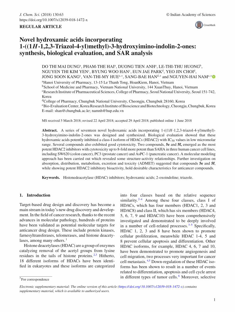
DO THI MAI DUNGa, PHAM-THE HAIa, DUONG TIEN ANHa, LE-THI-THU HUONGb,NGUYEN THI KIM YENc, BYUNG WOO HANc, EUN JAE PARKd, YEO JIN CHOId,JONG SOON KANGe, VAN-THI-MY HUEa,∗, SANG-BAE HANd,∗ and NGUYEN-HAI NAMa,∗aHanoi University of Pharmacy, 13-15 Le Thanh Tong, HoanKiem, Hanoi, VietnambSchool of Medicine and Pharmacy, Vietnam National University, 144 XuanThuy, Hanoi, VietnamcResearch Institute of Pharmaceutical Sciences, College of Pharmacy, Seoul National University, Seoul 151-742,KoreadCollege of Pharmacy, Chungbuk National University, Cheongju, Chungbuk 28160, KoreaeBio-Evaluation Center, Korea Research Institute of Bioscience and Biotechnology, Cheongju, Chungbuk, KoreaE-mail: [email protected]; [email protected]
MS received 5 March 2018; revised 22 April 2018; accepted 29 April 2018; published online 1 June 2018
Abstract. A series of seventeen novel hydroxamic acids incorporating 1-((1H -1,2,3-triazol-4-yl)methyl)-3-hydroxyimino-indolin-2-ones was designed and synthesized. Biological evaluation showed that thesehydroxamic acids potently inhibited a class-I isoform of HDACs (HDAC2) with IC50 values in low micromolarrange. Several compounds also exhibited good cytotoxicity. Two compounds, 5e and 5f, emerged as the mostpotent HDAC2 inhibitors with cytotoxicity up to 8-fold more potent than SAHA in three human cancer cell lines,including SW620 (colon cancer), PC3 (prostate cancer) and AsPC-1 (pancreatic cancer). A molecular modelingapproach has been carried out which revealed some structure-activity relationships. Further investigation onabsorption, distribution, metabolism, excretion and toxicity (ADMET) suggested that compounds 5e and 5f,while showing potent HDAC2 inhibitory bioactivity, hold desirable characteristics for anticancer compounds.
Keywords. Histonedeacetylase (HDAC) inhibitors; hydroxamic acids; 2-oxoindoline; triazole.
1. Introduction
Target-based drug design and discovery has become amain stream in today’s new drug discovery and develop-ment. In the field of cancer research, thanks to the recentadvances in molecular pathology, hundreds of proteinshave been validated as potential molecular targets foranticancer drug design. These include protein kinases,farnesyltransferases, telomerases, and histone deacety-lases, among many others.1
Histone deacetylases (HDAC) are a group of enzymescatalyzing removal of the acetyl groups from lysineresidues in the tails of histone proteins.2,3 Hitherto,18 different isoforms of HDACs have been identi-fied in eukaryotes and these isoforms are categorized
*For correspondence
Electronic supplementary material: The online version of this article (https:// doi.org/ 10.1007/ s12039-018-1472-x) containssupplementary material, which is available to authorized users.
into four classes based on the relative sequencesimilarity.2–4 Among these four classes, class I ofHDACs, which has four members (HDAC1, 2, 3 andHDAC8) and class II, which has six members (HDAC4,5, 6, 7, 9 and HDAC10) have been comprehensivelyinvestigated and demonstrated to be deeply involvedin a number of cell-related processes.3–5 Specifically,HDAC 1, 2, 3 and 8 have been shown to promotecellular proliferation, meanwhile HDAC 1-4, 5 and8 prevent cellular apoptosis and differentiation. OtherHDAC isoforms, for example, HDAC 4, 6, 7 and 10,have been demonstrated to promote angiogenesis andcell migration, two processes very important for cancercell metastasis.4,5 Down-regulation of these HDAC iso-forms has been shown to result in a number of eventsrelated to differentiation, apoptosis and cell cycle arrestin different types of tumor cells.6 Moreover, selective
1

63 Page 2 of 13 J. Chem. Sci. (2018) 130:63
Figure 1. Structures of some HDAC inhibitors.
suppression of the growth of tumor cells caused byHDAC inhibition has been clearly demonstrated notonly in vitro but also in a number of in vivo preclini-cal models and clinical settings.6 Design of compoundsto inhibit appropriate HDACs has, therefore, becomea very interesting approach in cancer drug design anddevelopment nowadays.7 As a result, a number ofHDAC inhibitors have been reported in the past decades.These inhibitors are diverse, from short-chain fattyacids (like butyrate, phenylbutyrate or valproic acid) tohydroxamic acids, or benzamides.8–14 To date, at least 4HDAC inhibitors have been approved for use in clinicalsettings. The first HDAC inhibitor approved by the U.S.FDA in October 2006 for the treatment of cutaneousT cell lymphoma (CTCL) was vorinostat (trade name,Zolinza®) (also known as SAHA or suberoylanilidehy-droxamic acid) (Figure 1). In 2009, the second HDACinhibitor, romidepsin (trade name, Istodax®) was alsoapproved by the U.S. FDA for the same indication. InFeb 2015, panobinostat (LBH-589, trade name Fary-dak®) was licensed by the US FDA for the treatmentof multiple myeloma.15 Also in 2015 chidamide (Epi-daza®) was approved by the Chinese FDA for relapsedor refractory peripheral T cell lymphoma.16 In addition,a number of other HDAC inhibitors such as PXD-01(belinostat), MS-27-527 (entinostat) (Figure 1) are cur-rently under different phases of clinical trials for severaltypes of cancer (Figure 1).
In continuation of our research program to developnovel hydroxamic acids as potential inhibitors ofHDACs and anticancer agents, we have designed, syn-thesized and evaluated several series of heterocyclic
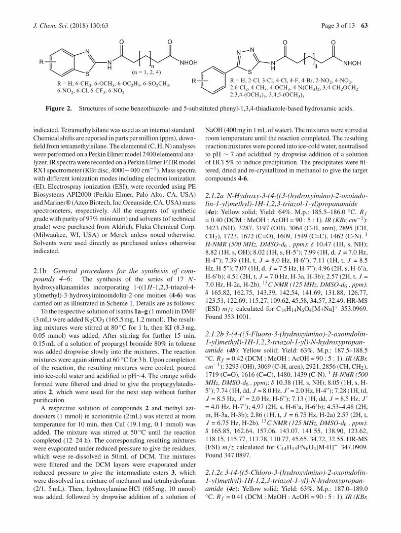
analogues of SAHA, which incorporated benzothiazoleor 5-aryl-1,3,4-thiadiazole systems (Figure 2). Thesecompounds exhibited very potent HDAC inhibitoryactivity as well as cytotoxicity.17–20 Several represen-tative compounds from these series also demonstratedvery potent antitumor activity in PC-3 prostate cancercells xenografted mice model.18 Encouraged by theseresults, we expanded our design to the new series of17 hydroxamic acids. In these hydroxamic acids the2-oxoindoline system is employed as a cap group andlinkers of different lengths incorporating a triazole moi-ety are probed. The current paper reports the results weobtained from the synthesis, biological evaluation andcomputational study on these novel hydroxamic acids.
2. Experimental
2.1 Chemistry
2.1a Chemicals and instruments: Thin layer chro-matography (TLC) was performed using Whatman® 250 µmSilica Gel GF Uniplates and visualized under UV light at 254and 365 nm. TLC was used to check the progress of reactionsand preliminary evaluation of compounds’ homogeneity.Melting points were measured using a Gallenkamp MeltingPoint Apparatus (LabMerchant, London, United Kingdom)and are uncorrected. Purification of compounds was carriedout using crystallization methods and/or open silica gel col-umn flash chromatography employing Merck silica gel 60(240 to 400 mesh) as stationary phase. Nuclear magnetic reso-nance spectra (1H NMR) were recorded on a Bruker 500 MHzspectrometer with DMSO-d6 as solvent unless otherwise
J. Chem. Sci. (2018) 130:63 Page 3 of 13 63
Figure 2. Structures of some benzothiazole- and 5-substituted phenyl-1,3,4-thiadiazole-based hydroxamic acids.
indicated. Tetramethylsilane was used as an internal standard.Chemical shifts are reported in parts per million (ppm), down-field from tetramethylsilane. The elemental (C, H, N) analyseswere performed on a Perkin Elmer model 2400 elemental ana-lyzer. IR spectra were recorded on a Perkin Elmer FTIR modelRX1 spectrometer (KBr disc, 4000−400 cm−1). Mass spectrawith different ionization modes including electron ionization(EI), Electrospray ionization (ESI), were recorded using PEBiosystems API2000 (Perkin Elmer, Palo Alto, CA, USA)and Mariner® (Azco Biotech, Inc.Oceanside, CA, USA)massspectrometers, respectively. All the reagents (of syntheticgrade with purity of 97% minimum) and solvents (of technicalgrade) were purchased from Aldrich, Fluka Chemical Corp.(Milwaukee, WI, USA) or Merck unless noted otherwise.Solvents were used directly as purchased unless otherwiseindicated.
2.1b General procedures for the synthesis of com-pounds 4–6: The synthesis of the series of 17 N -hydroxyalkanamides incorporating 1-((1H -1,2,3-triazol-4-yl)methyl)-3-hydroxyiminoindolin-2-one moities (4-6) wascarried out as illustrated in Scheme 1. Details are as follows:
To the respective solution of isatins 1a–g (1 mmol) in DMF(3 mL) were added K2CO3 (165.5 mg, 1.2 mmol). The result-ing mixtures were stirred at 80 ◦C for 1 h, then KI (8.3 mg,0.05 mmol) was added. After stirring for further 15 min,0.15 mL of a solution of propargyl bromide 80% in toluenewas added dropwise slowly into the mixtures. The reactionmixtures were again stirred at 60 ◦C for 3 h. Upon completionof the reaction, the resulting mixtures were cooled, pouredinto ice-cold water and acidified to pH∼4. The orange solidsformed were filtered and dried to give the propargylatedis-atins 2, which were used for the next step without furtherpurification.
A respective solution of compounds 2 and methyl azi-doesters (1 mmol) in acetonitrile (2 mL) was stirred at roomtemperature for 10 min, then CuI (19.1 mg, 0.1 mmol) wasadded. The mixture was stirred at 50 ◦C until the reactioncompleted (12–24 h). The corresponding resulting mixtureswere evaporated under reduced pressure to give the residues,which were re-dissolved in 50 mL of DCM. The mixtureswere filtered and the DCM layers were evaporated underreduced pressure to give the intermediate esters 3, whichwere dissolved in a mixture of methanol and tetrahydrofuran(2/1, 5 mL). Then, hydroxylamine.HCl (685 mg, 10 mmol)was added, followed by dropwise addition of a solution of
NaOH (400 mg in 1 mL of water). The mixtures were stirred atroom temperature until the reaction completed. The resultingreaction mixtures were poured into ice-cold water, neutralisedto pH ∼ 7 and acidified by dropwise addition of a solutionof HCl 5% to induce precipitation. The precipitates were fil-tered, dried and re-crystallized in methanol to give the targetcompounds 4-6.
2.1.2a N-Hydroxy-3-(4-((3-(hydroxyimino)-2-oxoindo-lin-1-yl)methyl)-1H-1,2,3-triazol-1-yl)propanamide(4a): Yellow solid; Yield: 64%. M.p.: 185.5–186.0 ◦C. R f
= 0.40 (DCM : MeOH : AcOH = 90 : 5 : 1). IR (KBr, cm−1):3423 (NH), 3287, 3197 (OH), 3064 (C-H, aren), 2895 (CH,CH2), 1723, 1672 (C=O), 1609, 1549 (C=C), 1462 (C-N). 1
H-NMR (500 MHz, DMSO-d6 , ppm): δ 10.47 (1H, s, NH);8.82 (1H, s, OH); 8.02 (1H, s, H-5’); 7.99 (1H, d, J = 7.0 Hz,H-4”); 7.39 (1H, t, J = 8.0 Hz, H-6”); 7.11 (1H, t, J = 8.5Hz, H-5”); 7.07 (1H, d, J = 7.5 Hz, H-7”); 4.96 (2H, s, H-6’a,H-6’b); 4.51 (2H, t, J = 7.0 Hz, H-3a, H-3b); 2.57 (2H, t, J =7.0 Hz, H-2a, H-2b). 13C NMR (125 MHz, DMSO-d6 , ppm):δ 165.82, 162.75, 143.39, 142.54, 141.69, 131.88, 126.77,123.51, 122.69, 115.27, 109.62, 45.58, 34.57, 32.49. HR-MS(ESI) m/z calculated for C14H14N6O4[M+Na]+ 353.0969.Found 353.1001.
2.1.2b 3-(4-((5-Fluoro-3-(hydroxyimino)-2-oxoindolin-1-yl)methyl)-1H-1,2,3-triazol-1-yl)-N-hydroxypropan-amide (4b): Yellow solid; Yield: 63%. M.p.: 187.5–188.5◦C. R f = 0.42 (DCM : MeOH : AcOH = 90 : 5 : 1). IR (KBr,cm−1): 3293 (OH), 3069 (C-H, aren), 2921, 2856 (CH, CH2),1719 (C=O), 1616 (C=C), 1480, 1439 (C-N). 1 H-NMR (500MHz, DMSO-d6 , ppm): δ 10.38 (1H, s, NH); 8.05 (1H, s, H-5’); 7.74 (1H, dd, J = 8.0 Hz, J ′ = 2.0 Hz, H-4”); 7.28 (1H, td,J = 8.5 Hz, J ′ = 2.0 Hz, H-6”); 7.13 (1H, dd, J = 8.5 Hz, J ′= 4.0 Hz, H-7”); 4.97 (2H, s, H-6’a, H-6’b); 4.53–4.48 (2H,m, H-3a, H-3b); 2.86 (1H, t, J = 6.75 Hz, H-2a) 2.57 (2H, t,J = 6.75 Hz, H-2b). 13C NMR (125 MHz, DMSO-d6 , ppm):δ 165.85, 162.64, 157.06, 143.07, 141.55, 138.90, 123.62,118.15, 115.77, 113.78, 110.77, 45.65, 34.72, 32.55. HR-MS(ESI) m/z calculated for C14H13FN6O4[M-H]− 347.0909.Found 347.0897.
2.1.2c 3-(4-((5-Chloro-3-(hydroxyimino)-2-oxoindolin-1-yl)methyl)-1H-1,2,3-triazol-1-yl)-N-hydroxypropan-amide (4c): Yellow solid; Yield: 63%. M.p.: 187.0–189.0◦C. R f = 0.41 (DCM : MeOH : AcOH = 90 : 5 : 1). IR (KBr,
63 Page 4 of 13 J. Chem. Sci. (2018) 130:63
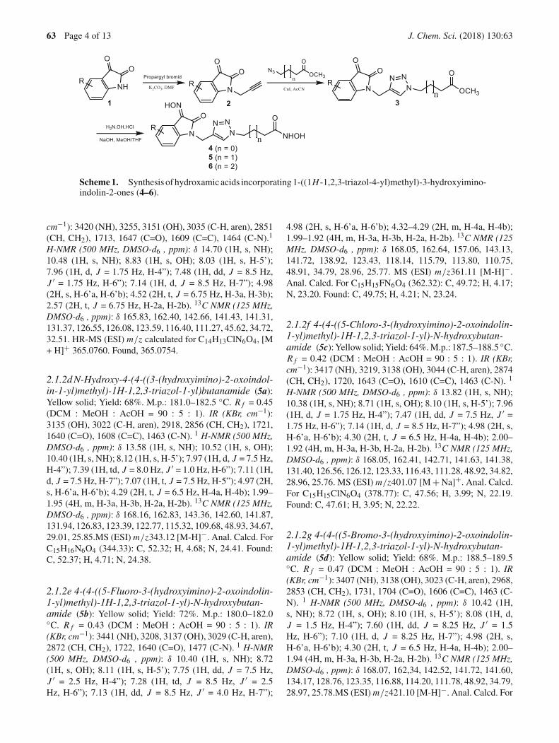
Scheme1. Synthesis of hydroxamic acids incorporating 1-((1H -1,2,3-triazol-4-yl)methyl)-3-hydroxyimino-indolin-2-ones (4–6).
cm−1): 3420 (NH), 3255, 3151 (OH), 3035 (C-H, aren), 2851(CH, CH2), 1713, 1647 (C=O), 1609 (C=C), 1464 (C-N).1
H-NMR (500 MHz, DMSO-d6 , ppm): δ 14.70 (1H, s, NH);10.48 (1H, s, NH); 8.83 (1H, s, OH); 8.03 (1H, s, H-5’);7.96 (1H, d, J = 1.75 Hz, H-4”); 7.48 (1H, dd, J = 8.5 Hz,J ′ = 1.75 Hz, H-6”); 7.14 (1H, d, J = 8.5 Hz, H-7”); 4.98(2H, s, H-6’a, H-6’b); 4.52 (2H, t, J = 6.75 Hz, H-3a, H-3b);2.57 (2H, t, J = 6.75 Hz, H-2a, H-2b). 13C NMR (125 MHz,DMSO-d6 , ppm): δ 165.83, 162.40, 142.66, 141.43, 141.31,131.37, 126.55, 126.08, 123.59, 116.40, 111.27, 45.62, 34.72,32.51. HR-MS (ESI) m/z calculated for C14H13ClN6O4, [M+ H]+ 365.0760. Found, 365.0754.
2.1.2d N-Hydroxy-4-(4-((3-(hydroxyimino)-2-oxoindol-in-1-yl)methyl)-1H-1,2,3-triazol-1-yl)butanamide (5a):Yellow solid; Yield: 68%. M.p.: 181.0–182.5 ◦C. R f = 0.45(DCM : MeOH : AcOH = 90 : 5 : 1). IR (KBr, cm−1):3135 (OH), 3022 (C-H, aren), 2918, 2856 (CH, CH2), 1721,1640 (C=O), 1608 (C=C), 1463 (C-N). 1 H-NMR (500 MHz,DMSO-d6 , ppm): δ 13.58 (1H, s, NH); 10.52 (1H, s, OH);10.40 (1H, s, NH); 8.12 (1H, s, H-5’); 7.97 (1H, d, J = 7.5 Hz,H-4”); 7.39 (1H, td, J = 8.0 Hz, J ′ = 1.0 Hz, H-6”); 7.11 (1H,d, J = 7.5 Hz, H-7”); 7.07 (1H, t, J = 7.5 Hz, H-5”); 4.97 (2H,s, H-6’a, H-6’b); 4.29 (2H, t, J = 6.5 Hz, H-4a, H-4b); 1.99–1.95 (4H, m, H-3a, H-3b, H-2a, H-2b). 13C NMR (125 MHz,DMSO-d6 , ppm): δ 168.16, 162.83, 143.36, 142.60, 141.87,131.94, 126.83, 123.39, 122.77, 115.32, 109.68, 48.93, 34.67,29.01, 25.85.MS (ESI) m/z343.12 [M-H]−. Anal. Calcd. ForC15H16N6O4 (344.33): C, 52.32; H, 4.68; N, 24.41. Found:C, 52.37; H, 4.71; N, 24.38.
2.1.2e 4-(4-((5-Fluoro-3-(hydroxyimino)-2-oxoindolin-1-yl)methyl)-1H-1,2,3-triazol-1-yl)-N-hydroxybutan-amide (5b): Yellow solid; Yield: 72%. M.p.: 180.0–182.0◦C. R f = 0.43 (DCM : MeOH : AcOH = 90 : 5 : 1). IR(KBr, cm−1): 3441 (NH), 3208, 3137 (OH), 3029 (C-H, aren),2872 (CH, CH2), 1722, 1640 (C=O), 1477 (C-N). 1 H-NMR(500 MHz, DMSO-d6 , ppm): δ 10.40 (1H, s, NH); 8.72(1H, s, OH); 8.11 (1H, s, H-5’); 7.75 (1H, dd, J = 7.5 Hz,J ′ = 2.5 Hz, H-4”); 7.28 (1H, td, J = 8.5 Hz, J ′ = 2.5Hz, H-6”); 7.13 (1H, dd, J = 8.5 Hz, J ′ = 4.0 Hz, H-7”);
4.98 (2H, s, H-6’a, H-6’b); 4.32–4.29 (2H, m, H-4a, H-4b);1.99–1.92 (4H, m, H-3a, H-3b, H-2a, H-2b). 13C NMR (125MHz, DMSO-d6 , ppm): δ 168.05, 162.64, 157.06, 143.13,141.72, 138.92, 123.43, 118.14, 115.79, 113.80, 110.75,48.91, 34.79, 28.96, 25.77. MS (ESI) m/z361.11 [M-H]−.Anal. Calcd. For C15H15FN6O4 (362.32): C, 49.72; H, 4.17;N, 23.20. Found: C, 49.75; H, 4.21; N, 23.24.
2.1.2f 4-(4-((5-Chloro-3-(hydroxyimino)-2-oxoindolin-1-yl)methyl)-1H-1,2,3-triazol-1-yl)-N-hydroxybutan-amide (5c): Yellow solid; Yield: 64%. M.p.: 187.5–188.5 ◦C.R f = 0.42 (DCM : MeOH : AcOH = 90 : 5 : 1). IR (KBr,cm−1): 3417 (NH), 3219, 3138 (OH), 3044 (C-H, aren), 2874(CH, CH2), 1720, 1643 (C=O), 1610 (C=C), 1463 (C-N). 1
H-NMR (500 MHz, DMSO-d6 , ppm): δ 13.82 (1H, s, NH);10.38 (1H, s, NH); 8.71 (1H, s, OH); 8.10 (1H, s, H-5’); 7.96(1H, d, J = 1.75 Hz, H-4”); 7.47 (1H, dd, J = 7.5 Hz, J ′ =1.75 Hz, H-6”); 7.14 (1H, d, J = 8.5 Hz, H-7”); 4.98 (2H, s,H-6’a, H-6’b); 4.30 (2H, t, J = 6.5 Hz, H-4a, H-4b); 2.00–1.92 (4H, m, H-3a, H-3b, H-2a, H-2b). 13C NMR (125 MHz,DMSO-d6 , ppm): δ 168.05, 162.41, 142.71, 141.63, 141.38,131.40, 126.56, 126.12, 123.33, 116.43, 111.28, 48.92, 34.82,28.96, 25.76. MS (ESI) m/z401.07 [M + Na]+. Anal. Calcd.For C15H15ClN6O4 (378.77): C, 47.56; H, 3.99; N, 22.19.Found: C, 47.61; H, 3.95; N, 22.22.
2.1.2g 4-(4-((5-Bromo-3-(hydroxyimino)-2-oxoindolin-1-yl)methyl)-1H-1,2,3-triazol-1-yl)-N-hydroxybutan-amide (5d): Yellow solid; Yield: 68%. M.p.: 188.5–189.5◦C. R f = 0.47 (DCM : MeOH : AcOH = 90 : 5 : 1). IR(KBr, cm−1): 3407 (NH), 3138 (OH), 3023 (C-H, aren), 2968,2853 (CH, CH2), 1731, 1704 (C=O), 1606 (C=C), 1463 (C-N). 1 H-NMR (500 MHz, DMSO-d6 , ppm): δ 10.42 (1H,s, NH); 8.72 (1H, s, OH); 8.10 (1H, s, H-5’); 8.08 (1H, d,J = 1.5 Hz, H-4”); 7.60 (1H, dd, J = 8.25 Hz, J ′ = 1.5Hz, H-6”); 7.10 (1H, d, J = 8.25 Hz, H-7”); 4.98 (2H, s,H-6’a, H-6’b); 4.30 (2H, t, J = 6.5 Hz, H-4a, H-4b); 2.00–1.94 (4H, m, H-3a, H-3b, H-2a, H-2b). 13C NMR (125 MHz,DMSO-d6 , ppm): δ 168.07, 162,34, 142.52, 141.72, 141.60,134.17, 128.76, 123.35, 116.88, 114.20, 111.78, 48.92, 34.79,28.97, 25.78.MS (ESI) m/z421.10 [M-H]−. Anal. Calcd. For
J. Chem. Sci. (2018) 130:63 Page 5 of 13 63
C15H15BrN6O4 (422.23): C, 42.57; H, 3.57; N, 19.86. Found:C, 42.59; H, 3.61; N, 19.85.
2.1.2h N-Hydroxy-4-(4-((3-(hydroxyimino)-5-methyl-2-oxoindolin-1-yl)methyl)-1H-1,2,3-triazol-1-yl)butana-mide (5e): Yellow solid; Yield: 71%. M.p.: 179.5–181.0 ◦C.R f = 0.41 (DCM : MeOH : AcOH = 90 : 5 : 1). IR (KBr,cm−1): 3280 (NH), 3184 (OH), 3068 (C-H, aren), 2908 (CH,CH2), 1713, 1673 (C=O), 1624, 1552 (C=C), 1476 (C-N). 1
H-NMR (500 MHz, DMSO-d6 , ppm): δ 13.44 (1H, s, NH);10.38 (1H, s, NH); 8.71 (1H, s, OH); 8.08 (1H, s, H-5’);7.83 (1H, s, H-4”); 7.20 (1H, d, J = 8.0 Hz, H-7”); 6.99(1H, d, J = 8.0 Hz, H-6”); 4.95 (2H, s, H-6’a, H-6’b);4.30 (2H, t, J = 6.5 Hz, H-4a, H-4b); 2.27 (3H, s, -CH3);1.98-1.93 (4H, m, H-3a, H-3b, H-2a, H-2b). 13C NMR (125MHz, DMSO-d6 , ppm): δ 168.08, 162.77, 143.59, 141.93,140.39, 132.14, 131.77, 127.38, 123.26, 115.35, 109.42,48.89, 34.67, 28.97, 25.78, 20.51. HR-MS (ESI) m/z calcu-lated for C16H18N6O4[M+Na]+ 381.1282. Found 381.1270.
2.1.2i N-Hydroxy-4-(4-((3-(hydroxyimino)-5-methoxy-2-oxoindolin-1-yl)methyl)-1H-1,2,3-triazol-1-yl)butan-amide (5f ): Yellow solid; Yield: 60%. M.p.: 180.0–182.0 ◦C.R f = 0.45 (DCM : MeOH : AcOH = 90 : 5 : 1). IR (KBr, cm−1):3281 (NH), 3187 (OH), 3071 (C-H, aren), 2916 (CH, CH2),1711, 1673 (C=O), 1630, 1596 (C=C), 1552, 1480 (C-N). 1 H-NMR (500 MHz, DMSO-d6 , ppm): δ 13.53 (1H, s, NH); 10.40(1H, s, NH); 8.09 (1H, s, H-5’); 7.58 (1H, d, J = 2.5 Hz, H-4”);7.04–6.98 (2H, m„ H-6”,H-7”); 4.94 (2H, s, H-6’a, H-6’b);4.30 (2H, t, J = 7.0 Hz, H-4a, H-4b); 3.72 (3H, s, -OCH3);2.00–1.92 (4H, m, H-3a, H-3b, H-2a, H-2b). 13C NMR (125MHz, DMSO-d6 , ppm): δ 168.05, 162.62, 155.21, 143.68,141.94, 136.24, 123.28, 116.86, 115.85, 113.09, 110.26,55.65, 48.90, 34.70, 28.97, 25.78. HR-MS (ESI) m/z cal-culated for C16H18N6O5[M-H]− 373.1266. Found 373.1241.
2.1.2j 4-(4-((7-Chloro-3-(hydroxyimino)-2-oxoindolin-1-yl)methyl)-1H-1,2,3-triazol-1-yl)-N-hydroxybutan-amide (5g): Yellow solid; Yield: 70%. M.p.: 187.5–188.5 ◦C.R f = 0.43 (DCM : MeOH : AcOH = 90 : 5 : 1). IR (KBr,cm−1): 3517, 3378 (NH), 3248 (OH), 3030 (C-H, aren), 2897(CH, CH2), 1717, 1664 (C=O), 1634, 1608 (C=C), 1441 (C-N). 1 H-NMR (500 MHz, DMSO-d6 , ppm): δ 10.41 (1H, s,NH); 8.72 (1H, s, OH); 8.07 (1H, dd, J = 7.75 Hz, J ′ = 1.0Hz, H-4”); 8.06 (1H, s, H-5’); 7.39 (1H, dd, J = 7.75 Hz,J ′ = 1.0 Hz, H-6”); 7.11 (1H, t, J = 7.75 Hz, H-5”); 5.31(2H, s, H-6’a, H-6’b); 4.29 (2H, t, J = 6.5 Hz, H-4a, H-4b);2.00–1.92 (4H, m, H-3a, H-3b, H-2a, H-2b). 13C NMR (125MHz, DMSO-d6 , ppm): δ 168.07, 163.64, 143.38, 142.19,138.46, 133.78, 125.84, 124.23, 122.49, 118.30, 114.80,48.88, 37.06, 28.96, 25.84. HR-MS (ESI) m/z calculated forC15H15ClN6O4[M-H]− 377.0770. Found 377.0748.
2.1.2k N-Hydroxy-5-(4-((3-(hydroxyimino)-2-oxoindo-lin-1-yl)methyl)-1H-1,2,3-triazol-1-yl)pentanamide(6a): Yellow solid; Yield: 68%. M.p.: 189.0–191.5 ◦C. R f
= 0.48 (DCM : MeOH : AcOH = 90 : 5 : 1). IR (KBr, cm−1):
3399, 3312 (NH), 3196 (OH), 3059 (C-H, aren), 2876 (CH,CH2), 1714, 1678 (C=O), 1608 (C=C), 1455 (C-N). 1 H-NMR(500 MHz, DMSO-d6 , ppm): δ 13.45 (1H, s, NH); 10.35 (1H,s, NH); 8.67 (1H, s, OH); 8.09 (1H, s, H-5’); 7.98 (1H, d, J =7.5 Hz, H-4”); 7.40 (1H, t, J = 7.5 Hz, H-6”); 7.12-7.06 (2H,m, H-5”, H-7”); 4.98 (2H, s, H-6’a, H-6’b); 4.29 (2H, t, J = 7.0Hz, H-5a, H-5b); 1.95 (2H, t, J = 7.5 Hz, H-2a, H-2b); 1.79–1.71 (2H, m, H-4a, H-4b); 1.45–1.39 (2H, m, H-3a, H-3b). 13CNMR (125 MHz, DMSO-d6 , ppm): δ 168.68, 162.74, 143.44,142.61, 141.80, 131.95, 126.85, 123.26, 122.73, 115.28,109.64, 49.05, 34.67, 29.22, 22.04. HR-MS (ESI) m/z cal-culated for C16H18N6O4[M-H]− 357.1317. Found 357.1313.
2.1.2l 5-(4-((5-Fluoro-3-(hydroxyimino)-2-oxoindolin-1-yl)methyl)-1H-1,2,3-triazol-1-yl)-N-hydroxypentan-amide (6b): Yellow solid; Yield: 69%. M.p.: 194.5–196.0 ◦C.R f = 0.48 (DCM : MeOH : AcOH = 90 : 5 : 1). IR (KBr,cm−1): 3396, 3320 (NH), 3172 (OH), 3058 (C-H, aren), 2876(CH, CH2), 1715, 1680 (C=O), 1630 (C=C), 1477 (C-N). 1
H-NMR (500 MHz, DMSO-d6 , ppm): δ 13.76 (1H, s, NH);10.34 (1H, s, NH); 8.67 (1H, s, OH); 8.08 (1H, s, H-5’); 7.75(1H, dd, J = 8.0 Hz, J ′ = 2.5 Hz, H-4”); 7.28 (1H, td, J =8.75 Hz, J ′ = 2.5 Hz, H-6”); 7.12 (1H, dd, J = 8.75 Hz, J ′ =4.0 Hz, H-7”); 4.98 (2H, s, H-6’a, H-6’b); 4.29 (2H, t, J = 7.0Hz, H-5a, H-5b); 1.95 (2H, t, J = 7.5 Hz, H-2a, H-2b); 1.76-1.73 (2H, m, H-4a, H-4b); 1.45–1.40 (2H, m, H-3a, H-3b).13C NMR (125 MHz, DMSO-d6 , ppm): δ 168.68, 162.61,158.00, 143.16, 141.64, 138.93, 123.29, 118.15, 115.77,113.83, 110.72, 49.06, 34.79, 31.51, 29.19, 22.03. HR-MS(ESI) m/z calculated for C16H17FN6O4[M-H]− 375.1222.Found 375.1229.
2.1.2m 5-(4-((5-Chloro-3-(hydroxyimino)-2-oxoindoli-n-1-yl)methyl)-1H-1,2,3-triazol-1-yl)-N-hydroxypenta-namide (6c): Yellow solid; Yield: 69%. M.p.: 194.5–196.0◦C. R f = 0.48 (DCM : MeOH : AcOH = 90 : 5 : 1). IR (KBr,cm−1): 3286 (NH), 3143 (OH), 3042 (C-H, aren), 2851 (CH,CH2), 1708, 1647 (C=O), 1608 (C=C), 1461 (C-N). 1 H-NMR(500 MHz, DMSO-d6 , ppm): δ 10.36 (1H, s, NH); 8.68 (1H,s, OH); 8.08 (1H, s, H-5’); 7.95 (1H, s, H-4”); 7.48 (1H, d,J = 8.0 Hz, H-6”); 7.14 (1H, d, J = 8.0 Hz, H-7”); 4.98(2H, s, H-6’a, H-6’b); 4.29 (2H, t, J = 7.0 Hz, H-5a, H-5b);1.95 (2H, t, J = 7.5 Hz, H-2a, H-2b); 1.77–1.72 (2H, m, H-4a, H-4b); 1.45–1.40 (2H, m, H-3a, H-3b). 13C NMR (125MHz, DMSO-d6 , ppm): δ 168.73, 162.44, 142.71, 141.57,141.38, 131.42, 126.59, 126.14, 123.31, 116.44, 111.29,49.09, 34.84, 31.53, 29.22, 22.06. HR-MS (ESI) m/z calcu-lated for C16H17ClN6O4[M-H]− 391.0927. Found 391.0938.
2.1.2n 5-(4-((5-Bromo-3-(hydroxyimino)-2-oxoindolin-1-yl)methyl)-1H-1,2,3-triazol-1-yl)-N-hydroxypentan-amide (6d): Yellow solid; Yield: 74%. M.p.: 201.5–203.0 ◦C.R f = 0.51 (DCM : MeOH : AcOH = 90 : 5 : 1). IR (KBr, cm−1):3420, 3289 (NH), 3147 (OH), 3031 (C-H, aren), 2862 (CH,CH2), 1708, 1646 (C=O), 1606 (C=C), 1462 (C-N). 1 H-NMR(500 MHz, DMSO-d6 , ppm): δ 10.34 (1H, s, NH); 8.67 (1H,s, OH); 8.08 (2H, s, H-5’, H-4”); 7.60 (1H, d, J = 7.75 Hz, H-
63 Page 6 of 13 J. Chem. Sci. (2018) 130:63
6”); 7.09 (1H, d, J = 7.75 Hz, H-7”); 4.98 (2H, s, H-6’a,H-6’b); 4.29 (2H, t, J = 6.0 Hz, H-5a, H-5b); 1.95 (2H,t, J = 6.5 Hz, H-2a, H-2b); 1.75–1.73 (2H, m, H-4a, H-4b); 1.45–1.41 (2H, m, H-3a, H-3b). 13C NMR (125 MHz,DMSO-d6 , ppm): δ 168.68, 162.31, 142.57, 141.73, 141.54,134.20, 128.79, 123.27, 116.86, 114.21, 111.76, 49.07,34.80, 31.51, 29.20, 22.04. HR-MS (ESI) m/z calculated forC16H17BrN6O4, [M-H]− 437.0422. Found 437.0421.
2.1.2o N-Hydroxy-5-(4-((3-(hydroxyimino)-5-methyl-2-oxoindolin-1-yl)methyl)-1H-1,2,3-triazol-1-yl)pentan-amide (6e): Yellow solid; Yield: 75%. M.p.: 199.5–201.6 ◦C.R f = 0.46 (DCM : MeOH : AcOH = 90 : 5 : 1). IR (KBr,cm−1): 3317 (NH), 3188 (OH), 3061 (C-H, aren), 2879 (CH,CH2), 1713, 1680 (C=O), 1622 (C=C), 1460 (C-N). 1 H-NMR(500 MHz, DMSO-d6 , ppm): δ 13.43 (1H, s, NH); 10.34 (1H,s, NH); 8.66 (1H, s, OH); 8.06 (1H, s, H-5’); 7.83 (1H, s,H-4”); 7.21 (1H, s, H-6”); 6.99 (1H, d, J = 6.0 Hz, H-7”);4.94 (2H, s, H-6’a, H-6’b); 4.29 (2H, s, H-5a, H-5b); 2.27(3H, s, -CH3); 1.95 (2H, s, H-2a, H-2b); 1.74 (2H, s, H-4a, H-4b); 1.42 (2H, s, H-3a, H-3b). 13C NMR (125 MHz,DMSO-d6 , ppm): δ 168.25, 163.25, 144.08, 142.45, 140.86,132.61, 132.25, 127.86, 123.74, 115.83, 109.89, 49.54, 35.17,32.00, 29.69, 22.52, 20.97. HR-MS (ESI) m/z calculated forC17H20N6O4, [M-H]− 371.1473. Found 371.1484.
2.1.2p N-Hydroxy-5-(4-((3-(hydroxyimino)-5-methoxy-2-oxoindolin-1-yl)methyl)-1H-1,2,3-triazol-1-yl)penta-namide (6f ): Yellow solid; Yield: 64%. M.p.: 196.0–197.5◦C. R f = 0.48 (DCM : MeOH : AcOH = 90 : 5 : 1). IR(KBr, cm−1): 3421 (NH), 3134 (OH), 3047 (C-H, aren), 2929,2838 (CH, CH2), 1718, 1628 (C=O), 1598 (C=C), 1480 (C-N). 1 H-NMR (500 MHz, DMSO-d6 , ppm): δ 10.35 (1H, s,NH); 8.67 (1H, s, OH); 8.05 (1H, s, H-5’); 7.57 (1H, d, J= 2.5 Hz, H-4”); 7.03–6.98 (2H, m, H-6”, H-7”); 4.94 (2H,s, H-6’a, H-6’b); 4.29 (2H, t, J = 7.0 Hz, H-5a, H-5b); 3.72(3H, s, -OCH3); 1.95 (2H, t, J = 7.0 Hz, H-2a, H-2b); 1.75–1.72 (2H, m, H-4a, H-4b); 1.45–1.40 (2H, m, H-3a, H-3b).13C NMR (125 MHz, DMSO-d6 , ppm): δ 168.74, 162.66,155.25, 143.71, 141.87, 136.25, 123.24, 116.90, 115.87,113.14, 110.28, 55.68, 49.07, 34.73, 31.54, 29.22, 22.06. HR-MS (ESI) m/z calculated for C17H20N6O5[M-H]− 387.1422.Found 387.1440.
2.1.2q 5-(4-((7-Chloro-3-(hydroxyimino)-2-oxoindolin-1-yl)methyl)-1H-1,2,3-triazol-1-yl)-N-hydroxypentan-amide (6g): Yellow solid; Yield: 73%. M.p.: 198.5–201.0 ◦C.R f = 0.47 (DCM : MeOH : AcOH = 90 : 5 : 1). IR (KBr, cm−1):3422 (NH), 3165 (OH), 3057 (C-H, aren), 2959, 2866 (CH,CH2), 1724, 1618 (C=O), 1608 (C=C), 1469 (C-N). 1 H-NMR(500 MHz, DMSO-d6 , ppm): δ 13.83 (1H, s, NH); 10.34 (1H,s, NH); 8.66 (1H, s, OH); 8.07 (1H, d, J = 7.0 Hz, H-4”); 8.03(1H, s, H-5’); 7.39 (1H, d, J = 8.0 Hz, H-6”); 7.11 (1H, t, J= 7.5 Hz, H-5”); 5.31 (2H, s, H-6’a, H-6’b); 4.29 (2H, t, J =7.0 Hz, H-5a, H-5b); 1.95 (2H, t, J = 7.25 Hz, H-2a, H-2b);1.77–1.71 (2H, m, H-4a, H-4b); 145–1.39 (2H, m, H-3a, H-3b). 13C NMR (125 MHz, DMSO-d6 , ppm): δ 168.67, 163.63,
143.26, 142.20, 138.45, 133.77, 125.84, 124.22, 122.44,118.29, 114.80, 49.01, 37.06, 31.50, 29.24, 22.01. HR-MS(ESI) m/z calculated for C16H17ClN6O4[M-H]− 391.0927.Found 391.0918.
2.2 Biological evaluation
2.2a Cytotoxicity assay: The cytotoxicity of the synthe-sized compounds was evaluated against three human cancercell lines, including SW620 (colon cancer), PC3 (prostatecancer), and AsPC-1 (pancreatic cancer). The cell lines werepurchased from a Cancer Cell Bank at the Korea ResearchInstitute of Bioscience and Biotechnology (KRIBB). Themedia, sera and other reagents that were used for cell culturein this assay were obtained from GIBCO Co. Ltd. (GrandIsland, New York, USA).The cells were culture in DMEM(Dulbecco’s Modified Eagle Medium) until confluence. Thecells were then trypsinized and suspended at 3 × 10⁴ cells/mLof cell culture medium. On day 0, each well of the 96-wellplates was seeded with 180 µL of cell suspension. The plateswere then incubated in a 5% CO2 incubator at 37 ◦C for 24h. Compounds were initially dissolved in dimethyl sulfoxide(DMSO) and diluted to appropriate concentrations by culturemedium. Then 20 µL of each sample of compounds, whichwere prepared as described above, were added to each wellof the 96-well plates, which had been seeded with cell sus-pension and incubated for 24 h, at various concentrations.The plates were further incubated for 48 h. Cytotoxicity ofthe compounds was measured by the colorimetric method, asdescribed previously.21 with slight modifications.22–24 TheIC50 values were calculated using a Probits method,25 takingaverage values from three independent determinations (SD ≤10%).
2.2b HDAC2 enzyme assay: The HDAC2 enzyme waspurchased from BPS Bioscience (San Diego, CA, USA).The HDAC enzymatic assay was performed using a Flu-orogenic HDAC Assay Kit (BPS Bioscience) according tothe manufacturer’s instructions. Briefly, HDAC2 enzymeswere incubated with vehicle or various concentrations of theassayed samples or SAHA for 30 min at 37 ◦C in the presenceof an HDAC fluorimetric substrate. The HDAC assay devel-oper (which produces a fluorophore in reaction mixture) wasadded, and the fluorescence was measured using VICTOR3
(PerkinElmer, Waltham, MA, USA) with excitation at 360 nmand emission at 460 nm. The measured activities were sub-tracted by the vehicle-treated control enzyme activities andIC50values were calculated using GraphPad Prism (Graph-Pad Software, San Diego, CA, USA).
2.3 Docking studies
An AutoDockVina program (The Scripps Research Insti-tute, CA, USA)was used for docking.26 The structure ofHDAC2 protein in complex with SAHA was obtained fromthe Protein Data Bank (PDB) (PDB ID: 4LXZ).27 The
J. Chem. Sci. (2018) 130:63 Page 7 of 13 63
coordinates of the compounds were generated by using theGlycoBioChem PRODRG2 Server (http://davapc1.bioch.dundee.ac.uk/prodrg/).28 For the docking studies, the grid mapswere centered on the SAHA binding site and comprised26 × 26 × 22 points with 1.0 Å spacing after SAHA wasremoved from the complex structure, as described in previousworks.17,18,20 The AutoDockVina program was performedusing eight-way multithreading and the other parameters weredefault settings.
2.4 Prediction of absorption, distribution,metabolism, elimination, and toxicity (ADMET)
In this work ADMET-related properties were computed usingVolsurf+ v.1.0.4 and the Chemistry Development Kit (CDK)software (http://cdk.sourceforge.net/). Compound topologywas saved as SMILES format (simplified molecular-inputline-entry system) using MarvinSketch and further submittedto Volsurf and CDK for molecular descriptor computation.ADMET descriptors comprise Blood-Brain Barrier distribu-tion (logBB), percentage of protein binding (PB, %), volumeof distribution (VD, L/kg) and metabolic stability againsthuman CYP3A4 enzyme (MetStab, %). The ability to interactwith cytochrome enzyme isoforms (substrate and/or inhibi-tion of CYP3A4, 1A2, 2C9, 2C19, and 2D6) was also checkedusing admetSAR server developed by Cheng et al. 29 Physic-ochemical descriptors including molecular weight (MW),partition coefficients (clogP, clogDpH=7.4), total polar surface
area (PSA, Å2), number of hydrogen bond donors and accep-tors, thermodynamic and pH-dependent aqueous solubility(mg/ml) were calculated. In addition, 3PRule was applied toclassify permeability across adenocarcinoma (Caco-2) cellmembrane30 - a model commonly used for intestinal absorp-tion estimation. The P-glycoprotein (P-gp, MDR1) substratestates were identified using chemoinformatic approachesdeveloped by Levatic et al.31 (available at http://pgp.biozyne.com). Lipinski’s Rule of Five (Ro5) was also applied to revealdrug-likeness of designed compounds.32 Finally, in vivo tox-icity was predicted by analyzing two risk factors (clogP andPSA) following the same criteria described by Hughes et al. 33
3. Results and Discussion
3.1 Chemistry
The targeted hydroxamic acids 4–6were synthesized viathree-step pathway, as illustrated in Scheme 1. The firststep was a nucleophilic substitution between isatins 1and propargyl bromide under basic conditions (K2CO3)
in dimethylformamide (DMF) with a catalytic amountof KI to furnish 1-propargylisatins 2. In the secondstep, a Click reaction between propargyl compounds2 and respective methyl azidoalkanoates (includingmethyl 3-azidopropanoate, methyl 4-azidobutanoate,methyl 5-azidovalerate) gave the intermediate esters 3.
This reaction proceeded smoothly in acetonitrile as asolvent and copper iodide as a catalyst. In most cases,the ester intermediates were precipitated by titration ofthe reaction residues, which were obtained by evapo-ration of acetonitrile, with cold water. The final step inthe pathway involved a nucleophilic acyl substitutionof hydroxylamine hydrochloride with the esters 3. Thisreaction occurred under alkaline conditions in a solventmixture which included tetrahydrofuran and methanolat 0–5 ◦C. The overall yields of compounds 4-6 weremoderate.
The structures of the synthesized compounds weredetermined straightforwardly based on analysis of spec-troscopic data, including IR, MS, 1H and 13C NMR(Supporting Information). The concurrent condensationof the hydroxylamine with the oxo group at position 3on the indoline ring under the above conditions occurredin all cases. This condensation has been observed andfully explained previously.19
3.2 Bioactivity
Due to limited funding resource, in this investigation thecompounds synthesized were preliminarily evaluatedfor histone deacetylase inhibition using HDAC2. Wechose HDAC2 because it has been shown that deacety-lation of a number of key histone proteins is regulatedprincipally by HDAC2 and HDAC3. HDAC2 has beendemonstrated to play very important role in promotingcellular proliferation, while inhibiting normal cell apop-tosis.34 In addition, the compounds were also evaluatedfor cytotoxicity against three human cancer cell lines,including SW620 (colon cancer), PC3 (prostate can-cer), and AsPC-1 (pancreatic cancer). The results arepresented in Table 1.
When designing these compounds, we initially envi-sioned that the triazole moiety would add more benefitsin hydrogen bonding with the amino acid regions inthe active binding sites of HDAC, while still servingas a part of a linker between the 2-oxoindoline andhydroxamic acid moieties. Due to shorter length ofthe C=N and C-N bonds compared to C-C bond, weestimated that if there were only 2C between the tri-azole and hydroxamic acids, the linker between the2-oxoindoline and hydroxamic acid moieties (in com-pounds 4a-c) would be shorter than 6C linker in SAHA.Therefore we decided to design series 5a-g with n =1 as the target compounds. Series 6a-g with one extraC was also synthesized to compare with series 5a-g.The results presented in Table 1 demonstrate that inseries 5a-g and 6a-g, with the exception of a 5-fluorinesubstituent, other substituents at either position 5 or 7
63 Page 8 of 13 J. Chem. Sci. (2018) 130:63
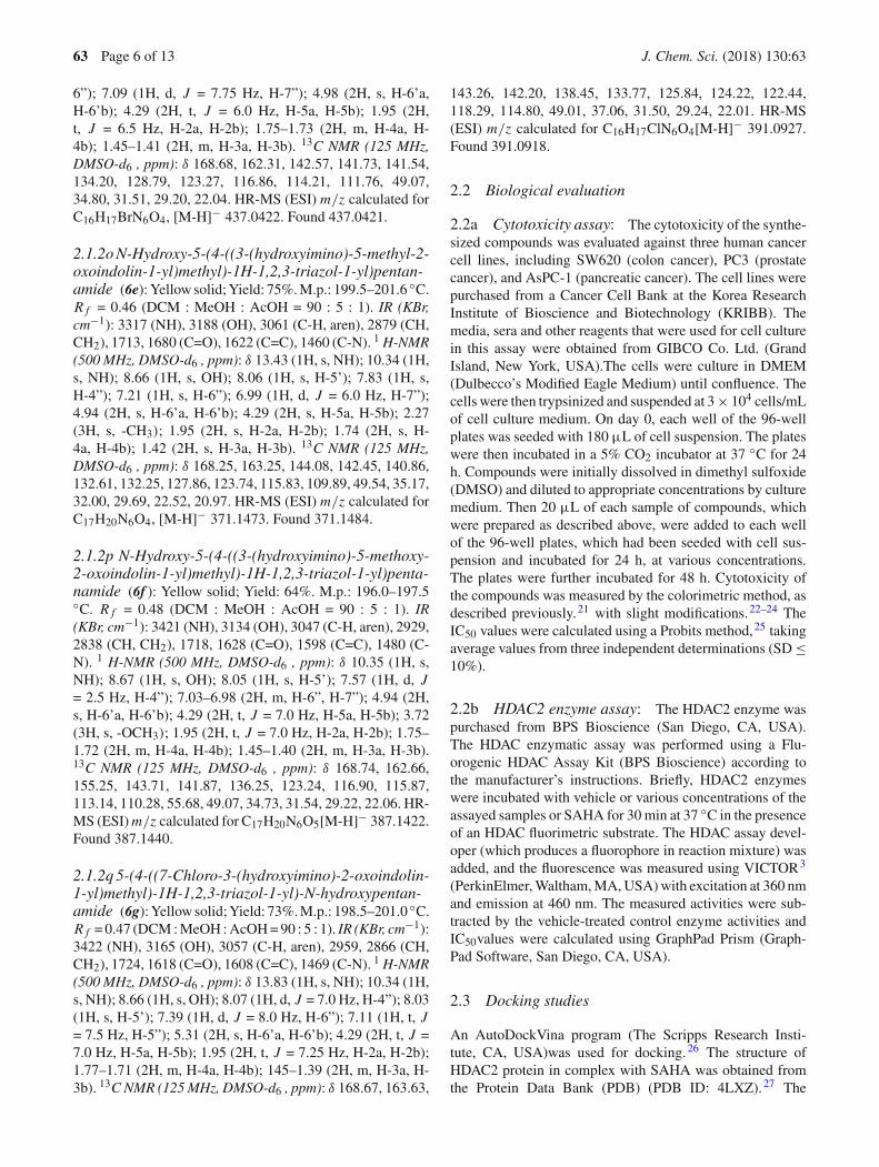
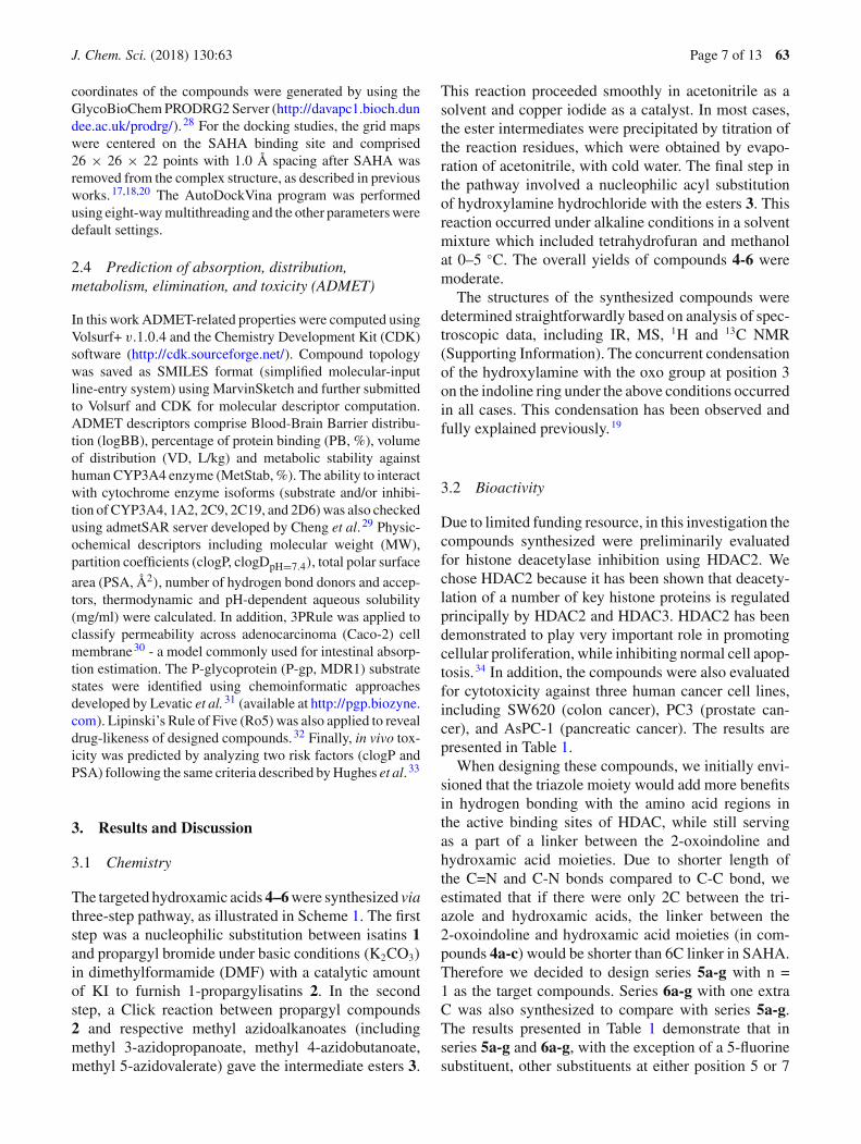
Table 1. Inhibition of HDAC activity and cytotoxicity of the compounds synthesized against several humancancer cell lines.
Cpd code R n LogP1 HDAC2 inhibition (IC50,2µM) Cytotoxicity (IC50,2 µM)/Cell lines3
SW620 PC3 AsPC-1
4a H 0 0.41 1.70 29.0 >30 >304b 5-F 0 0.61 6.24 >30 >30 >304c 5-Cl 0 1.05 2.80 >30 >30 >305a H 1 0.90 6.16 26.26 >30 26.875b 5-F 1 1.10 8.27 23.64 >30 >305c 5-Cl 1 1.54 1.72 13.46 16.28 11.605d 5-Br 1 1.79 3.53 2.93 6.08 3.015e 5-CH3 1 1.44 1.28 0.73 0.76 0.495f 5-OCH3 1 0.98 0.91 1.61 1.74 1.495g 7-Cl 1 1.54 5.08 10.58 9.27 12.906a -H 2 1.39 4.87 >30 >30 >306b 5-F 2 1.59 26.64 >30 >30 >306c 5-Cl 2 2.03 2.65 9.16 4.69 4.516d 5-Br 2 2.28 2.16 5.64 3.42 4.436e 5-CH3 2 1.94 3.52 >30 >30 >306f 5-OCH3 2 1.47 4.15 >30 >30 >306g 7-Cl 2 2.03 4.77 >30 >30 >30SAHA4 1.44 1.06 3.20 3.70 3.75
1Calculated by ChemDraw 9.0 software; 2The concentration (µM) of compounds that produces a 50% reductionin enzyme activity or cell growth, the numbers represent the averaged results from triplicate experiments withdeviation of less than 10%.; 3Cell lines: SW620, colon cancer; PC3, prostate cancer; AsPC-1, pancreatic cancer;4SAHA, suberoylanilide acid, a positive control.
on the 2-oxoindoline ring generally enhanced HDAC2inhibition. Also, in overall, series 5a-g inhibited theactivity of HDAC2 more potently compared to series6a-g. A similar trend was also observed with cytotox-icity when comparing series 5a-g and series 6a-g. Inseries 5a-g, it was found that the relationships betweenHDAC2 inhibition and cytotoxicity of the compoundswere relatively well correlated. Compounds 5d and5e,which were the most potent HDAC2 inhibitors in theseries, were also found to be the most cytotoxic againstall three cancer cell lines tested. In contrast, compounds5b and5g, which were the least potent HDAC2 inhibitorsin the series, were also among the least cytotoxic ones.From the results obtained, it is suggested that, whenconsidering the 4-methyl-1H -1,2,3-triazole moiety asa part of a linker between the 3-oxoindoline systemand hydroxamic acid group, an alkyl length of threecarbon connecting hydroxamic acid and triazole 1H -1,2,3-triazole moieties would be most favorable forbioactivities.
Among the compounds synthesized, compound 5fwas found to exhibit similar potency in term of HDAC2
inhibition compared to SAHA, meanwhile compound5eemerged as the most potential candidate with cytotoxi-city or up to 8-fold more potent than SAHA in all threecancer cell lines tested. It could be seen that in these twocompounds the correlation between HDAC2 inhibitorypotency and cytotoxicity was not finely observed. Somereasons might explain for this discrepancy. First, com-pound 5e could be more potently inhibit other types ofHDACs of class I and class II, which are also importantin promoting cell proliferation and inhibiting cellularapoptosis. Second, from logP values, it would be pos-sible that compound 5f (with lower logP value of 0.98)was not as good as compound 5e (with higher logP valueof 1.44, similar to that of SAHA) in penetrating throughcellular membrane.
3.3 Docking studies and structure-activityrelationships
It has been shown that histone-H3 and histone-H4deacetylation is regulated principally by HDAC2 and
J. Chem. Sci. (2018) 130:63 Page 9 of 13 63
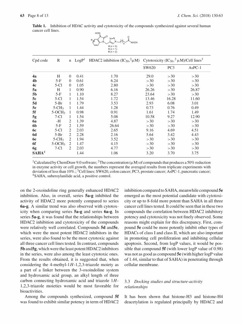
HDAC3.34 and the crystal structure of HDAC2 incomplex with SAHA (PDB ID: 4LXZ) has beenreported by Lauffer and co-workers.27 So we decidedto select the structure of SAHA-HDAC2 complex atemplate in docking experiments to study the structure-activity relationships of these hydroxamic acids andHDAC. Firstly, a control re-docking with co-crystalSAHA to the crystal structures of HDAC2 was executedfollowing the procedures reported previously.17,19,20 Theresults showed very similar interaction pattern betweenre-docked and the original SAHA in the crystal struc-ture (the all-atom root-mean-square deviation of 0.609Å). It was found from docking experiments that all ofthe hydroxamic acids synthesized were well located inthe active site of the enzyme with binding affinities(�G) between −6.7 and −8.1 kcal/mol, comparable orlower than that of SAHA (Table 2). It is noted that, insome instances the predicted binding affinities of thecompounds were not readily correlated to the experi-mental data obtained from HDAC2 inhibition assay. Forexample, the stabilization energies of predicted bind-ing modes on HDAC2 of compounds 5b and 5c werefound to be quite similar (−7.7 and −7.8 kcal/mol,respectively). These results were not explainable forthe 5-fold difference in the HDAC2 inhibitory effectsof compounds 5b and 5c. Especially in case of com-pound 5f, which has the same calculated stabilizationenergies as SAHA (−7.4 kcal/mol) was found to exhibitsimilar potency compared to SAHA in HDAC2 inhibi-tion (IC50 value was 0.91 µM vs. 1.06 µM of SAHA).Compounds 5e (�G = −8.1 kcal/mol) and5f were twomost potent in the series in term of HDAC2 inhibi-tion. Interestingly however, the length of aliphatic chainlinking hydroxamic group with triazole ring appearedto be an important factor. There was a remarkabledrop in the docking affinity ranges from butanamideto pentanamide moieties, which suggested the negativeeffect of lengthening the aliphatic chain beyond four-carbons.
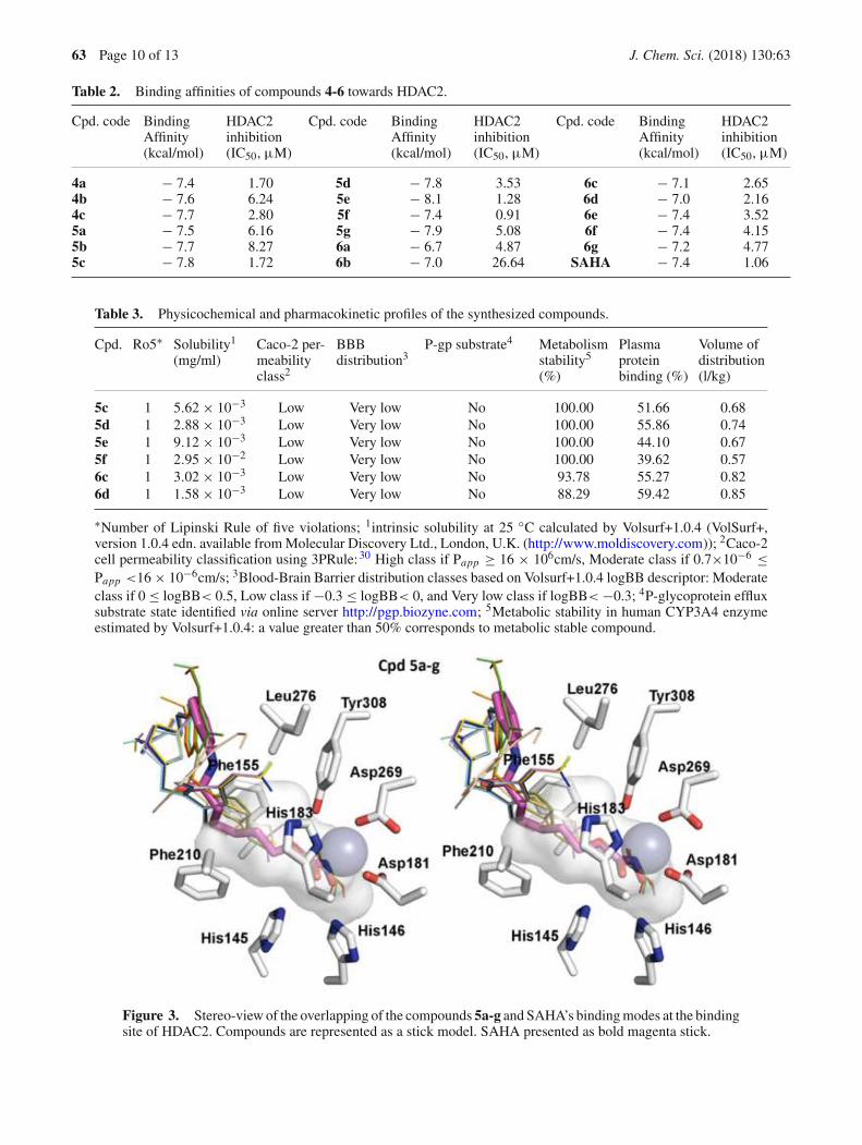
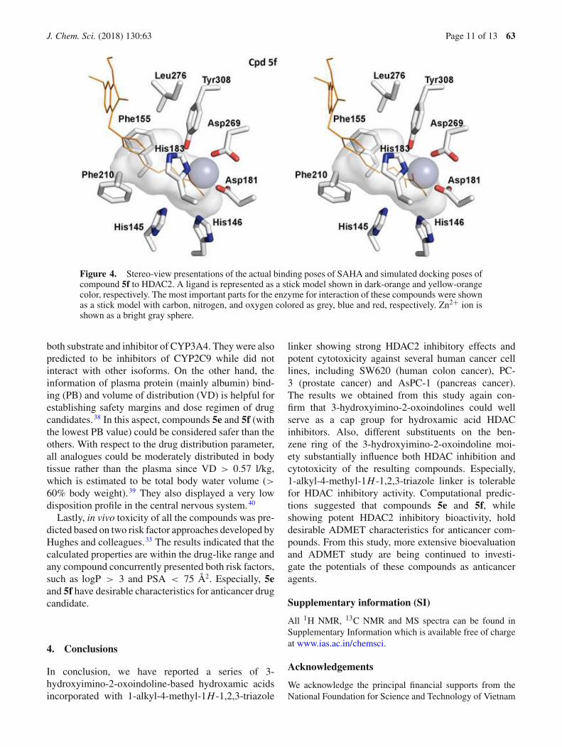
From the docking experiments, it was also found thata zinc ion (grey sphere), which was coordinated by threeresidues of HDAC2, including Asp181, His183 andAsp269, interacted with all the hydroxamic acid groupsin a similar manner as SAHA did. This zinc-chelatingfunctionality has been identified as the dominant fac-tor for inhibitor potency of synthesized hydroxamicderivatives. For all the compounds, it was found that a1-alkyl-4-methyl-1H -1,2,3-triazole linker between theindoline and hydroxamic acid moieties was tightlystacked between Phe155 and Phe210 residues of theenzymes (Figures 3, 4) and this pi-stacking interac-tion could be the key factor attributing to the goodbinding affinities of the compounds with HDAC2.
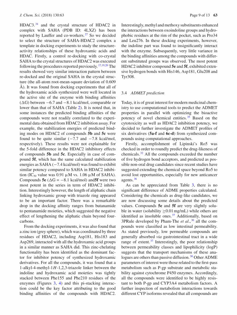
Interestingly, methyl and methoxy substituents enhancedthe interactions between oxoindoline groups and hydro-phobic residues at the rim of the pocket, such as Pro34and Leu276. In these docking experiments, however,the indoline part was found to insignificantly interactwith the enzyme. Subsequently, very little variance inthe binding affinities among the compounds with differ-ent substituted groups was observed. The most potentHDAC2 inhibitor compound 5e and 5f, exhibited exten-sive hydrogen bonds with His146, Asp181, Glu208 andTyr308.
3.4 ADMET prediction
Today, it is of great interest for modern medicinal chem-istry to use computational tools to predict the ADMETproperties in parallel with optimizing the bioactivepotency of novel chemical entities.35 Based on thecytotoxicity as well as HDAC2 inhibition potency, wedecided to further investigate the ADMET profiles ofsix derivatives (5a-f and 6c-d) from synthesized com-pounds using computational approaches.
Firstly, accomplishment of Lipinski’s Ro5 waschecked in order to roundly predict the drug-likeness ofchemicals.32 All the compounds exceeded the numberof five hydrogen bond acceptors, and predicted as pos-sible non-oral drug candidates since recent studies havesuggested extending the chemical space beyond Ro5 toavoid lost opportunities, especially for new anticanceragents.36
As can be appreciated from Table 3, there is nosignificant difference of ADME properties calculated.Considering the chemicals as drug-like candidates, weare now discussing some details about the predictedvalues. Compounds 5e and 5f are very slightly solu-ble in water (solubility ≥0.01 mg/mL) while others areidentified as insoluble ones.37 Additionally, based on3PRule developed by Pham-The et al., 30 all the com-pounds were classified as low intestinal permeability.As stated previously, low permeable compounds aregenerally absorbed via gastrointestinal tract in a widerange of extent.37 Interestingly, the poor relationshipbetween permeability classes and lipophilicity (logP)suggests that the transport mechanisms of these ana-logues are others than passive diffusion.30 Other ADMEparameters of interest were those related to the first-passmetabolism such as P-gp substrate and metabolic sta-bility against cytochrome P450 enzymes. Accordingly,all the compounds were identified to be highly resis-tant to both P-gp and CYP3A4 metabolism factors. Afurther inspection of metabolism interactions towardsdifferent CYP isoforms revealed that all compounds are
63 Page 10 of 13 J. Chem. Sci. (2018) 130:63
Table 2. Binding affinities of compounds 4-6 towards HDAC2.
Cpd. code BindingAffinity(kcal/mol)
HDAC2inhibition(IC50, µM)
Cpd. code BindingAffinity(kcal/mol)
HDAC2inhibition(IC50, µM)
Cpd. code BindingAffinity(kcal/mol)
HDAC2inhibition(IC50, µM)
4a − 7.4 1.70 5d − 7.8 3.53 6c − 7.1 2.654b − 7.6 6.24 5e − 8.1 1.28 6d − 7.0 2.164c − 7.7 2.80 5f − 7.4 0.91 6e − 7.4 3.525a − 7.5 6.16 5g − 7.9 5.08 6f − 7.4 4.155b − 7.7 8.27 6a − 6.7 4.87 6g − 7.2 4.775c − 7.8 1.72 6b − 7.0 26.64 SAHA − 7.4 1.06
Table 3. Physicochemical and pharmacokinetic profiles of the synthesized compounds.
Cpd. Ro5∗ Solubility1
(mg/ml)Caco-2 per-meabilityclass2
BBBdistribution3
P-gp substrate4 Metabolismstability5
(%)
Plasmaproteinbinding (%)
Volume ofdistribution(l/kg)
5c 1 5.62 × 10⁻³ Low Very low No 100.00 51.66 0.685d 1 2.88 × 10⁻³ Low Very low No 100.00 55.86 0.745e 1 9.12 × 10⁻³ Low Very low No 100.00 44.10 0.675f 1 2.95 × 10⁻² Low Very low No 100.00 39.62 0.576c 1 3.02 × 10⁻³ Low Very low No 93.78 55.27 0.826d 1 1.58 × 10⁻³ Low Very low No 88.29 59.42 0.85
∗Number of Lipinski Rule of five violations; 1intrinsic solubility at 25 ◦C calculated by Volsurf+1.0.4 (VolSurf+,version 1.0.4 edn. available from Molecular Discovery Ltd., London, U.K. (http://www.moldiscovery.com)); 2Caco-2cell permeability classification using 3PRule:30 High class if Papp ≥ 16 × 106cm/s, Moderate class if 0.7×10⁻⁶ ≤Papp <16 × 10−6cm/s; 3Blood-Brain Barrier distribution classes based on Volsurf+1.0.4 logBB descriptor: Moderateclass if 0 ≤ logBB< 0.5, Low class if −0.3 ≤ logBB< 0, and Very low class if logBB< −0.3; 4P-glycoprotein effluxsubstrate state identified via online server http://pgp.biozyne.com; 5Metabolic stability in human CYP3A4 enzymeestimated by Volsurf+1.0.4: a value greater than 50% corresponds to metabolic stable compound.
Figure 3. Stereo-view of the overlapping of the compounds 5a-g and SAHA’s binding modes at the bindingsite of HDAC2. Compounds are represented as a stick model. SAHA presented as bold magenta stick.
J. Chem. Sci. (2018) 130:63 Page 11 of 13 63
Figure 4. Stereo-view presentations of the actual binding poses of SAHA and simulated docking poses ofcompound 5f to HDAC2. A ligand is represented as a stick model shown in dark-orange and yellow-orangecolor, respectively. The most important parts for the enzyme for interaction of these compounds were shownas a stick model with carbon, nitrogen, and oxygen colored as grey, blue and red, respectively. Zn2+ ion isshown as a bright gray sphere.
both substrate and inhibitor of CYP3A4. They were alsopredicted to be inhibitors of CYP2C9 while did notinteract with other isoforms. On the other hand, theinformation of plasma protein (mainly albumin) bind-ing (PB) and volume of distribution (VD) is helpful forestablishing safety margins and dose regimen of drugcandidates.38 In this aspect, compounds 5e and 5f (withthe lowest PB value) could be considered safer than theothers. With respect to the drug distribution parameter,all analogues could be moderately distributed in bodytissue rather than the plasma since VD > 0.57 l/kg,which is estimated to be total body water volume (>60% body weight).39 They also displayed a very lowdisposition profile in the central nervous system.40
Lastly, in vivo toxicity of all the compounds was pre-dicted based on two risk factor approaches developed byHughes and colleagues.33 The results indicated that thecalculated properties are within the drug-like range andany compound concurrently presented both risk factors,such as logP > 3 and PSA < 75 Å2. Especially, 5eand 5f have desirable characteristics for anticancer drugcandidate.
4. Conclusions
In conclusion, we have reported a series of 3-hydroxyimino-2-oxoindoline-based hydroxamic acidsincorporated with 1-alkyl-4-methyl-1H -1,2,3-triazole
linker showing strong HDAC2 inhibitory effects andpotent cytotoxicity against several human cancer celllines, including SW620 (human colon cancer), PC-3 (prostate cancer) and AsPC-1 (pancreas cancer).The results we obtained from this study again con-firm that 3-hydroxyimino-2-oxoindolines could wellserve as a cap group for hydroxamic acid HDACinhibitors. Also, different substituents on the ben-zene ring of the 3-hydroxyimino-2-oxoindoline moi-ety substantially influence both HDAC inhibition andcytotoxicity of the resulting compounds. Especially,1-alkyl-4-methyl-1H -1,2,3-triazole linker is tolerablefor HDAC inhibitory activity. Computational predic-tions suggested that compounds 5e and 5f, whileshowing potent HDAC2 inhibitory bioactivity, holddesirable ADMET characteristics for anticancer com-pounds. From this study, more extensive bioevaluationand ADMET study are being continued to investi-gate the potentials of these compounds as anticanceragents.
Supplementary information (SI)
All 1H NMR, 13C NMR and MS spectra can be found inSupplementary Information which is available free of chargeat www.ias.ac.in/chemsci.
Acknowledgements
We acknowledge the principal financial supports from theNational Foundation for Science and Technology of Vietnam
63 Page 12 of 13 J. Chem. Sci. (2018) 130:63
(NAFOSTED, Grant number 104.01-2017.301). The workwas also partly supported by the project No. 2017R1A5A2015541 (from NRF, Korea, for extra-biology evaluation) andproject No. QG.16.14 (from VNU for L-T-T. Huong toperform ADMET prediction which was not supported byNAFOSTED grant).
Competing interestsThe authors report no conflict of interest.
Authors’ contributionsN-H.N proposed the work. N-H.N, DTMD, DTA, VTM Huemainly developed the synthesis studies. S-B.H, EJP, YJC andJSK performed the bioactivity testing assays. P-T.H, L-T-T.H,NTKY, BWH performed the docking simulations, ADMETand physicochemical computations. All authors have read andapproved the final manuscript.
References
1. Nam N H and Parang K 2003 Current targetsfor anticancer drug discovery Curr. Drug Targets 4159
2. Witt O, Deubzer H E, Milde T and Oehme I 2009 HDACfamily: What are the cancer relevant targets? Cancer Lett.277 8
3. Ruijter A J M, Gennip A H, Caron H N, Kemp S andKuilenburg A B P 2003 Histone deacetylases (HDACs):Characterization of the classical HDAC family Biochem.J. 370 737
4. Zwergel C, Valente S, Jacob C and Mai A 2015 Emergingapproaches for histone deacetylase inhibitor drug discov-ery Expert Opin. Drug Discov. 10 599
5. West A C and Johnstone R W 2014 New and emergingHDAC inhibitors for cancer treatment J. Clin. Invest. 12430
6. Hamm C A and Costa F F 2015 Epigenomes as thera-peutic targets Pharmacol. Ther. 151 72
7. Bolden J E, Peart M J and Johnstone R W 2006 Anti-cancer activities of histone deacetylase inhibitors Nat.Rev. Drug Discov. 5 769
8. Dallavalle S, Cincinelli R, Nannei R, Merlini L, MoriniG, Penco S, Pisano C, Vesci L, Barbarino M, Zuco V,De Cesare M and Zunino F 2009 Design, synthesis,and evaluation of biphenyl-4-yl-acrylohydroxamic acidderivatives as histone deacetylase (HDAC) inhibitorsEur. J. Med. Chem. 44 1900
9. Bracker T U, Sommer A, Fichtner I, Faus H, HaendlerB and Han H-S 2009 Efficacy of MS-275, a selectiveinhibitor of class I histone deacetylases, in human coloncancer models Int. J. Oncol. 35 909
10. Iyer S P and Foss F F 2015 Romidepsin for the Treat-ment of Peripheral T-Cell Lymphoma Oncologist 201084
11. Valente S and Mai A 2014 Small-molecule inhibitors ofhistone deacetylase for the treatment of cancer and non-cancer diseases: A patent review (2011–2013) Expert.Opin. Ther. Pat. 24 401
12. Jiyang L, Guangqiang L and Wenqing X 2013 Histonedeacetylase inhibitors: An attractive strategy for cancertherapy Curr. Med. Chem. 20 1858
13. Ververis K, Hiong A, Karagiannis T C, Licciardi P V2013 Histone deacetylase inhibitors (HDACIs): Multi-targeted anticancer agents Biologics 7 47
14. Qiu T, Zhou L, Zhu W, Wang T, Wang J, Shu Y andLiu P 2013 Effects of treatment with histone deacetylaseinhibitors in solid tumors: A review based on 30 clinicaltrials Future Oncol. 9 255
15. Raedler L A 2016 Farydak (Panobinostat): First HDACinhibitor approved for patients with relapsed multiplemyeloma Am. Health Drug Benefits 9 84
16. Guha M 2015 HDAC inhibitors still need a home run,despite recent approval Nat. Rev. Drug Discov. 14 365
17. Oanh D T K, Hai H V, Park S H, Kim H-J, Han B-W,Kim H-S, Hong J-T, Han S-B, Hue V T M and NamN-H 2011 Benzothiazole-containing hydroxamic acidsas histone deacetylase inhibitors and antitumor agentsBioorg. Med. Chem. Lett. 21 7509
18. Tung T, Kim O D T, Phuong D P T, Hue V T, Park SH, Han S W, Kim Y, Hong JT, Han S B and Nam NH 2013 New benzothiazole/thiazole-containing hydrox-amic acids as potent histone deacetylase inhibitors andantitumor agents Med. Chem. 9 1051
19. Nam N-H, Huong T L, Mai Dung D T, Phuong Dung PT, Kim Oanh DT, Quyen D, Thao L T, Park S H, KimK R, Han B W, Yun J, Kang J S, Kim Y and Han S-B 2013 Novel isatin-based hydroxamic acids as histonedeacetylase inhibitors and antitumor agents Eur. J. Med.Chem. 70 477
20. Nam N H, Huong T L, Dung D T M, Dung P T P, OanhD T K, Park S H, Kim K, Han S W, Yoon J E, Kang J S,Kim Y S and Han S B 2014 Synthesis, bioevaluation anddocking study of 5-substitutedphenyl-1,3,4-thiadiazole-based hydroxamic acids as histone deacetylase inhibitorsand antitumor agents J. Enzyme Inhib. Med. Chem. 29611
21. Skehan P, Storeng R, Scudiero D, Monks A, Mc MahonJ, Vistica D, Warren J T, Bokesch H, Kenney S andBoyd M R 1990 New colorimetric cytotoxicity assay foranticancer-drug screening J. Natl. Cancer Inst. 82 1107
22. Thuong P T, Na M K, Dang N H, Hung T M, Ky P T,Thanh T V, Nam N H, Thuan N D, Sok D E and Bae KH 2006 Antioxidant activities of Vietnamese medicinalplants Nat. Prod. Sci. 12 29
23. Ye G, Nam N-H, Kumar A, Saleh A, Shenoy D B,Amiji M M, Lin X, Sun G and Parang K 2007 Synthesisand evaluation of tripodal peptide analogues for cellulardelivery of phosphopeptides J. Med. Chem. 50 3604
24. You Y-J, Kim Y, Nam N-H and Ahn B-Z 2003Antitumor activity of unsaturated fatty acid esters of4′-demethyldeoxypodophyllotoxin Bioorg. Med. Chem.Lett. 13 2629
25. Wu L, Smythe A M, Stinson S F, Mullendore L A, MonksA, Scudiero D A, Paull K D, Koutsoukos A D, RubinsteinL V, Boyd M R and Shoemaker R H 1992 Multidrug-resistant phenotype of disease-oriented panels of humantumor cell lines used for anticancer drug screening Can-cer Res. 52 3029
26. Trott O and Olson A J 2010 AutoDock Vina: Improvingthe speed and accuracy of docking with a new scoring
J. Chem. Sci. (2018) 130:63 Page 13 of 13 63
function, efficient optimization, and multithreading J.Comput. Chem. 31 455
27. Lauffer B E L, Mintzer R, Fong R, Mukund S, Tam C, Zil-berleyb I, Flicke B, Ritscher A, Fedorowicz G, Vallero R,Ortwine D F, Gunzner J, Modrusan Z, Neumann L, KothC M, Lupardus P J, Kaminker J S, Heise C E and SteinerP 2013 Histone Deacetylase (HDAC) inhibitor kineticrate constants correlate with cellular histone acetylationbut not transcription and cell viability J. Biol. Chem. 28826926
28. Schuttelkopf A W and van Aalten D M F 2004 PRODRG:A tool for high-throughput crystallography of protein-ligand Complexes Acta Cryst. D 60 1355
29. Cheng F, Li W, Zhou Y, Shen J, Wu Z, Liu G, Lee P W andTang Y 2012 admetSAR: A comprehensive source andfree tool for assessment of chemical ADMET propertiesJ. Chem. Inf. Model 52 3099
30. Pham-The H, González-Álvarez I, Bermejo M, Gar-rigues T, Le-Thi-Thu H, Cabrera-Pérez M Á 2013 Theuse of rule-based and QSPR approaches in ADME pro-filing: A case study on Caco-2 permeability Mol. Inf. 32459
31. Levatic J, Curak J, Kralj M, Šmuc T, Osmak M andSupek F 2013 Accurate models for P-gp drug recogni-tion induced from a cancer cell line cytotoxicity screenJ. Med. Chem. 56 5691
32. Lipinski C A, Lombardo F, Dominy B W and Feeney PJ 2001 Experimental and computational approaches toestimate solubility and permeability in drug discoveryand development settings Adv. Drug Deliv. Rev. 46 3
33. Hughes J D, Blagg J, Price D A, Bailey S, De CrescenzoG A, Devraj R V, Ellsworth E, Fobian Y M, Gibbs M E,Gilles R W, Greene N, Huang E, Krieger-Burke T, LoeselJ, Wager T, Whiteley L and Zhang Y 2008 Physiochemi-cal drug properties associated with in vivo toxicologicaloutcomes Bioorg. Med. Chem. Lett. 18 4872
34. Pelzel H R, Schlamp C L and Nickells R W 2010 His-tone H4 deacetylation plays a critical role in early genesilencing during neuronal apoptosis BMC Neurosci. 1162
35. Edward H K and Li D 2002 Multivariate pharmaceuticalprofiling for drug discovery Curr. Top. Med. Chem. 2 87
36. Bradley C, Over B, Giordanetto F and Kihlberg J 2014Oral druggable space beyond the rule of 5: Insights fromdrugs and clinical candidates Chem. Biol. 21 1115
37. Pham-The H, Garrigues T, Bermejo M, González-Álvarez I, Monteagudo M C and Cabrera-Pérez M Á2013 Provisional classification and in silico study of bio-pharmaceutical system based on caco-2 cell permeabilityand dose number Mol. Pharmaceutics 10 2445
38. Bohnert T and Gan L-S 2013 Plasma protein bind-ing: From discovery to development J. Pharm. Sci. 1022953
39. Smith D A, Beaumont K, Maurer T S and Di L 2015Volume of distribution in drug design J. Med. Chem. 585691
40. Crivori P, Cruciani G, Carrupt P A and Testa B 2000Predicting blood-brain barrier permeation from three-dimensional molecular structure J. Med. Chem. 432204