numerical chromosomal abnormalities and nondisjunction lecture
TRANSCRIPT
Numerical ChromosomalAbnormalities andNondisjunction
Lecture 32:
• Meiosis I• Meiosis II• Centromere-linked markers
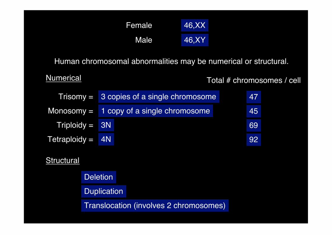
Female
Male
46,XX
46,XY
Human chromosomal abnormalities may be numerical or structural.
Numerical
Trisomy = 3 copies of a single chromosome 47
Monosomy = 1 copy of a single chromosome 45
Triploidy = 3N 69
Tetraploidy = 4N 92
Structural
Deletion
Duplication
Translocation (involves 2 chromosomes)
Total # chromosomes / cell
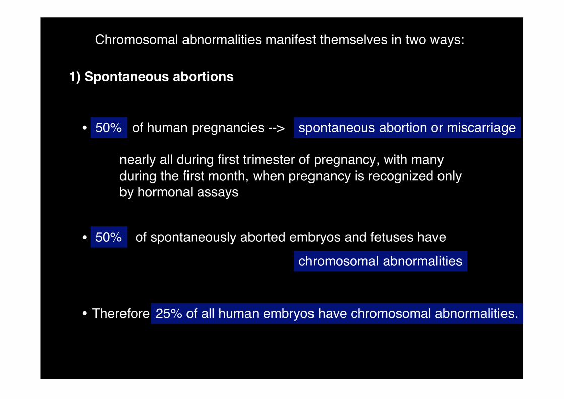
Chromosomal abnormalities manifest themselves in two ways:
1) Spontaneous abortions
of human pregnancies --> spontaneous abortion or miscarriage
of spontaneously aborted embryos and fetuses have
chromosomal abnormalities
nearly all during first trimester of pregnancy, with manyduring the first month, when pregnancy is recognized onlyby hormonal assays
25% of all human embryos have chromosomal abnormalities.
50%
50%
Therefore
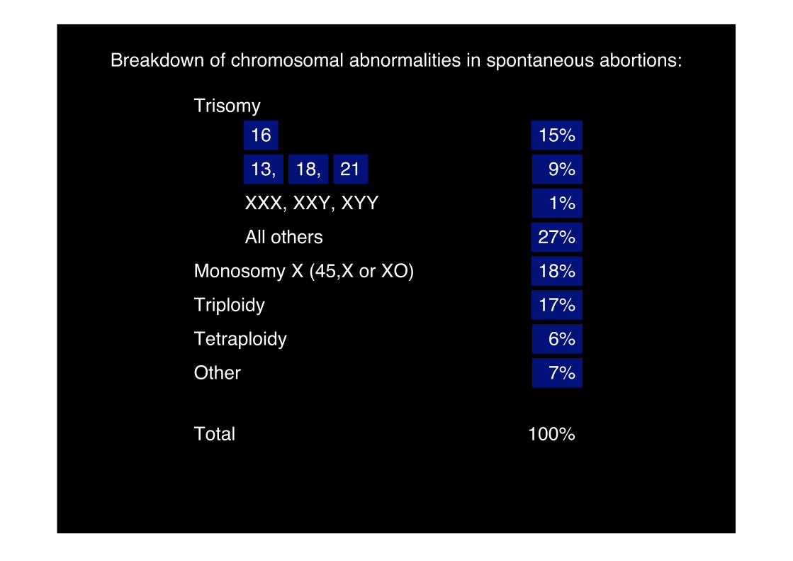
Trisomy
XXX, XXY, XYY
All others
Monosomy X (45,X or XO)
Triploidy
Tetraploidy
Other
Total
15%
9%
1%
27%
18%
17%
6%
7%
Breakdown of chromosomal abnormalities in spontaneous abortions:
100%
16
2118,13,
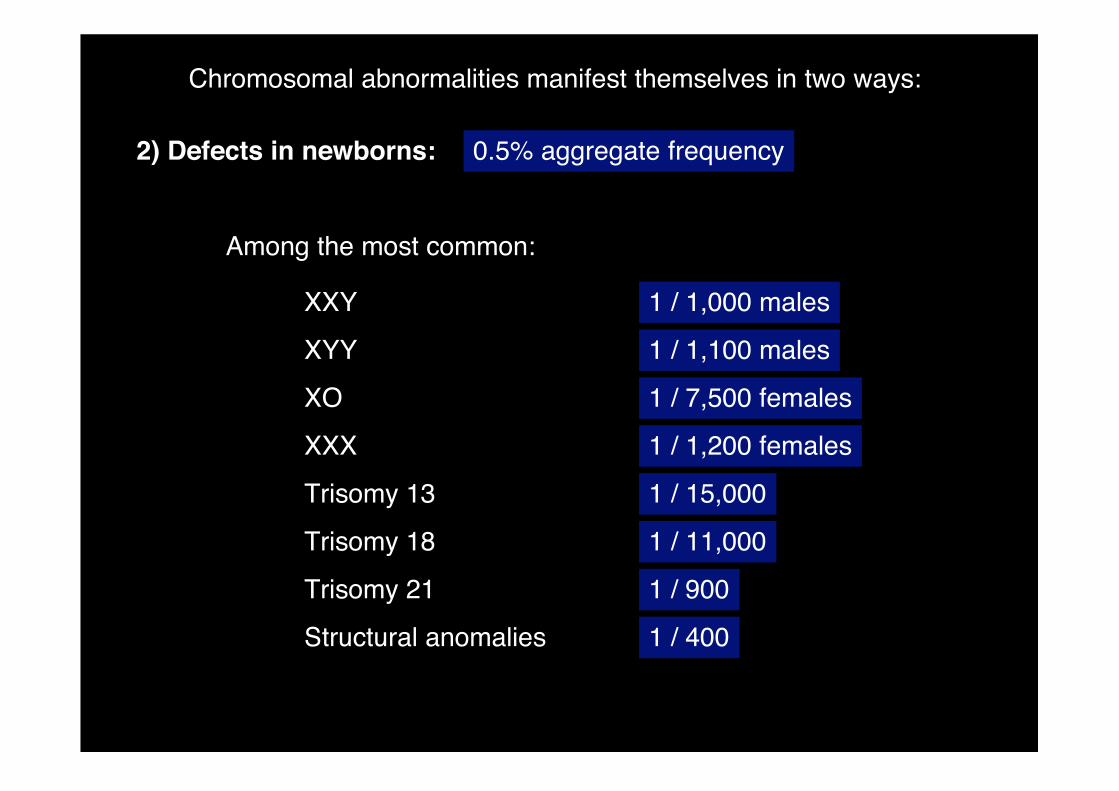
2) Defects in newborns: 0.5% aggregate frequency
Among the most common:
XXY
XYY
XO
XXX
Trisomy 13
Trisomy 18
Trisomy 21
Structural anomalies
1 / 1,000 males
1 / 1,100 males
1 / 7,500 females
1 / 1,200 females
1 / 15,000
1 / 11,000
1 / 900
1 / 400
Chromosomal abnormalities manifest themselves in two ways:
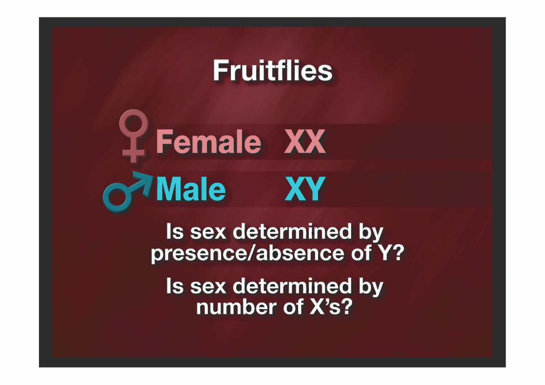
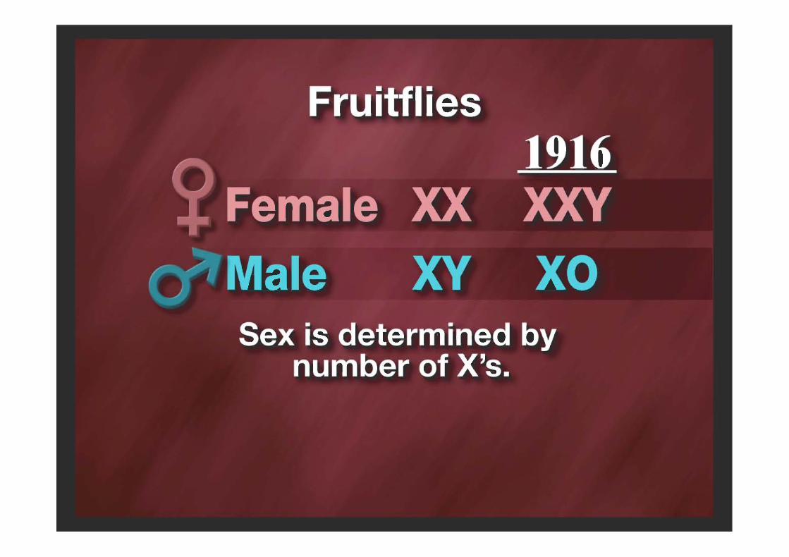
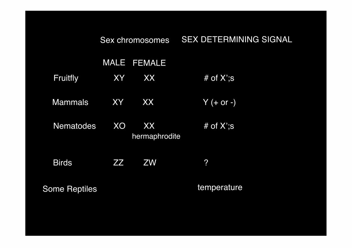
Fruitfly XY XX # of X’;s
Sex chromosomes SEX DETERMINING SIGNAL
MALE FEMALE
Mammals XY XX Y (+ or -)
Nematodes XO XX # of X’;shermaphrodite
Birds ZZ ZW ?
Some Reptiles temperature
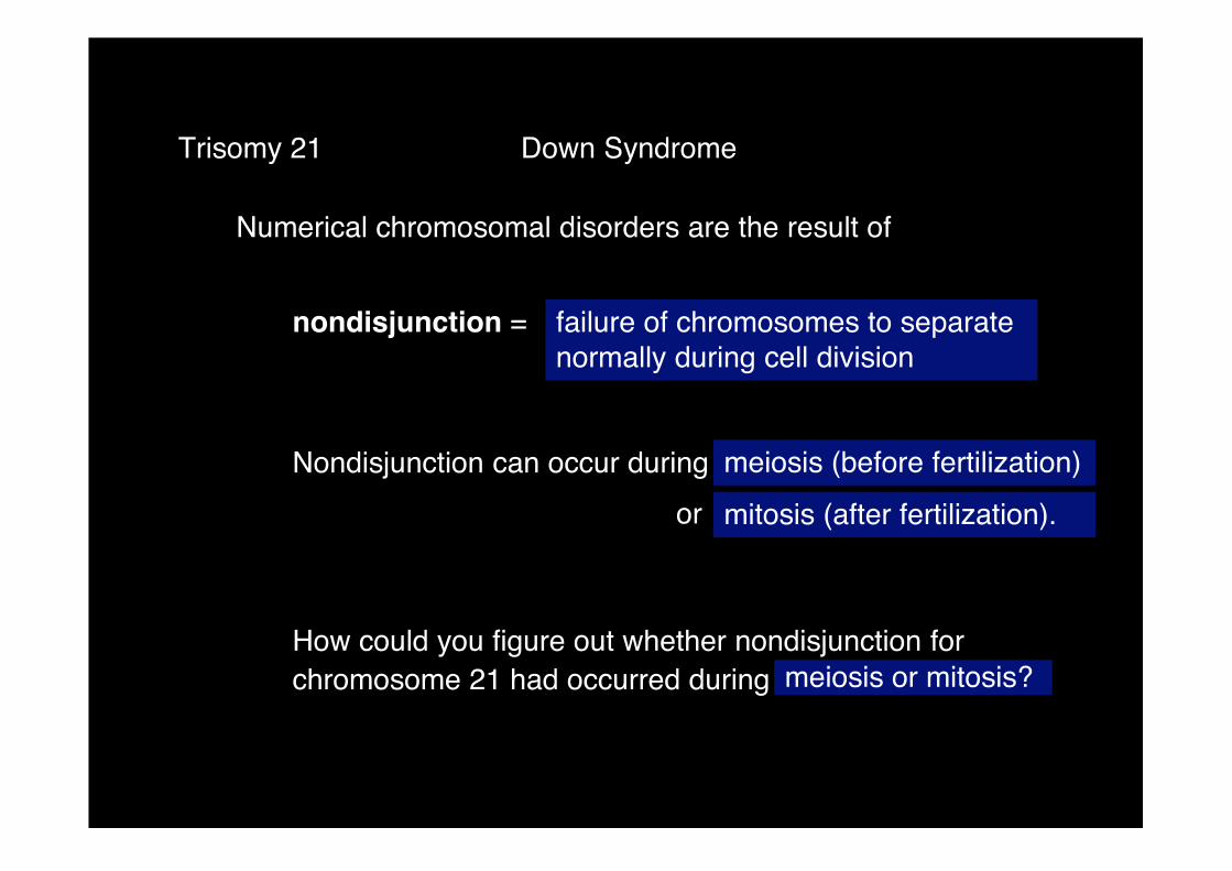
Numerical chromosomal disorders are the result of
Trisomy 21 Down Syndrome
nondisjunction = failure of chromosomes to separatenormally during cell division
Nondisjunction can occur during
How could you figure out whether nondisjunction forchromosome 21 had occurred during meiosis or mitosis?
or
meiosis (before fertilization)
mitosis (after fertilization).
�
�
�
�
�
�
�
�
�
�
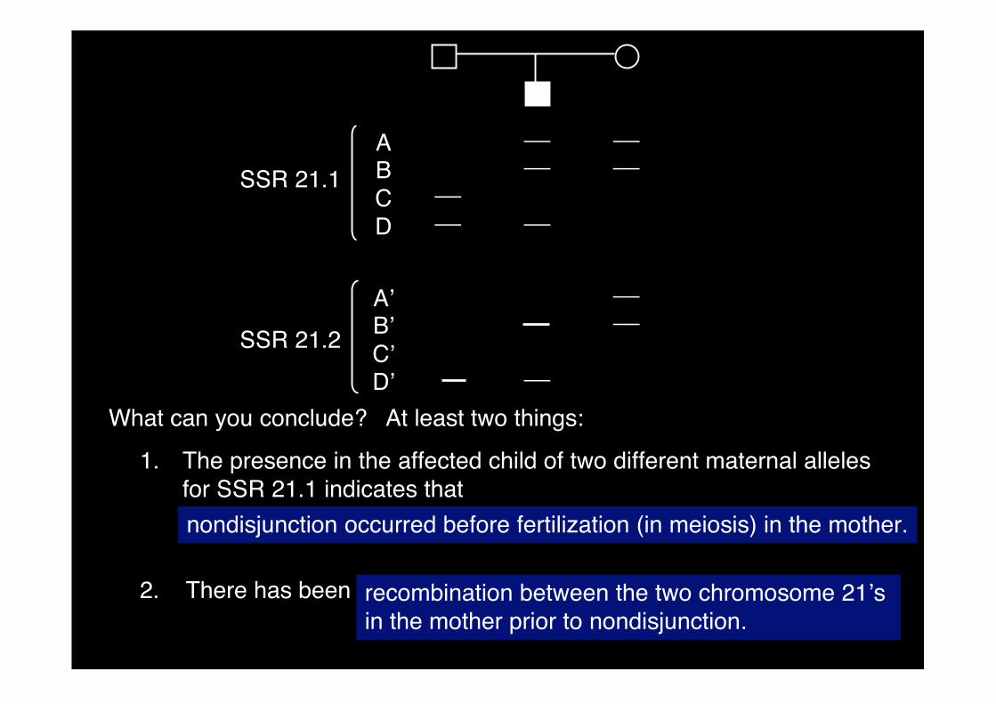
ABCD
SSR 21.1
A’B’C’D’
SSR 21.2
What can you conclude? At least two things:
1. The presence in the affected child of two different maternal allelesfor SSR 21.1 indicates that
recombination between the two chromosome 21’sin the mother prior to nondisjunction.
nondisjunction occurred before fertilization (in meiosis) in the mother.
2. There has been
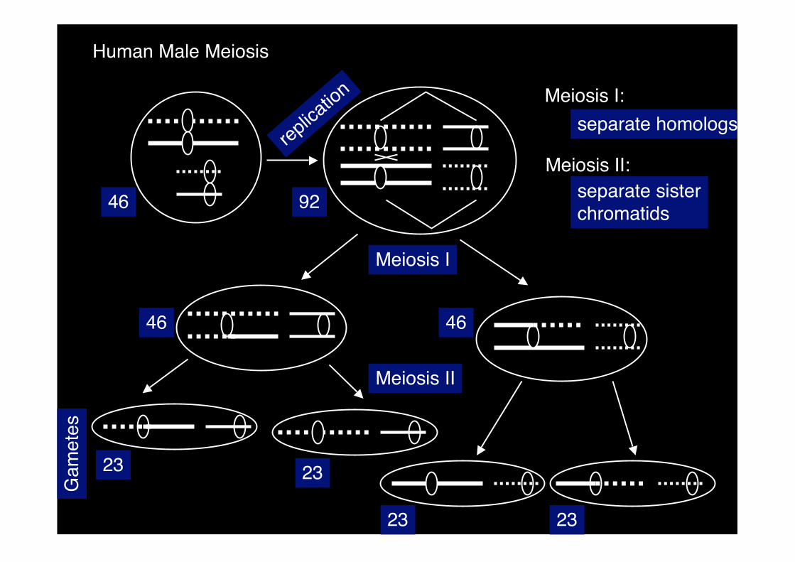
replic
ation
Meiosis I
Meiosis II
Gam
etes
46 92
46 46
23 23
2323
Meiosis I:
Meiosis II:
separate homologs
separate sisterchromatids
Human Male Meiosis
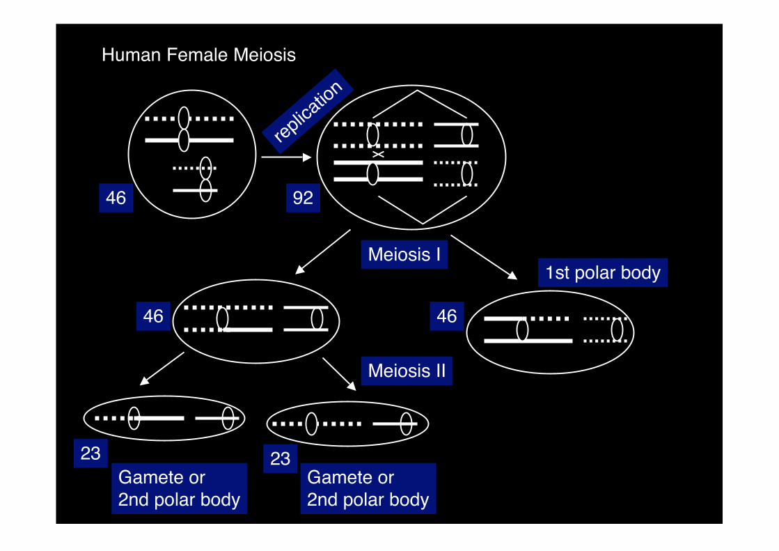
Human Female Meiosis
Gamete or2nd polar body
1st polar body
Gamete or2nd polar body
replic
ation
46 92
Meiosis I
Meiosis II
46 46
2323
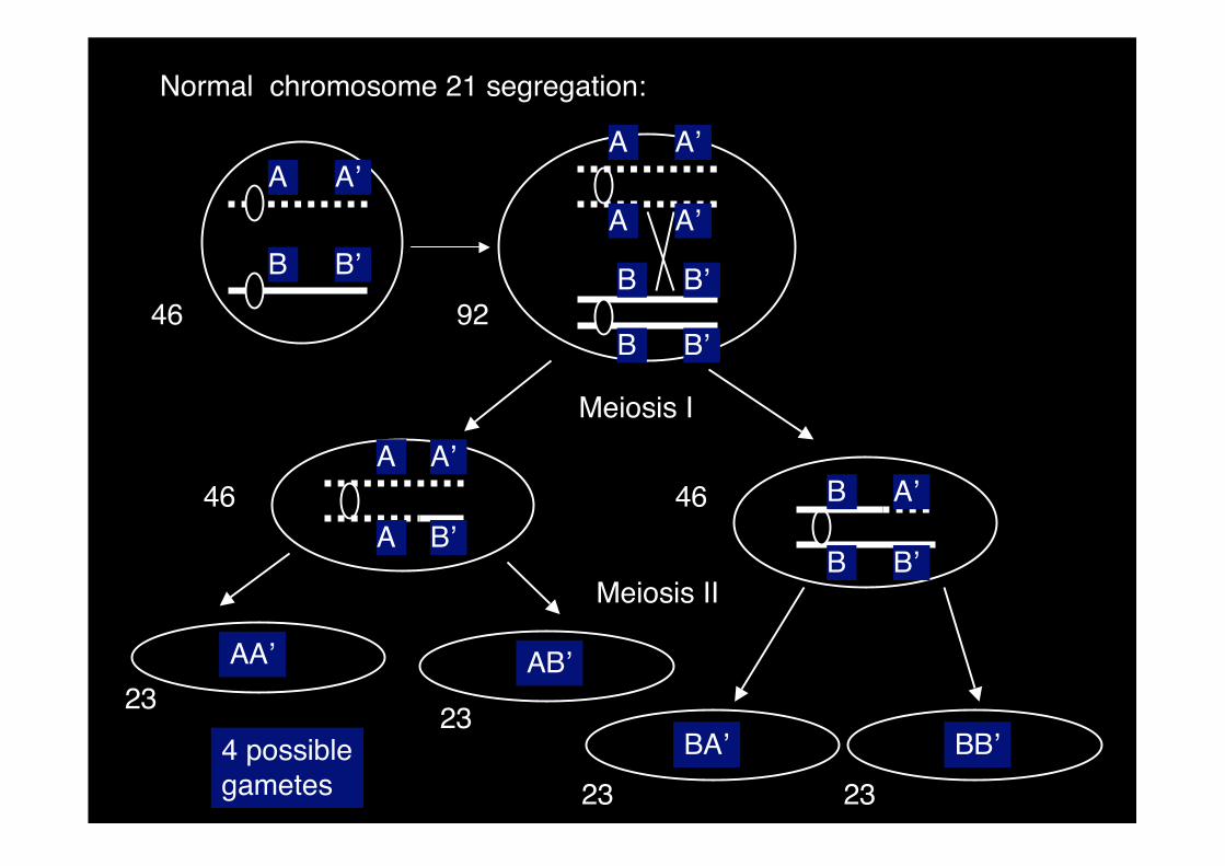
Normal chromosome 21 segregation:
Meiosis II
4 possiblegametes
46
46 46
Meiosis I
23 23
2323
92
A A’
B B’
A A’
A A’
B B’
B B’
A
A
A’
B’B B’
B A’
AA’ AB’
BA’ BB’
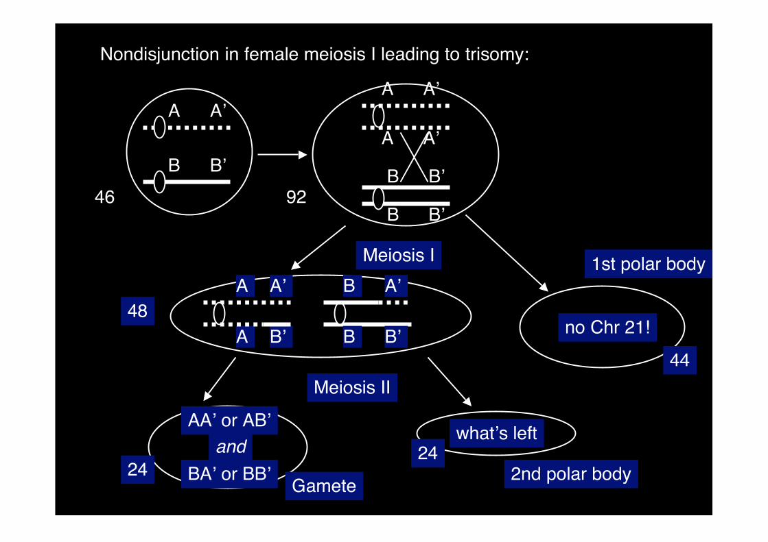
Nondisjunction in female meiosis I leading to trisomy:
Meiosis I
Meiosis II
Gamete
46 92
48
2424
A A’
B B’
A A’
A A’
B B’
B B’
A B’
AA’ or AB’what’s left
no Chr 21!
2nd polar body
B B’
B A’
BA’ or BB’
and
1st polar body
44
A’A
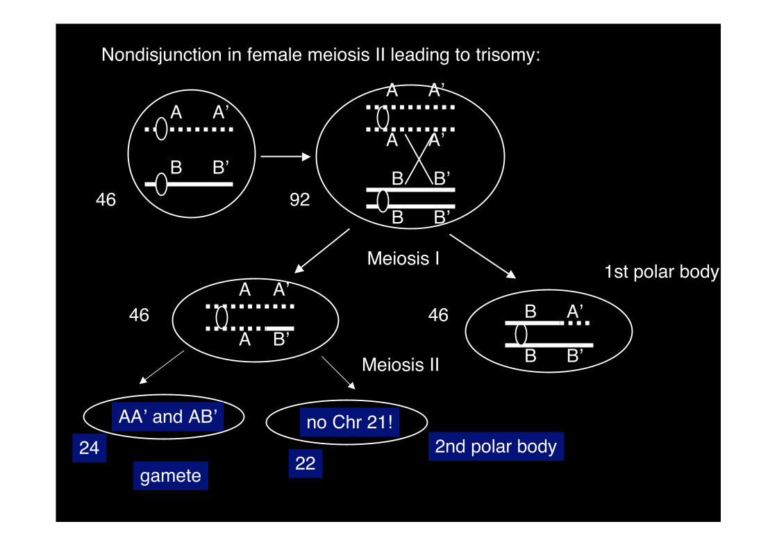
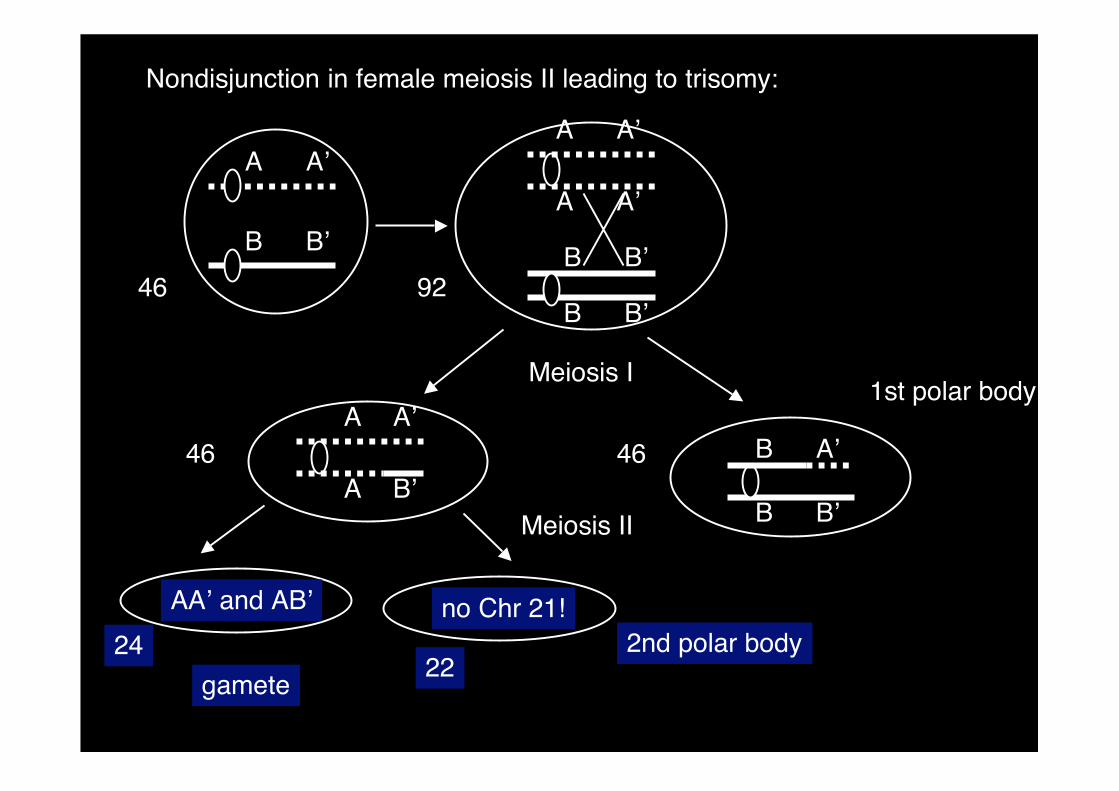
Nondisjunction in female meiosis II leading to trisomy:
Meiosis I
Meiosis II
gamete
46 92
46 46
2224
A A’
B B’
A A’
A A’
B B’
B B’
A
A
A’
B’B B’
B A’
AA’ and AB’ no Chr 21!
1st polar body
2nd polar body
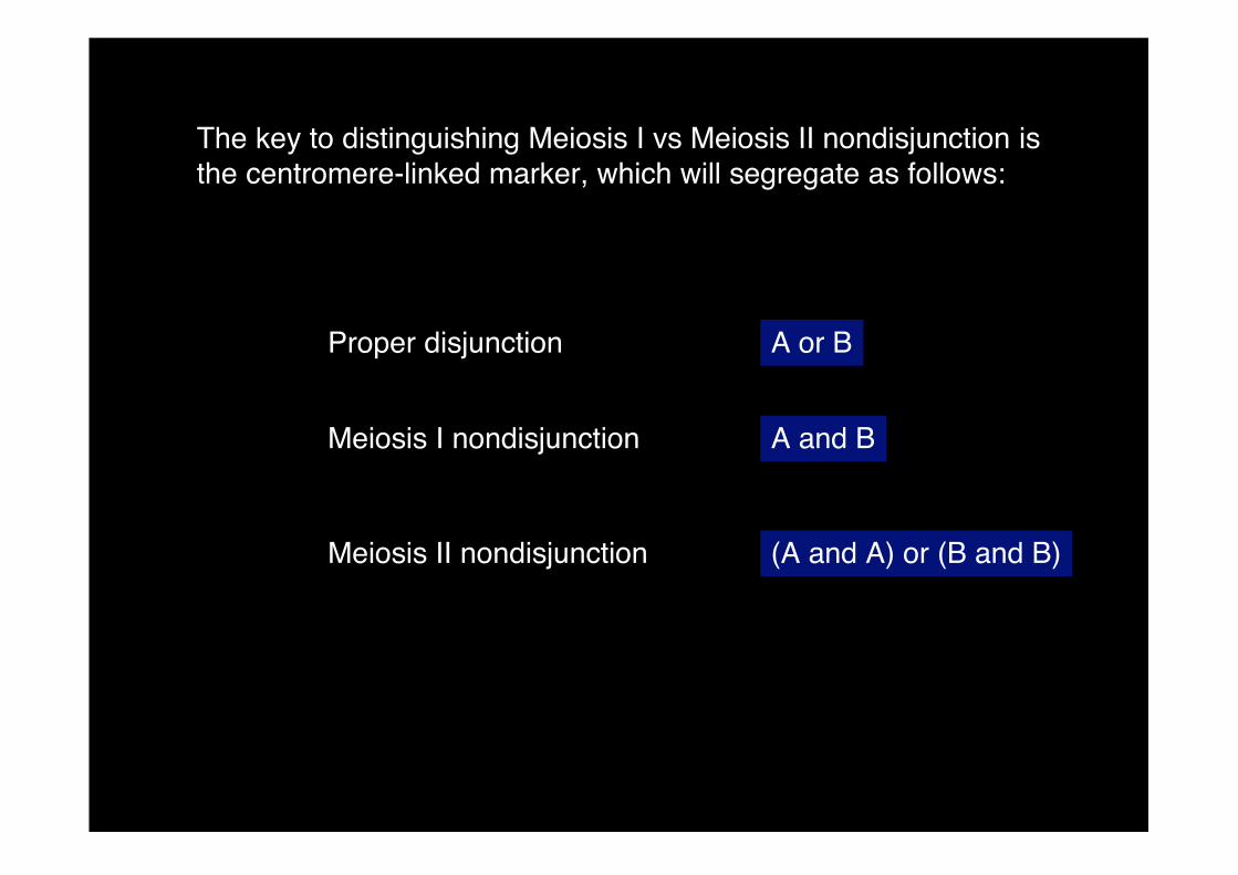
The key to distinguishing Meiosis I vs Meiosis II nondisjunction isthe centromere-linked marker, which will segregate as follows:
Proper disjunction
Meiosis I nondisjunction
Meiosis II nondisjunction
A or B
A and B
(A and A) or (B and B)

Nondisjunction in female meiosis I leading to trisomy:
Meiosis I
Meiosis II
Gamete
46 92
48
44
2424
A A’
B B’
A A’
A A’
B B’
B B’
A B’
AA’ or AB’what’s left
no Chr 21!
2nd polar body
B B’
BA’ or BB’
and
1st polar bodyA A’ B A’

The key to distinguishing Meiosis I vs Meiosis II nondisjunction isthe centromere-linked marker, which will segregate as follows:
Proper disjunction
Meiosis I nondisjunction
Meiosis II nondisjunction
A or B
A and B
(A and A) or (B and B)
Nondisjunction in female meiosis II leading to trisomy:
Meiosis I
Meiosis II
gamete
46 92
46 46
2224
A A’
B B’
A A’
A A’
B B’
B B’
A
A
A’
B’B B’
B A’
AA’ and AB’ no Chr 21!
1st polar body
2nd polar body
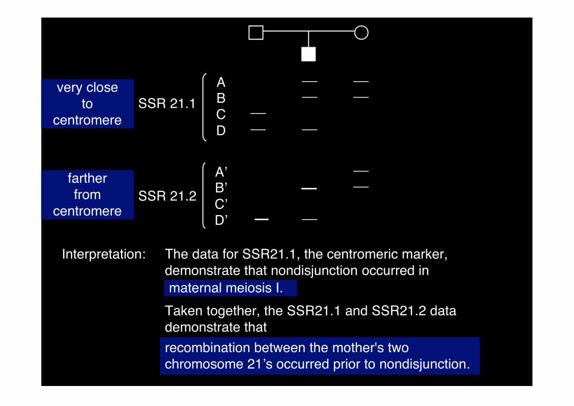
ABCD
A’B’C’D’
�
�
�
�
�
�
�
�
�
�
SSR 21.1
SSR 21.2
Interpretation: The data for SSR21.1, the centromeric marker,demonstrate that nondisjunction occurred inmaternal meiosis I.
Taken together, the SSR21.1 and SSR21.2 datademonstrate that
recombination between the mother's twochromosome 21’s occurred prior to nondisjunction.
very closeto
centromere
fartherfrom
centromere
ABCD
A’B’C’D’
�
�
�
�
�
�
�
�
�
�
SSR 21.1
SSR 21.2
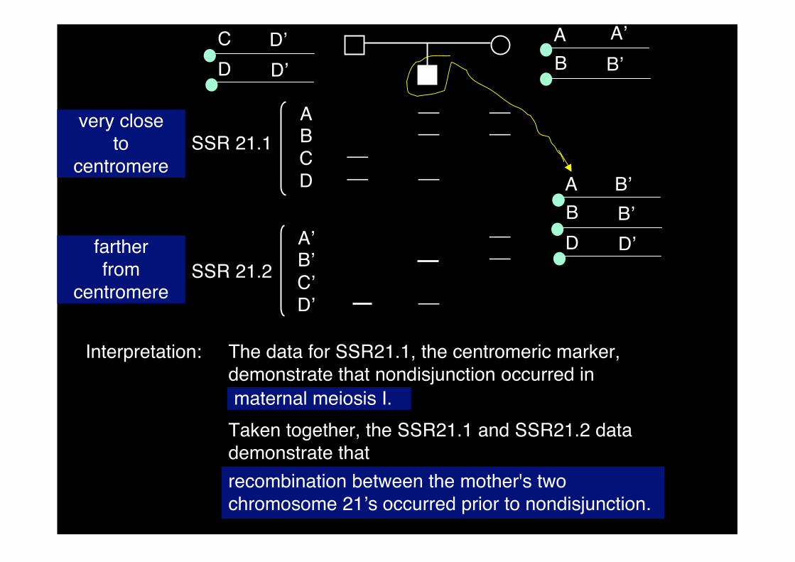
Interpretation: The data for SSR21.1, the centromeric marker,demonstrate that nondisjunction occurred inmaternal meiosis I.
Taken together, the SSR21.1 and SSR21.2 datademonstrate that
recombination between the mother's twochromosome 21’s occurred prior to nondisjunction.
very closeto
centromere
fartherfrom
centromere
A A’
B B’
A B’
B B’
D D’
C D’
D D’
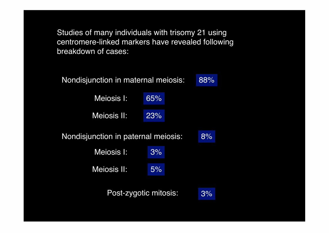
Studies of many individuals with trisomy 21 usingcentromere-linked markers have revealed followingbreakdown of cases:
Nondisjunction in maternal meiosis:
Nondisjunction in paternal meiosis:
88%
8%
Meiosis I:
Meiosis II:
65%
23%
Post-zygotic mitosis: 3%
Meiosis I:
Meiosis II:
3%
5%
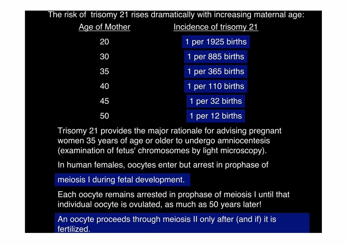
The risk of trisomy 21 rises dramatically with increasing maternal age:
Age of Mother
20
30
35
40
45
50
Incidence of trisomy 21
1 per 1925 births
1 per 885 births
1 per 365 births
1 per 110 births
1 per 32 births
1 per 12 births
Trisomy 21 provides the major rationale for advising pregnantwomen 35 years of age or older to undergo amniocentesis(examination of fetus' chromosomes by light microscopy).
In human females, oocytes enter but arrest in prophase of
meiosis I during fetal development.
Each oocyte remains arrested in prophase of meiosis I until thatindividual oocyte is ovulated, as much as 50 years later!
An oocyte proceeds through meiosis II only after (and if) it isfertilized.