october 31, 2016
TRANSCRIPT

Faall 2016 Cont
Monday, O
tinuin
October 31
g Educ
1, 2016
cation
n

10/20/2016
1
The Greatest Ocular Surface Disease and Dry Eye Course
Jack Schaeffer OD FAAOSchaeffer Eye Center
Birmingham , Alabama
Dr Jack L. Schaeffer
financial disclosure form
AlconAllerganAMO / AbbottBausch and LombCiba VisionCooper VisionEssilorHoyaInspireOptosOptovueZeis Vision
Membership Benefits
Help to build the premiere organization in providing OSD education and knowledge to all optometrists.
Access to invaluable downloadable practice management tools
Quarterly newsletter Membership certificate New industry products and services updates Access to private list serve weekly conversations
on ocular surface disease OSSOPT.com $54.00
www.ossopt.com
The OSD Wellness Symposium
Re invent the practice Prevent patient problems White eyes Perfect Vision Patient Referrrals Sunwear sales Decrease Contact lens dropout Look Better Feel better
The OSD Symposium
24 Doctors 22 Ods 2 MDS
Dry Eye Research Lectures Professors

10/20/2016
2
The OSD Wellness Initiative
OD’s Need education Staff Training Change the culture Inform the Public
I Care
The OSD Wellness Initiative
Pre Screening
Diagnosis
Treatment
Patient Education
The OSD Wellness InitiativeTech Driven
OSDI / Speed questionnaire History Topography / keratometry Visual Acuity
The OSD Wellness Initiative
Preventive Medicine
Dermatology Dentistry Psychology ( behavior modification)
DEWS
Dry eye is a multifactorial disease of the tears and ocular surface that results in symptoms of discomfort, visual disturbance, and tear film instability with potential damage to the ocular surface. It is accompanied by increased osmolarity of the tear film and inflammation of the ocular surface.
Dry Eye Estimated 20 to 30 million people in the
U.S. are thought to have early-stage signs or symptoms of dry eye
Affects women more commonly than men Difficult disease to understand and treat
Advanced dry eye effects roughly 6 million American females and 3 million American males
More common in older individuals (45 years or older)
Varied causes and severities Can be a stand-alone condition


10/20/2016
3
Dry eye is not just a disease,
it’s a complex, multi-factorial disorder.
Prause JU, Norn M. Relation Between Blink Frequency and Break-Up Time. Acta Ophthalmol. 1983; 61: 108-116.Cho P, Cheung P, Leung K, Ma V, Lee V. Effect of Reading on Non-Invasive Tear Break-Up Time and Inter-Blink Interval. Clin. Exp. Optom. 1997; 80: 62-8.Tsubota K, Seiichiro H, Okusawa Y, Egami F, Ohtsuki T, Nakamori K. Quantitative Videographic Analysis of Blinking in Normal Subjects and Patients with Dry Eye. Arch. Ophthalmol. 1996; 114(6): 715-720.Nally L, Ousler GW, Abelson MB. Ocular discomfort and tear film break-up time in dry eye patients: a correlation. IOVS 2000; 41(4): 1436. Collins M, Seeto R, Campbell L, Ross M. Blinking and Corneal Sensitivity. Acta Ophthalmologica 1989; 67(5): 525-531.Abelson MB, Holly FJ. A tentative mechanism for inferior punctate keratopathy. Am. J. Ophthalmol. 1977; 83: 866-869.Doane MG. Dynamics of the Human Blink. Ber. Disch. Ophthalmol. Ges. 1980; 77: 13-17.Kaneko K, Sakamoto K. Spontaneous Blinks as a Criterion of Visual Fatigue During Prolonged Work on Visual Display Terminals. Perceptual and Motor Skills 2001; 92(1): 234-250.
Factors Influencing Dry Eye Age Gender Arthritis Osteoporosis Gout Lens Surgery Contact Lens Wear Blink Disorders Lid Disease Nutritional Problems Rheumatoid Arthritis Thyroid Problems
LASIK Surgery Cosmetic Surgery Mechanical Disturbances Exposure Keratitis Entropion Ectropion Symblepheron Formation Large Lid Notches Lagophthalmos Incomplete Blinking Dellen Formation Illumination Systemic Medications
Time of Day Temperature Humidity Air Movement Allergies Change in
Environment Reading Preservatives in
Topical Eye Medications
Watching Movies Sleep
Sjogrens Non-Sjogrens
Auto-antibodies
Tear Deficient
Evaporative
Lacrimal Deficiency
Lacrimal Obstruction
Reflex
Oil Def. Lid Related Surface Change
Contact Lens
Dry Eye Etiology
NEI Workshop - Classification of Dry Eye (1995)
Tear Film Instability
Note that a patient may have one or more of these deficiencies—they are not mutually exclusive
Aqueous Deficiency Cause: insufficient tear production by
accessory and primary lacrimal glands Sign: low Schirmer (tear volume/flow) score,
tear meniscus height (better measurement)
Tear Film Instability (cont)
Mucin Deficiency Cause: insufficient or unhealthy mucin
production Sign: rapid tear film break-up time (TFBUT)
Lipid Deficiency Cause: meibomian gland dysfunction (MGD)
causing insufficient or unhealthy lipid production
Sign: irregular meibomian gland expression, fast TFBUT
DRUGS ASSOCIATED WITH DECREASED TEAR PRODUCTION
-Adrenergic-blocking, Anti-anginals and Anti-hypertensives
(e.g. Atenolol, Practolol, Propranolol)
Tricyclic Anti-depressants(e.g. Amittriptyline, Doxepin)
Oral Anti-histamines(e.g. Loratadine, Clemastine, Hydroxyzine, Ceterizine, Fexofenidine)
Alkylating Immunosuppressives(e.g. Busulfan, Cyclophosphamide)
Diuretics( T i t )

10/20/2016
4
Role Of Inflammation
Inflammation present in SS-KCS and non-SS KCS
Inflammation present in lacrimal glands, conjunctiva and meibomian glands
Mediated by proinflammatory cytokines in tears
Delayed tear clearance accentuates effect
Inflammation adversely affects neural transmission
PHYSIOLOGY OF THE DRY EYE
Pathologic Collagen vascular diseases or
Autoimmune diseases Rheumatoid Arthritis Lupus Erythematosis Sjogren’s Syndrome
0.4 % incidence 95-98% women
Fibromyalgia
PHYSIOLOGY OF THEDRY EYE
Marginal Contact lens wear--spk Keratoconus Associated with GPC and/or blepharitis Meibomian gland dysfunction(mgd) EBMD (map-dot dystrophy) Acne Rosacea (involves mgd, blepharitis,
dry eye and leads to rosacea keratitis)
PHYSIOLOGY OF THEDRY EYE
MEDICATION INDUCED AntihistaminesDiureticsDermatologic--i.e. Accutane SSRI’S (Selective Serotonin Reuptake
Inhibitors--i.e. Prozac, Paxil, Zoloft, Lexapro, (Welbutrin- to a lesser degree)
SSRI/NorEpi RI Combination—ie. Cymbalta
PHYSIOLOGY OF THEDRY EYE
HRT INDUCED Women on estrogen therapy (HRT) had a 69%
greater risk of dry eye syndrome Women on estrogen plus progesterone/progestin
had a 29% greater risk of dry eye syndrome Risk of dry eye increased 15% for every three
year interval on HRT 38% of Postmenopausal women in the U.S. use
HRT--translates into millions of women
Brigham and Woman’s Hosp. study—Nov. 2001, JAMA
Dry Eye Evaluation
Vision care Exam
CONVERSION
Medical Exam


10/20/2016
5
Examination
Adnexa
Lids / Lid Margins
Tears
Conjunctiva
Cornea
EXAMINATION
ADNEXA Dermatological Inflammation
Dermatochalasis
LIDS/ LID MARGINS Infectious
Inflammatory
Allergic
Physiologic( Lagophthalmos)
DIAGNOSTIC TESTS EXTERNAL EXAMINATION THE CRANIAL NERVE FUNCTION
For a 7th nerve palsy w/incomplete blink on one side Leads to asymmetric dry eye or exposure
keratitis
THE HANDS For typical arthritic changes suggestive of
Rheumatoid or Osteoarthritis Heberden’s Nodes--Nodular Swelling of
Distal Joints
DIAGNOSTIC TESTS
EXTERNAL EXAMINATION SKIN
For Acne Rosacea The nose/forehead for men The cheeks for women The eyelid margins for pustules, redness
and teleangectasia
THE LID MARGINS For blepharitis/meibomitis
Lid Disease
Blepharitis
Lid Wiper Epitheliopathy LWE
Meibomian Gland Disease MGD
GPC
To be covered later in presentation
EXAMINATION
CONJUNCTIVA Goblet Cell function (ekc/post-op)
Staining
Mechanical abnormalities

10/20/2016
6
EXAMINATION
CORNEA Staining
Topographical
Hypoxia
Secondary Infectious/Inflammatory
Dystrophy
DIAGNOSTIC TESTS
TEAR EVALUATION Tear Meniscus TFBUT Evidence of Fluorescein Staining Tear Consistency-i.e. thickness,
debris, evidence of meibomiangland oil and sebaceous secretions
Shirmers
DIAGNOSTIC TESTS Schirmer--w/ or w/o anesthetic Phenol Red Thread Test Zone Quick-represents fluid present in
the conjunctival sac
Fluorescein Staining Rose Bengal Staining Lissamine Green Staining Tear Osmolarity Collagen Plugs
Schaeffer Shirmer
Always do this as the last test
Place strip in any part of the eye
Count to three
remove
Tear Osmolarity
InflammaDry
RPS Technologies

10/20/2016
7
Dry Eye Disease Cycle of Inflammation1
Dry eye is often hidden until patients have progressed and experienced symptoms
Dry eye symptoms overlap with other ocular surface diseases, complicating diagnosis
Numerous clinical diagnostics exist, with no single method preferred
Most ECPs use one or multiple tests, symptom assessment and patient history to diagnose[1] Definition and Classification of Dry Eye. Report of the Diagnosis and Classification Subcommittee of the Dry Eye Work Shop (DEWS). Ocular Surface 2007;5:75‐92.
Dry Eye Disease and MMP‐9
Matrix metalloproteinases (MMP) are proteolyticenzymes that are produced by stressed epithelial cells on the ocular surface1
MMP‐9 in Tears
Non‐specific inflammatory marker
Normal range between 3‐41 ng/ml
More sensitive diagnostic marker than clinical signs1
Correlates with clinical exam findings1
Ocular surface disease (dry eye) demonstrates elevated levels of MMP‐9 in tears1
[1] Chotiakavanich S, de Paiva CS, Li de Quan, et al. Invest Ophthalmol Vis Sci 2009; 50(7): 3203‐3209.
Dry Eye Disease and MMP‐9
Increased concentrations of MMP‐9 can be found in other diseases or conditions, including:
Ocular rosacea
Meibomian gland disease
Sjögren’s syndrome
Corneal ulcers
Corneal erosions
Importance of Detecting MMP‐9
Identifying elevated levels of MMP‐9 facilitates better management of… Patients who present with signs or symptoms of dry eye
Patients having ocular surgery such as LASIK or cataract surgery
When elevated levels of MMP‐9 are not tested, confirmed, and treated prior to ocular surgery, the following complications may occur: Less accurate pre‐surgical measurements lead to worse visual acuity outcomes1
Mild dry eye becomes severe dry eye
Asymptomatic dry eye becomes symptomatic, chronic dry eye2
Epithelial ingrowth or LASIK flap slippage3
[1] Trattler W, Goldberg D, Reilly C. Incidence of concomitant cataract and dry eye: prospective health assessment of cataract patients. Presented at: World CorneaCongress; April 8,2010;Boston,MA. [2] Ambrosio R. J Refract Surg 2008; 24:396‐407. [3] Fournie PR, Gordon GM, Dawson DG, et al. Arch Ophthalmol 2010; 128:426‐436.
Normal Levels of MMP‐9
Literature meta‐analysis supports that normal levels of MMP‐9 (ng/ml) in human controls range from 3‐41 ng/ml
[1] Acera A, Rocha G, Vecino E, et al. Inflammatory markers in the tears of patients with ocular surface disease. Ophthalmic Res. 2008 Oct; 40(6):315‐21. [2] Chotikavanich S, de Paiva CS, Li de Q, et al. Production and activity of matrix metalloproteinase‐9on the ocular surface increase in dysfunctional tear syndrome. Invest Ophthalmol Vis Sci. 2009 Jul; 50(7):3203‐9. [3] Solomon A, Dursun D, Liu Z, et al. Pro‐ and anti‐inflammatory forms of interleukin‐1 in the tear fluid and conjunctiva of patients with dry‐eye disease. Invest Ophthalmol Vis Sci. 2001;42(10):2283‐92. [4] Leonardi A, Brun P, Abatangelo G, et al. Tear levels and activity of matrix metalloproteinase (MMP)‐1 and MMP‐9 in vernal keratoconjunctivitis. Invest Ophthalmol Vis Sci. 2003;44(7):3052‐8.[5] Lema I, Sobrino T, Durán JA, et al. Subclinical keratoconus and inflammatory molecules from tears. Br J Ophthalmol. 2009;93(6):820‐4. [6] Honda N, Miyai T, Nejima R, et al. Effect of latanoprost on the expression of matrix metalloproteinases and tissueinhibitor of metalloproteinase 1 on the ocular surface. Arch Ophthalmol. 2010;128(4):466‐71. [7] Markoulli M, Papas E, Cole N, et al. The effect of contact lens wear on the diurnal profile of matrix metalloproteinase‐9 and its inhibitor in the tear film.Poster presented at the 6th International Conference on the Tear Film and Ocular Surface: Basic Science and Clinical Relevance. Florence, Italy. 24 Sept 2010.
MMP‐9 and Dry Eye Severity1
[1] Chotiakavanich S, de Paiva CS, Li de Quan, et al. Invest Ophthalmol Vis Sci 2009; 50(7): 3203‐3209.

InflammaDry® Limit of Detection
Normal levels of MMP‐9 in human tears ranges from 3‐41 ng/ml
NEGATIVE TEST RESULTMMP‐9 < 40 ng/ml
POSITIVE TEST RESULTMMP‐9 ≥ 40 ng/ml

InflammaDry 4‐Step Process
* Release the lid after every 2‐3 dabs. Allow the sampling fleece to rest along the conjunctiva for 5 seconds.
*

10/20/2016
8
InflammaDry Product Overview
Detects elevated levels of MMP‐9 in tear fluid
Rapid: 10 minute results
Easy to use: can be performed by a nurse or technician
In‐office: point‐of‐care immunoassay test aids in diagnosis at the time of office visit
Low cost: no additional equipment required
InflammaDry Intended Use
InflammaDry is a rapid, immunoassay test for the visual, qualitative in vitro detection of elevated levels of the MMP‐9 protein in human tears from patients suspected of having dry eye. InflammaDry is to be used to aid in the diagnosis of dry eye, in conjunction with other methods of clinical evaluation. This test is intended for prescription use at point‐of‐care sites.
Ocular Surface DiseaseSecondary to Systemic Disease
Systemic Disease Diabetes Rheumatoid Arthritis Sjogren’s syndrome
Thyroid Eye Disease Rosacea Sleep Apnea Graft Vs Host DiseaseMany others
TBUT vs ABMD Causes of Clinical Dry Eye
Mucin deficiency Goblet cell dysfunction
Epithelial surface disease
Aqueous deficiency Lacrimal gland dysfunction
Keratoconjunctivitis sicca
Meibum deficiency Meibomian gland disease
Evaporative dry eye

10/20/2016
9
Developing a Specialty Ocular Surface Disease
Practice
Lid Disease
Lid Disease
We cannot treat the dry eye until we understand and treat
LWEMGDBlepharitisEpihora
IT IS ALL ABOUT THE LIDS
What is OCuSOFT® Lid Scrub™?
• Mild eyelid cleanser that effectively removes oil, debris and desquamated(dead) skin from the eyelids
• Recommended for routine daily eyelid hygiene and maintenance
• Ocusoft lid scrubs BID 1 week preop cataract surgery eradicatedStaph epidermidis equal to topical 5% Betadine intraoperatively¹
¹Jackson M. Endophthalmitis Prophylaxis: Ocusoft Lid Scrub Plus vs. Topical Betadine(ESCRS Barcelona 2010 presentation and OSN supersite)
57
OcuSoft Tea Tree Kit
Contains Tea Tree Oil + Buckthorn seed oil
Ung QHS
OcuSoft Cleansers
58
59
BlephEx Treatment

10/20/2016
10
60
Baby Shampoo…..really a myth
It is the traditional method taught in school but is has disadvantages which include:
• Requires Mixing and Diluting (Convenience?)
• Poor Patient Compliance (actually is irritating to eye)
• Long Term Use Will Make the Skin Dry
• More Professional Treatments are Available
Overview and Summary Recent Clinical Findings
The disease can present alone, classified as primary Sjögren’s, or subsequent to another autoimmune condition (e.g. rheumatoid arthritis), which is classified as secondary Sjögren’s1,2
Sjögren’s is one of the most common autoimmune diseases1
It currently takes 4.7 years to receive an accurate diagnosis3
While the immune response is largely directed to the exocrine glands (lacrimal and salivary), systemic effects are seen in 30-70% of patients1
1. Tincani A, et al. Novel aspects of Sjögren’s Syndrome in 2012. BMC Med Apr 4 2013;11:93. doi: 10.1186/1741-7015-11-93. 2. American Academy of Ophthalmology Preferred Practice Pattern – Dry Eye, 2011. 3. http://www.sjogrens.org.
Traditional Serological Disease Markers for Sjögren’s
1. Tincani A, et al. Novel aspects of Sjögren’s Syndrome in 2012. BMC Med Apr 4 2013;11:93. doi: 10.1186/1741-7015-11-93.
All layers of the tear film may be affected since Sjögren’s is a chronic, progressive disease1
o Patient evaluation should include:
Medical and ocular history
Tear volume
Tear film distribution and stability
Clearance of the tear film
Myth: “There are only a few patients in my practice”

Dry Eye
Aqueous Deficient EvaporativeCombination
Sjögren’s Dry Eye Non-Sjögren’s Dry EyeIntrinsic Extrinsic
Primary
Secondary
Lacrimal Deficiency
Lacrimal Gland Duct Obstruction
Reflex Block
Systemic Drugs
Meibomian OilDeficiency
Disorders of Lid Aperture
Low Blink Rate
Drug Action e.g. isotretinoin
Vitamin A Deficiency
Topical Drugs Preservatives
Contact Lens Wear
Ocular Surface Disease e.g. allergy
• The ocular manifestation of Sjögren’s (primary or secondary) can present as aqueous-deficient dry eye alone, or in combination with evaporative dry eye1,2
o At least 25MM patients diagnosed with Dry Eyeo Patients with Dry Eye symptoms see ECP first
• Major dry eye classification scheme2
Myth: “There are only a few patients in my practice”
1. Tincani A, et al. Novel aspects of Sjögren’s Syndrome in 2012. BMC Med Apr 4 2013;11:93. doi: 10.1186/1741-7015-11-93. 2. American Academy of Ophthalmology Preferred Practice Pattern – Dry Eye, 2011.

10/20/2016
11
Disease progression can vary, so prognoses can also vary1
o Symptoms range from mild dry eye/mouth to severe organ damage and/or lymphoma
o Symptoms may remain stable, worsen or improve in cycles
o As the disease progresses, debilitating fatigue and joint pain can significantly impair quality of life
Early detection and treatment may assist in preventing complications2
However, it currently takes 4.7 years to receive an accurate diagnosis2
1. http://www.ninds.nih.gov/disorders/sjogrens/sjogrens.htm. 2. http://www.sjogrens.org/home/about-sjogrens-syndrome/diagnosis.
Ocular symptoms are frequently the first to present in patients with Sjögren’s, enabling ECP’s an opportunity to identify disease before systemic development
Early diagnosis and treatment may delay the progression of disease1
Active research is ongoing for additional therapeutic options for Sjögren’s:1,2
o Biological therapeutic agents (e.g. monoclonal antibodies)
o Antimalarials
o Vitamin D supplementation
o Immunosuppressants
1. Tincani A, et al. Novel aspects of Sjögren’s Syndrome in 2012. BMC Med Apr 4 2013;11:93. doi: 10.1186/1741-7015-11-93. 2. Ramos-Casals M, Brito-Zeron P. Emerging biological therapies in primary Sjogren’s Syndrome. Rheumatology 2007;46:1389-1396.
Sjogren’s syndrome is currently defined by:Ocular symptoms – dry eyesOral symptoms – dry mouthOcular signs – abnormal Schirmer’s test or Rose Bengal or Lissamine Green stainingOral signs – decreased salivary gland flowHistopathology showing lymphocytic infiltration of salivary or lachrymal glandsAutoantibodies – anti-Ro and/or anti – La, ANA, RFExclude – hepatitis C, HIV, neck radiation, sarcoidosis, graft versus host disease, lymphoma, anti-cholinergic drugs
Other manifestations include:– Lung disease – usually a lymphocytic interstitial pneumonia– Kidney disease – usually mild tubular disease, but may have glomerular disease– Peripheral neuropathy– Vasculitis involving skin, bowel, muscle, nerve and occasionally other organs– Vasculopathy, especially with secondary anti-phospholipid antibodies– 5% of patients develop non-Hodgkin lymphomas
Sjogren’s syndrome leads to:Corneal abrasions and other Keratopathies
Blepharitis
Uveitis
Other ocular infections
Dental caries
Other infections of the mouth
Systemic involvement in Sjogren’s syndrome may lead to:
Respiratory dysfunction
Renal dysfunction
Lymphoma
71CONFIDENTIAL 72CONFIDENTIAL
The Sjö™ In-Office Testing Kit

10/20/2016
12
19 Year Old With Severe Dry Eyes?
D. B.
• 19 y. o. b. m. presents w mother c/o OU red and “infected” x 2 weeks, also very dry. Mother states last occurrence “tubes had to be put in”.
• PMHx: Mild “Behavioral” Cerebral Palsy, Epilepsy, Asthma, Deafness.
• POHx: L DCR w Crawford Tubes 2001 secondary to Dacryocystitis.
D. B.
• Allergies: Bactrim = Rash
• Meds: Depakote qd, Risperdal qd, Tylenol prn, Sudafed prn.
• Normal Pregnancy and birthweight.
• Dev. Milestones were delayed.
D. B.
• Oc. Meds.: Ciprofloxacin gtts qd, Refresh Liquigel qd.
• BVa: 20/40 OD, 20/50 OS
• Fundus: Normal OD, OS
Work Up
• CBC w/ Diff.
• LFTs
• Vitamin Panel
Treatment
• Pres. Free Tears q1-2h
• Topical Vitamin A ung 0.1% qid
• OTC Multi-vitamin qd
• Vitamin A 3000 mcg/d (10,000 IU)
Tseng SC. Topical retinoid treatment for various dry-eye disorders. Opthalmology 1985 92(6):717-727

10/20/2016
13
Lab Results
• Vitamin A 22 (26-72 mcg/dL)
• Vitamin B12 159 (200-1100 pg/mL)
• Vitamin D 12 (20-100 ng/mL)
PCP Treatment
• Vit. B12 injections
• Vit. D 400 IU qd OTC
• Continue Vit. A 10,000 IU qd
Childhood Xerophthalmia
• Congenital
• Alacrima
• Ectodermal Dysplasia
• Allgrove Syndrome (Triple A)
• Cystic Fibrosis
• Endocrine
• D. M.
• Thyroid Dz
Childhood Xerophthalmia
• Immunological
• Sjogren Syndrome
• GvHD
• Juvenile Idiopathic Arthritis
• Dermatologic
• Epidermolysis bullosa
• Acne Rosacea
• TEN (Toxic Epidermal Necrosis)
Childhood Xerophthalmia
• Post Infectious
• HTLV-1, EBV, HIV
• Medications
• Β blockers, Retinoids, Valproic acid**
• Nutritional
• Malabsorption, Poor dietary habits**
Vitamin A Deficiency• Nyctalopia• Xerophthalmia
• Bitot’s Spots• Xerostomia• Pruritis• Anemia• Humoral and Cell Med. Immune
Dysfunction• Excessive Bone Deposition• Mortality

10/20/2016
14
W. H. O.• Most common cause of preventable
blindness in the world.1
• Est. 52,000 children go blind every year in India.2
2. Rahamathullaji, L, Underwood, BA, Thulasiraj, RD et al. Reduced mortality among children in Southern India receiving a small weekly dose of vitamin A. New England Journal of Medicine 1990, 323 : 929-35
1. Severe malnutrition: report of a consultation to review current literature Geneva, World Health Organization, 6-7 September 2004
Advanced Recalcitrant PEK
Autologous Serum Amniotic Membrane
Autologous Serum
Contains Epithelial Growth Factor (EGF) Transforming Growth Factor 8 (TGF8) Fibronectin Vitamin A Other Cytokines
Autologous Serum
Blood Draw at Lab Spin down to plasma @4000 rpm for 20
minutes Deliver to Compounding Pharmacy 2:1 Filtered Compounding with BSS 8 Bottles Frozen until used
Autologous Serum
1 gtt q2h from morning until bedtime Keep Vial Refrigerated Keep Additional Vials Frozen Until Use 8 Straight Weeks Evaluate After 6-8 Weeks Possible Additional Course
Autologous Serum Cost
Lab Draw $30 Compounding Pharmacy $120 $150 for 8 Vials
IF Patient delivers Serum to Pharmacy

10/20/2016
15
Autologous Serum Cost
Lab Draw $30 Compounding Pharmacy $120 Virology Testing $210 Freeze and Shipment To/From
Compounding Pharmacy Approximately $450 to $550 for 8 Vials
Sutureless Amniotic Membrane
ProKera – Amniotic Membrane for wound healing Cryopreserved
Bio Optix Dry Membrane
Biological Scaffolding
Helps initiate an active healing process by providing proteoglycans and growth factors
Collagens, fibronectin and lamillin Cryopreserved membrane contains heavy-
chain hyaluronic acid Inhibits proinflammatory cells Suppress T Cells
Persistent Corneal Defect Recurrent Cornea Erosion Corneal Ulcer Pterygium Graft Bullous Keratopathy Band Keratopathy
Sutureless Amniotic Membrane
ProKera – Amniotic Membrane for wound healing Corneal Ulcer Bullous Keratopathy Folds in Descemet’s Chemical Burns Mechanical Complications 2ary to graft Disruption of surgical wound Non-healing surgical wound
Inflammation’s Effect on Healing
Inflammation: the first sign of wound healing & is also the hallmark symptom of all ocular surface diseases
Uncontrolled inflammation leads to: Chronic pain and discomfort/irritation Delayed healing, more tissue damage Vision-threatening complication, e.g., scar/haze
Effective control of inflammation is an important strategy to promote healing and minimize the risk of scar/haze
Non-Resolved Inflammation
Tissue Damage
Controlling Inflammation is Key to Preventing Tissue Damage!Controlling Inflammation is Key to Preventing Tissue Damage!

• The amniotic membrane is the innermost lining of the placenta (amnion)
• Amniotic membrane shares the same cell origin as the fetus• Stem cell behavior
• Structural similarity to all human tissue
The Amniotic Membrane

Ocular Surface Disease
Corneal Inflammation
Keratitis
Conjunctival Inflammation
Conjunctivitis
Eyelid Inflammation
Blepharitis
Inflammation is the Hallmark of All Ocular Surface Diseases

PROKERA® utilizes the proprietary CryoTek™ cryopreservation process that maintains the active extracellular matrix of the amniotic membrane which uniquely allows for regenerative healing.
PROKERA® is the only FDA-cleared therapeutic device that both reduces inflammation and promotes scar less healing
PROKERA® can be used for a wide number of ocular surface diseases with severity ranging from mild, moderate, to severe
PROKERA®: BIOLOGIC CORNEAL
BANDAGE

10/20/2016
16
100
Insertion of Pro-Kera
Remove from inner pouch Rinse with saline (prevents stinging from
preservation media Apply topical anesthesia Hold upper lid and have patient look down Insert into superior fornix Slide under lower eyelid Check for centration
CONFIDENTIAL AND PRIVILEGED Property of Bio-Tissue, Inc. Do not reproduce or distribute.
Continue medications
Apply Temporary Tarsorrhaphy(PRN)- Tape- Tegaderm- “Breathe-Right”
nasal strips
Post-Treatment Protocol

10/20/2016
17
SCLERAL LENSES
Punctal / Lacrimal Occlusion
Rationale for occlusion therapy: Diminishes tear drainage from the ocular surface Enhances contact time between tears & ocular surface Utilizes “normal tears” Natural complement of proteins, enzymes, buffers, etc.
Multiple modalities, manufacturers, products Collagen, silicone, acrylic polymers Intracanalicular vs. punctal occlusion
LACRISERT®
(hydroxypropyl cellulose ophthalmic insert)
A Novel Approach to Treating
Dry Eye Syndrome
Please see full Prescribing Information.
LACRISERT(hydroxypropyl cellulose ophthalmic insert)
Lacrisert [package insert]. Aton Pharma, Inc.: Lawrenceville, NJ; 2007.
Indicated in patients with moderate to severe dry eye syndrome (DES), including keratoconjunctivitis sicca.
Indicated especially in patients who remain symptomatic after an adequate trial of therapy with artificial tear solutions.
Indicated for patients with exposure keratitis, decreased corneal sensitivity, and recurrent corneal erosions.
M G D
Meibomian Gland Dysfunction

©KNichols 2012
Current Dry Eye Definition “Dry eye is a multifactorial disease of the tears and ocular surface that results in symptoms of discomfort, visual disturbance, and tear instability with potential damage to the ocular surface. It is accompanied by increased osmolarity of the tear film and inflammation of the ocular surface.”

©KNichols 2012
DEWS—Classification of Dry Eye
80%20% 5% 65% 35%

©KNichols 2012
TFOS International MGD Workshop
• Over 65 International clinicians, scientists, and industry participants
• 2+ year process
• Published in March 2011, IOVS
• #1 Most downloaded IOVS article for the last 12 months
• Downloaded over 5500 times
• All MGD workshop reports are in the “top 10”
• Translation into 12 languages
• www.tearfilm.org

10/20/2016
18
Meibomian Gland Dysfunction
Level one Treatment: Available to all Doctors Medical : In office and home Procedures
Level two Treatment: Specialized equipment needed
Meibomian Gland Dysfunction
1 Manual Expression
2 Miboflow
3 Lipiflow
Tear Conservation
M G D
Meibomian Gland Disease
Meibomian Gland Dysfunction and Management
Kelly K. Nichols, OD, MPH, PhD
FERV ProfessorUniversity of Houston College of Optometry
Chair, TFOS International Meibomian Gland Workshop©KNichols 2012
Meibomian Gland Dysfunction
• The TFOS Report of the International Meibomian Gland Dysfunction Workshop– Etiologies
– Definition/ Classification
– Epidemiology
– Clinical characteristics
– Diagnosis/ Management
– Contact lenses, surgical implications

10/20/2016
19
©KNichols 2012
Lecture Descriptionwww.tearfilm.org
©KNichols 2012
Anatomy, Physiology and Pathophysiology of the
Meibomian Gland
Erich Knop, M.D., Ph.D. (Chair)Nadja Knop, M.D., Ph.D.Thomas J. Millar, Ph.D.Hiroto Obata, M.D.
David A. Sullivan, Ph.D.
©KNichols 2012
Meibomian Gland DysfunctionDefinition & Classification
J. Daniel Nelson, M.D. (Co‐Chair)
Jun Shimazaki, M.D., Ph.D. (Co‐Chair)
Jose M. Benitez‐del‐Castillo, M.D., Ph.D.
Jennifer Craig, Ph.D., MCOptom
James P. McCulley, M.D.
Seika Den, M.D., Ph.D.
Gary N. Foulks, M.D.
Evaluation, Diagnosis and Grading of Severity of
Meibomian Gland Dysfunction
Alan Tomlinson, MCOpt, Ph.D. (Chair) E. Ian Pearce, Ph.D. Anthony J. Bron, F.R.C.S. Richard Yee, M.D.Donald R. Korb, O.D. Norihiko Yokoi, M.D., Ph.D.Shiro Amano, M.D., Ph.D. Reiko Arita, M.D., Ph.D. Jerry R. Paugh, O.D. Murat Dogru, M.D.
©KNichols 2012
Management and Therapy of Meibomian Gland
Dysfunction
Gerd Geerling, M.D. (Chair) Terrence O’Brien, M.D. Joseph Tauber, M.D. Maurizio Rolando, M.D.Christophe Baudouin, M.D., Ph.D. Kazuo Tsubota, M.D.Eiki Goto, M.D. Kelly K. Nichols, O.D., M.P.H., Ph.D.Yukihiro Matsumoto, M.D.
©KNichols 2012
Current Practice Patterns*
• Lid hygiene, warm compresses and lid massage• Cleaning of the lid margin with baby shampoo, cotton buds or wet towels, daily for 5‐15 minutes
• Lubricants in cases with additional dry eye• Topical antibiotic oint (moderate to severe)• Systemic tetracyclines/ derivatives in recurrence• Incision and curettage with optional steroid injection in chalazion
*Excerpted from Moorfields Manual, Wills Eye Manual (Guidelines for posterior blepharitis and meibomitis)

©KNichols 2012
• Large sebaceous glands
• No direct contact to hair follicles
• Located in the tarsal plates
• Upper and lower eye lids
Meibomian Gland ‐ ANATOMY
Modified and colored from Krstic H. Human microscopic anatomy. Springer Medizin Verlag 1991, (reproduced from Knop N & Knop E Ophthalmologe 2009; 106:872–883)

©KNichols 2012
Meibomian Gland – PATHOLOGY• Obstructive MGD leads to a progressive ductal DILATATION and acinar ATROPHY
Fom Knop E & Knop N. Meibom-Drüsen Teil IV. Funktionelle Interaktionen in der Pathogenese der Dysfunktion (MGD). Ophthalmologe.2009;106:980–987

©KNichols 2012

Testing Summary
• Symptoms (no validated survey)
• Expression (not widely accepted)
– Quality/ Quantity
• Lid assessment
– Redness (difficult to grade)
– Irregularity
– MG location
• Staining (fluorescein)
– Photography
• Aq. Production (© 1903)

©KNichols 2012
Stages of MGD

©KNichols 2012
DISEASE STAGINGStage MGD grade Symptoms Corneal
Staining
1
+ (minimally altered expressibility and secretion quality)
Asymptomatic None
2
++ (mildly altered expressibility and secretion quality)
Minimal to Mild None to limited
3
+++ (moderately altered expressibility and secretion quality)
ModerateMild to moderate; mainly peripheral
4
++++ (severely altered expressibility and secretion quality)
MarkedMarked; central in
addition
“PLUS DISEASE” Co‐existing or accompanying disorders of the ocular surface and/ or eyelids

10/20/2016
20
©KNichols 2012
Current Practice Patterns
• World‐wide variation
• Underreporting difficult to assess patterns
• Underdiagnosis common, clinical follow‐up irregular
• Lid warming and hygiene common
• Many use artificial lubricants
• Most Common Rx: Systemic tetracycline or derivatives (less frequent in EU/Japan)
– 2nd most common Rx: topical antibiotic or antibiotic‐steroid combination
©KNichols 2012
Stage =
I 2 3 4 Plus‐Disease+Inform patient (about dietary / environmental / medication effects)± Eyelid hygiene (warming / expression)
+Eyelid hygiene (warming / expression), Advise re: potential benefits of ambient humidity / n‐3 fatty acid,± Lubricant/lipid, topical azithromycin, tetracycl. derivatives
+ Oral tetracyclines± Ointment (pm), cyclosporine/steroid for DE
+ Anti‐inflammatory therapy for DE
Recommended Staged Therapy
+ Steroids, CL, Surgery
Design and Conduct of Clinical Trials
Penny A. Asbell, M.D.(Chair)Fiona Stapleton, M.Sc., O.D., Ph.D.
Kerstin Wickström, Ph.D.Esen Akpek, M.D.
Pasquale Aragona, M.D., Ph.D.Reza Dana, M.D., M.Sc., M.P.H.
Michael A.Lemp, M.D.Kelly K. Nichols, O.D., M.P.H., Ph.D. ©KNichols 2012
Existing Clinical TrialsKey Issues Findings
Trial objective Majority interventional treatment trials. 1/3 comparative (hot compresses or artificial tears).
Trial design /Methodology
Primarily small trials (<40 subjects) of short (<3 months) duration. Most prospective, 3 randomized controlled design, & 2 were double masked.
Study population Chronic disease but selection criteria not uniformly defined; lid changes & symptoms most common clinical characteristics.
Inclusion criteria No specific and consistent criteria; most common are lid margin signs (80%), dry eye findings (50%), symptoms of discomfort/foreign body sensation (46%).
Exclusion criteria Classification of exclusion criteria in three different categories:1) Ocular disease related/CL wear (most common);2) Iatrogenic ( e.g surgery, 1/3 studies);3) Systemic disease related/pregnancy (15%).
n = 26
Meibomian Glands
Modified sebaceous gland
30-40 glands exist in upper tarsus 20-40 glands exist in the lower tarsus Secretion stimulus not fully
understood Secretion of meibomian oil increases with
testosterone; decreases with estrogen Oil expelled by mechanical force on gland
during blinking Not all glands secreting simultaneously
141
Treatment of MGD/NOMGD
In-Office Therapy Manual Expression Off-Label Pharmacotherapy
Oral tetracycline/doxycycline Topical Antibiotics – erythromycin, tobramycin Topical Steroids – dexamethasone
142
At Home Therapy– Warm compresses– Eyelid Scrubs
– Self expression

Structure of a Stable Tear Film
139
Aqueous/Mucin complex
Corneal Epithelium
Glycocalyx
Amphiphilic Lipid Layer
Nonpolar Lipid layer
-mucin bound complex responsible for the integration of aqueous layer with corneal epithelium
-mucins are distributed throughout this layer, rather than in distinct aqueous and mucin layers
- complex - over 100 different species of lipid
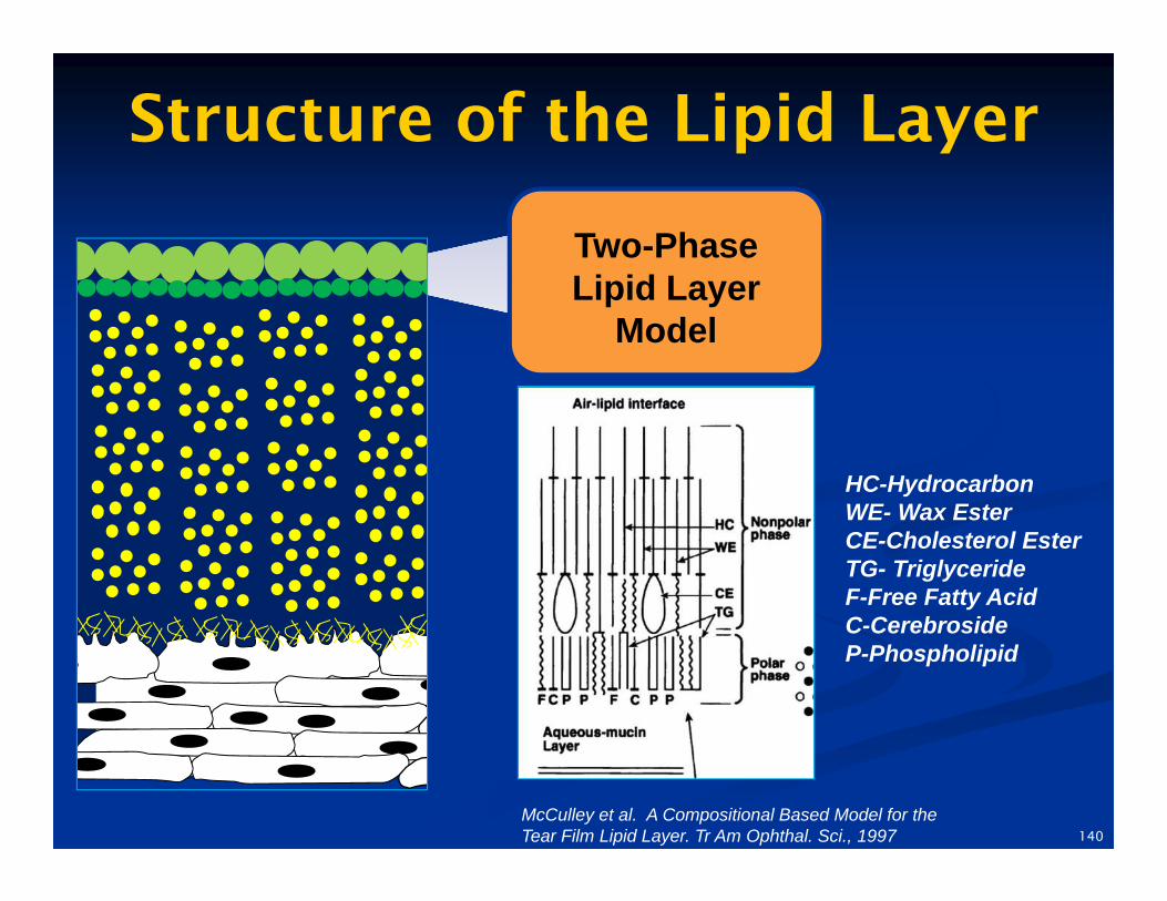
Structure of the Lipid Layer
Two-Phase Lipid Layer
Model
HC-HydrocarbonWE- Wax EsterCE-Cholesterol EsterTG- TriglycerideF-Free Fatty AcidC-CerebrosideP-Phospholipid
McCulley et al. A Compositional Based Model for the Tear Film Lipid Layer. Tr Am Ophthal. Sci., 1997 140
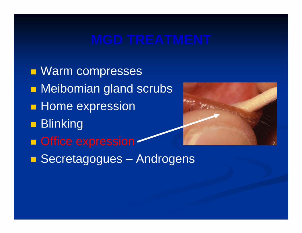
MGD TREATMENT
Warm compresses
Meibomian gland scrubs
Home expression
Blinking
Office expression
Secretagogues – Androgens


10/20/2016
21
Collins Expressor Forceps (Item 98610)For aggressive expression of the Meibomian gland.
Livengood Expressor PaddlesAngled (Item 98620) & Flat (Item 98630)
For mild or gentle expression of the Meibomian gland.
New! Ophthalmic Surgical Instruments Maskin Expressor
$ 575
Rhein Medical
BRUDER EYE COMPRESSESMicrowave Activated
Bruder Eye Hydrating Compress and Stye Compress conveniently provide an effective yet natural and drug-free way to help provide and maintain proper eye moisture. BENEFITS FEATURES• Replenishes Moisture Naturally• Relieves Dryness• Refreshes Tired Eyes• Provides Drug Free Relief
• Ready in Minutes from the Microwave• Naturally Hydrating• Washable & Reusable• Clean Moist Heat• Soft Conforming Design• Non-Allergenic• Dust-Free
08.10
BRUDER STYE COMPRESSItem #34170
BRUDER EYE HYDRATING COMPRESSItem #34160
WARNING
Hot compresses can change the corneal tissues and structure
Possible Link to Keratoconus
Evidence Based Medicine
Schaeffer Eye Protocol1) OSD Evaluation
1) Includes test expression2) All staining
2) RTC expression1) At home heat with eye medibeads2) 15-20 minutes in waiting room with Bruden’sheat pack ( or rear wait) 3) Expression 1 of 34) RTC 2 weeks
Meibomian Gland Expression
Fees: $189 / $25
Out of pocket
Covers 3 Office visits
$68.00 Per visit after initial three visits
99213 / 99212
Dry eye progress check before expression
MGD EXPRESSION

10/20/2016
22
MGD
Maskin Expressor
Maskin Probe
1)$ 158 box ( 10)
2) 1,2,4,6 MM intraductals
3) Aluminum Handle $104


10/20/2016
1
The Greatest Anterior Segment Disease and
Contact lens complications course ever
Jack Schaeffer OD FAAO
FINANCIAL DISCLOSURE FORM
DR JACK L. SCHAEFFER
I HAVE RECEIVED HONORARIUM, COMPENSATION, OR SERVE AS AN ADVISOR TO THE
FOLLOWING COMPANIES
•
• ALCON
• ALLERGAN
• AMO/ABBOTT
• ARCTIC/DX
• ATON
• BAUSCH AND LOMB
• COOPERVISION
• ESSILOR
• ISTA
• HOYA
• OPTOVUE
• OPTOS
• VISTAKON
• ZEIS VISION
Dilation Vs Optomap
• The two together delivers a the highest level of Comprehensive Eye Care
• If you have to choose just one:
DILATE, DILATE, DILATE
Telephone Consultations
30 YO WF
Telephone symptoms: sore upper lid, painful spot on lid
Internal Hordeolum??
Ready to Dx on telephone: decided to see the patient
Bacterial Conjunctivitis?
Extremely Tender Upper lid
Upper lid swelling
Excessive Mucous production
Bacterial ConjunctivitisOrbital Cellulitis?
Tx:
PO
Augmentin PO 875 Mg Bid
OcularZymaxid OS q 2 h

10/20/2016
2
Day 2Facial Pain Headache
Fever
Referral to PCP, R/O Orbital Cellulitis
Dx Severe Sinus infection:Contd Meds PO ( Augmentin)
Antibiotic Injection in office
Sinus infection
Lid swelling with Pain
Lid Disease- Infection
Treatment• Keflex 500 Mg BID
– Cephalexin
• Bactrim: double strength: BID – Trimethoprim/ Sulfamethoxazol
• Augmetin 875 mg BID
• Miboflow
• Hot compress ( Written instructions)
MiBoFlow
Caniliculitis/DacryocystitisTreatment
• Keflex 500 Mg BID– Cephalexin
• Bactrim: double strength: BID – Trimethoprim/ Sulfamethoxazol
• Augmetin 875 mg BID
• Hot compress ( Written instructions)
• MiBo Flow

10/20/2016
3
Doctor number 3
• 68 YO female
• Pain discomfort 2 years OU
• OD > OS
• 3 rd doctor
• Treatment
• Restasis BID
Concretions (lithiasis)
• White to yellow nodules superficially buried within and beneath the palpebral conjunctiva
• Asymptomatic unless enlarge, protrude
• Pathophysiology- inclusion cysts filled with keratin and epithelial debris- very little calcium Concretions only
n=35Concretions + MGD n=15
Severe Dry Eye 43% 47%
TBUT < 10 seconds 51% 60%
Haici P et al Dry eye syndrome in patients with conjunctival concretions. Cesk Slov Oftalmol 2006
Concretions Management
• Asymptomatic- neglect (@ 6% become symptomatic
• Symptomatic– Fine tipped forceps delivery
– 25 ga needle
– Education R.E. recurrence
BRUDER Dry Heat Glass Bead Sanitizer
Suitable for all metal instruments including the Bruder Meibomian Gland Expression Instruments
Fast acting and easy to use:
• Chamber size: : 1 5/8" Diameter x 2 1/2" Deep.
• Chamber with glass beads heats to 250 °C in approximately 30 minutes
• Sanitizes in 30 seconds
• Electrical
NOTE: Glass Beads Sanitizers are not FDA approved as sterilizers. Glass Bead sanitizers are a quick, easy and accurate alternative to traditional methods of sterilization and sanitizing.
Item #98200 Sanitizer with 1 bag of glass beads.
Item #98201Replacement Beads. Contain 2 refills.
Bruder Instrument Trays
Autoclavable instrument trays are ideal for instrument storage or transport.
Available in two convenient sizes.
Item #98301Instrument Tray Large ‐ 4”
4" x 6 1/2" x 3/4"
Item #98300Instrument Tray Small – 2 1/2”
2 1/2" x 6" x 3/4"
Item #98610 COLLINS Expressor ForcepsGERMAN STAINLESSFor mild to aggressive expression of Meibomian gland. 95mm Forceps with closed paddles
Item #98620 LIVENGOOD Expressor Paddle ‐ AngledGERMAN STAINLESSFor mild or gentle expression of the Meibomian gland. 75mm oval blades with 12 degree angle. Non‐slip knurled handle.
Item #98630 LIVENGOOD Expressor Paddle – StraightGERMAN STAINLESSFor mild or gentle expression of the Meibomian gland. 75mm flat oval blades. Non‐slip knurled handle.
Meibomian Gland Expression Offering
COLLINS Forceps
LIVENGOOD Forceps can be used together or in tandem.
Sold separately.
BRUDER Surgical Instrument Line

10/20/2016
4
BRUDER Surgical Instrument Line
Item #98703 BRUDER Jeweler Forceps 3
Item #98704 BRUDER Jeweler Forceps 4
Item #98705 BRUDER Jeweler Forceps 5
Item #98707 BRUDER Jeweler Forceps 7
Bruder Surgical Instruments ship in storage cases.
Popular Jeweler Forceps
?
• We will discuss this later
HZO
• More on Zoster later
Allergic Dermatitis
• Elocon
• Mometasone Crème
• Lotemax ung
Corneal Toxicity
Organic Soap splashed in eye
Moroccan Oil based soaps
Trauma

10/20/2016
5
Corneal Abrasion
• Debridement of the Cornea
• Techniques
• Instruments
• Bandage Contact lenses
BRUDER Surgical Instrument LineItem #98650 BRUDER Epilation Forceps
These forceps feature non-slip jaws/tips and an easy-grip, no slip handle for precise eyelash removal. German stainless.
Item #98651 KARPECKI Punctal Plug ForcepsThis instrument has a groove on the inside tip to hold the plug solidly in place during the procedure. Also if necessary the instrument can be turned 90 degrees to a flat side to push the plug into place. German stainless.
Item #98652 KARPECKI Bandage Lens ForcepsThis instrument has a narrow, but rounded tip. The application of a special coating instead of serration assures the bandage will not slip when being removed. Slide the forceps under the edge of the bandage lens and easily pick it off the eye. German stainless.
Item # 98653 KARPECKI Debrider The instrument has a slightly curved tip with a “crisp” edge on both sides. The edge is just right to remove the keratin easily by sliding the instrument, curve forward, along the eyelid in a single direction. German stainless.
Specialty Instrument Offering
• Amniotic membrane is the innermost lining of the placenta and shares the same cell origin as the fetus
o AM Contains cytokines and growth factors
• Cryopreserved/Active amniotic membrane is a biologic therapy that:
o Promotes regenerative healing
o Reduces inflammation
o Minimizes scar formation
o Inhibits angiogenesis
o Minimizes pain
Amniotic Membrane (AM): An Emerging Clinical Option
Extracellular matrix (ECM) components found in cryopreserved amniotic membrane regulate and promote regenerative tissue processes1,2,3
Key components
• Heavy chain hyaluronic acid
• Proteoglycans
• Growth factors
• Collagens (types I, III, IV, V and VI)
• Fibronectin
• Laminin
Key Amniotic Membrane Components
1. Rinastiti M, et al. Int J Oral Maxillofac Surg. 2006;35:247-251. 2. Jin CZ, et al. Tissue Eng. 2007;13:693-702. 3. Niknejad H, et al. Eur Cell Mater. 2008;15:88-99. 4. He H, et al. J Biol Chem. 2009;284:20136-20146. 5. Data on file, Bio-Tissue, Inc., 2012. 6.Hopkinson A, et al. Invest Ophthalmol Vis Sci. 2006;47:4316-4322.
Direct inhibition of pro-inflammatory cells 4,5
• Suppresses T-cell activation• Dose-dependently inhibits giant cell
formation
Biological scaffoldingBiological scaffolding
Regenerative healing 6
Product Specifications
Outer Ring Diameter:
21.6 mm 21.6 mm 21.6 mm
Inner Ring Diameter:
17.9 mm 15.5 mm 15.5 mm
Device Height 0.7 mm 1.1 mm 1.1 mm
Tissue Thickness
Single Layer Single Layer Multiple Layers
Ring Description
Ring & Elastomeric Band System
(polycarbonate)
Dual Ring System(polycarbonate)
Dual Ring System(polycarbonate)
Recommended Treatment Tips
Pre-Treatment
• Rinse PROKERA® with saline to prevent stinging from preservation media
• Topical medications may be used while the PROKERA® is place (PRN)
• Temporary Tarsorrhaphy (PRN)
o Tape
o “Breathe-Right”
o Nasal Strips
Post-Treatment
• Follow-up within 1 week (10 day global period)
• During the healing process the membrane will thin or dissolve
• PROKERA® is easily removed in the office once the healing is completed

10/20/2016
6
Developing a Specialty Practice
Cornea Disease Recurrent Erosion
Recurrent Erosion
EBMD / ABMD
• 44 y. o. w. f. c/o 2 wk hx fbs / pain OS
• PCP tx w/ Gentamicin OS tid x 12 days
• Pain is worse today Va affected
• Pt c/o similar sx in past lasting days.
• 20/25 OD, 20/60 OS w/o Rx
• First Eye Exam!
Case 2
• 50 YOF
• Woke up with discomfort
• Feels like something is in my eye
Two unusual cases
• 1 ) 49 yo female– RCE 10 years ago OS , loss of 90%
epithelium
– Finger nail
– 2008 OD ?? RCE vs ABMD
10/20/2016
7
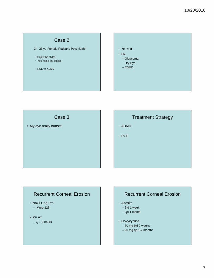
Case 2
– 2) 38 yo Female Pediatric Psychiatrist
• Enjoy the slides
• You make the choice
• RCE vs ABMD
• 78 YOF
• Hx– Glaucoma
– Dry Eye
– EBMD
Case 3
• My eye really hurts!!!
Treatment Strategy
• ABMD
• RCE
Recurrent Corneal Erosion
• NaCl Ung Pm – Muro 128
• PF AT – Q 1-2 hours
Recurrent Corneal Erosion
• Azasite – Bid 1 week
– Qd 1 month
• Doxycycline – 50 mg bid 2 weeks
– 20 mg qd 1-2 months

10/20/2016
8
Recurrent Corneal ErosionLong Term Therapy
• Restasis– Tid
• Fresh –Kote– Qid
• Lacriserts ?
• Treatment
• Nsaid
• Bandage Contact lens– Antibiotic??
– How often
– RTC daily until healed
– Remove and fresh lens and leave in place 3 days?
Recurrent Corneal Erosion
• Bandage Contact lenses
• Antibiotic ung
• Change lens how often
• See patient how often
46
Bacte-viral Conjunctivitis?
• 66 y. o. b. m. c/o 3 d hx of “running, redness, soreness”, OS. AT no help.
• Bilat. Pseudophakia
• No meds., chronic sinusitis.
• 20/20 OD, 20/40 OS, IOP 16,22.
• (-) PAN
? Bacterial Conjunctivits?
• Tx: Besivance OS qid
Zithromax 250 mg 5 d dose pk
• 2 d F/U: 20/50 Va, dec. mucopurulent discharge, 2+ chemosis / injection,
(+) PAN.
• Tx: Besivance qid, finish Zithromax

10/20/2016
9
?Viral?
• 5 d F/U: OS “still swollen but not as sore”. Pt. thinks OD is “catching the infection”.
• OD 20/25, OS 20/40
• Mild follicles OU
• SEIs OU
EKC
• 7d: 20/30 OD, OS• Resolving SEIs• IOP: 20, 21
• 14d: 20/20, 20/25• Mild SEI OS, OD clear• IOP 16,17• D/C Durezol OU
Take Home
• Follow your instincts.
• If it looks bacterial tx it as such.
• Treat aggressively.
• If clin. pic. changes, change with it.
• Patients can have two pathological conditions at the same time.
Epidemic Keratoconjunctivitis
RPS Adeno Detector
Prospective, masked, multi‐center clinical trial in U.S. and Europe
186 consecutive patients examined all cases of acute conjunctivitis and compared to both cell culture and PCR
25% of all acute conjunctivitis confirmed as Adenovirus
RPS Detector
89% Sensitive vs. 91% Cell Culture
94% Specificity vs. 100% Cell Culture
Four Step Test Process
Step 1: Collect Sample
Step 2: Transfer sample to strip
Step 3: Dip test cassette into buffer solution for 15 seconds
Step 4: In less than 10 minutes read test results
RPS Adeno Detector showing a positive result for Adenovirus (2 redlines)

10/20/2016
10
Common Adeno Symptoms
Colds
Pharyngitis
Bronchitis
Pneumonia
Diarrhea
Conjunctivitis
Fever
Cystitis
R h ill
HEKC
A new type of acute keratoconjunctivitis developed throughout Southeast Asia*
Singapore in the summer of 1970.
It was highly contagious and probably was transmitted from person to person by the hand to eye route.
Sixteen cases, diagnosed by viral isolation or serologic study, or both.
*Yang YF, Hung PT, Lin LK, Green IJ, Hung SC.M. Epidemic hemorrhagic keratoconjunctivitis. J Ophthalmol. 1975 Aug;80(2):192-7
Similar to EKC
*Adeno are non-enveloped, double-stranded DNA
*Non enveloped enhances transmission by allowing prolonged survival after dissication*On dry surfaces, steel, viruses remain infective up to 5 weeks
* Penetrate normal barriers to infection* Less than 5% of the US population have antibodies effective against any given serotype
Treatment EKC
• 1 lubricants
• 2 combo antimicrobial / steriod
• 3 Steroid
• 4 Betadine
• 5 Zirgan
• Contagious ? How long
Zirgan™ Product Overview PH3186 6/10© Bausch + Lomb IncorporatedZIRGAN is a trademark of
Zirgan™(Ganciclovir Ophthalmic Gel) 0.15%
Product Background
Please see full prescribing information for Zirgan® provided at this presentation
60Zirgan™ Product Overview PH3186 6/10© Bausch + Lomb IncorporatedZIRGAN is a trademark of Laboratories Théa Corporation licensed by Bausch & Lomb Incorporated
Zirgan™ (ganciclovir ophthalmic gel) 0.15%Indication and Usage
Zirgan is a topical ophthalmic antiviral that is indicated for the treatment of acute herpetic keratitis
(dendritic ulcers).
Important Risk Information
Zirgan is indicated for topical ophthalmic use only.
Please see full prescribing information for Zirgan® provided at this presentation

10/20/2016
11
61 Zirgan™ Product Overview PH3186 6/10
© Bausch + Lomb IncorporatedZIRGAN is a trademark of
Zirgan™ (ganciclovir ophthalmic gel) 0.15%Dosage and Administration
• The recommended dosing regimen for Zirgan is 1 drop in the affected eye 5 times per day (approximately every 3 hours while awake) until the corneal ulcer heals, and then 1 drop 3 times per day for 7 days.
Please see full prescribing information for Zirgan® provided at this presentation
Zirgan
• 1 drop 5x/day until ulcer “heals”
• Then 1 drop tid for 7 days
• 5 gram tube, available early 2010
EKC treatmentMelton/ Thomas
• Povidone- Iodine 5% ( betadine)– Broad spectrum microbiocide
– Indicated for “Irrigation of the ocular surface”
– OFF LABEL USE• Anesthetize with proparacaine
• Instill 1-2 drops NSAID
• Instill several drops of betadine in eye ( close eye)
• Swap excess over lid margin
• After one minute irrigate with saline
• Instill 1-2 drops NSAID
• Rx Lotemax or Zylet or Tobadex ST qid 4 days
– No reports of adverse reactions
– Avoid if allergic to iodine
– Betadine 5% ophthalmic prep soln ( 30 ml opaque)
– 99070 supply code
64
19yoF Red Eye OD
• Red Eye x 3 days with no pain, today was the first day with irritation
• Recently had Staph infection in leg, off antibiotics less than a week ago ( Bactrim)
• VA sc 20/20- OD 20/25 OS
65
Treatment
• Zylet qid OD
• RTC 1 day
• Some improvement over the new few days, but minimal.
66
Treatment
• D/c Zylet qid OD, begin Besivance q1h OD
• Differentials?
Pt showed significant improvement, at 1-day follow up

10/20/2016
12
67
Differentials?
• Herpes Simplex Keratitis
• Adenovirus
• Solution Hypersensitvity
• MRSA
• Remember staph in fection leg treated with Bactrim
• Nursing student
68
Whats Next?
• Diagnosis
• Treatment
THYGESSONS
70
Thygessons
• Possible Thygeson’s
When all else fails: Thygessons Vs HSV
• Discontinue ALL meds
71
Thygeson’s SPK• Described by Phillips Thygeson in 1950
• Slightly elevated corneal lesions, minimal staining
• Usually bilateral, Second to third decade
• Noted corneal sensitivity decreased but not as severe as herpes
• Mild conjunctival involvement, worse with exacerbations
• Appearance similar to EKC described by Fuchs 72
Thygeson’s SPK• Lesions in basal epithelial layer /
Bowman’s layer
• Debris from necrosis / degenerated epicells
• Increased Langerhans cell density
• Part of inflammatory response- Type II

10/20/2016
13
73
Thygeson’s SPK Treatment: Anecdotal
• Cyclosporin 2% in olive oil (8 patients)
• Supratarasal injection triamcinolone (1 case-chronic 6+ years)
• Trifluridine (6 eyes)
• PRK in myopic patient had lesions recur in periphery (untreated area) vscentral (treated area)
• Rimexolone 1% for reversing dendriticcell density (4 patients)
74
Thygeson’s SPK
• Steroid Use
• Loteprednol 0.2%, 0.5%
• Cyclosporine 0.05% Long Term
75
Back to the case…
• D/c All meds
• Lesions healed in 1 week
• No recurrences since October
Plaquenil Keratopathy
Vortex Keratopathy or Cornea
Verticillata
Clinical features:
• Symptoms: the corneal changes are rarely of any visual significance.
• Signs:
– Symmetric, bilateral, whorl-like pattern of powdery, white, yellow or brown corneal epithelial deposits
– Appears in a vortex fashion in the inferocentral cornea and swirls outwards sparing the limbus
• Occurs in Fabry's disease and in patients being treated with a variety of drugs including amiodarone, chloroquine, amodiaquine,
meperidine, indomethacin, chlorpromazine and tamoxifen.
Staph. Hypersensitivity
• Treatment– Warm compresses
– Lid hygiene with commercial lid cleanser
– Broad spectrum topical antibiotic
– Antibiotic ointment
– Topical steroid*
– Oral tetracycline antibiotics if >10 y. o.
Krachmer JH, Mannis MJ, Holland EF, eds. Cornea. 2nd ed. Philadelphia: Elsevier Mosby; 2005. p. 1235-1238.Rapuano CJ, Luchs JI, Kim T. Anterior Segment: The Requisites in Ophthalmology. St. Louis: Mosby; 2000. p. 165-168.

10/20/2016
14
Staphylococcus Hypersensitivity
• 58 YOM
• Custom Toric Soft C/L– +4.00-3.00
– +3.00-375
• Pain OS 4-5 days
• Presents wearing CL
Phylctenular Kerato-Conjunctivitis
PKC / Staph. Hypersensitivity
• Non-infectious hypersensitivity• Phlyctenules (phlyctena)
– histiocytes, lymphocytes, plasma cells, neutrophils
• Microbial association– Staph. Aureus– Myco. Tuberculosis– Chlamydia trachomatis– Neisseria gonorrhea– Coccidiodes immitis– Bacillus spp.– Herpes simplex virus– Leishmaniasis Ascaris lubricoides– Hymenlepsis nana– Candida spp.
Differential Diagnosis
• Staphylococcal keratitis with phlyctenule
• Microbial keratitis (Mycobacterium tuberculosis)
• Inflamed pterygium
• CIN (Conjunctival Intra-epithelial Neoplasia)
• Chronic FB
Phlyctenular Kerato-Conjunctivits
• Tx: Pred Forte OD q2h x 2 d then qid, Gatifloxacin OD qid x 1 wk., warm compresses, lid hygiene.
• D/C topical allergy meds.
• Doxycycline 50mg bid x 2 mo.
• PPD (-)

10/17/2016
1
Glaucoma in Review
Ashley M. Speilburg, OD, FAAO
Glaucoma
A degenerative disease of the optic nerve characterized by ganglion cell axon death, excavation of the optic nerve, nerve fiber bundle defects and visual field loss.
Primary vs. secondary vs. developmental
Open angle vs. closed angle
GlaucomaPRIMARY GLAUCOMAS
Open Angle Forms:
Primary Open Angle Glaucoma
Low Tension Glaucoma
Closed Angle Forms
Primary Angle Closure
Acute Angle Closure
Plateau Iris Configuration
Plateau Iris Syndrome
SECONDARY GLAUCOMAS
Open Angle Forms:
Neovascular glaucoma
Irido‐corneal‐endothelial (ICE) syndrome
Posterior polymorphous dystrophy
Epithelial drown growth
Fibrous down growth
Inflammatory membranes
Malignant tumors
Neurofibromatosis
Juvenile xanthogranuloma
Pigmentary Glaucoma
Exfoliation (psuedoexfoliation)
Uveitis
Malignant melanoma
Uveitis
Lens Induced Glaucoma
Alpha‐chymotrypsin
Vitreous in anterior chamber
Healon
Post‐surgical
Edema
Glaucomacyclitic crisis
Scleritis
Alkali Burn
Angle recession glaucoma
Intraocular foreign bodies
Steriod induced glaucoma
Carotid cavernous fistula
Cavernous sinus thromboisis
Retrobulbar tumors
Graves disease
Superior vena cava obstruction
Mediastinal tumors
Sturge‐Weber syndrome
Famial episcleral venous pressure eleva
GlaucomaClosed Angle Forms
Neovascular glaucoma
ICE syndrome
Posterior polymorphous dystrophy
Trauma
Uveitis
Aniridia
Miotic induced
Phacomorphic
Subluxed lens
Aphakic or psuedophakic
Iris bombe due to 3600 posterior synechia
Malignant glaucoma (ciliary block glaucoma)
Choroidal detachment / Choroidal effusion
Melanoma
Retinoblastoma
Cysts of iris and ciliary body
Melanoma
Retinoblastoma
Cysts of iris and ciliary body
DEVELOPMENTAL GLAUCOMAS
Primary developmental glaucomas:
Congential glaucoma
Juvenile glaucoma
Secondary developmental glaucomas:
Retinopathy of prematurity
Post‐traumatic
Tumor related
Inflammatory induced
Neurofibromatosis
Rubella
Sturge‐Weber syndrome
Associated with congential anomaliesor syndromes:
Aniridia
Axenfeld ‐ Rieger Syndrome
Chromosome abnormalities
Homocystinuria
Lowe’s syndrome
Marfan’s syndrome
Microcornea
Microspherophakia
Nanophthalmos
Secondary Glaucoma
Glaucomatous damage from increased IOP that is a direct result of some other ocular or systemic abnormality.
Pigmentary
Exfoliation
Angle recession
– Neovascular
– Uveitic
– ICE syndromes
Primary Open Angle Glaucoma
Chronic Open Angle Glaucoma or Open Angle Glaucoma
Characterized by:
Adult onset
IOP >21 mmHg
Open angle of normal appearance
Glaucomatous optic nerve damage
Characteristic visual field loss

10/17/2016
2
Normal Tension Glaucoma
Low Tension Glaucoma
Characteristic optic nerve and visual field changes in the absence of IOP over 21mmHg.
Up to 33% of open angle glaucoma
Few unique features
PPA
Disc hemorrhages
h/o migraines
h/o reynaud’s phenomenon
Ocular Hypertension
IOP repeatedly > 21 mmHg in the absence of glaucomatous optic nerve damage, visual field loss and RNFL defects.
7% of population over 40 have IOP > 21 mmHg
Only ~1% of which have glaucoma
10% of OHTN will develop POAG at 5 years
Gonioscopy!
To properly diagnose any type of glaucoma gonioscopy is key!
Open angle vs. closed angle
Primary vs. secondary
Risk Factors
Ocular
IOP
Pachymetry (CCT)
Optic nerve
Myopia (high)
Non‐ocular
Older age
Race
Family History
Diabetes
Hypertension (or rather over treatment of HTN)
OPP
Age
Prevalence for glaucoma increases with age
1% < 40 yo
3‐8x higher > 70 yo
Longer exposure to high IOP?
Other factors (vascular, connective tissue integrety)
Race
Baltimore Eye Study
1.7% vs. 5.6% at 40+
11% at 80+
St. Lucia Study
Caribbean blacks 14%
LALES
Latinos 5% (8% 60‐70yo ; 15% 70+)
10/17/2016
3
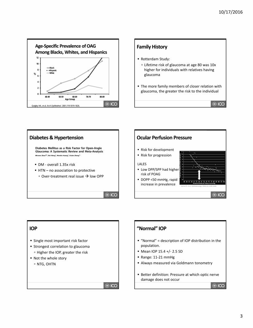
Age‐Specific Prevalence of OAGAmong Blacks, Whites, and Hispanics
0
2
4
6
8
10
12
40-49 50-59 60-69 70-79 80-89
BlackHispanicWhite
Quigley HA, et al. Arch Ophthalmol. 2001;119:1819-1826.
%
Age Group
Family History
Rotterdam Study:
Lifetime risk of glaucoma at age 80 was 10x higher for individuals with relatives having glaucoma
The more family members of closer relation with glaucoma, the greater the risk to the individual
Diabetes & Hypertension
DM ‐ overall 1.35x risk
HTN – no association to protective
Over‐treatment real issue low OPP
Ocular Perfusion Pressure
Risk for development
Risk for progression
LALES
Low DPP/SPP had higher risk of POAG
DOPP <50 mmHg, rapid increase in prevalence
Varma R, et al. Ophthalmology. 2004;111:1439-1448.
IOP
Single most important risk factor
Strongest correlation to glaucoma
Higher the IOP, greater the risk
Not the whole story
NTG, OHTN
“Normal” IOP
“Normal” = description of IOP distribution in the population.
Mean IOP 15.4 +/‐ 2.5 SD
Range: 11‐21 mmHg
Always measured via Goldmann tonometry
Better definition: Pressure at which optic nerve damage does not occur

10/17/2016
4
Characteristics of IOP
Increases slightly with age Normal change not associated with the development of glaucoma
Symmetrical between eyes: ~2‐4 mmHg Greater than this can be a sign of disease
Diurnal fluctuation (4‐6mmHg) Exaggerated in glaucoma patients
Highest in overnight hours Single IOP measurement in office is insufficient
Pachymetry
Thinner CCT increases risk of POAG in patients with OHTN
Anatomic “weakness”?
True IOP higher than measured
Still a factor in NTG
How much?
The Ocular HypertensionTreatment Study (OHTS)
Supported by the National Eye Institute, National Center on Minority Health and Health Disparities, Research to Prevent Blindness, and Merck Research Laboratories
The Ocular Hypertension Treatment Study: A Randomized Trial Determines That Topical Ocular Hypotensive Medication Delays orPrevents the Onset of Primary Open-Angle Glaucoma
Arch Ophthalmol.2002;120(6):701-713.
OHTS
Provided solid evidence that lowering IOP in patients with OHTN reduces the risk of developing POAG.
Identified risk factors associated with the development of glaucoma.
OHTS
Treatment reduced risk of development of glaucoma from 9.5% (observation arm) to 4.4% (treatment arm) at 5 years.
Treatment: 20% reduction and <24 mmHg
Modest goal
OHTS and Corneal Thickness
For all IOP’s, a thinner cornea increased the risk of developing glaucoma at 5 yrs
IOP <555 >555‐<588 >588
>25.75 36% 13% 6%
>23.75‐<25.75 12% 10% 7%
<23.75 17% 9% 2%
CCT Microns
High risk Low risk

10/17/2016
5
OHTS Risk Factors
• Age: 20% – 40% increase per decade
• IOP: > 26 mmHg
• Vertical C/D: > 0.5
• CCT: < 555 µm
OHTS Conclusions
Conclusions:
In patients with elevated IOP, topical ocular hypotensive
treatment was effective in delaying the probability of onset of
POAG1
For patients with a moderate to high risk of developing POAG,
IOP‐lowering treatment should be considered. This does not
imply that all borderline or elevated IOP patients should
receive treatment1
Some predictors of developing POAG were found to be:
baseline IOP, age, vertical cup‐to‐disc (C/D) ratio, and lower
central corneal thickness2
1. Kass et al. Arch Ophthalmol. 2002; 2. Gordon et al. Arch Ophthalmol. 2002.
Optic Nerve
C/D ratio, RNFL, NRR
Associate with increased vertical C/D ratio
No single cut off for C/D
Larger cup in larger discs
Asymmetry
Focus on integrity of NRR and RNFL
Myopia?
2x more common among POAG
Selection bias?
Very high myopia (>‐14D) are high risk
Flat, obliquely inserted
VF confounded by retinopathy in MD
Global Risk Assessment
What is global risk assessment?
Estimates a patient’s overall risk based on multiple rather than a single risk factor
Ideally based on evidence from well‐controlled clinical trials and long‐term studies
How is it used?
Helps guide treatment decisions for optimal patient care
Global Risk Calculator for OHTN
Enhanced accuracy of identifying those most at risk for POAG
Individualize who is treated
Developed from OHTS study data
Identify the global risk of developing glaucoma in next 5 years
Identify who will benefit from treatment vs those who only need observation

10/17/2016
6
Online OHTS Risk Calculator
5 key risk factors considered
http://ohts.wustl.edu/risk/calculator.html
Treatment Recommendations
**Suggested guidelines only**
Expert Panel Recommendations
< 5% No treatment
5‐15% Treatment optional
>15% Treatment recommended
Patterns of damage
– Arching pattern divided by horizontal raphe
• Structural and functional change must correlate
• Ganglion cell axons damage at ONH
• Patters of damage should reflect retinal anatomy of RNFL
Types of VF defects
Pics of nasal step
Arcuate
Paracentral
Severity
American Glaucoma Society staging system
VF Interpretation
• Reliability Indices• Fixation losses• False positives• False negatives
• Global Indices• Mean deviation• Pattern standard
deviation• Visual field index
• Glaucoma Hemifield Test• Total deviation plot• Pattern deviation plot

10/17/2016
7
Fixation Losses False Negatives
10/17/2016
False Positives
10/17/2016
Global Indices
• Mean Deviation
• Difference between threshold values and age‐match normal value
10/17/2016
Global Indices
• Pattern Standard Deviation
• Represents localized VF loss
• Corrects for media opacities
10/17/2016
Global Indices
Visual Field Index Staging index
Less affected by cataract than MD
Improved correspondence to patterns of ganglion cell loss
Weights central points higher due to higher density of ganglion cells 100% in normal VF
0% in perimetrically blind VF
10/17/2016

10/17/2016
8
Glaucoma Hemifield Test
• Compares mirror‐image point clusters of common glaucomatous VF loss above and below the horizontal midline
1. “Within Normal Limits”2. “Borderline”3. “Outside Normal Limits4. “Abnormally High Sensitivity”5. “General Reduction of Sensitivity” Figure 5‐5, The Field Analyzer Primer, 4th ed.
Single Field Analysis
• ID correct demographics
• Review Reliability Indices
• Compare TD and PD
• Review Global Indices & GHT
Single Field Analysis Single Field Analysis
OCT and Glaucoma
Excellent diagnostic tool to aide in ONH and RNFL assessment
Mild to moderate stage disease
Loses usefulness in severe stage disease
Floor effect
OCT Interpretation
• Key Parameters• Compared to normative data
• RNFL Thickness Map• RNFL Deviation Map
• Deviation from normal• En face fundus image
• NRR Thickness• RNFL TSNIT Graph• RNFL Quad & Clock Hour
• Compared to normative data
• Bscans• RPE & disc black• ILM & cup red

10/17/2016
9
OCT Interpretation
Scan quality
Signal strength > 7
No loss of signal areas
No motion artifacts
Review Key Parameters
Color coded based on normative data base
OCT Interpretation
Review displays
RNFL Thickness
RNFL Deviation
TSNIT
Quad/clock hour
B‐scans
Structure‐Function Correlation
We’ve got to have it!
OCT and HVF
HVF and ONH
OCT and ONH
Target IOP
A pressure at which additional damage is considered unlikely to take place
Risk factors & IOP How many, how strong and how high?
Current level of damage/ severity of disease
Greater the damage, the lower the IOP
Progression rate (if known)
No exact method
May (and should) be modified as needed
Target IOP
Collaborative Normal Tension Glaucoma Study
Showed 30% reduction in IOP reduced risk of progression
IDed risk factors for progression (migraine, female sex, disc heme)
Advanced Glaucoma Intervention Study
VF stability achieved with average IOP of 12 mmHg and always IOP <18 mmHg
Early Manifest Glaucoma Trial
Tx reduces risk of progression in early POAG
10% reduced risk for every 1mmHg lower
Risks for progression: (higher IOP, age, XFG, bilateral, worse VF)
Treatment: Traditional Medications4 Classes Available medications
PGA Latanoprost, Travatan Z (travaprost), Lumigan, Zioptan
BB Timolol soln and gfs (Betimol, Istalol, Timoptic), levobunolol (Betimol)
A‐agonist Brimonidine (Alphagan P)
CAI Dorzolamide, Azopt
Fixed Combos Available medications
BB + CAI Dorzolamide‐timolol, Cosopt PF
BB + A‐agonsit Combigan
A‐agonist + CAI Simbrinza

10/17/2016
10
Treatment: Traditional Medications4 Classes MOA Side Effects Contraindications
PGA30% ↓QD
Increasesuveoscleraloutflow
Darkening of skin, iris,growth of lashes, loss of periorbital fat, stinging, hyperemia
Caution if used monocularly
BB25+% ↓QD
Decreases AqHproduction
Bradycardia, bronchiolar constriction, arrhythmia,heart block, sexual dysfunction
Asthma, COPD, bradycardia, congestiveheart failure
A‐agonist20‐25% ↓BID‐TID
Decreases AqHproduction
Low BP, orthostatic hypotension, allergic reactions up to 25%
Children
CAI20% ↓BID‐TID
Decreases AqHproduction
Allergic reactions, stinging, taste disturbances
Caution in truesulfonamide allergies, sickle‐cell disease
Treatment: Medication Selection
Consider safety and efficacy
PGA first line for most
Review history to determine best drug for patient
Compliance and adherence are required
Balance safety/efficacy with acceptable SE and dosing schedule
Treatment: New Medications
Rho Kinase (Rock) Inhibitors
Increase outflow through TM
Lowers episcleral venous pressure
NET inhibition reduces fluid production
May be ideal for NTG
Rhopressa (QD)
Roclatan (Rhopressa + latanoprost)
Treatment: New Medications
Vesneo (latanoprostene bunod) (QD)
Latanoprost + nitric oxide donating moiety
Increases outflow through TM and uveoscleral
May positively impact perfusion pressure
CRL from FDA: no safety or efficacy concerns
Treatment: Lasers
Laser trabeculoplasty
ALT and SLT
Different lasers, different MOAs
Similar efficacy
SLT may be more repeatable
Treatment: Laser Trabeculoplasty
Questions:
What is the success rate?
How much does it lower IOP?
How long does it last?
Does the laser matter?
How does it compare to medical/surgical Tx?
Is it repeatable?

10/17/2016
11
Treatment: Laser Trabeculoplasty
What is the success rate?
Effective ~70‐75% of the time
How much does it lower IOP?
20‐25% on average
Treatment: Laser Trabeculoplasty
How long does the effect last?
~80% success at 1 year
~40% at 2 years?
~50% success at 5 years
~20% at 10 years
Does the laser matter?
No significant difference in IOP or complications
Treatment: Lasers
How does it compare to medical/surgical Tx?
SLT Med – SLT vs latanoprost same at 12 mo
GLT – better than timolol at 2 years; similar results at 7 years
Moorfields – Trabeculectomy > medical > trabeculoplasty
Is it repeatable?
ALT – not already treated areas
SLT – yes, but probably less effective
Treatment: MIGS
Micro Invasive Glaucoma Surgery
Ab interno microincision
Minimal trauma
Effective
High safety profile
Rapid recovery
Treatment: MIGS
Newer procedures
iStent
CyPass Micro‐Stent
Xen Gel Stent
Treatment: MIGS
iStent (Glaukos)
FDA approved 2012
Indicated for use in conjunction with cataract surgery in adult patients with mild‐moderate open‐angle glaucoma currently treated with ocular hypotensive medications.
Allows aqueous direct route to Schlemm’s canal via bypass of TM

10/17/2016
12
Treatment: MIGS
CyPass Micro‐Stent (Alcon)
FDA approved, late July 2016
Implanted at time of CE in patients with mild‐moderate open‐angle glaucoma
Reduces IOP by increasing suprachoroidaloutflow
Treatment: MIGS
XEN Gel Stent (AqueSys) Investigational device in US Investigated for use as stand alone procedure in patients with glaucoma refractory to medications Soft compressible silicone tube, 6mm long, ~ thick as a human hair Bypasses conventional outflow pathway by filtering aqueous into subconjunctival space Suspected advantages: lower IOP, suitable for more advanced glaucoma
Progression Monitoring
Structure vs function?
WGA Consensus Both should be evaluated
Assess risk factors for progression
Stein JD et al. Trends in use of ancillary glaucoma tests for patients with open‐angle glaucoma from 2001‐2009. Ophtholmology. 2012;119:748‐758.
Risk Factors for Progression
Older age
Bilateral disease
Higher mean IOP
Disc hemorrhage
Thinner CCT with higher baseline IOP
More advanced VF loss at baseline
Low OPP
Exfoliation syndrome
Visual Field Progression
Overview reports
Changes in global indices
MD
PSD
Progression analysis software
Guided Progression Analysis (HFA, Carl Zeiss Meditek)
Event & Trend
Visual Field Progression
Event vs. Trend Analysis
Event – to identify statistically significant worsening of the VF
Point by point basis
Best for ID of small, localized change
Trend – quantify any observed rate of change
Better for overall worsening
Requires more tests for a reliable slope (5+)

10/17/2016
13
Event Analysis
Based off progression criteria from EMGT Sensitive and specific compared to expert eval
Compares individual tests points on PD of successive tests, to the average of two baselines
Flags points that vary by more than the expected variability of similarly damaged VFs.
Open and shaded triangles ID statistically significant change from baseline.
Figure 6‐4, The Field Analyzer Primer, 4th ed.
Event Analysis
Considerations
Each test point has 5% change of
being falsely flagged Look for repeatedly depressed points
Look for clusters of points typical of glaucoma
Extension of existing defects and central points more likely to be “real” change
Not calculated for VFs with MD < ‐20 dB
Trend Analysis
Linear regression analysis of the of the VFI
Plots VFI vs age
Calculates rate of progression after 5+ tests
Projects into future if 5+ tests available
What could happen if present trend continues
Trend Analysis
Considerations
Need at least 5 tests for a meaningful slope
More is better
Minimally affected by small, focal defects
‐1 ‐ 1.5% per year probably max acceptable rate of progression
Depends on age of patient and extent of VF loss
Requires sufficient frequency of testing
WGA Consensus on Progression of Glaucoma 2011
Baseline data collection – first two years
In clinical practice, at least two reliable VFs is optimal in the first six months.
At least two further VFs should be performed within the next 18 months.
Repeat VFs sooner if possible progression
Follow‐up data collection ‐ after the initial two years
The frequency of follow‐up VFs should be based on the risk of clinically significant progression (based on extent of damage and life expectancy).
Low – Moderate Risk: 1x per year
High Risk: 2x+ per year
Always repeated sooner if progression is suspected
Weinreb RN et al. Progression of Glaucoma: The 8th Consensus Report of the World Glaucoma Association. Amsterdam: Kugler Publications; 2011.
Structural Progression
Serial disc and RNFL photography: still valuable
Disc hemorrhages, PPA, steroscopic eval
C/D estimates insufficient for monitoring structural change
OCT: objective, reproducible measurements and quantitative assessment of disc and RNFL
Requires good image quality
Weinreb RN et al. Progression of Glaucoma: The 8th Consensus Report of the World Glaucoma Association. Amsterdam: Kugler Publications; 2011.

10/17/2016
14
OCT Progression Analysis
Statistical analysis of sequential OCT scans to detect progression
Event and Trend Analysis
OCT Event Analysis
Compares follow up scan to baselines
IDs change when difference is greater than a predetermined threshold based on test‐retest variability
Image alignment is critical
More susceptible to artifact
OCT Trend Analysis
Regression line between parameter and time
Provides rate of change
Various parameters
RNFL thickness (average, inferior, superior)
Rim area or cup‐to‐disc area
Less susceptible to artifact
Requires large number of tests for reliable slope
OCT Progression Patterns
Widening of a defect
Depening of an existing defect
Inferior‐temporal defects
Reliable baselines
Reliable
Free of artifact
Ideally within 6 months of each other
Change baselines when treatment is altered
When Progression is Suspected
Repeat the test Confirm if change is real
Is the change typical of glaucoma?
What is the rate of change?
Is the rate of change acceptable compared to the patient’s life expectancy and stage of disease?
Not all statistically significant change is clinically meaningful

10/17/2016
15
Adjusting Therapy
Establish new target IOP
Rate over which progression took place
Additional risk factors
Age of patient
Severity of damage
Adjusting Therapy: Options
Additional medication
Often jump to a fixed combination
Laser
Open angle?
MIGS
Mild‐moderate glaucoma
Need CE also?
Trabeculectomy/ GDI
Significant additional IOP lowering is needed on
Rapid progression in younger patient
Questions?
Email: [email protected]