ocular complications due to cancer treatment
TRANSCRIPT

Contents
6.1 Introduction . . . . . . . . . . . . . . . . . . . . . 826.2 Eyelids, Periorbital Skin and Tear Film . . . . . . 82
6.2.1 Anatomy and Physiology . . . . . . . . . . 826.2.2 Acute Radiation Effects . . . . . . . . . . . . 836.2.3 Chronic Radiation Effects . . . . . . . . . . 836.2.4 Chemotherapy . . . . . . . . . . . . . . . . 846.2.5 Medical and Nursing Management . . . . . 84
6.3 Conjunctiva . . . . . . . . . . . . . . . . . . . . . 856.3.1 Anatomy and Physiology . . . . . . . . . . 856.3.2 Acute Radiation Effects . . . . . . . . . . . . 856.3.3 Chronic Radiation Effects . . . . . . . . . . 856.3.4 Chemotherapy . . . . . . . . . . . . . . . . 856.3.5 Medical and Nursing Management . . . . . 85
6.4 Cornea . . . . . . . . . . . . . . . . . . . . . . . . 866.4.1 Anatomy and Physiology . . . . . . . . . . 866.4.2 Acute Radiation Effects . . . . . . . . . . . . 866.4.3 Chronic Radiation Effects . . . . . . . . . . 866.4.4 Chemotherapy . . . . . . . . . . . . . . . . 876.4.5 Medical and Nursing Management . . . . . 87
6.5 Lens . . . . . . . . . . . . . . . . . . . . . . . . . . 876.5.1 Anatomy and Physiology . . . . . . . . . . 876.5.2 Acute Radiation Effects . . . . . . . . . . . . 876.5.3 Chronic Radiation Effects . . . . . . . . . . 876.5.4 Chemotherapy . . . . . . . . . . . . . . . . 886.5.5 Medical and Nursing Management . . . . . 88
6.6 Uvea: Iris, Ciliary Body and Choroid . . . . . . . . 896.6.1 Anatomy and Physiology . . . . . . . . . . 896.6.2 Acute Radiation Effects . . . . . . . . . . . . 896.6.3 Chronic Radiation Effects . . . . . . . . . . 896.6.4 Chemotherapy . . . . . . . . . . . . . . . . 896.6.5 Medical and Nursing Management . . . . . 89
6.7 Sclera . . . . . . . . . . . . . . . . . . . . . . . . . 906.7.1 Anatomy and Physiology . . . . . . . . . . 906.7.2 Acute Radiation Effects . . . . . . . . . . . . 906.7.3 Chronic Radiation Effects . . . . . . . . . . 906.7.4 Chemotherapy . . . . . . . . . . . . . . . . 906.7.5 Medical and Nursing Management . . . . . 90
6.8 Optic Nerve and Retina . . . . . . . . . . . . . . . 906.8.1 Anatomy and Physiology . . . . . . . . . . 906.8.2 Acute Radiation Effects . . . . . . . . . . . . 906.8.3 Chronic Radiation Effects . . . . . . . . . . 906.8.4 Chemotherapy . . . . . . . . . . . . . . . . 916.8.5 Medical and Nursing Management . . . . . 92
6.9 Orbital Bones and Tissue . . . . . . . . . . . . . . 926.9.1 Anatomy and Physiology . . . . . . . . . . 926.9.2 Acute Radiation Effects . . . . . . . . . . . . 926.9.3 Chronic Radiation Effects . . . . . . . . . . 926.9.4 Chemotherapy . . . . . . . . . . . . . . . . 936.9.5 Medical and Nursing Management . . . . . 93
6.10 Conclusion . . . . . . . . . . . . . . . . . . . . . . 93References . . . . . . . . . . . . . . . . . . . . . . . . . . 93
Ocular Complications Dueto Cancer Treatment
Chapter 6 81
Michael Ober · Camille A. Servodidio ·David Abramson
06_Schwartz_Occular 27.01.2005 8:31 Uhr Seite 81

Chapter 682 M. Ober · C. A. Servodidio · D. Abramson
6.1 Introduction
The eye is composed of many tissues that vary great-ly in their sensitivity to cytotoxic therapy. This chap-ter highlights the ocular complications of cancertreatment and discusses the relevant anatomy andmedical management. Each section discusses the ba-sic anatomy and physiology of a specific area of theeye (Fig. 6.1), common radiation and chemothera-peutic complications and therapeutic management.
6.2 Eyelids, Periorbital Skin and Tear Film
6.2.1 Anatomy and Physiology
The thinnest skin in the body is located on the outersurface of the eyelids. It is devoid of subcutaneous fatallowing for the accumulation of fluid to manifestrapidly as swelling. The upper and lower eyelids con-tain fibrous connective tissue, known as the tarsalplates, which function as structural support. Theeyelashes are located on the anterior portion of theeyelids and aid in protection of the eye.
The tear film covers the anterior surface of theconjunctiva and cornea. It serves the vital role ofsupplying the cornea with moisture, nutrients, en-zymes, immunoglobulins and protein signals, as wellas allowing the maintenance of a clear, non-kera-tinized epithelium in the visual axis. Furthermore,the tear film comprises the smooth outer refractivecoating essential to vision by filling in corneal irreg-ularities. The tear film consists of three layers. Theaqueous layer is produced by the accessory lacrimalglands found in the conjunctiva. Meibomian glandslocated within the tarsal plates produce an oily layerthat sits on top of and acts to stabilize the aqueouslayer. The goblet cells of the conjunctiva produce thethird, or mucous, layer. The overall function of thetear film is vitally dependant on each of these indi-vidual layers, and a deficiency in any layer willadversely affect the entire ocular surface.
The tears drain from the ocular surface via twopuncta located on the medial aspect of the upper andlower lid margin. The puncta lead to the canaliculithat empty into the lacrimal sac and, in turn, into the nose via the nasal-lacrimal duct.
Figure 6.1
Cross-sectional anatomy of the eye
06_Schwartz_Occular 27.01.2005 8:31 Uhr Seite 82

Chapter 6 83Ocular Complications Due to Cancer Treatment
6.2.2 Acute Radiation Effects
Madarosis, or loss of eyelashes, and erythema are thefirst side effects of radiation therapy (RT) involvingthe eye. Usually, eyelashes will grow back; however,permanent loss does occur. Erythema can occurwithin days of treatment (generally after doses of atleast 20–30 Gy) and usually persists for a few days.Dermatitis is the most common acute side effect ofRT. Dry dermatitis of irradiated skin can occur withdoses greater than 20 Gy and often leads to desqua-mation. Moist dermatitis, with exposure of the der-mis and associated serum leakage, can occur after thefourth week of RT following doses of 40 Gy or more,
fractionated over a 4-week period. Blisters and ede-ma may precede moist dermatitis. Symptoms includeredness, peeling, burning, itching and pain [1] (Fig. 6.2).
6.2.3 Chronic Radiation Effects
The late effects of RT to the eyelids following dosesfrom 30–60 Gy include madarosis, telangiectasia (di-lated, tortuous blood vessels; Fig. 6.3), hyperpigmen-tation, depigmentation, ectropion, hyperkeratosis, at-rophy, necrosis, ulceration and punctual occlusion.Although rarely seen today, lid deformities, such asectropion (out-turning of eyelid margin), entropion
Figure 6.2
Side effects of chemotherapy and radiation of the eye
06_Schwartz_Occular 27.01.2005 8:31 Uhr Seite 83

Chapter 684 M. Ober · C. A. Servodidio · D. Abramson
(in-turning of eyelid margin) and atrophy or con-tracture, are seen when the tarsus has been includedin the radiation field. The time of onset ranges from 2months to greater than 5 years after treatment. De-struction or occlusion of the puncta may occur whenthe medial portions of the eyelid are irradiated,which leads to impaired tear drainage. Lid necrosis,exacerbated by excess sun exposure in areas previ-ously irradiated, may develop months to years aftertreatment [2, 3].
6.2.4 Chemotherapy
Many chemotherapeutic agents, such as cyclophos-phamide, ifosfamide and methotrexate, alter the nor-mal tear film physiology either by causing inflamma-tion of the lacrimal glands or by being excreted di-rectly into tears, which leads to dry-eye symptomsand inflammation around the eyelids and anteriorsegment of the eye [4]. Patients treated with alkyl sul-fonates, including busulfan and nitrosourea, havealso reported developing dry eyes [5]. Both 5-fluo-rouracil [6] and docetaxel [7] have been associatedwith stenosis of the punctum and tear (canalicular)drainage system. In addition, some patients receiving5-fluorouracil develop excessive lacrimation along
with cicatricial eyelid malpositioning. Intravenousdoxorubicin has also been associated with excessivelacrimation. Paleness of the periorbital skin has beenreported following mithramycin infusion, whiledrooping of the upper eyelid, known as ptosis, hasbeen reported following long-term corticosteroid use [8].
6.2.5 Medical and Nursing Management
The management for eyelid complications due tocancer treatment consists mainly of skin care, includ-ing the use of ultraviolet protection, meticulous hy-giene with mild soaps, the use of skin lubricants,avoiding skin sensitizing drugs (i.e. tetracyclines)and occasionally corticosteroid and/or antibioticcreams. Ptosis, tear drainage or eyelid position mayrequire minor surgical manipulation by an ophthal-mologist and should be referred in clinically signifi-cant cases [9]. The mainstay of dry eye therapy con-sists of tear replacement with artificial tears dropsand ointment. Patients with symptoms or at riskshould be encouraged to use liberal amounts of arti-ficial tears. Unpreserved artificial tears are preferred,especially when they are used more than four timesper day, due to the fact that the preservatives them-
Figure 6.3
Telangiectasia of the conjunctivalblood vessels
06_Schwartz_Occular 27.01.2005 8:31 Uhr Seite 84

Chapter 6 85Ocular Complications Due to Cancer Treatment
selves can be irritating to the cornea, conjunctiva andeyelids. Further aids include punctal occlusion, warmcompresses to eyelids and, in advanced cases, cy-closporine drops. Patients with continued sympto-matic or refractory dry eyes should be referred to aneye care professional without delay, as the conse-quences of hesitating could be permanent vision loss.
6.3 Conjunctiva
6.3.1 Anatomy and Physiology
The conjunctiva is a thin, transparent mucous mem-brane that lines both the posterior aspect of the eye-lids (palpebral conjunctiva) and the anterior surfaceof the eye (bulbar conjunctiva). The folds betweenthe palpebral and bulbar conjunctiva are known asthe superior and inferior fornices, respectively. Tis-sue is redundant in the fornices to allow for adequatemovement of the globe. The main lacrimal gland,which functions during reflex tearing, empties intothe superior fornix, while the accessory lacrimalglands, supplying basal tear secretion, are foundthroughout the conjunctiva, concentrating in the for-nices.
The conjunctiva contains a stratified non-kera-tinized epithelium overlying a stroma, known as thesubstantia propria. Goblet cells supplying the mucinlayer of the tear film are found intermixed with theepithelial cells. Besides acting as a physical barrier,the conjunctiva aids in host defenses by hosting im-mune cells as well as colonizing bacteria.
6.3.2 Acute Radiation Effects
Conjunctival inflammation (conjunctivitis), whichmanifests as vascular injection with clear or mucoiddischarge tends to occur 1–3 weeks after the start ofradiation treatment. Edema of the conjunctiva,known as chemosis, may occur simultaneously or inisolation and usually lasts for a few days. Affectedconjunctiva may also ulcerate leading to an increasedrisk of infection. The duration of these signs may beprolonged when RT doses over 30 Gy are used [1, 2,10].
6.3.3 Chronic Radiation Effects
Late effects of RT to the conjunctiva include pro-longed injection, telangiectasis, symblepharon (ad-hesions between the bulbar and palpebral conjuncti-va) and subconjunctival hemorrhage, shortening ofthe fornices, loss of goblet cells, keratinization andnecrosis. Exposure to 30–50 Gy results in prolongedconjunctival injection, which develops in 1–2 years,followed by telangiectatic vessels 3–6 years later.These fragile vessels tend to rupture with minor trau-ma, resulting in subconjunctival hemorrhage.
Chronic ulceration of the conjunctiva can be seenfollowing treatment with 60 Gy. This leads to symble-pharon formation, resulting in shortening of the for-nices, trichiasis (turning of lashes onto the ocularsurface) and eyelid malpositioning. Goblet cell lossoccurs at relatively low doses, resulting in tear film in-stability and dry eye symptoms, while doses over50 Gy may result in keratinization of the conjunctiva.These keratin plaques constantly irritate adjacentcornea, occasionally causing scarring and visual loss.Necrosis may occur after radioactive plaque therapyfor retinoblastoma patients, where doses to the con-junctiva between 90–300 Gy are used [1–3, 10].
6.3.4 Chemotherapy
Conjunctivitis is a commonly reported symptom fol-lowing induction therapy with many medications, in-cluding cyclophosphamide, ifosfamide, nitrosoureas,cytosine arabinoside, doxorubicin, methotrexate,deoxycoformycin and mitomycin. 5-fluorouracil isalso associated with conjunctivitis and eye irritation.This usually occurs concurrently with the initiationof therapy and resolves within two weeks of treat-ment cessation. The immunosuppressive effects ofcorticosteroids are believed to facilitate opportunis-tic infections throughout the eye, including bacterial,viral and fungal conjunctivitis [11].
6.3.5 Medical and Nursing Management
Antibiotic eye drops, sometimes in combination withcorticosteroids, are used for prolonged conjunctivitisand for conjunctival ulceration. Artificial teardrops
06_Schwartz_Occular 27.01.2005 8:31 Uhr Seite 85

Chapter 686 M. Ober · C. A. Servodidio · D. Abramson
often aid chronic conjunctival irritation by providingthe lubrication necessary to replace lost tear volumeand dilute toxic chemotherapeutic metabolites ex-creted into the tear film. Vitamin A ophthalmic oint-ment (tretinoin 0.01% or 0.1%) may reverse squa-mous metaplasia and loss of vascularization fromscar formation [12]. Patients with infectious conjunc-tivitis should be instructed to wash their hands fre-quently and take great care in interactions withothers to prevent the spread of communicable dis-eases. In addition, sunglasses for protection from thesun and wind may be helpful in reducing symptoms.Severe conjunctival reactions, such as symblepharonand forniceal shortening, may require ophthalmolog-ic manipulations such as symblepharon lysis on arepeated basis; or, alternatively, mucous membranegrafting with forniceal reconstruction may be neces-sary. Ophthalmologic referral is therefore indicated.
6.4 Cornea
6.4.1 Anatomy and Physiology
The cornea is the transparent, avascular anterior por-tion of the eye that refracts and transmits light to theinner structures of the eye. Along with the overlyingtear film, it provides approximately two thirds of therefracting power of the eye. The conjunctiva bordersthe cornea in an area known as the limbus. This re-gion contains corneal stem cells; therefore, compro-mising this zone leads directly to the loss of cornealtransparency and often its integrity. The cornea is an avascular tissue and thus depends on the limbalvessels along with the tear film and aqueous fluidfrom the anterior chamber for nutrients and wasteremoval.
The cornea consists of five specialized layers,including, from anterior to posterior: epithelium,Bowman’s membrane, stroma, Descemet’s membraneand endothelium. The epithelium is stratified, non-keratinized and replaces itself every 5–7 days. Thestroma contains approximately 90% of the overallcorneal thickness, including a specialized superficialregion known as Bowman’s membrane. Descemet’smembrane is a tough, thickened basement mem-brane secreted by the endothelium. The endothelial
cells form a monolayer, which controls corneal hy-dration via ionic pumps. Small changes in cornealhydration (thickness) drastically change the opticalproperties of the cornea; therefore, the endothelialpumps are essential to maintaining clear vision. En-dothelial cells can migrate to fill an area with damage,but they do not regenerate; therefore, all loss of en-dothelial cells is permanent. Inflammation of thecornea, known as keratitis, also increases the cornealthickness and blurs vision.
6.4.2 Acute Radiation Effects
The corneal epithelium is adversely affected after RTdoses of 10–20 Gy. Early effects include epithelial de-fects, keratitis and decreased corneal sensation.When the tear film production or integrity is re-duced, the epithelial cells become fragile and looselyadherent to themselves and the underlying stromalbed, resulting in epithelial defects. Patients with thisproblem will complain of ocular discomfort, foreignbody sensation, excess reflex tearing and blurryvision. Acute keratitis is often self-limited followingexposure to 30 Gy, but, following treatment with up to50Gy, it may persist for months, along with conjunc-tivitis . Decreased corneal sensation may result fromnerve damage and be exacerbated by impaired reflextearing, which, in turn, diminishes the blink rate anddelays complaints from the patient [1, 10].
6.4.3 Chronic Radiation Effects
Late RT effects on the cornea include chronic epithe-lial defects, neovascularization, keratinization, ede-ma, ulceration and perforation. Epithelial defectsmay persist for months when radiation causes dam-age to corneal epithelial stem cells, accessory tearglands, goblet cells and/or corneal nerves. The cornearesponds to these non-healing areas with neovascu-larization and keratinization, both of which tem-porarily or permanently decrease visual acuity. Ab-normal blood vessels and chronic inflammation maylead to lipid deposition within the corneal stroma,further worsening vision. Damage to lacrimal glands,goblet cells and corneal sensation impairs host de-fenses by limiting the cornea’s contact with tears and
06_Schwartz_Occular 27.01.2005 8:31 Uhr Seite 86

Chapter 6 87Ocular Complications Due to Cancer Treatment
their accompanying nourishment, lubrication, im-munoglobulins and enzymes. Colonization and inva-sion of the corneal surface by bacteria may accelerateulceration and perforation [1, 3, 10, 13].
6.4.4 Chemotherapy
Patients develop keratitis following treatment withmany chemotherapeutic agents, including chloram-bucil, cyclophosphamide, methotrexate, nitrosoureas,5-fluorouracil and deoxycoformycin [4]. Punctatecorneal opacities and keratitis will occur acutely withcytosine arabinoside therapy, usually resolving ap-proximately four weeks after completion. Both vin-cristine and vinblastine have been associated withcorneal hypoesthesia [14]. Patients undergoing long-term tamoxifen treatment may acquire whirl-likecorneal inclusions known as verticillata [15]. In addi-tion, the immunosuppressive effects of corticos-teroids are believed to facilitate opportunistic infec-tions throughout the eye, resulting in bacterial, viraland fungal keratitis, as well as in corneal ulcers.
6.4.5 Medical and Nursing Management
Artificial tears and ointment are important in main-taining a healthy cornea following insults from can-cer treatment. Patients using these solutions morethan four times daily should consider unpreservedformulations. Antibiotic drops are recommended forepithelial defects. Corticosteroid (dexamethasone)eye drops are often given prophylactically with an-timetabolite treatment, especially cytosine arabi-noside, to reduce corneal and conjunctival irritation.Steroid drops may also be used with specific types ofsterile infiltrates for keratitis. Corneal infections andulcerations are treated with administration of antibi-otic eye drops as frequently as every 15 minutes. Ban-dage contact lenses, along with antibiotic drops, maybe used for non-healing epithelial defects. Emer-gency surgical intervention may be required whencorneal perforation is pending or apparent, or whenpermanent central corneal scarring becomes evident.Patients should be instructed to avoid factors thatmay contribute to eye irritation or dryness, such asfans, wind, smoke or low-humidity situations.
6.5 Lens
6.5.1 Anatomy and Physiology
The lens is the second clear, avascular refracting sur-face of the eye. It lies posterior to the iris and is sus-pended circumferentially by a ligament known as thezonule. This encapsulated structure is devoid ofnerves and vasculature and thus depends on theaqueous and vitreous humor for nutrients. Through-out life, the mitotically active cells located within theanterior periphery of the lens migrate inward towardthe denser nucleus in the center. The cells of the lensare never shed; rather, they are incorporated into thenucleus. Thus, injured cells leave permanent, visibledefects. For this reason, the crystalline lens is partic-ularly susceptible to the formation of a cataract aftercancer treatment. A cataract simply refers to the lossof optical clarity within the lens, a condition that canvary widely in severity.
6.5.2 Acute Radiation Effects
On rare occasion, transient myopia may occur in theweeks following RT as a result of increased water con-tent within the lens.
6.5.3 Chronic Radiation Effects
The posterior subcapsular cataract is the characteris-tic late complication of RT (Fig. 6.4). The lens is the most radiosensitive structure within the eye because of its perpetual mitotic activity and inabilityto remove injured cells or disperse heat efficiently.The report on cataracts following radiation therapyin 1957 by Merriam and Focht yielded results thatremain clinically relevant today. They found thethreshold for cataract development to be a singleexposure to 200 rads, fractionated doses of 400 radsover 3 weeks to 3 months, or a total dose of 550 radsdivided over more than 3 months. Furthermore,they reported that patients receiving a single treat-ment of 200 rads, fractionated doses of >1000 radsover 3 weeks to 3 months, or 1100 rads over greaterthan 3 months, developed cataracts 100% of the time [16]. The lens in children less than one year
06_Schwartz_Occular 27.01.2005 8:31 Uhr Seite 87

Chapter 688 M. Ober · C. A. Servodidio · D. Abramson
of age is more sensitive to radiation, as compared to the adult lens, presumably due to higher mitoticactivity [10].
6.5.4 Chemotherapy
The cataract is the most frequently reported side ef-fect associated with corticosteroid use. The incidenceof steroid-induced cataracts ranges from 15–52%,depending on dose and duration [17]. Although vari-able, the approximate threshold for cataract forma-tion is 10 mg prednisone daily for one year [18]. Itshould be noted that steroid-induced cataracts havebeen reported following treatment with systemic,inhaled, topical and skin formulations. Some patientstreated with busulfan [19] also acquire cataracts, asdo those receiving topical mitomycin C. Patientstaking tamoxifen have been found to have a higherproportion of a specific class of cataract (posteriorsubcapsular) following years of treatment, which alsomay be indicative of lenticular toxicity [4, 20].
6.5.5 Medical and Nursing Management
At the present time, there are no known medicaltreatments for the reversal of cataracts. Prevention ofcataracts is best accomplished by fractionation of theRT dose, lens shielding during treatment and limitingexposure to toxic medications. Once a clinically sig-nificant cataract develops, surgical extractions andobservation become the only options. Cataract ex-traction is elective in the vast majority of situationsand depends upon the patient’s and family’s desires.
Visual pathways in the brain develop only during afinite period of time. When the central nervous sys-tem is presented with altered visual stimuli duringthis critical period, such as through an opaque lens,the potential visual acuity is reduced. When this phe-nomenon occurs, it is termed amblyopia. The vitaltime begins before or at birth and is believed to endbetween age 7 and 13. Once development is complete,alterations in the visual system no longer change thepotential vision. When identified early in its course,amblyopia is potentially reversible. Visually impair-ing complications in children must therefore berecognized early.
Figure 6.4
Radiation-induced cataract
06_Schwartz_Occular 27.01.2005 8:31 Uhr Seite 88

Chapter 6 89Ocular Complications Due to Cancer Treatment
6.6 Uvea: Iris, Ciliary Body and Choroid
6.6.1 Anatomy and Physiology
The uvea consists of three structures with a commonembryologic origin: the iris, ciliary body andchoroid. The iris acts as the light aperture of the eye.It is a muscular membrane with a central circularopening (the pupil). Despite the wide variation in iriscolor on the anterior surface, the posterior surface ofthe normal iris characteristically contains a thick lay-er of heavily pigmented cells that act to absorb andthus limit the influx of light. The size of the pupil iscontrolled by the autonomic nervous system withinput from both sympathetic and parasympatheticsystems.
The ciliary body is a muscular structure locatedposterior to the iris and peripheral to the lens. Theciliary body produces the aqueous humor, the fluidthat fills the anterior segment of the eye. This fluiddrains through a structure known as the trabecularmeshwork located anterior to the iris. As a result, thefluid must travel through the pupil in order to exit theeye. Any disruption to this flow will result in a back-up of fluid and increased pressure within the eye,known as glaucoma. The ciliary body is also respon-sible for adjusting the tension on the zonule that al-lows for lens accommodation. The choroid, locatedbetween the retina and sclera, is the posterior seg-ment of the uveal tract. It is a highly vascular struc-ture that supplies the outer retina with oxygen.
6.6.2 Acute Radiation Effects
Uveitis is an early effect of RT. It is caused by an in-crease of vascular permeability, which leads to a leak-age of protein and inflammatory cells [2]. Iritis (in-flammation of the iris) is dose-related and can occurafter a fractionated dose of greater than 60 Gy over5–6 weeks.
6.6.3 Chronic Radiation Effects
Iris neovascularization, posterior synechiae (adhe-sions between the iris and the lens) and iris atrophyare the major long-term complications of RT. Iris
neovascularization, also known as rubeosis iritis,occurs several months to years following RT withfractionated doses of 70–80 Gy over 6–8 weeks. Theabnormal vessels that result from this condition cangrow into the trabecular meshwork, thereby causingintractable glaucoma. Rubeosis iritis is believed to becaused by retinal ischemia, resulting in the liberationof vascular growth factors throughout the eye. Poste-rior synechiae can also cause glaucoma by preventingfluid produced behind the iris from reaching the tra-becular meshwork located anterior to the iris. Irisatrophy has been reported three years after highdoses of beta-irradiation with 170–250 Gy [2, 3].
6.6.4 Chemotherapy
Corticosteroid treatment is known to cause an eleva-tion in intraocular pressure with the associated de-velopment of glaucoma. Several factors may influ-ence a patient’s susceptibility to steroid-inducedglaucoma, including older age, genetic predispositionto glaucoma and length and increased dose of treat-ment [21–23]. Generally, therapy for at least twoweeks is required for increased intraocular pressureto manifest.Although the increased pressure inducedby corticosteroids usually resolves with cessation ofthe therapy, irreversible glaucoma has also beendemonstrated [24]. In addition, corticosteroids havebeen implicated in facilitating infective uveitis.
Severe uveal reactions have been reported follow-ing intracarotid treatment with chemotherapeuticagents, including one case, with intracarotid cisplatininfusion, of serous retinal detachment [25]. In addi-tion, one report found that 25% of patients treatedwith intracarotid mechlorethamine, a nitrogen mus-tard compound, developed an ipsilateral necrotizinguveitis [26].
6.6.5 Medical and Nursing Management
The medical management of non-infectious uveitisincludes steroid ophthalmic drops and dilation drops(often Cyclogyl) to reduce inflammation,paralyze theciliary body for pain control and pull the iris awayfrom the lens. Beta-blocker, alpha-agonist, carbonicanhydrase inhibitors and prostaglandin analog eye
06_Schwartz_Occular 27.01.2005 8:31 Uhr Seite 89
Chapter 690 M. Ober · C. A. Servodidio · D. Abramson
drops all aid in lowering intraocular pressure. Photo-coagulation of the iris (peripheral iridotomy) is occa-sionally needed to restore aqueous flow from produc-tion by the ciliary body to drainage in the trabecularmeshwork. Severe, unresponsive glaucoma mayrequire surgical intervention to create an alternativepathway for aqueous drainage.
6.7 Sclera
6.7.1 Anatomy and Physiology
The sclera is an acellular, avascular, collagenous pro-tective layer of the eye. It is continuous with thecornea at the limbus and covered anteriorly by theconjunctiva. The superficial coating of the sclera,known as the episclera, consists of a loose, transpar-ent, vascular coating.
6.7.2 Acute Radiation Effects
The sclera may become inflamed 2–4 weeks after the initiation of RT. This condition is transient andusually resolves on its own.
6.7.3 Chronic Radiation Effects
The sclera is able to tolerate doses of RT up to 900 Gyfrom an iodine or cobalt plaque when administeredover a period of four days to one week. Thinning,melting or atrophy of the sclera can occur severalyears after fractioned RT doses of 20–30 Gy. Thesescleral conditions are uncommon after RT for child-hood tumors treated with external beam radiation,unless extremely high doses are used. Scleral perfora-tion may also occur, although it is rare [2].
6.7.4 Chemotherapy
There are no reported scleral complications whenchemotherapy agents are used systemically; however,mitomycin C, which is used topically as adjunct treat-ment for ocular surface tumors, may lead to scleralulceration, scleritis and scleral calcification [4].
6.7.5 Medical and Nursing Management
Scleritis may benefit from systemic corticosteroidtherapy. More severe reactions, such as scleral melt-ing and ulceration, require close observation, treat-ment with antibiotic drops and surgical repair withscleral grafting. Eye protection and the importance ofavoiding trauma should be emphasized to patients.
6.8 Optic Nerve and Retina
6.8.1 Anatomy and Physiology
The retina is a thin, transparent structure that func-tions to convert light energy into electrical stimuli forthe brain to interpret. The macula, located temporalto the optic disc, is responsible for central vision andcontains the highest concentration of photoreceptors.The blood–retina barrier, which is analogous to theblood–brain barrier, protects the retina. It is very sen-sitive to changes in vascular permeability that can leadto swelling of the retinal layers (i.e. macular edema).
The optic nerve contains 1,100,000 axons from thesuperficial layer of the retina. These axons leave theeye through an area known as the optic disc and com-prise the pathway through which visual stimuli reachthe brain.
6.8.2 Acute Radiation Effects
RT, either with 20–35 Gy fractionated over 2–4 weeksor in doses in excess of 50 Gy, has been reported toproduce a transient retinal edema [1, 2].
6.8.3 Chronic Radiation Effects
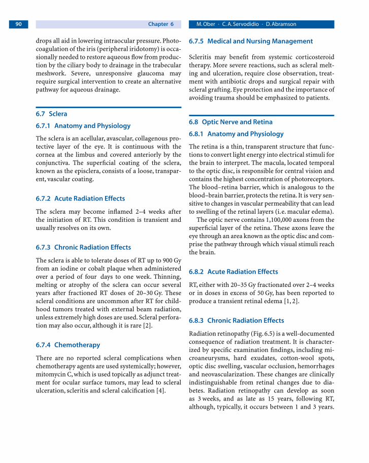
Radiation retinopathy (Fig. 6.5) is a well-documentedconsequence of radiation treatment. It is character-ized by specific examination findings, including mi-croaneurysms, hard exudates, cotton-wool spots,optic disc swelling, vascular occlusion, hemorrhagesand neovascularization. These changes are clinicallyindistinguishable from retinal changes due to dia-betes. Radiation retinopathy can develop as soon as 3 weeks, and as late as 15 years, following RT,although, typically, it occurs between 1 and 3 years.
06_Schwartz_Occular 27.01.2005 8:31 Uhr Seite 90

Chapter 6 91Ocular Complications Due to Cancer Treatment
Although as little as 15 Gy of external beam radiationhas led to signs of retinopathy, 30–60 Gy are usuallyrequired. In the authors’ experience, fewer than 5% ofchildren treated with external beam radiation forretinoblastoma develop radiation retinopathy. 50 Gyis regarded as the threshold for the development ofretinopathy following radioactive plaque exposure.Either a history of diabetes mellitus or concurrenttreatment with chemotherapy is believed to increasesusceptibility to radiation retinopathy [3, 27].
6.8.4 Chemotherapy
The optic nerve and retina are common sites forchemotherapeutic complications. Retinal hemor-rhages, cotton wool spots and optic disc edema haveall been reported following systemic nitrosoureas[28], while intracarotid infusion has been implicatedin optic neuritis and atrophy [29]. In some patientstreated systemically, cisplatin has produced opticneuritis, papilledema and retinal toxicity that mani-fests as color blindness [30]. Intracarotid infusioncan lead to visual loss from severe retinal and/or op-tic nerve ischemia, pigmentary retinopathy or exuda-tive retinal detachment [31]. Carboplatin has led to
visual loss due to retinopathy and optic neuropathy[32]. Intrathecal methotrexate has been reported tocause optic nerve atrophy, optic neuropathy, retinalpigment changes and retinal edema [33]. Patientstreated with Tamoxifen for a period greater than ninemonths are susceptible to a crystalline retinopathyand visual impairment, although the visual impair-ment is generally reversible with cessation of treat-ment. In addition, bilateral optic neuritis with retinalhemorrhages has been reported within three weeksof Tamoxifen initiation [34].
Corticosteroids have been implicated in the devel-opment of pseudotumor cerebri and its associatedoptic nerve swelling. In addition, the immunosup-pressive effects of corticosteroids have been linked to retinal infections. Plant alkaloids vincristine andvinblastine may lead to visual loss and double visionsecondary to optic neuropathy, optic atrophy andcranial nerve palsies [35, 36].Acute optic neuropathy,along with cranial nerve palsy, may also follow 5-fluorouracil treatment [4]. In addition, visual loss in the form of optic nerve damage has been attributed to fludarabine, cyclosporine, paclitaxel,nitrogen mustards and intrathecal cytosine arabi-noside [37–40].
Figure 6.5
Radiation retinopathy
06_Schwartz_Occular 27.01.2005 8:31 Uhr Seite 91

Chapter 692 M. Ober · C. A. Servodidio · D. Abramson
6.8.5 Medical and Nursing Management
Retinal hemorrhages and cotton wool spots as part ofradiation retinopathy will resolve without treatment;however, they are clear indications of retinal damageand are cause for ophthalmologic referral. Retinaledema manifests as blurred vision when it affects themacula. It is diagnosed by careful slit lamp biomi-croscopy with the aid of fluorescein angiography.Current treatment options include laser photocoagu-lation and corticosteroids. Neovascularization (bothiris and retinal) is a manifestation of chronic retinalischemia and is also treated with laser photocoagula-tion. Because diabetes mellitus and hypertension canmimic and/or potentiate radiation retinopathy, strictcontrol of blood sugar and blood pressure should beemphasized.
The treatment of optic disc edema and optic neu-ropathy is controversial. While the use of systemiccorticosteroids and pressure-lowering medicationsmay be effective, observation is also a viable option.
6.9 Orbital Bones and Tissue
6.9.1 Anatomy and Physiology
The orbital cavity is composed of seven bones: themaxilla, palatine, frontal, sphenoid, zygomatic, eth-moid and lacrimal bones. They form the shape of aquadrilateral pyramid with the apex forming posteri-orly and the medial walls parallel. The soft tissues ofthe orbit consist of the extraocular muscles, orbitalfat, fascia and vascular structures. The function of theorbital bones is to protect the eye, while the soft tis-sues act to cushion the eye and optic nerve duringmovement.
6.9.2 Acute Radiation Effects
There are no known acute radiation effects to theorbital bones.
6.9.3 Chronic Radiation Effects
Suppression of bony growth remains the most com-mon orbital complication of chronic RT. The result isespecially noticeable in patients treated at a youngage for retinoblastoma or rhabdomyosarcoma.A hol-lowing of the temporal bone, stunted vertical growthof the orbit and saddle nose (flattening and shorten-ing of the bridge of the nose) are typical featureswhich occur years after a dose of 40–70 Gy to theorbit, fractionated over a 3- to 7-week time period [2](Fig. 6.6). The bony effects of radiation are reducedwhen treatment is delayed until 6 months or, evenbetter, one year of age [3]. Furthermore, advanced ra-diation techniques allow greater precision in tissuelocalization, thus sparing anterior segments of theeye and uninvolved bone.
Anophthalmic socket syndrome, or soft tissue at-rophy, and contracture of the socket following re-moval of the eye, has been documented after radio-therapy in patients treated for retinoblastoma [41].Osteonecrosis rarely results after very high doses ofradiotherapy, but may be associated with concurrentorbital infections. Most devastatingly, second, non-
Figure 6.6
Orbital bone suppression
06_Schwartz_Occular 27.01.2005 8:31 Uhr Seite 92

Chapter 6 93Ocular Complications Due to Cancer Treatment
ocular cancers may also develop in the radiationfield, especially in retinoblastoma patients who arepredisposed to tumor formation [42].
6.9.4 Chemotherapy
Intracarotid carboplatin concurrent with intra-venous etoposide may produce severe visual losssecondary to severe orbital inflammation and opticnerve ischemia [43].
Both 5-fluorouracil and methotrexate therapyhave also led to clinically significant periorbital ede-ma. Corticosteroids have been shown to cause a pro-trusion of the globe known as exophthalmos [44].Paralysis of the eye muscles (ophthalmoplegia) hasbeen reported with cyclosporine [45] and vin-cristine, due to cranial nerve palsy [14].
6.9.5 Medical and Nursing Management
There is no medical treatment to reverse the retarda-tion of bone growth. Osteonecrosis may require sur-gical debridement and antibiotics. Anophthalmicsocket syndrome is very difficult to treat and some-times requires orbital reconstruction surgery.Anophthalmic sockets with ocular prosthesis requireregular care and cleaning with gentle soaps. The orbititself must be examined by a medical professional pe-riodically for the development of second malignan-cies. Finally, counseling should be available to pa-tients regarding the disfiguring effects of radiationon bone growth.
6.10 Conclusion
The present and future outlook for the treatment ofchildren with primary ophthalmic tumors and othertumors involving the eye and its bony structures isencouraging. Cancers that were once uniformly fatalare today viewed as treatable. Newer techniques inradiotherapy, which provide the ability to conservevision and spare non-involved bone, together withadvancements in chemotherapy and surgery, offernot only a longer lifespan, but also, improved qualityof life.
References
1. Haik BH, Jereb B, Abramson DH et al. (1983) Ophthalmicradiotherapy. In: Iliff NT (ed) Complications in ophthalmicsurgery. Churchill Livingstone, New York, pp 4449–4485
2. Brady LW, Shields J, Augusburger JJ et al (1989) Complica-tions from radiation therapy to the eye. Front Radiat TherOncol 23:238–250
3. Ober MD, Beaverson K, Abramson DH Ocular complica-tions. In: Wallace H, Green D (eds) Late effects of childhoodcancer. Arnold, London
4. Al-Tweigeri T, Nabholtz JM, Mackey JR (1996) Ocular toxi-city and cancer chemotherapy. A review. Cancer 78:1359–1373
5. Sidi Y, Douer D, Pinkhas J (1977) Sicca syndrome in apatient with toxic reaction to busulfan. JAMA 238:1951
6. Straus DJ, Mausolf FA, Ellerby RA (1977) Cicatricialectropian secondary to 5-fluorouracil therapy. Med PediatrOncol 3:15–19
7. Esmaeli B, Valero V, Ahmadi A et al (2001) Canalicularstenosis secondary to docetaxel (taxotere). A newly recog-nized side effect. Ophthalmology 108:994–995
8. Miller D, Pecxon JD (1965) Corticosteroid and functions inthe anterior segment of the eye. Am J Ophthalmol 59:31
9. Seiff SR, Shorr N, Adams T (1985) Surgical treatment ofpunctual-canalicular fibrosis from 5-fluorouracil therapy.Cancer 56:2148–2149
10. Donnenfeld ED, Ingraham HJ,Abramson DH (1993) Effectsof ionizing radiation on the conjunctiva, cornea, and lens.In: Alberti WE, Sagerman RH (eds) Medical radiology.Radiotherapy of intraocular and orbital tumors. Springer,Berlin Heidelberg New York, pp 261–270
11. Palmer ML, Hyndiuk RA (2000) Toxicology of corticos-teroids and other antiinflammatory agents. In: Albert DM,Jakobiec FA (eds) Principals and practice of ophthalmolo-gy, 2nd edn. Saunders, Philadelphia, PA, pp 399–416
12. Tseng SC (1986) Topical treatment for severe dry-eye disor-ders. J Am Acad Dermatol 15:860–866
13. Blondi FC (1958) The late effects of x-radiation on thecornea. Trans Am Ophthalmol Soc 56:413–450
14. Albert DM, Wong VG, Henderson ES (1967) Ocular compli-cations of vincristine therapy.Arch Ophthalmol 78:709–713
15. Kaiser-Kupfer MI, Lippman ME (1978) Tamoxifen retino-pathy. Cancer Treat Rep 62:315–320
16. Merriam GR, Focht EF (1957) A clinical study of radiationcataracts and the relationship to dose. Am J Roentgenol77:759–785
17. Braver DA, Richards RD Good TA (1967) Posterior subcap-sular cataracts in steroid treated children. Arch Ophthal-mol 77:161
18. Loredo A, Rodriguez RS, Murillo L (1972) Cataracts aftershort-term corticosteroid treatment. N Engl J Med 286:160
06_Schwartz_Occular 27.01.2005 8:31 Uhr Seite 93

Chapter 694 M. Ober · C. A. Servodidio · D. Abramson
19. Podos SM, Canellos GP (1969) Lens changes in chronicgranulocytic leukemia. Possible relationship to chemo-therapy. Am J Ophthalmol 68:500–504
20. Gorin MB, Day R, Costantino JP (1998) Long-term tamox-ifen citrate use and potential ocular toxicity.Am J Ophthal-mol 125:493–501
21. Armaly MF (1966) The heritable nature of dexamethasone-induced ocular hypertension. Arch Ophthalmol 75:32–35
22. Armaly MF (1963) Effect of corticosteroids on intraocularpressure and fluid dynamics. I. Effect of dexamethasone inthe normal eye. Arch Ophthalmol 70:482–490
23. Armaly MF (1963) Effect of corticosteroids on intraocularpressure and fluid dynamics. II. The effect of dexametha-sone in the glaucomatous eye. Arch Ophthalmol 70:492–499
24. Spaeth GL, Rodriques MM, Weinreb S (1977) Steroid in-duced glaucoma. A: Persistent elevation of intraocularpressure. B: Histopathological aspects. Trans Am Ophthal-mol Soc 75:353–381
25. Margo CE, Murtagh FR (1993) Ocular and orbital toxicityafter intra-carotid cisplatin therapy. Am J Ophthalmol 116:508–509
26. Anderson B, Anderson B (1960) Necrotizing uveitis inci-dent to perfusion of intracranial malignancies with nitro-gen mustard and related compounds. Trans Am Ophthal-mol Soc 58:95–104
27. Brown GC, Shields JA, Sanborn G et al (1982) Radiationretinopathy. Ophthalmology 89:1494–1501
28. Shingleton BJ, Bienfang DC, Albert DM et al (1982) Oculartoxicity associated with high-dose carmustine. ArchOphthalmol 100:1766–1772
29. Miller DF, Bay JW, Lederman RJ (1985) Ocular and orbitaltoxicity following intra-carotid injection of BCNU and cis-platin for malignant gliomas. Ophthalmology 92:402–406
30. Ostrow S, Hohn D, Wiernik PH et al (1978) Ophthalmolog-ic toxicity after cis-dichlorodiamine platinum (II) therapy.Cancer Treat Rep 62:1591–1593
31. Margo CE, Murtagh FR (1993) Ocular and orbital toxicityafter intra-carotid cisplatin therapy. Am J Ophthalmol 116:508–509
32. Rankin EM, Pitts JE (1993) Ophthalmic toxicity during car-boplatin therapy. Ann Oncol 4:337–338
33. Millay, RH, Klein ML, Shults WT (1986) Maculopathy asso-ciated with combination chemotherapy and osmotic open-ing of the blood brain barrier. Am J Ophthalmol 102:626–632
34. Ashford AR, Donev I, Tlwarl RP (1988) Reversible oculartoxicity related to tamoxifen therapy. Cancer 61:33–35
35. Albert DM, Wong VG, Henderson ES (1967) Ocular compli-cations of vincristine therapy.Arch Ophthalmol 78:709–713
36. Shurin SB, Rekate HL, Annable W (1982) Optic atrophy in-duced by vincristine. Pediatrics 70:288–291
37. Porges Y, Blumen S, Fireman Z et al (1998) Cyclosporine-in-duced optic neuropathy, ophthalmoplegia, and nystagmusin a patient with Crohn’s disease. Am J Ophthalmol 126:607–609
38. Chun JH, Leyland-Jones BR, Caryk SM et al (1986) Centralnervous system toxicity of fludarabine phosphate. CancerTreat Rep 70:1225–1228
39. Capri G, Munzone E, Tarenzi E et al (1994) Optic nerve dis-turbances: a new form of paclitaxel neurotoxicity. J NatlCancer Inst 86:1099–1101
40. Margileth DA, Poplack DG, Pizzo PA (1977) Blindness dur-ing remission in two patients with acute lymphoblasticleukemia. A possible complication of multimodality thera-py. Cancer 39:58–61
41. Abramson DH (1988) The diagnosis of retinoblastoma.BullNY Acad Med 64:283–317
42. Abramson DH, Frank CM (1998) Second non-ocular tu-mors in survivors of bilateral retinoblastoma: a possibleage effect on radiation-related risk. Ophthalmology. 105:573–579; discussion 579–5780
43. Lauer AK, Wobig JL, Shults WT et al (1999) Severe ocularand orbital toxicity after intracarotid etoposide phosphateand carboplatin therapy. Am J Ophthalmol 127:230–233
44. Van Dalen JT, Sherman MD (1989) Corticosteroid-inducedexophthalmos. Doc Ophthalmol 72:273–277
45. Bixenman WW, Nicholls JV, Warwick OH (1968) Oculomo-tor disturbances associated with 5-fluorouracil chemother-apy. Am J Ophthalmol 83:604–608
06_Schwartz_Occular 27.01.2005 8:31 Uhr Seite 94