ocular morbidity in mechanical injuriesrepository-tnmgrmu.ac.in/3865/1/2203008subhalakshmia.pdf ·...
TRANSCRIPT

OCULAR MORBIDITY IN MECHANICAL INJURIES
DISSERTATION SUBMITTED TO
THE TAMILNADU DR.M.G.R. MEDICAL UNIVERSITY,
CHENNAI, TAMILNADU
M.S. DEGREE EXAMINATION OF
BRANCH III OPHTHALMOLOGY
MARCH 2013

CERTIFICATE
This is to certify that this dissertation entitled Ocular morbidity
in mechanical injuries submitted by Dr. A. Subhalakshmi to the
faculty of Ophthalmology The Tamil Nadu Dr. MGR Medical University,
Chennai in partial fulfillment of the requirement for the award of M.S
Degree Branch III (Ophthalmology), is a bonafide research work carried
out by her under our direct supervision and guidance.
Dr. A. R. Anbarasi Dr. V . Chittibabu
Additional Professor Prof & Head of the Department
Department of Ophthalmology, Department of Ophthalmology
Tirunelveli Medical College, Tirunelveli Medical College,
Tirunelveli. Tirunelveli.
THE DEAN
Tirunelveli Medical College,
Tirunelveli.

ACKNOWLEDGEMENT
I express my heartful gratitude to The Dean, Tirunelveli Medical
College, Tirunelveli, for all the facilities provided for the study.
I take this opportunity to express my profound gratitude to
Dr.A.Meenakshisundaram, M.S., and Dr.V.Chittibabu, M.S., Prof. and
HOD, Dept of Ophthalmology for his support, guidance, advice and
constant encouragement all through this study.
I am highly thankful to Dr.M.Elangovan, M.S.,
Dr.A.R.Anbarasi, M.S., Associate Prof., Department of Ophthalmology,
TVMCH who helped me to sharpen my critical perceptions by offering
most helpful suggestions and corrective comments.
I am highly obliged to Dr.J.Kishore Kumar Jacob, M.S.,
Dr. D.Anandhi, M.S, and Dr. S.B.Sivathanu, M.S, and Dr.M.Rita Hepsi
Rani, M.S, Assistant Professors, Department of Ophthalmology , TVMCH
who helped me by offering their valuable suggestions and for being with
me to support all my endeavour throughout the study.
My special thanks to my Co-Postgraduate colleagues
Dr.Bhavaani Preetti, Dr.K.Saranya for their help and peer less support.
My special thanks to my teachers in Department of Neuro Surgery.

I thank all those patients who participated in the study, for their co-
operation which made this study possible.
I owe my thanks to Ms. E.Meera Devi, for her immense help in
analyzing the data and preparing the manuscript.
Last… But not the least,
I am most indebted to my beloved family, my friends and The
Almighty…
TABLE OF CONTENTS
Sl.No Title Pg.No
1. Introduction 1
2. Aim of the study 2
Part I 3. Anatomy of eye 3
4. Mechanical injuries of eye 9
5. Review of literature 54
Part II 6. Materials and methods 57
7. Observation (Statistical analysis) 60
8. Discussion 73
9. Conclusion 78
10. Proforma 80
11. Bibliography 84
12. Master Chart 87

OCULAR MORBIDITY IN MECHANICAL INJURIES
INTRODUCTION
Mechanical injury to eye can occur in a variety of ways and
produce myriad clinical sequelae. In this era of high speed traffic and
industrialization, incidences of injuries are increasing in general. Like
any other part of the body, eyes are also not exempt from these
injuries, in spite of being protected by lids, projected margins of orbit,
the nose and cushion of fat from behind.
Estimates of the incidence of eye trauma vary widely.
Ocular injury due to mechanical trauma is one of the major health
problems in India. Children at play in recreational activities, young
men at work in urban factories, construction sites and rural
agricultural settings and older people who suffer falls commonly
suffer the consequences of ocular injury. The majority of injuries are
sustained by active and productive individuals. Unfortunately, these
injuries are often vision threatening and the lifestyle and future of the
injured individual is irrevocably altered.

AIM OF THE STUDY
Aim of the study was to prospectively determine the effect of
mechanical injuries on the
o Anterior segment of the eye
o Posterior segment of the eye
o Visual pathway and neuro-ophthalmic system
o Orbit and adnexa
and visual outcome of the same.

ANATOMY OF EYE
Each eyeball is a cystic structure kept distended by pressure
inside it. The eyeball is not a sphere but an oblate spheroid. The
anterior and posterior poles are the central points of maximal
convexities of the eye ball . The equator of the eyeball lies at the
midplane between the two poles.
Dimensions of adult eye ball:
Anteroposterior diameter 24 mm
Horizontal diameter 23.5 mm
Vertical diameter 23 mm
Circumference 75 mm
Volume 6.5 mm
Weight 7gm
Layers of eyeball:
Eyeball contains three layers:
Outer fibrous layer
Vascular layer (uveal tissue)
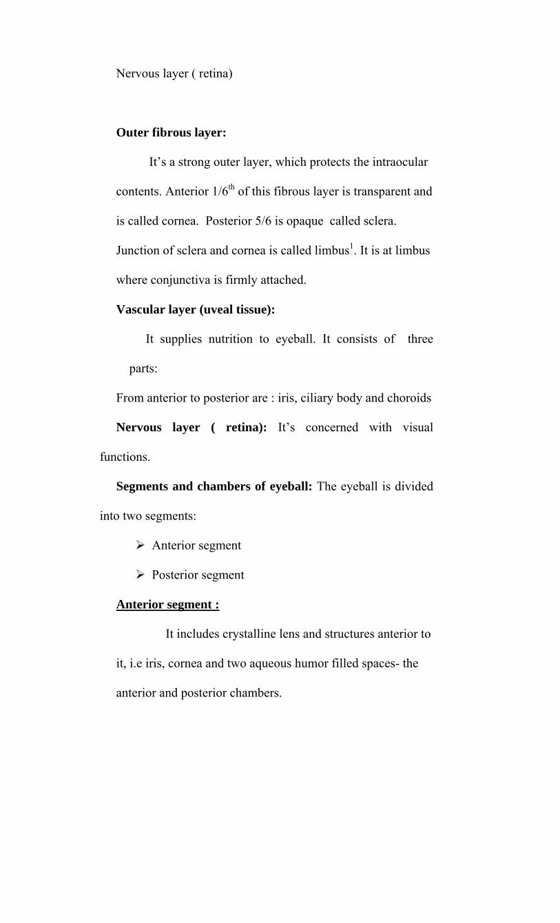
Nervous layer ( retina)
Outer fibrous layer:
It’s a strong outer layer, which protects the intraocular
contents. Anterior 1/6th of this fibrous layer is transparent and
is called cornea. Posterior 5/6 is opaque called sclera.
Junction of sclera and cornea is called limbus1. It is at limbus
where conjunctiva is firmly attached.
Vascular layer (uveal tissue):
It supplies nutrition to eyeball. It consists of three
parts:
From anterior to posterior are : iris, ciliary body and choroids
Nervous layer ( retina): It’s concerned with visual
functions.
Segments and chambers of eyeball: The eyeball is divided
into two segments:
Anterior segment
Posterior segment
Anterior segment :
It includes crystalline lens and structures anterior to
it, i.e iris, cornea and two aqueous humor filled spaces- the
anterior and posterior chambers.

Anterior chamber: It is bounded anteriorly by back of the
cornea and posteriorly by iris and ciliary body2. The anterior chamber is
about 2.5 mm deep in centre in normal adults and 1mm deep in
periphery. It contains about 0.25 ml of aqueous humor.
Posterior chamber: It is a triangular space containing 0.06ml of
aqueous
humor. It is bounded anteriorly by the posterior surface of iris and part of
ciliary body, posteriorly by crystalline lens and its zonules and laterally
by ciliary body.
Posterior segment:
It includes structures posterior to lens i.e, vitreous humor,
retina, choroids and optic nerve.
ANATOMY OF ORBIT
The two bony orbits are quadrangular truncated pyramids situated
between the anterior cranial fossa above and the maxillary sinuses
below. Each orbit is formed by seven bones namely: frontal, ethmoid,
lacrimal, palatine, maxilla, zygomatic and sphenoid.
The depth of orbit is 42mm along medial wall and 50 mm along the
lateral wall. The base of the orbit is 40mm width and 35mm height.

WALLS OF ORBIT
Medial wall:
The medial wall of orbit is quadrilateral in shape and is formed by the
frontal process of the maxilla, the lacrimal bone, the orbital plate of
ethmoid bone and the body of sphenoid bone.
Relations:
Medial to medial wall lie anterior ethmoidal air sinuses,
middle meatus of nose, middle and posterior ethmoidal sinuses and
sphenoidal air sinus.
Orbital index= height/width x
100
Orbital index shows racial
variation
Orbital index >89 Megasenes (eg. Orientals)
Orbital index between 83-89 Mesosenes (eg. Caucasians)
Orbital index <83 Microsenes
Volume of orbit 29ml
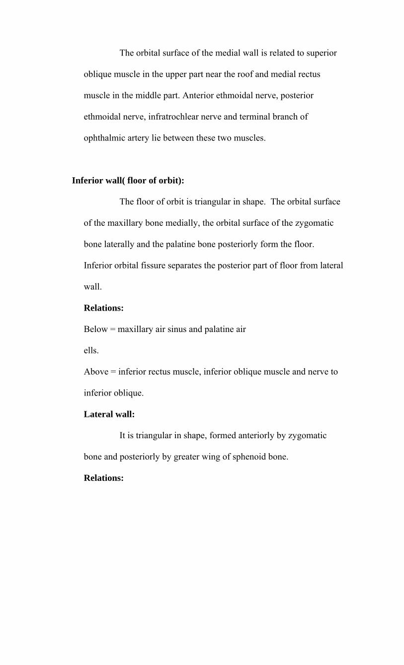
The orbital surface of the medial wall is related to superior
oblique muscle in the upper part near the roof and medial rectus
muscle in the middle part. Anterior ethmoidal nerve, posterior
ethmoidal nerve, infratrochlear nerve and terminal branch of
ophthalmic artery lie between these two muscles.
Inferior wall( floor of orbit):
The floor of orbit is triangular in shape. The orbital surface
of the maxillary bone medially, the orbital surface of the zygomatic
bone laterally and the palatine bone posteriorly form the floor.
Inferior orbital fissure separates the posterior part of floor from lateral
wall.
Relations:
Below = maxillary air sinus and palatine air
ells.
Above = inferior rectus muscle, inferior oblique muscle and nerve to
inferior oblique.
Lateral wall:
It is triangular in shape, formed anteriorly by zygomatic
bone and posteriorly by greater wing of sphenoid bone.
Relations:

Laterally—It separates the orbit from temporal fossa anteriorly and
from middle cranial fossa posteriorly
Medially—related to lateral rectus, lacrimal nerve and vessels,
zygomatic nerve and the communication between zygomatic and
lacrimal nerves.
Roof:
It is triangular in shape and is formed mainly by the orbital plate
of frontal bone. Behind this it is formed by lesser wing of sphenoid
bone.
Relations:
Above: Frontal lobe of cerebrum and meninges
Below: periorbita, frontal nerve, levator palphebrae superioris,
superior rectus, superior oblique, trochlear nerve and lacrimal gland.
At the junction of the roof and the medial wall are anterior and
posterior ethmoid canals.
At the junction of roof and lateral wall is a gap posteriorly, the
superior orbital fissure
Base of orbit:
The anterior open end of the orbit is referred to as base. It is
bounded by orbital margins.
Superior orbital margin- formed by orbital arch of frontal bone

Lateral orbital margin- by zygomatic process of frontal bone and
zygomatic bone.
Inferior orbital margin- by the zygomatic bone laterally and maxilla
medially
Medial orbital margin- below by anterior lacrimal crest on the frontal
process of maxilla and above by frontal bone.
Apex of orbit:
It is the posterior end of the orbit. Here the four orbital walls
converge. The apex has two orifices- the optic canal and the superior
orbital fissure3
MECHANICAL INJURIES TO EYE
The Ocular Trauma Classification Group has attempted to
develop a uniform classification system for mechanical injuries to the
eye, based on primary evaluation.
Injuries are broadly divided into two categories
A. open globe – full- thickness defects in the corneoscleral coat of
the
eye
B. closed globe – ocular injury without a full – thickness defect of
the

coats.
Open globe injuries:
It is further subdivided into
1. ruptures of globe with an inside to outside break in the ocular coats.
2. laceration is used to denote full thickness outside to inside break in
ocular coats.
This can be further sub – divided
into
a. penetrating injury -if the object traverses the coat only once
b. perforating injury -if both an entry and exit wound are present.

Modes of open globe injuries:
a. Trauma by sharp and pointed instruments like needles, knives, nails,
arrows, screw-drivers, pens, pencils, compasses, glass pieces and so
on.
b. Trauma by foreign bodies traveling at high speed such as bullet
injuries and iron foreign bodies in lathe workers.
Closed globe
injuries:
It usually follows blunt trauma. It is subdivided into:
1. contusion or concussion injuries
2.Superficial foreign bodies are frequently found on the cornea and
conjunctiva
3.Lamellar laceration – partial thickness injury of the
coats.
Modes of closed globe injury:
a.Direct blow to the eye ball by fist, ball or blunt instruments like
sticks and big stones
b.Accidental blunt trauma to eyeball may also occur inroad
side accidents, automobile accidents, injuries by agricultural and
industrial instruments / machines and fall upon the projecting blunt
objects4
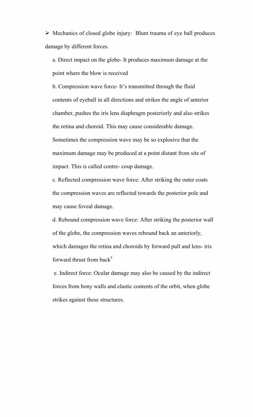
Mechanics of closed globe injury: Blunt trauma of eye ball produces
damage by different forces.
a. Direct impact on the globe- It produces maximum damage at the
point where the blow is received
b. Compression wave force- It’s transmitted through the fluid
contents of eyeball in all directions and strikes the angle of anterior
chamber, pushes the iris lens diaphragm posteriorly and also strikes
the retina and choroid. This may cause considerable damage.
Sometimes the compression wave may be so explosive that the
maximum damage may be produced at a point distant from site of
impact. This is called contre- coup damage.
c. Reflected compression wave force: After striking the outer coats
the compression waves are reflected towards the posterior pole and
may cause foveal damage.
d. Rebound compression wave force: After striking the posterior wall
of the globe, the compression waves rebound back an anteriorly,
which damages the retina and choroids by forward pull and lens- iris
forward thrust from back5
e. Indirect force: Ocular damage may also be caused by the indirect
forces from bony walls and elastic contents of the orbit, when globe
strikes against these structures.

For descriptive purposes, mechanical injuries can be classified
broadly into:
1. Anterior segment injuries
2. Posterior segment injuries
3. Neuro ophthalmic manifestations of trauma
4. Orbital and adnexal injuries
ANTERIOR SEGMENT INJURIES
Anterior segment trauma is a common cause of ocular
morbidity and constitutes an important segment of ophthalmic
practice.
A. Subconjunctival haemorrhage:
Blood under conjunctiva has a fiery red appearance. Inability to
discern the posterior limit of subconjunctival haemorrhage indicates
orbital or intracranial injury. It is managed by reassurance and further
imaging to rule out serious injury.
B. Superficial foreign bodies:
Foreign body may be present in the superior tarsal sulcus. It is
detected by eversion of upper lid. Double eversion using Desmarre
retracter is necessary to reveal foreign bodies in deep fornices.

Iron foreign body may embed in corneal stroma, when a rust
ring may be formed. A corneal foreign body may be removed under
topical anaesthesia at the slit lamp using foreign body spud or 25
guage needle. After foreign body removal, topical anaesthetic drops
are applied hourly, and pressure patch applied if epithelial abrasion
is significant. Patient are closely monitored till epithelium heals.
C. Corneal abrasion:
It is a break in corneal epithelium due to minor trauma which
stains with fluorescein. Hourly broad spectrum topical antibiotics,
cycloplegics and pressure patch ensure healing within 24 hours.
Damage to epithelial basement membrane adhesion complexes may
lead to recurrent corneal erosions. This causes acute onset of pain on
awakening . Signs vary from irregular area of raised epithelium causing
focal break in tear film to a full thickness epithelial defect with elevated
grey margins.
It is treated by epithelial debridement, pressure patch, copious
lubricants for 6-8 weeks, bandage contact lens for 2 months or
intrastromal punctures or photo therapeutic keratectomy.6
D. Hyphema:
It is common in young males , usually due to injury to peripheral
iris and anterior ciliary body in the form of iridodialysis, cyclodialysis,
disruption of major arterial circle, arteries of ciliary body and choroidal
vessels.
Raised IOP, vascular spasm and formation of fibrin clot can stop
the hyphema. The clot may have a bilobed configuration with contiguous
blood in anterior and posterior chamber called 8 ball hyphema.
Fibrin degradation products are then cleared through the normal
trabecular outflow pathways and by iris vessels to a small extent
Grades of hyphema-
Grade I - blood level less than 1/3 of anterior chamber (incidence
>50%) Grade II - blood level 1/3 to ½ of anterior chamber
Grade III - blood level ½ to near total
Grade IV blood occupying total anterior chamber ( incidence < 10%)
A complete eye evaluation is required to assess concomitant injury and
occult globe rupture. Dilated fundoscopy is done at the time of
presentation. Vision, hyphema status and IOP are documented in every
follow up visits. Scleral depression is avoided upto one month to avoid
rebleeding.
Rebleeding occurs commonly between 2 to 5 days after trauma. It
is indicated by the presence of bright red layered blood over clotted
blood and increased size of hypema. It indicates poor pronosis.

Corneal blood staining and optic neuropathy due to persistent
hyphema and elevated IOP lead to permanent visual loss .
(i) Corneal blood staining occurs due to combination of elevated IOP,
endothelial dysfunction and anterior chamber blood.
Slit lamp examination shows yellow granular changes and stroma
shows reduced fibrillar definition.
(ii)Optic neuropathy- occurs if IOP is persistently elevated > 35 mm
of Hg for 7 days. So to prevent optic neuropathy, intervention should be
done if IOP > 35 mm of Hg.
Hyphema is best managed:
Medically by
a) 1% Atropine eye drop 3 times a day- gives comfort in the setting of
iritis
b) 1% prednisolone acetate eye drop – helps to minimize discomfort in
iritis
c) Antifibrinolytic agents – Aminocaproic acid 50mg/ kg orally every
4hrs
upto 30g/day for 5 days. It competitively inhibits conversion of
plasminogen to plasmin, which degrades fibrin clot.
Hb keratocyte Hemosiderin deposition
Keratocyte death death

• Surgical management- 5% of patients may require anterior chamber
wash out, usually performed through two clear corneal incision. As the
clot retracts in 4-7 days, the ideal time for AC wash is 7 days.
Other criteria for early surgical intervention are:
1. IOP > 50mm of Hg for 5 days
2. IOP > 35mm of Hg for 7 days to prevent optic nerve damage
3. IOP > 25mm of Hg for 5 days in total or near total hyphema to
prevent
corneal blood staining
4.IOP averages >25mm of Hg for 24 hours in patients with sickle
haemoglobinopathies
Sickle cell anaemia patients with hyphema should be managed
aggressively. Sickle cells are elongated and rigid that reduces their
egress through trabecular outflow pathways. Even small hyphema can
clog the trabecular meshwork leading to raised IOP. Consequent hypo
perfusion can lead to anterior segment hypoxia, acidosis and
hypercarbia, thus encouraging sickling. These patients are also
predisposed to optic nerve damage and central retinal artery occlusion
with a marginal raise in IOP. Therefore, IOP should be monitored and
treated aggressively by topical beta adrenergic antagonists and AC
wash out. Carbonic anhydrase inhibitors are avoided.

Approximately, 75% of patients with hyphema regain visual
acuity of greater than 20/50. Long term visual loss in hyphema is
more commonly due to traumatic cataract, posterior pole inury and
optic nerve damage than to complications related directly to blood
itself.
E. Iris injury
• Tears of iris sphincter and pupillary frill are frequent and may
cause an irregular unreactive and chronically dilated pupil.
• Dialysis of the iris root produces a D shaped pupil and may
result in acute hyphema.
• Damage to trabecular meshwork and formation of peripheral
anterior synechiae may lead to elevated IOP7.
• Less frequently, a cyclodialysis may occur leading to chronic
hypotony.
Management:
• Sunglasses may decrease glare and photophobia. Tinted contact
lenses or those with an artificial pupil may decrease these
symptoms
• The argon and Nd YAG lasers can be used to recenter an
eccentric pupil. Laser sphinterotomy may open an eccentric or
secluded pupil toward visual axis.

• Mc Cannel suture using 10 0 polypropylene to suture iris
dialysis8
F. Traumatic glaucoma:
(i)Immediate glaucoma may be caused by clogging the trabecular
meshwork by red blood cells or inflammatory cells. Patients with
large hyphema may have pupillary block and acute angle closure 9.
(ii) An intermediate term glaucoma may be caused by ghost cell
glaucoma occurring 3-4 weeks after vitreous haemorrhage. Rigid
khaki coloured non pliable ghost red blood cells may percolate
forward and clog the trabecular meshwork.
(iii)Late glaucoma –
a. frequently associated with angle recession. It is identified by,
broadened ciliary band by gonioscopy, AC appearing abnormally
deep and fibrotic changes in the angle
b. pupillary block
c. peripheral anterior synechiae
d. scarring of trabecular meshwork
e. injuries to lens – phacomorphic, phacolytic and
phacoanaphylactic
glaucoma.
Management
1. Topical corticosteroids are useful to minimize inflammation, prevent

formation of anterior and posterior synechiae and prevent
scarring of
trabecular meshwork
2. Acute IOP elevations treated medically with aqueous suppressants
3. In cases of pupillary block, laser peripheral iridotomy, hyphema
wash out
or lens surgery may be necessary to treat acute IOP elevations
4. Chronic glaucoma should be treated with medical therapy.
5. In angle recession glaucoma- laser trabeculopasty has low success
rate due
to intrinsic damage to trabecular meshwork.
6. Peripheral anterior synechiae- Laser gonioplasty may be useful.
Filtration surgery needed if the above said measures fail to control
IOP.
G. Lens injury:
Blunt trauma to eye may lead to contusion cataract and lens
subluxation or dislocation. Rupture of capsule lead to rapid
opacification of lens. Distortion of the globe with equatorial scleral
expansion may also cause capsular rupture or result in zonular
dehiscence with consequent lens subluxation or dislocation.
a. Contusion cataracts – patient presents with deterioration of visual
acuity.

b. Complete lens dislocation into vitreous render the eye
functionally aphakic
c. Zonular dehiscence may be signalled by iridodonesis and
phacodonesis. In addition anterior chamber may show prolapsed
vitreous. Lens capsule should be examined for anterior or posterior
capsule rupture. Special attention should be devoted for lens induced
IOP elevations and inflammation.
d. Pupillary block may occur with subluxated or dislocated lens or
with an intumescent cataract following capsular rupture. Release of
lens proteins may cause subsequent phacolytic glaucoma secondary to
high molecular weight proteins, lens particles and macrophages
clogging the aqueous outflow channels. Such findings usually suggest
immediate surgical intervention.
Management:
The anatomic position and stability of the lens dictate the
method by which it is managed surgically.
(i) If the capsule and zonules are intact or minimally subluxated a
standard extracapsular extraction or phacoemulsification procedure
can be performed.
(ii) If zonular damage is significant but lens is free of vitreous,
intracapsular extraction may be performed through the limbus10

(iii) If vitreous has prolapsed around the lens, vitreous must be first
removed. Extreme caution must be taken not to exert vitreous traction
during lens delivery.
(iv) In cases with significant vitreous disruption and posterior lens
dislocation, parsplana vitrectomy- lensectomy approach is preferred.
The placement of an IOL, selection of IOL type and implantation
technique must be tailored to the individual case. If posterior capsule
is intact without vitreous loss or undue anterior segment disruption,
then posterior chamber IOL can be implanted. In the absence of
posterior capsule support flexible anterior chamber IOL can be
implanted. Transcleral sutured posterior chamber lenses or glued
IOLs can be used if adequate vitrectomy is performed.

H. Corneoscleral lacerations
a. Cultures should be taken from
1.wound margins
2. a portion of any excised tissue
3.intraocular foreign bodies
Specimens are inoculated directly into blood and chocolate agar
plates, fluid thioglycolate broth or cooked meat broth from anaerobes,
as well as on Sabouraud’s medium for fungus.
b. Intravenous antibiotic started immediately. Broadspectrum
coverage is afforded by a combination of a cephalosporin or
vancomycin for gram positive coverage and an aminoglycoside for
gram negative coverage. Topical antibiotics are better avoided in open
globe injury. If the wound is self sealing with minimal uveal prolapse,
a topical antibiotic is administered.
c. Surgical management- The primary objective of initial surgery is
complete water tight closure of the globe. Secondary goals include
removal of disrupted lens and vitreous, avoidance of uveal and
vitreous incarceration in the wound, removal of intraocular foreign
bodies and restoration of normal anatomical relationship.
d. Non perforating corneal lacerations All suspected non
perforating corneal lacerations should be carefully examined to rule

out occult perforation of Descemet`s membrane. Siedel `s test is
performed with 2% fluorescein to check for microscopic leaks.
(i) Small perforating injuries may self seal. When the wound is
somewhat unstable , bandage contact lens can be used to support the
wound and promote reepithelialisation by shielding the wound from
lid movement. For small self sealing corneal lacerations, a bandage
contact lens may be sufficient to protect and support the wound as it
heals, upto 3 to 6 weeks. Topical antibiotics and cycloplegics are used
with bandage contact lens in place.
(ii)Long partial thickness wound and in those with significant wound
override or gape need suturing.
(iiv)Puncture wounds with small amount of central tissue loss and for
wounds that require suture placement in visual axis, cyanoacrylate
tissue adhesive is applied under topical anaesthesia at a slit lamp or
under operating microscope. Immediately after the adhesive has
adequately dried bandage soft contact lens is applied.
(iv) Large lacerations (greater than 2 to 3 mm) , displaced wounds,
wounds with loss of corneal tissue and laceration with accompanying
iris or lens incaraeration must be brought to the operating room for
definitive surgery and suture placement. Surgery is done under
general anaesthesia if there is no medical contraindication. If the
wound is relatively water tight and the anterior chamber formed, it

may be sutured directly. The ultimate aim of corneal suturing is
definitive placement of corneal sutures to make the wound watertight,
minimize scarring and reconstruct the native nonastigmatic corneal
contour. Monofilament 10-0 nylon sutures are used, sutures
approximately 90% deep in stroma and of equal depth on both sides
of the wound. The wound should be checked for leaks by gentle
pressure on the globe with dry cellulose sponge.
(v) Corneal laceration with iris incarceration After an extensive
corneal laceration anterior chamber shallows , with consequent iris
incarceration or prolapse. If prolapsed the tissue must be critically
evaluated . . Healthy appearing tissue may be reposited.
Obviously devitalized tissue should be exised. If foreign body is
present within the tissue it is certainly excised. Moreover the tissue
should be evaluated for surface epithelialisation. If it is present, it may
lead to seeding of anterior chamber leading to epithelial proliferation. Iris
reposition done by direct sweeping with a spatula or irrigating canula
through the paracentesis site, followed by suturing of the corneal
laceration.
(vi) Corneal laceration with Lens involvement: Lens disruption is
difficult to diagnose due to fibrinous anterior chamber reaction, pupillary
membrane and corneal edema. It is better to intervene after treating
corneal edema.
A lens with a disrupted capsule and flocculent capsular material
liberated into the anterior chamber is preferably removed to prevent
ongoing phacogenic postoperative inflammation. If the lens is clearly
cataractous it may be removed in the initial operation. However a
stable lens with intact capsule and without liberated cortical material
can be treated in secondary procedure.
(vii)Corneal laceration with vitreous involvement: Violation of
vitreous face usually accompanies corneal laceration with lens
involvement. The primary goal is to relieve any vitreous incarceration in
the traumatic or surgical wounds. It is important to remove disrupted
vitreous from anterior chamber to minimize subsequent corneal
endothelial damage. Preferably automated microvitrectomy
instrumentation is used. After vitrectomy, dry cellulose sponges are
useful to identify vitreous strands remaining in the anterior chamber or at
laceration site. At the conclusion of surgery, the pupil should be round
without peaking, wound free of incarceration and the vitreous behind the
iris.
(viii)Simple corneoscleral laceration: Lacerations extending beyond
the limbus into sclera need to be explored to delineate the full extent of
the wound. For large laceration with unstable globe, sutures may be
placed before definitive exploration of the wound to restore structural
integrity. The limbus is first approximated to restore correct anatomic

relationship. The prolapsed iris is next reposited and corneal wound is
closed. The extent of scleral laceration is then ascertained. The laceration
occasionally extends far posteriorly, sometimes upto optic nerve. In these
cases with poor prognosis, it is probably preferable to leave the more
posterior extent of the wound unsutured than to distort the globe
excessively.
(ix)Corneoscleral lacerations with uveal and vitreousprolapse:
Vitreous prolapsing through a scleral wound is easily secured with dry
cellulose sponges and cut flush with the sclera. Care is taken to avoid
traction on vitreous. A vitrectomy instrument can be used to excise
vitreous from wound.
Uveal tissue often prolapses from gaping scleral lacerations. In
severe cases prolapsed tissue is excised to allow for wound closure. Any
tissue removed should be identified histolgically. Evidence of retina in
excised portion indicates poor prognosis.
The preferred method for scleral closure over prolapsed uvea is a
zippering technique. The wound is closed from the anterior limbal end
with interrupted sutures, that are successively placed posteriorly. The
sutures are relatively closely spaced, with the goal to oversew the uveal
tissue with the sclera.
I. Anterior segment intra ocular foreign bodies: Although search for
an intraocular foreign body must be made in all eyes with penetrating

injuries, foreign bodies are more common after activities involving
striking metal on metal.
An entry wound through cornea may be small and self sealing, and
the one through sclera may be obscured by subconjunctival
haemorrhage. Anterior segment intraocular foreign body may lodge in
the angle and is signaled by, focal corneal edema overlying an angle
in that area. Gonioscopy can be performed to confirm and localize the
foreign body. A foreign body may embed on the lens and be seen as
focal cataract formation. Radiographic examination, including plain
radiographs, CT scanning and MRI may be helpful in detecting an
intraocular foreign body.
Foreign body in the angle or in the iris or in the lens can be removed
through a limbal incision placed either directly over the object or
across the anterior chamber. In some cases of metallic foreign body,
magnet can be used for extraction
Anterior segment reconstruction:
Despite meticulous primary repair of a penetrating corneoscleral
injury, normal wound healing mechanisms may lead to devastating
late complications in eye trauma. Such sequelae include
corneal scarring, astigmatism
glaucoma

Vitreous incarceration within ocular wounds and associated
chronic inflammation – cystoid macular edema, retinal
detachment, infection.
Pupillary or cyclitic membrane
conjunctival scarring and symblepheran
ocular surface epithelial damage leading to persistent
epithelial defects, stromal vascularisation, sterile stromal
ulceration
Procedures done are- Lensectomy is frequently combined with
open sky vitrectomy.
- Goniosynechiolysis
- Reconstructive iridoplasty
- Membrane removal and iridoplasty should be
done before donor cornea is placed and sutured.
2. POSTERIOR SEGMENT TRAUMA
It includes any changes induced in the eye by injury that affects the
vitreous body, retina, choroids, optic nerve and sclera. The injury
could be due to direct effect of blunt trauma, perforating trauma,
electromagnetic trauma or posterior segment sequelae of remote
trauma.
A. Scleral rupture:

The sclera is resistant to severe injury but with severe blunt injury
rupture can occur.
a. Direct rupture is uncommon.
b. Indirect rupture occur at the site away from the site of impact in an
area of scleral weakness.11
Most scleral ruptures are solitary but multiple ruptures can occur.
The most common sites of rupture are the superior nasal and superior
temporal quadrants between coreneal limbus and equator.
Scleral ruptures are occult and not easily documented by slit lamp
examination. Signs of occult scleral rupture are visual acuity of light
perception or no light perception, reduced ductions, ocular hypotony,
hyphema, severe chemosis and bullous subconjunctival haemorrhage.
Ophthalmoscopy may be limited by media opacities including
hyphema, cataract and vitreous haemorrhage. Ultrasonography and
computed tomography may show a shrunken globe but actual rupture
is not reliably demonstrated.
The only definitive way to rule out scleral rupture is by careful,
controlled exploration of globe. Eyes with scleral rupture is often
severely injured internally, with choroidal and retinal tearing,
suprachoroidal haemorrhage, vitreous haemorrhage, teraing of ciliary
body and avulsion of optic nerve. Internal ocular injuries associated
with scleral rupture make ocular reconstruction and visual

rehabilitation challenging. Success is usually measured by globe
salvage and ambulatory vision . Early primary repair is an important
first step in management. Vitrectomy is effective in dealing with
intraocular haemorrhage with scleral rupture.
Scleral perforations include single perforations, double
perforations multiple perforations accompanied by retained
intraocular foreign body. It usually involves severe trauma to the lens,
choroids, ciliary body and retina. Profound visual loss is often the
unfortunate result of scleral perforation, despite wound closure,
vitrectomy , scleral buckling and use of vitreous substitutes.
B. Choroid rupture:
The choroid is susceptible to rupture from the effects of blunt trauma
applied to the globe. The rupture may occur
a. directly, at the site of application of the force
b. indirectly on the opposite side of the globe.
The choroids is susceptible to rupture because of inelastic characters
of Bruch`s membrane.
Posterior choroidal ruptures are curvilinear, concentric to optic nerve
and may be accompanied by intraretinal and subretinal haemorrhages.
Some posterior choroidal ruptures are subtle and can be diagnosed
only with the aid of fluorescein angiography. Early hypoflurescence

from choriocapillary injury and late hyperfluorescence in a curvilinear
pattern concentric with optic disc are characteristic.
Choroidal ruptures at the site of direct application of force often
occur in infero temporal quadrant, because the bony orbit doesn’t
protect the eye well in that quadrant. When choroidal ruptures are
accompanied by retinal rupture, it is called retinitis sclopetaria,
chorioretinitis sclopetaria or chorioretinitis plastica sclopetaria.
Severe subretinal fibrosis with complete atrophy and disruption of
choroid, retinal pigment epithelium and retina occur.
C. Traumatic retinal breaks
This occur when the vitreous is violently shifted away from retina.
Any area of strong vitreoretinal adhesion is likely to be the site of
retinal tearing. The vitreous base, lattice degeneration, previous
chorioretinal scars, the fovea and retinal blood vessels are common
sites of retinal tear.
Direct or coup forces applied to the retina can result in retinal
necrosis and atrophy. Often massive chorioretinal scarring occur at
the margins of the retinal atrophy after the haemorrhage clears. As a
result , retinal detachment does not occur.

All nonfoveal traumatic retinal tears deserve treatment with
cryotheraphy, laser photocoagulation or scleral buckling unless
chorioretinal scarring adequately surrounds the tear.
D.Retinal dialysis
It is the most common traumatic retinal break, refers to a retinal
break occurring at the anterior edge of ora serrata and posterior edge of
which is attached to the vitreous base. Inferior temporal and superior
nasal quadrants are the most common areas for retinal dialysis.
The retina and vitreous are tightly adherent at the vitreous base and
as the vitreous base is avulsed into vitreous cavity retina follows,
creating a tearing of retina at or near ora serrata.
The challenge of management of retinal dialysis is early
recognition. Accurate diagnosis requires skill at indirect
ophthalmoscopy with scleral depression. If retinal dialysis without
retinal detachment is observed, cryotherapy or laser prophylactic
therapy is indicated. If retinal detachment is present, scleral buckling
achieves a reattachment rate of 98%
E.Traumatic retinal detachment:
Its commonly seen more frequently in young male patients.
Traumatic retinal detachment is associated with superior nasal dialysis in
22%, inferior temporal dialysis in 31%, giant retinal tears in 16%, retinal
flap tears in 11% and tears associated with lattice degeneration in8%

Traumatic retinal detachments are managed by pneumatic retinopexy ,
scleral buckling or vitrectomy depending on the retinal breaks and degree
of proliferative vitreoretinopathy.
F. Commotio retinae:
Although the retina may be torn during blunt trauma, severe
nontearing injury can also occur in a coup or contre coup location.
The term applied to blunt nontearing retinal injury is commotio retina
or Berlins edema. Commotio retina has a whitish gray appearance
with a cherry red spot that can be observed several hours after blunt
trauma and is occasionally accompanied by intra retinal haemorrhage
retinal pigment epithelial detachment or choroidal rupture. Visual
acuity is very much reduced or may be unaffected. No effective
treatment for commotio retina is known.
G.Vitreous changes in blunt trauma:
The vitreous can be injured in blunt trauma by disinsertion or
opacification.
a. Disinsertion occur at vireous base, optic nerve, retinal vessels,
fovea, lattice degeneration and chorio retinal scars. The retina is
usually severely injured and dominates the clinical picture. The
avulsed vitreous base has the appearance of a loose clothline, a

hammock or a ribbon suspended loosely through vitreous cavity. It is
pathognomonic of blunt trauma.
b. Vitreous opacification usually results after haemorrhage from
torn retinal, choroidal or ciliary body vessels. Most vitreous
haemorrhage clear spontaneously but eyes with dense haemorrhage
may need vitrectomy to clear the visual axis. Observation for upto six
months may be rewarded for spontaneous clearing and avoidance of
intraocular surgery. The goal of surgery in a severely lacerated globe
is prevention of secondary complications such as endophthalmitis,
retinal detachment, cyclitic membrane and toxic injury from a
retained foreign body.
Repair of posterior segment laceration is divided into primary and
secondary repair.
a. Primary repair refers to immediate efforts to restore the external
anatomic integrity of the globe. It should be attempted as soon as
possible after injury. Ocular surgery may be delayed by associated
life threatening injuries to the head, chest or abdomen.
b. Secondary repair refers to surgical steps taken to restore
anatomical integrity of the eye. Cataract extraction, drainage of
haemorrhagic choroidal detachment, vitrectomy, removal of an
intraocular foreign body, injection of intraocular antibiotics and
scleral buckling may be performed.
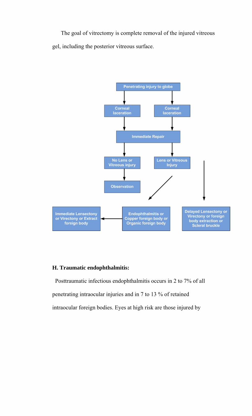
The goal of vitrectomy is complete removal of the injured vitreous
gel, including the posterior vitreous surface.
H. Traumatic endophthalmitis:
Posttraumatic infectious endophthalmitis occurs in 2 to 7% of all
penetrating intraocular injuries and in 7 to 13 % of retained
intraocular foreign bodies. Eyes at high risk are those injured by

foreign objects contaminated by soil or vegetable matter.
Staphylococcus and Bacillus species are the most causes of
posttraumatic endophthalmitis.
I. Intraocular foreign body
Penetration of the globe with foreign bodies are frequent.
Seriousness of such injuries depend on the retention and toxicity of
the intraocular foreign body12
Foreign bodies types: 1. Metallic –a. magnetic (eg) iron
b. non magnetic (eg) copper
2. Non metallic – plastic, wood
Ion poisoning of intraocular epithelial structures is seen with copper,
iron, lead zinc and nickel. In general, metals with low redox potential
such as iron and copper have the greatest potential for metallosis.
Modes of damage:
a. mechanical effects – depends on size, velocity and type of foreign
body. Foreign bodies of size greater than 2mm cause extensive
damage
b. introduction of infection- pieces of the wood and stones carry a
greater chance for infection
c. reactions of foreign body- depends on chemical nature of foreign
body. It can be of four types.
(i) no reaction- usually with inert substances like glass, plastic
(ii) local irritative action leading to encapsulation of foreign
body
(iii) suppurative reaction is excited by copper, mercury
particles, nickel , zinc etc
(iv)specific reaction – iron causing siderosis bulbi, copper
alloys producing chalcosis
Management of intraocular foreign body:
To come to a correct diagnosis following steps are followed:
1. History
2. Thorough ocular examination
3. X ray orbit antero-posterior and lateral views
4. Localisation of foreign body by CT scan, ultrasound
Treatment: IOFB should always be removed except when it is inert
and probably sterile or when little damage has been done to the vision
and the process of removal may destroy sight. Method of removal
depends on the site of lodgement of the foreign body
1. FB in anterior chamber: removed through a corneal inciscion
directed towards
2. FB entangled in iris tissue – removed by performing sectoral
iridectomy of the part containing the FB
3. FB in the lens: ECCE with intraocular lens implantation done
4. FB in the vitreous and retina – removed by posterior route

Magnetic FB- by pars plana sclerotomy using forceps or external
magnet
Non magnetic FB- removed by forceps with parsplana vitrectomy
Algorithm of management of intraocular foreign body
3. NEUROPHTHALMIC MANIFESTATIONS OF TRAUMA
A.Optic nerve injury:
The clinical manifestations of optic nerve trauma include
(i) decreased visual acuity
(ii) visual field defects
(iii) dyschromotopsia
(iv) relative afferent pupillary defect
(v) occasionally fundoscopic abnormalities of optic disc.
In patients with visual complaints, the visual acuity range from
normal to perception of light. Patients with normal acuity may have
visual field defects that spare fixation. Damage to optic chiasma may
occur secondary to blunt trauma. Therefore careful examination of
visual field in the fellow eye will detect such lesions.
Trauma to optic nerve may be direct or indirect.
a. Direct injury commonly occurs in the form of penetration injuries
or severe blunt trauma to the orbit and globe. Laceration or
transection of the nerve may occur when a variety of objects and
projectiles penetrate the eye, orbit or cranium. In these cases, loss of
vision is immediate and usually complete. Tractional forces on the
globe may cause avulsion of optic nerve head. Retained orbital
foreign bodies or fragments of orbital fractures may impinge on optic
nerve.
Optic nerve sheath haemorrhage and traumatic arachnoid cysts
may also compress the optic nerve. Increased tissue pressure caused
by blood or air within the confines of orbit may compromise visual
functions by optic nerve compression and possibly vascular perfusion
b. Indirect traumatic optic neuropathy is defined as traumatic loss of
vision that occurs without external or initial ophthalmoscopic
evidence of injury to the optic nerve. Loss of vision may be

immediate or delayed. The optic disc may appear normal or
edematous, depending on whether the injury involves the posterior or
anterior portions of nerve respectively13. Optic atrophy can be
identified within one week in anterior lesions and in 3-6 wk in
posterior lesions.
Management:
a. High dose intravenous methylprednisolone is effective in
reducing the morbidity due to optic nerve trauma. The rationale for
corticosteroid treatment is reduction of tissue edema, resulting in
increased vascular perfusion. Additionally, membrane stabilization is
thought to reduce tissue necrosis.
A reasonable regimen would be IV methyl prednisolone in a 1g
loading dose, followed by 250- 500 mg every 6hrs for 3-5 days.
Medical treatment must be instituted as soon as the diagnosis of
traumatic optic neuropathy is confirmed clinically. Following
institution of treatment, the patient is observed for approximately 48-
72 hrs. If visual function improves on corticosteroid therapy,
conversion to a tapering course of oral therapy after 48 hrs. If there is
no response, optic canal decompression is considered. Required
b. Surgical intervention is needed when there is delayed loss of
vision while the patient is receiving corticosteroids or during tapering
of corticosteroids which implies a compressive lesion. It involves

decompression of optic nerve at the level of optic canal. Optic nerve
sheath fenestration near the neuroocular junction is recommended for
an intrasheath optic nerve hematoma. Timely decompression of such
hematomas by lateral canthotomy or direct drainage may be a sight
saving maneuver.
Surgical decompression of optic canal is indicated if neuroimaging
confirms the presence of bone fragments or foreign bodies impinging
on optic nerve.
B. Chiasmal lesions
Traumatic lesions of optic chiasm occur but are uncommonly
treated, because the trauma that produces such lesions are usually fatal.
Mechanisms for causing damage to the optic chiasm include:
a. disruption of the chiasmal blood supply by shearing, thrombosis or
spasm of nutrient vessels.
b. Penetrating injuries or fractures may result in laceration or tearing of
the chiasm.
c. Compression of the chiasm may occur by herniation of the gyrus
rectus or by a dilated third ventricle secondary to hydrocephalus.
Associated damage to the hypothalamic-pituitary axis, resulting in
diabetes insipidus and multiple cranial nerve palsies may be present.
There is no effective treatment for optic chiasmal trauma. Primary
optic atrophy is usually noted 6-8 wks following injury.

C. Retrochiasmal lesions:
Lesions of the posterior visual pathway are not uncommon and
may involve the optic tract, geniculocalcrine pathway, occipital cortex
or surrounding visual association areas. Patients may complain of
blurred vision in a portion of their visual field.
All retrochiasmal lesions produce homonymous visual field
defects. Generally more anterior the lesion, more incongruous will be
the field defects. Lesions placed more posteriorly produce congruous
field defects. Cortical blindness is a condition that occurs when there
is bilateral involvement of occipital visual cortex. Most common
cause of cortical blindness is a penetrating injury of the occiput like
gunshot wound that result in destruction of brain tissue. Less
commonly, closed head injury may cause cortical blindness. This
usually results from blow of concussive force to occiput. Visual loss
may be immediate or delayed. These patients may experience a
gradual return of vision without permanent sequelae.
Neuroimaging can be extremely valuable in demonstrating the extent
of these lesions, but clinical examination gives more clues.
D. Oculomotor nerve injury
Traumatic third nerve injury may be partial or complete. The patient
may complain of oblique diplopia and blurring near vision as a result

of decreased accomodation. Varying degrees of ptosis in combination
with impaired adduction, elevation and depression is noted on
examination. With pupil involvement, there is efferent pupillary
defect. Over time abberant regeneration may occur in the form of
pseudo Von Grafe, pseudo Duane, pseudo Argyl Robertson Pupil.
Management- 1/3 rd spontaneously recover within 6-12 months.
Residual ptosis and ophthalmoparesis are managed surgically.
E. Trochlear nerve injury
The fourth nerve is vulnerable to trauma due to following reasons:
a. longest cranial nerve
b. slender
c. exits dorsally from brain
The most common site of trochlear nerve injury is the portion
traversing the subarachnoid space. Contrecoup motion of brainstem
during trauma results in contusion of one or both trochlear nerves by
the tentorial incisura. Patients usually complain of vertical binocular
diplopia that increases on down gaze. Patient may also have a head
tilt to the side opposite the paresis. The Bielschowsky head tilt test is
useful in localizing dysfunction to the superior oblique muscle –
ipsilateral hypertropia worsening on opposite gaze and same side
head tilt

The prognosis of functional recovery in fourth nerve palsy is
relatively good. Approximately 40% of patient may recover within 8
months after injury. If sufficient recovery has not occurred by 6 to 12
months and base up prism prove unsatisfactory, then surgical
correction is considered.
F. Abducent nerve injury
It occurs in severe head injury with loss of consciousness.
a.The most common site of injury to abducent nerve is where it passes
beneath the petroclinoid ligament to pierce the dura on entering the
posterior cavernous sinus.
b. Next common site is in the cavernous sinus. There will be
associated other cranial nerve palsies
c. Trauma to tectum of pons can result in damage to the abducent
nucleus. Because the genu of fascial nerve loops over abducens nucleus,
there may be concomitant facial nerve palsy.
d.Increased intracranial pressure from posttraumatic hydrocephalus or
cerebral edema may compromise the abducens nerve as it traverses the
subarachnoid space. Thus it can be a false localizing sign14.
Approximately 40 – 50% of patients with traumatic sixth nerve
paresis spontaneously improve within 6 – 12 months. Surgery may be
considered if recovery is incomplete after 6 – 12 months and the
patient is symptomatic even after conservative measures

G. Central disorders of ocular motility:
Trauma to internuclear and supranuclear pathways of the ocular
motor system in the brainstem and cerebrum may be manifested by a
variety of motility disturbances. The features of ocular motility
disturbances may be:
- Internuclear ophthalmoplegia occurring in trauma to
mescencephalic region- MLF
- Skew deviation occuring in disruption of vestibular input
- Horizontal gaze palsy occuring in lesions at pontine level
- Vertical gaze palsy occur in damage to rostral interstitial nucleus
of the MLF
- Convergence spasm or insufficiency, Impaired accommodation,
Disorders of fusion, Acquired esotropia occuring as a part of dorsal
midbrain syndrome
H. Facial nerve injury
Trauma to facial nerve with subsequent impairment of orbicularis
oculi function and reflex lacrimation may lead to exposure
keratopathy or corneal blindness.
Sites of facial nerve injury:
a. Facial nerve nucleus or fascicles- in brainstem injury
b. Intracranial portion of facial nerve in fallopian canal- in
temporal bone fracture

c. Extracranial portion of facial nerve- in blunt or penetrating
injuries
Ocular manifestations of facial nerve palsy:
Lagophthalmos
Exposure keratitis
Keratoconjunctivitis sicca
Impaired blink
Lower lid retraction
Paralytic ectropion
Epiphora
Brow ptosis
Abberant regeneration
Management:
• Exposure keratitis- topical lubricants and lid tapping
• Tarsorraphy- if conservative measures are insufficient
• Use of gold weights for upper lid in combination with
surgical correction for lower lid laxity provides long term
functional and cosmetic results
D. ORBITAL AND ADNEXAL INJURIES
A. Periocular lacerations :
Careful inspection of periocular lacerations provides abundant
clues to the nature and extent of the injury. The general considerations

include the location and depth of injury, tissue loss, impaired function
and presence of foreign body.
a.Marginal lid laceration:
Lacerations lateral to puntum- unlikely to involve canaliculus
Lacerations medial to punctum- likely to be associated with
canalicular laceration
b.Non marginal laceration:
Superficial- involving skin and orbicularis
Deep - 1. upper eyelid involves septum, levator aponeurosis and
muscle, lacrimal gland, trochlea and superficial neurovascular
bundles.
2. lower eyelid the septum and inferior oblique muscle may
be
injured.
3. medial canthus: canaliculus, lacrimal sac, medial canthal
tendon
4. lateral canthus: lateral retinaculum, facial nerve may be
injured.
Complex eyelid lacerations may present with large gaping wounds.
As the intact portion of eyelids contract, large gaps develop in the
eyelid contour giving the appearance of an avulsion. In these cases of

pseudo- tissue loss, primary repair is possible. However in case of
tissue loss, an effort should be made to save tissue if available.
In penetrating injuries to upper eyelid, it is important to assess
levator function preoperatively. In the setting of acute soft tissue
edema, the levator function is often underestimated. But a careful
examination can help to distinguish a direct levator injury from a
traumatic third cranial nerve injury.
It is important to have a high index of suspicion for the presence
of foreign body. Based on the history and initial inspection of the
patient, the appropriate imaging modality help to determine the
location, depth and possibly the nature of foreign body prior to
commencing surgical repair.
Treatment of periocular injuries:
Acute treatment: Wounds should be thoroughly cleaned. Adequate
debridement of particulate matter prevents the late sequelae of dermal
tattooing.
Timing of repair:
In cases of isolated periocular trauma, timing of repair is not
critical. More emergent intervention needed in case of associated
globe rupture. In most cases, a delay of 12- 36 hrs from time of injury
doesn’t alter surgery results.
Laceration repair:

Superficial non marginal injury repair- can be repaired primarily
without difficulty. Marginal full thickness injury – primary objective is
to restore the local anatomy with appropriate alignment of the eyelid
margin, tarsus and skin. Eyelid alignment is best achieved by using the
eyelash line, gray line and meibomian units as local landmarks. These
structures can be adequately positioned with two to three 6-0 or 7-0
sutures placed along eyelid margin.
c. Levator lacerations:
If a levator muscle complex injury is suspected, it is important to
explore the muscle and aponeurosis through an upper eyelid crease
incision. If the aponeurosis is detached from the tarsus, repair is directed
at suturing the cut end of the aponeurosis to the upper anterior face of
tarsus with either silk or polyglactin suture.
d. Lateral canthus injury:
If the attachment of lateral canthal tendon is disrupted, repair
should be directed posteriorly using a permanent suture anchored to
the periosteum. If there is no periosteum, the tendon can be fixed to
the orbital rim through appropriately drilled holes.
e. Medial canthal injury:
Lacerations extensive enough to lacerate the medial canthal tendon
are also associated with canalicular injuries. The medial canthal tendon is
optimally repaired with a permanent suture fixed to the periosteum

At the level of posterior lacrimal crest, if the bone or periosteum
is not intact, transnasal wiring of medial canthus is the possible
solution.
f. Canalicular injury:
In the majority of cases, there is usually no doubt that the
canalicular system is disrupted based on disruption of wound. In cases
that are not readily apparent, probing of the canalicular system and
inspection of wound for direct visibility of the probe help establish
the diagnosis. In cases where depth of wound is not seen, canalicular
information may provide additional information.
The general concept of canalicular repair is to pass an internal stent
to bridge the laceration and to reestablish continuity of the disrupted
canalicular system. Stents can be monocanalicular – Viers rod,
Silastic tube or bicanalicular- pigtail, nasolacrimal probe.
g. Tissue loss:
(i) Superficial avulsion:
In cases of tissue loss anterior to the septum, primary
granulation is considered. When allowed to granulate, these wounds
require more attention than primary repair, as the epithelialisation is
prolonged and there is increases risk of infection.
(ii) Deep tissue loss:

Large confluent loss of the anterior lamella of the eyelids
including skin and muscle, which exposes underlying fat is not
suitable for granulation. These wounds should be covered with full-
thickness skin grafts or musculocutaneous flaps in acute setting.
B. Injuries to orbit:
Based on the patterns of fracture, the orbit is divided into four
zones:
1. supraorbital area of the frontal bone
2. nasoethmoid area
3. zygoma
4. internal orbit
Thin section CT imaging is the gold standard in diagnosis of
orbital injuries. CT scan not only provide precise documentation of
skull features, but also gives the information about the soft tissues of
the globe, orbit and cranial vault.

Type of fracture Associated soft tissue injury
Frequency X-ray view CT finding Clinical signs Management
Supra orbital frontal fractuies
Forehead contusion or laceration
Less common
Antero posterior view Fracture of supra orbital frontal bone
Ptosis, proptosis Fractured segmentsreplaced anatomicaheld with wires
Nasoethmoorbital fractures
Edema over medial canthal region
Common AP view – fracture of nasal bone, fracture of the junction of the frontal process of maxilla with frontal bone, fracture of medial orbit, fracture of infra orbital rim
Same as x-ray Nasal flattening with loss of dorsal height, increased angle between lip and collumella
Reduction and fixation of all fractured segmentsMay need acute bografting.
Zygoma fracture Periorbital and subconjuctival ecchymosis
More common
Water’s view fracture through inferior orbital rim Caldwell view displacement of zygomatico frontal articulation
CT precisely define displacement
Swelling of cheek and face with contour irregularities, loss of malar prominence, step deformity at the zyomaticomaxillray articulation
Open reduction andfixation with miniature metal plates
Continued:
Type of fracture Associated soft tissue injury
Frequency X-ray view CT finding Clinical signs
Internal orbital fractures: (i) orbital apex fractures
Periorbital contusion
Less common CT through orbital apex – fracture at or adjacent to optic canal
CSF leaks, traumatic optic neuropathy, carotid cavernous fistula
(ii) orbital roof fracture Periorbital contusion, lid laceration
Common in young children
Anteroposterior view show fracture
Same as X-ray CSF rhinorrhea, pneumocephalus, sub periosteal haematoma, ptosis
(iii) medial orbital wall fracture
Depressed nasal bridge, traumatic telecanthus
Less common Anteroposterior view show fracture
Fracture involving frontal process of maxilla, lacrimal bone, ethmoid bone along medial wall or orbit
Epistaxis, CSF rhinorrhoea, orbital haematoma, damage to lacrimal drainage system

(iv) Orbital floor fractures :
It can be: Direct fractures- involves orbital rim called impure
fracture
Indirect fractures ( blow out)- don’t involve orbital rim
An orbital blow out fracture should be suspected in any patient who
received periorbital blow forceful enough to cause ecchymosis.
Physical examination reveals
1. Eyelid signs- ecchymosis, edema
2. Diplopia with limitation of upgaze, downgaze or both- due to
entrapment of the inferior rectus or its adjacent septa in the
fracture
3. Enophthalmos and ptosis of the globe- These occur with large
fractures in which the orbital soft tissues prolapse into the
maxillary sinus.
4. Hypoesthesia in the distribution of the infraorbital nerve
5. Emphysema of the orbit and eyelids
Xray anteroposterior view of orbit and facial bone shows tear drop
sign
CT scans with coronal or saggital views are usually indicated to guide
treatment. The majority of blowout fractures don’t require immediate

intervention. They are observed for 7-10 days so that swelling and
orbital haemorrhage can subside. Blowout fracture in paediatric
patients, where attempted ocular excursions can stimulate the
oculocardiac reflex leading to bradycardia, nausea and pain,
immediate intervention is needed.
Indications for surgical intervention are:
1. Diplopia with limitation of upgaze or downgaze within 30
degrees of primary position, positive traction test 7-10 days after
injury and with radiological confirmation of fracture of orbital
floor.
2. Enophtholmos that exceeds 2mm and that is cosmetically
unacceptable to the patient
3. Large fractures involving at least half of the orbital floor,
particularly when associated with large medial wall fractures
Surgery is best done within two weeks for easy manipulation of the
scar tissue and fibrosis of entrapped tissue. The surgical correction
can be made through an infraciliary approach or conjunctival incision
combined with lateral cantholysis. The steps are,
(i) elevation of periorbita from orbital floor
(ii) release of prolapsed tissue from the fracture
(iii) placement of an implant over the fracture to prevent recurrent
adhesions Complications of blowout surgery include, decreased visual

acuity or blindness, diplopia, undercorrection or overcorrection of
enophthalmos, lower eyelid retraction, infraorbital nerve
hypoesthesia, infection, extrusion of implant, lymphedema and
damage to lacrimal pump.
REVIEW OF LITERATURE
Ocular trauma due to mechanical injuries is one of the major
cause of visual impairment world wide. There are in excess of 2
million cases of ocular trauma per year in the world and of these
about 40,000 cases sustain severe visual impairment. The incidence
of ocular trauma are increasing due to modernization and increased
industrialization. So clinical study of ocular morbidity due to
mechanical injuries in patients attending the tertiary hospital is done
in this thesis. This study includes history, clinical examination,
investigations and the morbidity is assessed qualitatively and
quantitatively.
Ocular injuries are classified anatomically into anterior
segment, posterior segment, neuroophthalmic injuries and orbital and
adnexal injuries. The nature of mechanical injury, occupation of the

individual, duration between injury and examination, initial visual
acuity and final visual acuity are studies.
A retrospective study on epidermiology of adult eye injuries by
Ksenijia Karaman et al states that initial visual acuity predicted the
visual outcome. The severity of ocular injury was independent of the
age of the individual. Penetrating injury to eye leads to more ocular
morbidity than blunt injury. Similar results were found by De Juan et
al.
A prospective study on the profile of ocular trauma at tertiary
eye care by D.V.Singh et al revealed that males were injured more
(88.5%). One third of injuries occurred in pediatric age group. This
is supported by the retrospective study in a tertiary eye care in Eastern
India by Dr.Sucheta Parija et al, who had found similar ratio of
involvement among males and females. Right eye involved in 50.1%,
left eye in 46.8% and both eyes involved in 3.1%. Facial injury
associated with ocular injury was seen in 20.1% of eyes. 3% of
patients were able to reach tertiary centre within 48 hours.
A prospective study on expected effect of treatment on the ratio
of visual deficiency after ocular trauma by Virgilio Lima-Gomez et al
showed that the most frequent feature of poor visual prognosis is
afferent papillary defect and globe rupture. A retrospective study on
pediatric ocular trauma by M.R.Shoja et al states that, high frequently

of ocular injuries occur at streets and roads. A major group of
children belong to poor socioeconomic class. In this study
penetrating injuries predominate over blunt injuries. This may be due
to the fact that ocular injuries treated as out patient injuries were not
included.
A retrospective study by Mohammed Yashir Arafat et al on
injuries to eye has stated that, the incidence of ocular trauma in males
are 84% and in females are 16% which is comparable to the study by
D.V.Singh et al. The study also had found that right eye is involved
in 66% and left eye in 34%. The post treatment visual acuity between
6/6 and 6/60 was 74%.
Atkari et al in this study had found that males are move
involved 55.8% and females 44.2%. This is comparable to other
studies. In his study he had noted left eye involvement (59.9%) more
than right eye (42.7%). Bilateral involvement is rare (5.6%). This is
in contrast to other studies, where right eye involvement outnumbered
left eye. The common age group involved was 16-35 years, which is
the active period of life. Most of them were farmers (32.1%) and
house wives (26.2%). His study also shows that only 43.3% came
for treatment within 7 days and very minimal of 3.09% reported for
treatment within 6 hours.

The visual outcome of perforating globe injuries depends on
the type of injury sustained. Injuries due to sharp objects cause
localized damage and visual prognosis is better. Blunt injuries
usually cause globe disorganization and so visual prognosis is poor.
Enucleation was performed previously and now it has declined
because of better understanding of pathophysiology, improved
surgical techniques and antibiotic use to prevent sepsis.
MATERIALS AND METHODS
The prospective study was conducted on 100 cases of ocular
trauma attending Tirunelveli Medical College Hospital. Patients with
ocular injury attending ophthalmology outpatient department were
randomly included in the study.
Inclusion criteria were:
o Patients of all ages

o Both males and females
o All co-operative patients
o Irrespective of economic status
Exclusion criteria:
o Unconscious patients
o Patients not co-operative for examination and
procedures
o Terminally ill patients
A detailed patient data including age, sex, occupation,
residential area, nature of injury like road traffic accident, assault,
accidental fall, accidental injuries due to stone, needle pricks etc..,
were taken. The presenting complaints, time interval between injury
and examination, directly came for treatment or referred from
peripheral hospital and initial medical assistance taken were also
recorded.
Patients unaided visual acuity at the time of presentation was
recorded using Snellen’s chart. Orbit, eyelid and adnexa were
examined by diffuse illumination. Lid injuries, periorbital contusion
and injuries around orbit were recorded. Slit-lamp examination was
done to patients with trauma attending our out patient department and
also to trauma patients of emergency department who needed slit
lamp examination.

Conjunctiva examined for any injury like tear and sub-
conjunctival haemorrhage. Cornea examined for abrasion, tear,
foreign body, extent and depth of injury. Anterior chamber reaction in
the form of flare and cells were noted and recorded. Iris examined for
sphinter tear and iridodialysis. Pupil examination done to note direct
and indirect light reflexes, mydriasis, relative afferent papillary defect
and pupil distortions. Lens examination done to look for any
penetrating injury, traumatic cataract, subluxation and dislocation.
Posterior segment examination done by direct ophthalmoscopy
in all patients provided the anterior segment did’nt preclude fundus
examination. +90D examination and indirect ophthalmoscopy done
for needed patients. Optic disc, macula and retina were examined for
disc edema, macular hole or haemorrhage, retinal haemorrhage, tear,
detachment and infarcts.
Intraocular pressure recorded by Applanation tonometry or non
contact tonometry in trauma patients attending our department and
Schiotz tonometry done to others.
B scan done to assess the posterior segment in patients with
blunt and penetrating injury to eye ball and in those patients for whom
fundus examination cant be done due to hazy media. CT scan facial
bones and X- ray orbit done to needed patients. Final diagnosis was
made. Treatment given to these patients either medically or surgically

depending on the case. Final visual acuity on discharge was recorded.
Causes for poor visual recovery in the patients included in the study
were noted. All the datas are included in a proforma and analysed. All
medico-legal cases are duly entered in accident register.

OBSERVATION
STATISTICAL ANALYSIS
The study subjects were analyzed and interpreted in
terms of their demographic characteristics, type of injury, type of
morbidity, eye/ eyes involved in the injury and visual outcome. The
continuous variables were interpreted by Students’ ‘t’ test and
categorical variables were interpreted by χ2 (Chi-square) test where
ever applicable. The above statistical procedures were performed by
IBM SPSS statistics 20. The P- values less than 0.05 (P<0.05) were
defined as statistically significant in two-tailed test.
RESULTS AND OBSERVATION
The different type of injury in the persons included in the
study were classified according to their age and sex.

Table-1.Classification based on age with sex.
Age (years) Male Female Total
No % No % No %
0-19 14 17.1 5 27.8 19 19.0
20-39 38 46.3 4 22.2 42 42.0
40-59 23 28.1 7 38.8 30 30.0
60+ 7 8.5 2 11.1 9 9.0
Total 82 100.0 18 100.0 100 100.0
Mean ± SD 34.9±16.0 35.7±18.2 35.1±16.3
Range 8-80 8-65 8-80
‘t’ 0.186 -
Significance P>0.05
Table.1. Classification based on age & sex
14
5
38
4
23
77
205
1015
20
2530
3540
0-19 20-39 40-59 60+
MaleFemale

The above table-1 shows the percentage distribution in the
study subjects according to their sex with age. The mean age of males
were 34.9±16.0 and females were 35.7±18.2 (years). They were not
significantly differed in respect of their age (P>0.05). The mean age
of the total subjects was 35.1±16.3 years with range 8-80 years. The
Males were 82% and females were 18%.

Table-2. Sex wise distribution of accidents.
Accident Gender
χ2 Df Sig. Male Female Total
Accidental
Fall 17 4 21
6.785 5 P>0.05
Assault 19 4 23
RTA 2W 34 6 40
RTA 4W 6 1 7
Others 6 3 9
Total 82 18 100
Table.2. Sex wise distribution of accidents
17
4
19
4
34
6 6
1
6
3
0
5
10
15
20
25
30
35
Accidental fall Assault RTA 2W RTA 3W Others
MaleFemale

The sex wise accidents were shown in the above table -2. Among the
injuries, road traffic accidents were 47% , assault – 23%, accidental
fall – 21%. In the road traffic accidents, accidents by 2 wheeler was
85% . The results revealed that there was no significant relationship
between the gender with accidents (P>0.05).

Table-3. Sex wise distribution of affected eyes.
Eyes Gender χ2 Df Sig.
Male Female Total
Right 36 15 51
9.787 2 P<0.01 Left 30 3 33
Both 16 0 16
Total 82 18 100
Table.3. Sex wise distribution of affected eyes
36
15
30
3
16
005
1015
20
2530
3540
Right Left Both
MaleFemale
The above table-3 associates sex of patients with eyes
affected. The females of 83.3% (15) were affected with right eyes and
43.9% (36) of males were affected with right eyes. Similarly, 19.5%
(16) of males only were affected with both eyes. The above

associated eyes with gender was statistically significant (P<0.01).
Table-4.Time of examination since admission.
Time of
examination
(hrs)
Type of admission
χ2 Df Sig. Direct Referred Total
0-6 12 2 14
13.856 2 P<0.001 6-12 25 21 46
12+ 12 28 40
Total 49 51 100
Table.4. Time of examination since admission
12
2
25
21
12
28
0
5
10
15
20
25
30
0 To 6 6 To 12 12+
DirectReferred
The admission with duration was related in the above table -4. The
direct admission was examined early compared to referred cases.
These referred cases has got initial medical attention in the peripheral

hospitals.
Table-5. Laterality of injury.
Injury Eyes
χ2 Df Sig. Right Left Both Total
Accidental
fall 13 6
221
6.690 10 P>0.05
Assault 12 8 3 23
RTA 2W 18 15 7 40
RTA 4W 2 3 2 7
Others 6 1 2 9
Total 51 33 16 100
Table.5. Laterality of injury
13
6
2
12
8
3
18
15
7
23
2
6
12
0
2
4
6
8
10
12
14
16
18
Accidentalfall
Assault RTA 2W RTA 4W Others
Right LeftBoth
The accidents wise affected eyes were shown in the above table -5.

The results revealed that there was no significant relationship between
the accidents with affected eyes (P>0.05).
Table-6. Percentage distribution of type of injury.
Type of Injury No %
AS 23 23%
AS+PS 7 7%
ORBIT 14 14%
Neuro 3 3%
Combined injury 53 53%
Total 100 100%
Table 6. Percentage distribution of type of injury
14%
3%
53%
23%
7%
AS
AS+PS
ORBIT
NEURO
COMBINEDINJURY
The above table -6 describes the Combination of type of injury.
This table shows that combined injury involving more than one

segment accounts to 53%. Anterior segment injury alone is next
commonly involved.
Table-7. Visual outcome in Traumatic optic neuropathy
Sex Presenting
vision
PL and more
Presenting
vision
No PL
Improved
visual outcome
Males 14 3 6
Females 4 2 1
Total 18 5 7
Table.7 Visual outcome in Traumatic optic neuropathy
14
4 3
2
0
2
4
6
8
10
12
14
Presenting vision PL & more Presenting vision No PL
MaleFemale
This table shows 18 patients had traumatic optic neuropathy.
Initial visual acuity of PL and above were 18, and no PL at

presentation were 5. Of these 7 patients showed improvement with
treatment. All these 7 patients had PL and above visual acuity at
presentation.
Table- 8: Cause of open globe injury:
Mode of injury No of persons
RTA 3
Accidental injury 15
Assault 1
Total 19
Table 8. Cause of open globe injury
15
3
19
1%
Mode of injuryRTAAccidental injuryAssault
Open globe injury noted in 19 patients. Common cause for open globe

injury was accidental injury with sharp objects. Of these 6 patients
developed endophthalmitis. Two of them had intraocular foreign
body.
Table-9. Category of vision at different intervals.
Vision At admission At discharge
At 1 month
follow up
Persons Persons Persons
Good 61 68 69
Moderate 2 3 2
Poor 9 4 5
Blind 28 25 24
Total 100 100 100
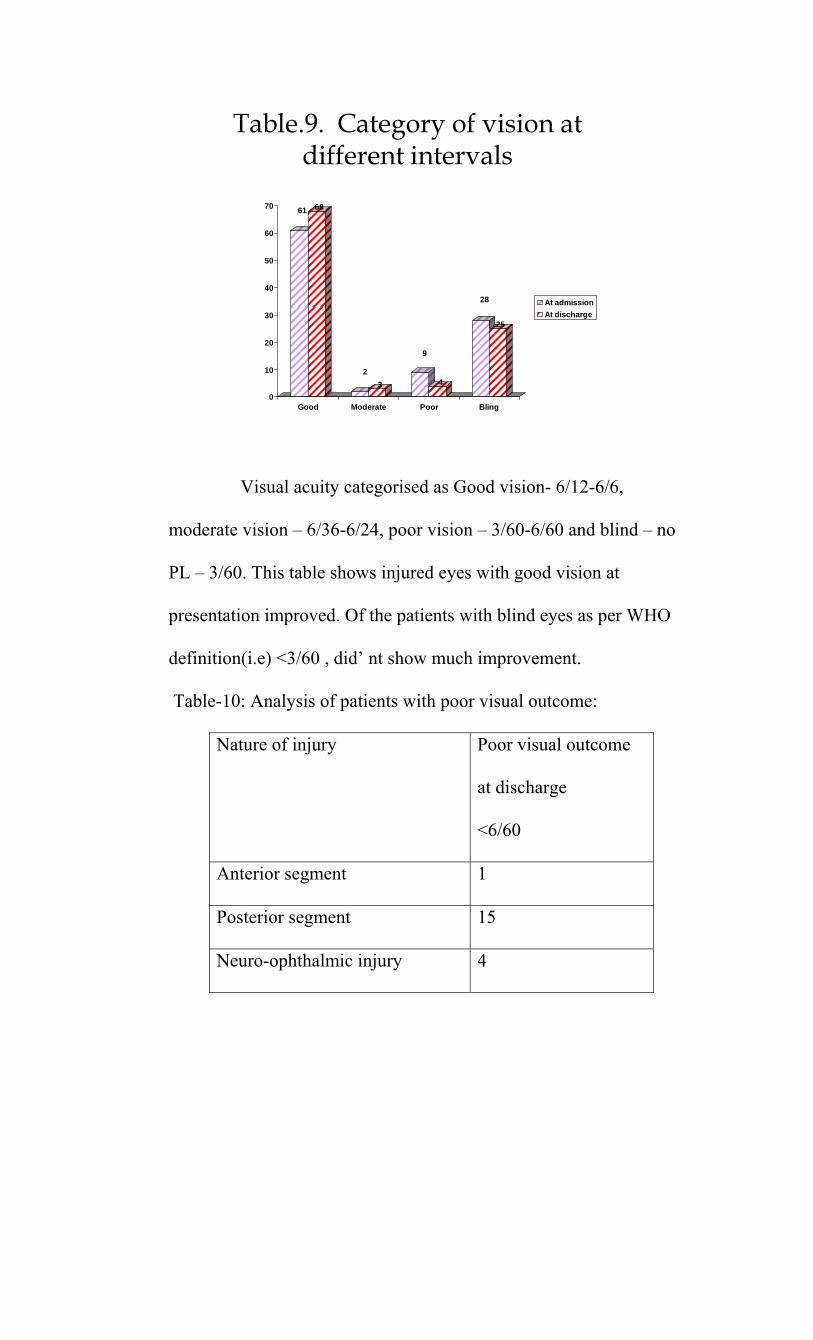
Table.9. Category of vision at different intervals
61 68
23
9
4
28
25
0
10
20
30
40
50
60
70
Good Moderate Poor Bling
At admissionAt discharge
Visual acuity categorised as Good vision- 6/12-6/6,
moderate vision – 6/36-6/24, poor vision – 3/60-6/60 and blind – no
PL – 3/60. This table shows injured eyes with good vision at
presentation improved. Of the patients with blind eyes as per WHO
definition(i.e) <3/60 , did’ nt show much improvement.
Table-10: Analysis of patients with poor visual outcome:
Nature of injury Poor visual outcome
at discharge
<6/60
Anterior segment 1
Posterior segment 15
Neuro-ophthalmic injury 4

Orbit and adnexal injury -
Combined injury 9
Total 29
Table.10. Analysis of patients with poor visual outcome
15
1
9
0
4
Anterior segment
PosteriorsegmentNeuro-ophthalmicinjuryOrbit and adnexalinjuryCombined injury
The cause for visual acuity of < 6/60 is shown in this table. Posterior
segment injury showed poor visual outcome in 15 cases. Orbit and
adnexal injury and anterior segment injury alone had good visual
outcome.
DISCUSSION

Ocular injuries are one of the common cause of ocular
morbidity nowadays. It has been estimated that, 1.6 million people are
blind in the world due to ocular trauma, 2.3 million people are
visually impaired due to trauma and 19 million people are unilaterally
blind due to ocular trauma. Males are more affected than females and
ocular injuries are common in younger age groups.
In this study, common age group involved was between 20 – 39
years 42%. This is comparable to study by Atkari et al17. This age
group is the economically productive age group and ocular injury to
this age group cause a great deal of economic loss to the country.
Although students (22%) formed the major subgroup, drivers (17%) ,
coolies (22 %)and farmers (14%) were also involved. But study by
Atkari et al showed most of the involved patients were farmers
(32.1%). High incidence in students may be due to their increased
involvement in risk activities. They play hazardous and dangerous
games. This could be the reason for high incidence in students.

Males are more commonly affected (82%) compared to
females of (18%.) Male predominance and peak age involvement in
our study were similar to other studies. This is due to the fact that,
males are more involved in outdoor activities. A prospective study on
the profile of ocular trauma at tertiary eye care by D.V.Singh et al18
showed similar results. This is also supported by the retrospective
study in a tertiary eye care in Eastern India by Dr. Sucheta Parija et
al19 . This is comparable to the demographic profile noted in a study
in South India by Pieramici DJ, Sternberg P Jr, Aaberg TM et al20 .
Involvement of males more than females, who are the bread winner
for most of the families will have an impact on productivity of our
nation for sure.
The proportion of right eye involvement (51%) was more than
left eye (33%), which is in agreement with other studies. Similar
results were noted in a study by D.V.Singh et al.

In our study among the mode of trauma, road traffic
accident(47%) was the major cause for mechanical injuries to eye.
Assault was the cause for 33%, accidental falls for 21% . Of the road
traffic accidents, two wheeler accidents were responsible for 40%.
Similar results were seen in De Juan E et al21 study on penetrating
ocular injuries. This may be due to increased vehicular traffic and
increased population. Rash driving is the major reason for two
wheeler accidents.
In our study, 49% of patients presented directly to our hospital
and 51% had initial medical assistance and referred from peripheral
hospitals. Of the directly presented patients, 12 patients have come
after 12 hours. In our study 14% have reported within 6 hours of
injury. But in Atkari et al study only 3.09% patients have reported
within 6 hours. Now due to increased communication and transport
facilities, more patients are able to reach tertiary care hospitals earlier.
Initial hours after injury are important as early treatment helps in
speedy recovery. We noted that patients in whom treatment was
initiated within 12 hours had better visual acuity.

Based on injuries sustained, anterior segment alone was
involved in 23%, anterior and posterior segment in 7%, neuro-
ophthalmic manifestations alone in 3%, orbital and adnexal injury
alone in 14% and combined injuries in 53%. Anterior segment and
orbital injuries alone had better visual outcome and less morbidity
than neuro-ophthal, posterior segment and combined injuries. This
was in agreement with study by Virgilio Lima Gomey 22
In our study, 18 patients had traumatic optic neuropathy. Of
these 7 patients (39%)showed improvement with steroid treatment.
This is comparatively less than other study by J.A.Mauriello et al23,
which showed 56.25% with steroid treatment. This difference may be
due to delayed presentation and so delay in initiation of steroid
treatment. We could find that patients with at least PL vision on
presentation improved with treatment. Of the 7 patients who showed
visual improvement, all had vision of PL and above.

In our study, open globe injury was found in 19% of patients
and in them 6 patients (30.1%) developed endophthalmitis. Similar
incidence was noted in a study by D.V.Singh et al. This is
comparatively high compared to study by Thompson SW, Rubsamen
PE et al24 . The delayed presentation to the tertiary centre may be the
reason for this high occurrence. In our study we had two patients with
open globe injury , who subsequently developed endophthalmitis after
foreign body removal. This was due to late presentation to our
hospital, after 48 hours. Lens capsule injury is associated with
endophthalmitis. This is seen in one patient, in our study. This is also
noted in studies by Reynolds DS et al25 and Schmidseder E, et al26.
The presence of intraocular foreign body, lens capsule injry and
delayed presentation are risk factors for developing endophthalmitis.
In our study, 71% had good vision at discharge and one
month follow up. Patients who presented with good visual acuity had
good visual outcome. This is also shown in study by Sternberg P Jr et
al and also by Ksenija Karaman et al27 . The presenting visual acuity
is one of the important prognostic factors for good visual outcome.

Poor visual outcome of <6/60 were seen in 29 % in our study.
The major cause for this is found to be posterior segment injury alone
( 51%). Combined injury accounted for (31%). Poor visual outcome
in posterior segment injury is due to high incidence of vitreous
disturbance, incarceration of retinal tissue, vitreous haemorrhage and
associated retinal and choroids detachment. This is in agreement with
study by Cleary PE, Ryan SJ et al28 .

CONCLUSION
Ocular injury due to mechanical trauma can cause
morphological and functional impairment to ocular structures and
vision.
In our study, younger age group between 20 – 39 years are
commonly involved in ocular injury due to mechanical trauma.
Students are more involved in mechanical injuries. This age
group should be focused and made aware of preventive aspects
against mechanical injury. Children should be guarded from
using sharp objects. Children should be allowed to play under
parent supervision.
Males are more involved than females in our study. This is due
to males being involved in outdoor activities for occupation.
So, to prevent injuries during occupation wearing of protective
eye shields while working should be encouraged.
While considering the mode of injury, road traffic accidents
were responsible for ocular injury in many patients. Of the
road traffic accidents, two wheelers predominated. To prevent
this, people should be made aware of wearing helmets which

could reduce ocular injuries to a great extent. To reduce injuries
due to four wheeler accidents, wearing of seat belts should be
given priority and should be enforced by law.
Early institution of treatment, once diagnosis is made, reduced
ocular morbidity in our study. So people should be made aware
of seeking early medical help. This can be achieved by mass
media to disseminate ocular trauma related information.
Posterior segment injury and open globe injury had poor visual
outcome. These patients should be referred to retina surgeon
and tertiary referral centre at the earliest. So primary
ophthalmologists should diagnose this earlier and refer to
tertiary hospitals where such facilities are available. protective
measures should be taken in working places and
It is necessary to implement preventive measures to prevent
mechanical trauma to eye. Wearing seat belts and helmets
should be made compulsory. Legislation alone is not successful
for this effort. Education regarding safety measures and
measures to be taken if injury occurred should be done through
mass media. These measures will reduce ocular morbidity due
to mechanical trauma to a great extent.

PROFORMA
Serial No:
Hospital No: MLC / Non MLC Date:
Name: Age: Sex:
Occupation and income: Date of admission:
Date of surgery:
Date of discharge:
Address:
Complaints: Duration between trauma and examination:
0 to 6hrs / 6 to 12hrs / 12 to 24hrs /
days/weeks
Hospital details:
RTA/assault/other injuries
Right eye/left eye/both eyes
Loss of consciousness – yes/no
Painful/painless
Visual disturbance – yes/no
Movements of eye – possible/restricted
Diplopia – yes/no
Watering of eye – yes/no
Photophobia – yes/no
Head ache – yes/no
Vomiting – yes/no
Swelling
ENT symptoms – bleeding nose /tinnitus
CLINICAL EXAMINATION:
Right eye Left eye
EOM
Anterior segment examination:
(SLE)
Extra ocular movements
Lid
Conjunctiva
Cornea
Limbus
Anterior chamber
Iris
Pupils

Lens
Duct
Tension
Posterior segment examination
Fundus – Media
Disc margins
C : D ratio
Vessels
Haemorrhages
Macula
Periorbital region
Swelling /ecchymosis/emphysema
Orbital margins
Infra orbital anesthesia
Vision
Colour vision
Field of vision
Others
ENT examination
Central nervous system examination:
Conscious/unconscious
Oriented / disoriented

Memory
Speech
Cranial nerve examination
Motor system
Sensory system
Speciality opinion:
Neuro surgery/neuro medicine/plastic surgery/orthopedics
Investigations:
X-ray skull
CT scan Brain and Orbit
MRI scan brain and Orbit
Others
Final clinical diagnosis:
Treatment: Medical/surgical/others
Follow up visits:
Statistical analysis

BIBLIOGRAPHY
1. A.K Khurana, Indu Khurana Anatomy and Physiology of Eye –
Second edition
2. Parsons’ Diseases of the Eye, Twenty – first Edition.
3. BSCE American Academy of Ophthalmology , Section 7, 2010-2011
4. Jack K Kanski, Brad Bowling Clinical ophthalmology 2011
5. Albert & Jackobie Practice and Principles of Ophthalmology
6. Ferenc Kuhn Dante J, Pieremici Ocular Trauma Principles and Practice
7. Lead Editors Myron Yanoff Opthalmology, Third Edition
8. Shield’s Text book of Glaucoma Sixth Edition
9. Roger F Steinert’s Cataract Surgery 2009
10. Benjamin Boyd and Solomon Boyd - Modern Management of Ocular
Trauma
11. L. C Dutta, Modern Ophthalmology Third Edition
12. Rupseh Agarwal Girish Rao Rishikesh Naigaonkar,Xialing Ou, Sathish
Desai, Prognostic Factors For Vision Outcome Following Open
Globe Injuries.

13. Neuro-ophthalmology – Aravind Eye Care System.
14. Walsh and Hyot’s Clinical Neuro-Ophthalmology: THE ESSENTIALS –
Second Edition.
15. Diseases of the Orbit, A Multidisciplinary Approach, Jack Rootman,
Second Edition.
16. Stallard’s Eye Surgery, M.J. Roper-Hall- Seventh Edition.
17. Adhikari R.K.et al , Ocular Trauma in Rapti Eye Hospital Dang, 1993
18. D.V.Singh , Yog Raj Sharma, R.V. Azad, Dinesh Talwar, Rajpal,
Profile of Ocular Trauma at Tertiary Eye Care Vol. 7 No. 1, January –
March 2005.
19. Dr. Sucheta Parija, Dr. Mamata samal et al, A Retrospective Study in
A Tertiary Eye Care Centre in Eastern India, 2010
20. Sternberg P Jr, De Juan E Jr, Michel R G, Auer C Multivariate Analysis
of Prognostic Factors in Penetrating Ocular Injuries- Am J Ophthalmology
1984, 98: 467-472
21. De Juan E, Sternberg P, Michels RG, Penetrating ocular injuries;
types of injuries and visual results. Ophthalmology 1983; 90: 1318-22.
22. Virgilio Lima-Gomez and Dulce Milagros Razo-Blanco Hernandez –
Expected effect of treatment on the rate of visual deficiency after
ocular trauma- 2010
23. J. A. Mauriello , J. de Luca, A. Krieger, M. Schulder and L. Frohman-
Management of Traumatic Optic Neuropathy Study- Br. J.

Ophthalmology 1992 June -76(6) 349-352
24. Thompson SW, Rubsamen PE, et al , Endophthalmitis after penetrating
ocular trauma: risk factors and visual acuity outcomes. Ophthalmology
1995; 102: 1696-1701.
25. Reynolds DS, Flynn HW Jr. Endophthalmitis after penetrating ocular
trauma. Curr Opin Ophthalmol 1997; 8 (3): 32-38.
26. Schmidseder E, Mino de Kaspar H, Klauss V, Kampik A. Post-
traumatic endophthalmitis after penetrating eye injuries. Risk factors,
microbiological diagnosis and functional outcome. Ophthalmology
1998; 95 (3): 153-57.
27. Ksenija Karaman et al, Epidemiology of Adult Eye Injuries in Split-
Dalmation County, Croatian Medical Journal2004.
28. Cleary PE, Ryan SJ. Method of production and natural history of
experimental posterior segment penetrating eye injury in rhesus monkey.
Am J Ophthalmol 1979; 88: 221-31.

KEY WORDS TO MASTER CHART
RTA – Road traffic accident
RE – Right eye
LE – Left eye
BE – Both eyes
AS injury – Anterior segment injury
PS injury – Posterior segment injury
Neuro – Neuro ophthalmic injury
VA on A – Visual acuity on admission
VA on D – Visual acuity on discharge
SCH – sub conjunctival haemorrhage
# - Fracture
EL – Eye lid
AC – Anterior chamber
HM – Hand movements
PL – Perception of light
NO PL – No perception of light
MC – Mature cataract
FTCT – Full thickness corneal tear
RD – Retinal detachment

CD – Choroidal detachment
IOFB – Intraocular foreign body
VH – Vitreous haemorrhage
IViAB – Intravitreal antibiotics
RCS – Retina choroids sclera
K tear – Corneal tear
PCIOL – Posterior chamber intraocular lens
P&B - Pad and bandage
EDH – Extra dural haemorrhage
SDH – Sub dural haemorrhage
# BB – Fracture both bones

S.NO NAME
AGE
SEX
RE LE
BE
TYPE OF INJURY
DIRECT
REFER RAL
0to 6 6 to 12
1 Subramanian 50 M Y accidental fall
2 Abuthakir 18 M
y
RTA 2 wheeler y y
3 Sangeetha 8 F y RTA 2 wheeler y y
4 Kuttiraj 48 M y y RTA 2 wheeler y y
5 Kalliappan 44 M y assault y y
6 Vinoth 26 M y RTA 2 wheeler y y
7 Thangavel 57 M y RTA 2 wheeler y y
8 Muthukrishnan 47 M y RTA 2 wheeler y
9 Mytheen pitchai 28 M y RTA 2 wheeler y
10 Mohammed nasar 40 m y assault y Y
11 Jeyaraman 27 M y RTA 4 wheeler y y
12 Ganesan 20 M y RTA 2 wheeler y
13 Chelliah 57 M y accidental fall y y
14 Shunmugathai 43 F y accidental fall RTA y y
15 Gandhi 60 M y accidental fall y
16 Packiaraj 42 M y assault y y
17 Karupasamy 36 M y thorn prick y

18 Krishnan 80 M y assault y
19 Muthuraj 18 M y RTA 2 wheeler y
20 Balasubramanian 40 M y RTA 2 wheeler y
21 Ravi 18 M y RTA 2 wheeler y
22 Kothaiammal 65 F y acci hit in a iron pipe y y
23 Banumathi 40 F y accidental fall y
24 Subbiah 66 M y hit by goat horn y
25 Kumar 22 M y accidental fall y y
26 Kalleswari 37 F y RTA 3 wheeler y y
27 Santhan 37 M y RTA 2 wheeler y
28 Chidambaram 35 M y assault y
29 Karthikeyan 38 M y RTA 2 wheeler y y
30 Sollamuthu 29 M y hit by goat horn y
31 Aaron 47 M y
assault stamped in chest y y
32 Mariraj 32 M y RTA 2 wheeler y
33 Sundari 56 F y RTA 2 wheeler y
34 Bagyanathan 37 M y assault y y
35 Ramakrishnan 35 M y RTA 2 wheeler y
36 Guna 23 M y RTA 2 wheeler y
37 Vinoth kumar 15 M y RTA 2 wheeler y y

38 Kannan 23 M y RTA 2 wheeler y y
39 Manikandan 25 M y RTA 4 wheeler y y
40 Gomathi 16 F y RTA 4 wheeler y
41 Malaiarasan 19 M y RTA 2 wheeler y
42 Athilingam 65 M y assault with stick y y
43 Sudali 21 F y RTA 2 wheeler y y
44 Saroja 35 F y assault y y
45 Alwar 55 M y assault hit by stay wire y y
46 Yesudass pandidurai 28 M y
RTA 2 wheeler y
47 Sankaranarayanan 56 M y assault y y
48 Muthammal 61 F y assault y y
49 Sundaram 55 F y RTA 2 wheeler y y
50 Ramesh 25 M y RTA 4 wheeler y y
51 Summons 51 M y assault y y
52 Saravanan 40 M y RTA 2 wheeler y y
53 Pathirakali 28 F y assault y y
54 Ramachandran 45 M y RTA 2 wheeler y y

55 Balaselvakumaran 19 M y
RTA 2 wheeler y y
56 Sudali 40 M y assault y
57 Shunmugavel 31 M y accidental fall y y
58 Muthusamy 49 M y assault - bucket y y
59 Thangapalam 65 M y assault- stone y y
60 Kandasamy 76 M y RTA 2wheeler y
61 Mariselvam 18 M y RTA 2wheeler y
62 Karim 21 M y assault-cricket ball y
63 Senthilkumar 27 M y RTA 2wheeler y y
64 Sankar 22 M y RTA 2wheeler y
65 Muthukrishnan 23 M y RTA 4 wheeler y y
66 John Vincent 36 M y RTA 2wheeler y y
67 Sannasi 60 M y fall from height y
68 Manikandan 17 M y RTA 2wheeler y
69 Rossammal 43 F y acci injury by drumstick y y
70 Muthumani 31 M y RTA 2wheeler y y
71 Mookammal 50 F y assault y y
72 Narayanan 13 M y assault acci y y

73 Puthiavan 45 M y RTA 2wheeler y
74 Murugan 45 M y RTA2wheeler
75 Ananthakumar 31 M y RTA4wheeler y
76 Muthupandi 40 M y assault hand y y
77 palani 29 M y assault stone y y
78 Sukthar 20 M y
RTA compression injury chest y y
79 Mariappan 20 M y RTA2wheeler y y
80 Senthilmeena 22 M y RTA2wheeler y y
81 Ganesan 39 M y acci fall y y
82 Kannan 36 M y acci injury by stick y
83 Rajeshwari 45 F y acci injury iron bar y
84 Muthusivamani 8 M y
penetrating injury pencil RE y y
85 Kaleeswari 18 F y
penetrating injury needle RE y y
86 Sakthinarayanan 10 M y acci injury stick y
87 Pothi 35 M y assault stick y
88 Subash 12 M y acci injury stone y y
89 Srinivasagam 45 M y acci injury plastic piece y y
90 Sheik seyed 25 M y acci injury with stick y
91 Xavier 29 M y acci injury stick y

92 chandramohan 22 M y acci injury stick y
93 Singarajan 55 M y acci injury stone y
94 Balasubramanian 25 M y acci injury with iron FB y y
95 Vijayakumar 35 M y
acci injury with metal blade y
96 Deivanayagam 12 M y acci injury needle stick y
97 Jebna 11 F y injury with tyre y y
98 Vijayakumar 11 F y injury with stick y y
99 Dinesh 16 M y RTA 2wheeler y y
100 Subbiah 56 M y RTA 4 wheeler y y