ocular pathology basics - nc state veterinary medicine · ocular pathology basics christopher m...
TRANSCRIPT

1
Ocular Pathology Basics
Christopher M Reilly, DVM, MAS, DACVP
Basic Science Course
June 5‐6, 2018
NC State University

2
Outline
• General tips
–Be nice to your pathologist
–What the heck am I looking at?
• Specific lesions not covered elsewhere
• Stains

3
Be nice to your pathologist
• Help them help you
–History, when known
– Specific instructions when needed
–Description/diagrams for focal lesions
• Package specimens appropriately
• Don’t put big things in cassettes

4
What the heck am I looking at?
• You gotta know normal: – www.youtube.com/watch?v=5n4nfMFb‐BU ‐ overview
– www.youtube.com/watch?v=bkGVB2CMXnY ‐ fibrous tunic
– www.youtube.com/watch?v=SI‐kfQae49o ‐ anterior uvea
– www.youtube.com/watch?v=r7cpMQqFqNc ‐ choroid/tapetum
– www.youtube.com/watch?v=bwMEEfFq3eU ‐ retina and optic nerve
– www.youtube.com/watch?v=lnLKaD675tU ‐ glaucoma
– www.youtube.com/watch?v=osYVARsMbUo ‐ some bird stuff

5
The basics:
• Pink = protein = eosinophilic
– Cytoplasm/matrix/granules
• Blue = nuclei = basophilic
• Purple = cytoplasm/matrix = amphophilic
• Fat/Water = clear (washes out)
– except early/moderate corneal edema
• PIgments = their natural color
– Melanin, hemosiderin, hematoidin

6
Example – cells v matrix v pigment

7
Mechanism Overviews/Examples
• Intracellular accumulations
• Extracellular accumulations
• Necrosis v Apoptosis
• Tissue Degenerations
• Inflammation
• Neoplasia
• Aging
• Special stains*

8
Intracellular accumulation
• Water – Acute cellular swelling
• Other stuff
– Lipid – e.g. lipid corneal dystrophy
• Also may be extracellular
–Hemosiderin – e.g. chronic hemorrhage
– Lipofuscin – e.g. age, storage disease (e.g. neuronal lipofuscinosis)

9
Corneal Epithelial Edema

10
Hemosiderin
Hematoidin

11
Extracellular Accumulations
• Edema
• Generally clear space
– Uvea, retina, orbital tissues, dermis/lids
• Cornea ‐ unique appearance of edema
• Overhydration of GAGs between collagen fibers = “washed out” appearance of expected artifactualcorneal clefting
• When severe, fibers are wispy, irregular, and variably stained

12
Normal Canine Cornea

13
Corneal Edema

14
Mineralization
• Dystrophic
–Due to cell death, normal calcium
• Metastatic
–Hypercalcemia, normal tissue
• Mineral is basophilic in tissue section
–Often shatters/fragments with sectioning

15
Stromal/BM mineralization
“Band Keratopathy”

16
Von Kossa Stain

17
Necrosis
• Cellular swelling, then loss–Usually demarcated groups of cells
• Hypereosinophilia ‐ denatured protein–Nuclear changes – karyorrhexis
• Eventual loss of nuclear detail
– Secondary inflammation
• Connective tissue relatively spared

18
Necrosis ‐ retina
More viable
Necrotic

19
Necrosis ‐ cornea
• Can’t occur to extracellular matrix (ECM)
• CAN occur in stromal tissue

20
Apoptosis
• Programmed cell death
– Usually individual cells
• Can also be part of development
– Anterior segment, lymphoid development
• Intrinsic or extrinsic signals
• Classically: shrunken cells with uniform nuclear fragmentation
• No overt inflammation

21
Apoptotic keratinocytes
www.studyblue.com

22
Tissue Degeneration
• Atrophy – loss of tissue bulk
– Senile iris atrophy
–Optic nerve atrophy – often with gliosis
• Proliferation can accompany atrophy
–Phthisis bulbi – widespread along with disorganization

23
Corpora Nigra

24
Atrophic Corpora Nigra

25
Optic nerve atrophy

26
Phthisis bulbi
• The end stage of severe ocular dz
• Must be differentiated from microphthalmia
• Criteria:
– Shrinkage
–Atrophy
–Disorganization

27
Phthisis bulbi

28
Phthisis, Cat

29
Other: Solar ElastosisApoptotic squamous cell (sunburn cell)

30
Inflammation
• Can affect any or all of the eye
• Classified by:
– Location
–Chronicity
–Cell type(s)
– Etiologies

31
Inflammation
• Includes both the fluid (edema, flare) and cellular (infiltrate, cell) events
–Corneal edema can be non‐inflammatory (endothelial disease)
– Fluid dysregulation, however, may lead to inflammatory changes
• Most eye diseases have some inflammatory or immune component

32
Types
• Neutrophilic–Acute, innate
– Tissue destruction, necrosis• keratomalacia
–Cavities/chambers
– Surfaces
https://bcrc.bio.umass.edu/courses/fall2011/biol/biol523/content/neutrophil

33
Fibrinosuppurative exudate

34
Lymphocytic plasmacytic
• Chronic, adaptive
– At least a couple days
– Tissue response (e.g. uvea)
• Often perivascular, sometimes nodular
• Etiologically nonspecific
–Proportion can help (plasma cells in FIP)

35
Lymph/plasma cell/Mott cell

36
Eosinophilic
• Acute or chronic
• Allergy, foreign body, parasites
• Immune/idiopathic
– Eosinophilic keratitis
• Grossly characteristic
– Granular on corneal surface
• Luna’s stain can highlight

37
Luna’s Stain, Eos

38
Granulomatous
• Variably strict definitions
– True granulomas
– Granulomatous inflammation – sheets
• Plump, activated, interdigitated
– Histiocytic infiltrates
• Idiopathic/Immune mediated histiocytoses
– Common, confusing, poorly understood

39
Granulomatous
• Search for etiology
– Fungal – stains: GMS, PAS, BCG IHC
–Mycobacterial ‐ Fite’s, Ziehl‐Neelsen AF, BCG
– Foreign body ‐ polarized light for plants, plastic, suture, hairs, cotton
• Wrong diagnosis = wrong treatment
– Steroids v antimicrobials
• May need fresh tissue for culture
– Think before you fix

40
Granuloma
*

41
Fibrosis
• Common end result of inflammation
• Indicates chronicity
• Corneal fibrosis/scarring
• Uvea is resistant
–But chambers and surfaces prone
• Pre‐iridal, cyclitic, retrocorneal, vitreal, epiretinal

42
Corneal Fibrosis

43
Dystrophy
• Inherited*, non‐inflammatory, bilateral lesions
–Corneal opacities in vet med
• Endothelial dystrophy
–Better characterized in humans – Boston Terriers, Dachshunds, Chihuahuas
• Often secondary – true dystrophy?

44
Corneal endothelial dystrophy
DM
Flattened Endothelium with irregular matrix

45
Neoplasia
• Round cell: leukocytes (melanoma?)
– Sheets
– No real pattern, architecture
• Epithelial – polygonal, cuboidal, columnar
–Nests, cords, tubules, acini, “islands”
• Mesenchymal – spindle cells, mostly
–More vague patterns, streams, whorls, etc

46
Aging
• Variably significant, can be subtle
–Nuclear sclerosis, senile cataract
– Thickening of DM and lens capsule
–Hyaline material in ciliary body
–Cystic degeneration of ocualr
–Asteroid hyalosis

47
Asteroid Hyalosis, dog
Lens
Vitreous

48
Ciliary Hyalinization

49
Systemic Disease
• Metabolic
–Diabetes mellitus (cataract, uveitis)
–Hypertension (retinal hemorrhage, etc)
• Neoplasia–Metastasis
• Infection – FIP, systemic mycoses, West Nile Virus

50
Common Special Stains I
• Periodic Acid‐Schiff
– Starch (glycogen, glycoproteins, fungi)
–Magenta
• Alcian Blue (Alb)
–Mucopolysaccharides, GAGs
– Bright blue• i.e. vitreous
• Along with PAS
– Deep Blue ‐ Cartilage

51
Common Special Stains
• Periodic Acid‐Schiff ‐ PAS
– Carbohydrates (BM, fungus, cellular debris, mucus, Lipofuscin)
– Magenta
– Often done w/ Alcian Blue
• Grocott’s Methenamine Silver ‐ GMS
– Similar to PAS, can stain differently
– Black – can be confusing in pigmented eyes
– Light green counterstain
PAS-positive vascular deposits in diabetic vasculopathy

52
Neurnal Ceroid Lipofuscinosis
H&E
RGCs

53
Neuronal Ceroid Lipofuscinosis
PAS
RGCs

54
Common Special Stains II
• Tissue Gram stain
– Typically Brown & Brenn (B&B)
• Gram +, Gram – (often weak)
• Yellow background
• Other techniques better for some
–Brown and Hopps for Klebsiella spp.

55
B&B Tissue Gram Stain

56
Common Special Stains III
• Von Kossa
– Phosphate, black
• Prussian Blue (or Gomori’s, Perl’s)
– Iron (hemosiderin)
– Ferrugination – iron in blood vessels adjacent to dead neurons (can look like mineral)
• Masson’s Trichrome
–Muscle = red; Collagen = Blue; Cytoplasm = Pink; Nuclei = Blue/black

57
Von Kossa Stain

58
Hemosiderin/Prussian Blue
https://www.google.com/url?sa=i&source=images&cd=&ved=2ahUKEwjL4Ye8lbrbAhXCiVQKHfdPD14Qjhx6BAgBEAM&url=http%3A%2F%2Fslideplayer.com%2Fslide%2F8027407%2F&psig=AOvVaw1wmD0gXZIKNvGiR7mQon35&ust=1528207143078519

59
Optic nerve, Trichrome

60
Common Special Stains III
• Luna’s Stain – eosinophil granules
–Red/brown
• Giemsa/Toluidine blue – metachromatic
–Mast cells (purple)
–Bacteria (pink)

61
Luna’s Stain, Eos

62
Giemsa
63
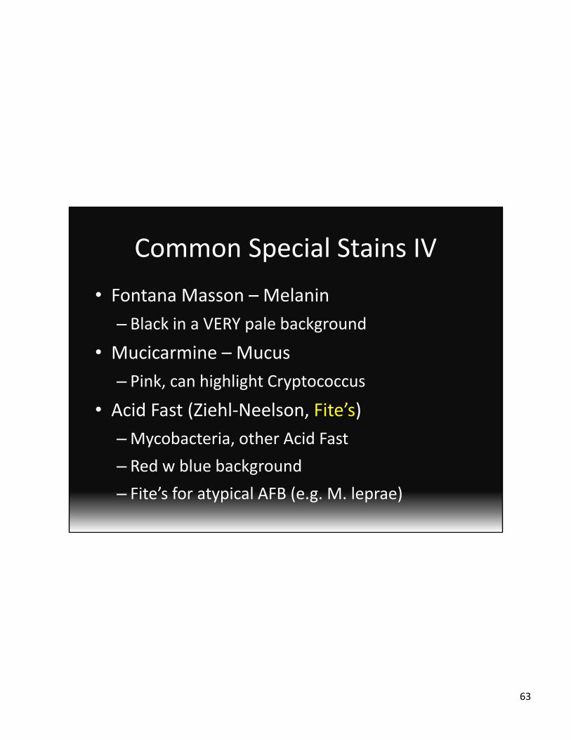
Common Special Stains IV
• Fontana Masson – Melanin
– Black in a VERY pale background
• Mucicarmine – Mucus
– Pink, can highlight Cryptococcus
• Acid Fast (Ziehl‐Neelson, Fite’s)
–Mycobacteria, other Acid Fast
– Red w blue background
– Fite’s for atypical AFB (e.g. M. leprae)

64
Mucicarmine

65
Special Stains V
• Oil Red O, Sudan Black– Lipid, can’t be on processed tissue
–Can do fixed, unprocessed
• Phosphatongstic acid‐hematoxylin
– Fibrin, black
• Vierhoff‐van Gieson– Elastin, black

66
Special Stains VI
• Luxol Fast Blue
–Myelin, blue
• Bodian’s
–Axons, black
• Combos:
– LFB/HE, LFB/Bodians, LFB/PAS

67
Luxol Fast Blue ‐ Myelin