the pathology of ocular trauma - jcahpodocuments.jcahpo.org/atpo/pdf/atpo_handouts/atap1140.pdf ·...
TRANSCRIPT

The Pathology of Ocular Trauma Frank Scribbick, MD
San Antonio, TX
One of the most common reasons for loss of vision, especially in younger patients is ocular trauma. The consequences of trauma can result in no or permanent loss of vision. This lecture will outline the various types of trauma, and the pathological changes that result in vision loss.
Blunt trauma
Scribbick/ATPO/San Francisco 2009 1

Contusions that result in simple corneal abrasions will heal quickly and only leave a scar if bowman’s layer is violated.
Scribbick/ATPO/San Francisco 2009 2
Blunt trauma can also cause changes in the retina called commotion retinea, these changes represent swelling of the retina and almost always resolve.
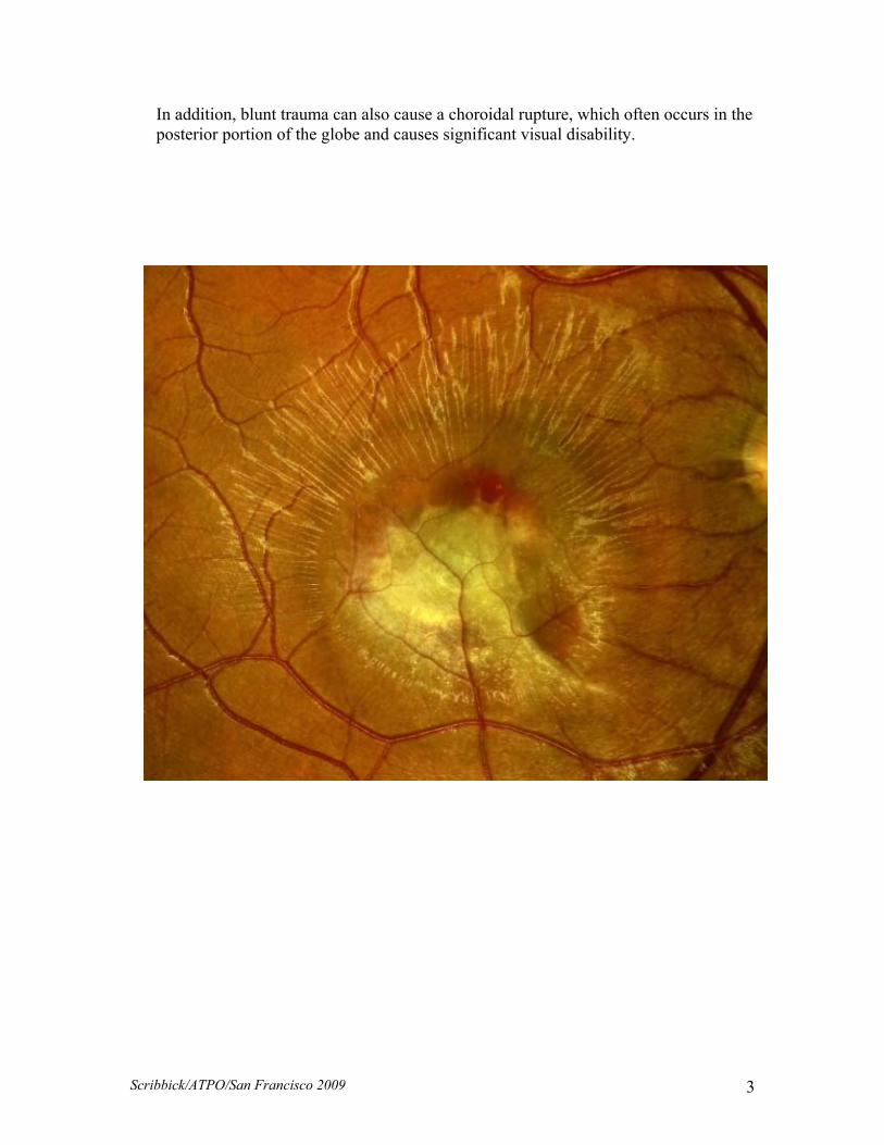
In addition, blunt trauma can also cause a choroidal rupture, which often occurs in the posterior portion of the globe and causes significant visual disability.
Scribbick/ATPO/San Francisco 2009 3

More severe blunt trauma can cause angle recession and bleeding in the anterior chamber. The angle recession is caused by the increase in lateral pressure toward the angle as the globe in compressed in the anterior-posterior direction and expanded in the horizontal direction. This angle recession will result in healing with scarring over the angle and glaucoma later in life. This force can also completely tear the iris from its root or separated the ciliary body from it’s attachment at the scleral spur.
Scribbick/ATPO/San Francisco 2009 4

Blunt trauma severe enough to cause angle recession will always result in a hyphema, or blood in the anterior chamber. The blood in the anterior chamber will resolve on its own, or may need to be surgically removed. In some patients the blood with increased pressure can cause blood staining of the cornea.
Scribbick/ATPO/San Francisco 2009 5

Blunt trauma can also cause a traumatic cataract which often has a “flower or petalloid apperanece” that may or may not be visually significant. If cataract extraction is necessary, care must be taken during the removal because the same trauma may have also weakened the zonules. Blunt trauma can also cause a rupture of the globe, and this usually occurs at the weakest part of the sclera, the limbus and behind the extraocular muscle insertions. These ruptures can also cause disorganization of the intraocular contents, and in some cases loss of the contents through the rupture site.
Scribbick/ATPO/San Francisco 2009 6

Sharp trauma
The eye is subject to various types of sharp trauma. These include knives, pencils, darts, screw drivers, etc. Sharp projectiles like BBs, bullets, arrows and flying metal fragments can also penetrate the globe. Any of the mechanisms that result in globe penetration will cause significant disruption of the intraocular contents.
Scribbick/ATPO/San Francisco 2009 7

Corneal lacerations require immediate repair, and will result in corneal scarring. A corneal transplant may be required after the globe is stable. A penetrating injury that violates the lens capsule will result in an immediate cataract and an intense inflammatory reaction known as phacoantigenic uveitis. Retinal disruption and detachment usually with some degree of intraocular bleeding can also occur after a sharp penetrating injury.
Scribbick/ATPO/San Francisco 2009 8

Intraocular foreign bodies The eye can be penetrated by a variety of projectile foreign bodies. Most often it is metallic. Iron foreign bodies can cause deposition of iron within the epithelium of the intraocular structures. This is called sideroisis. A similar change occurs with intraocular hemorrhage, with the hemosiderin also deposited in the epithelium. Both of these conditions are toxic to the eye, and the intraocular foreign body will need to be removed. Intraocular copper foreign bodies cause a condition called chalcosis, with copper deposited in tissues; this is also toxic to the eye.
Scribbick/ATPO/San Francisco 2009 9

These are pictures of lenses from patients that had an intraocular copper foreign body on the left, and an iron foreign body on the right. T Intraocular vegetable matter can result in a devastating fungal endophthalmitis.
Scribbick/ATPO/San Francisco 2009 10
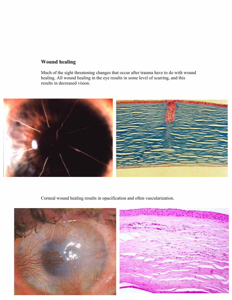
Wound healing Much of the sight threatening changes that occur after trauma have to do with wound healing. All wound healing in the eye results in some level of scarring, and this results in decreased vision.
Scribbick/ATPO/San Francisco 2009 11
Corneal wound healing results in opacification and often vascularization.

Penetrating injuries to the globe, and this includes surgical wounds, that are not properly closed and apposed can result in epithelial down growth.
Scribbick/ATPO/San Francisco 2009 12

Retinal trauma Retinal detachments and tears cans release pigmented and inflammatory cells that can form proliferating membranes that lead to redetachments and retinal distortions.
Scribbick/ATPO/San Francisco 2009 13

Sympathetic ophthalmia Sympathetic ophthalmia is one of the most dreaded complications of open globe trauma. This occurs after certain intraocular antigens are exposed to the immune system after open globe trauma, and the body mounts an immune response to these antigens. The problem is that these antigens are present in the “healthy” non-traumatized eye, and when this happens the body attacks the good eye. This is prevented by removing a traumatized eye within two weeks of the injury if there is no hope of useful sight in the future.
Scri 14bbick/ATPO/San Francisco 2009