ocular pathology case conference
DESCRIPTION
OCULAR PATHOLOGY CASE CONFERENCE. Chirag Patel, M.D. (PGY-2) Hilary Nikols, M.D. (PGY-3) Mark Becher, M.D. Louise Mawn, M.D. October 20, 2008. Case #3. Case #3 (cont’d). Case 3 : Basal Cell Carcinoma. Case 3 : Basal Cell Carcinoma. Case #5. Case #5 (cont’d). Case #5 (cont’d). - PowerPoint PPT PresentationTRANSCRIPT
OCULAR PATHOLOGY OCULAR PATHOLOGY CASE CONFERENCECASE CONFERENCE
Chirag Patel, M.D. (PGY-2)Chirag Patel, M.D. (PGY-2)
Hilary Nikols, M.D. (PGY-3)Hilary Nikols, M.D. (PGY-3)
Mark Becher, M.D.Mark Becher, M.D.
Louise Mawn, M.D.Louise Mawn, M.D.
October 20, 2008October 20, 2008
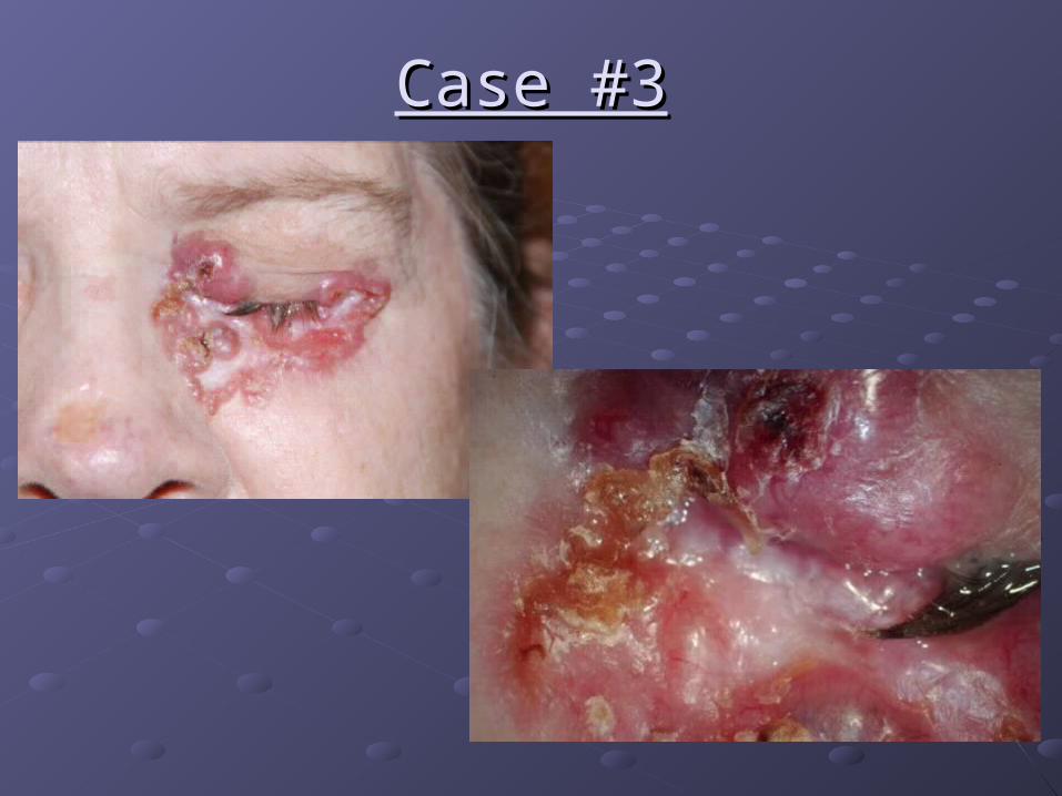
Case #3Case #3

Case #3 (cont’d)Case #3 (cont’d)

Case 3Case 3: Basal Cell Carcinoma: Basal Cell Carcinoma

Case 3Case 3: Basal Cell Carcinoma: Basal Cell Carcinoma

Case #5Case #5

Case #5 (cont’d)Case #5 (cont’d)

Case #5 (cont’d)Case #5 (cont’d)

Case 5Case 5: capillary hemangioma: capillary hemangioma

Case 5Case 5: capillary hemangioma: capillary hemangioma

Case #6Case #6

Case #6 (cont’d)Case #6 (cont’d)

Case #6 (cont’d)Case #6 (cont’d)

Case 6Case 6: vascular malformation, : vascular malformation, possibly arteriovenouspossibly arteriovenous
masson
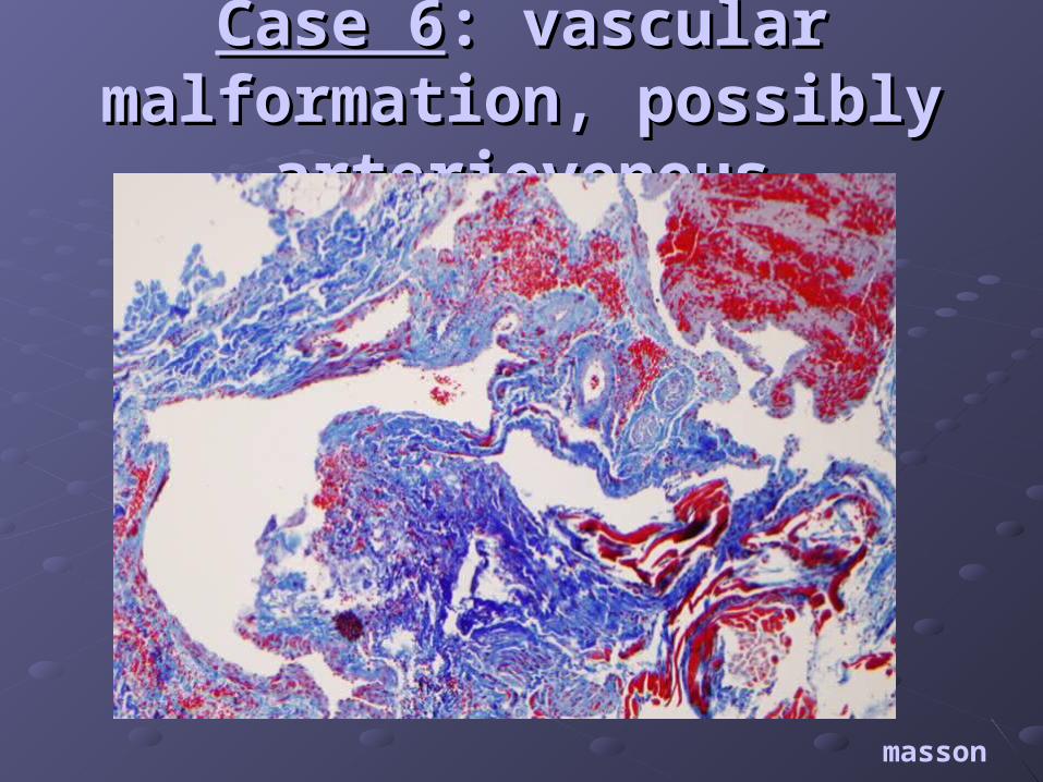
Case 6Case 6: vascular malformation, : vascular malformation, possibly arteriovenouspossibly arteriovenous
masson

Case 6Case 6: vascular malformation, : vascular malformation, possibly arteriovenouspossibly arteriovenous
masson

Case #7Case #7

Case #7 (cont’d)Case #7 (cont’d)
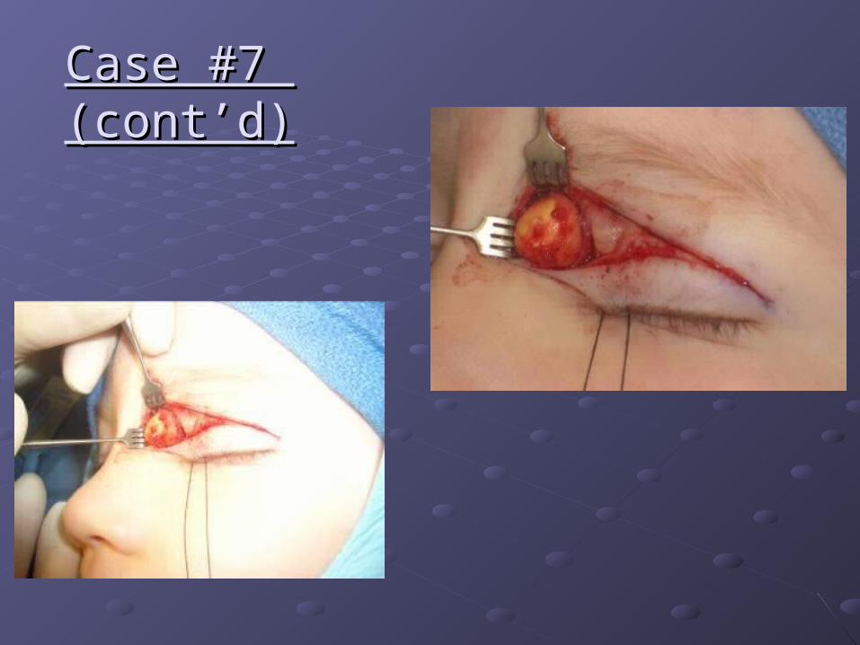
Case #7 Case #7 (cont’d)(cont’d)

Case 7Case 7: dermoid: dermoid
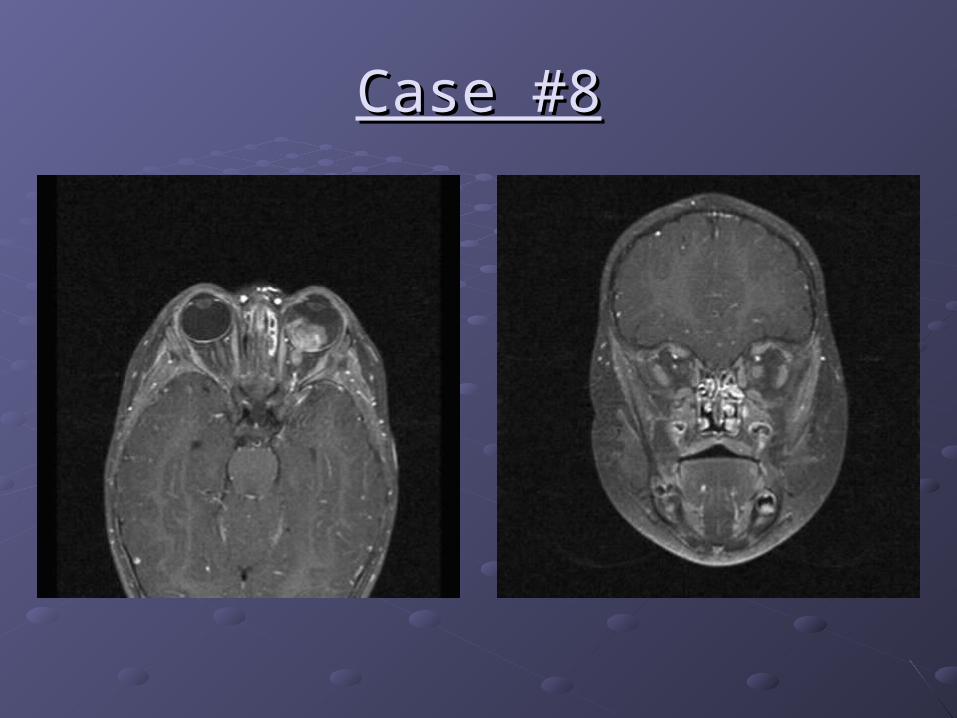
Case #8Case #8

Case 8Case 8: Retinoblastoma: Retinoblastoma
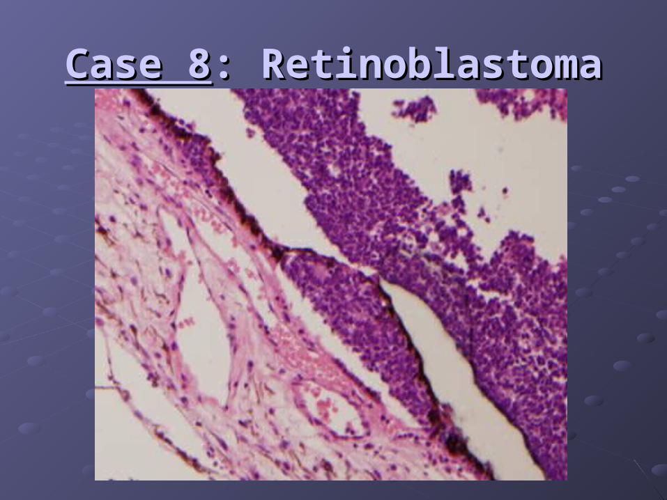
Case 8Case 8: Retinoblastoma: Retinoblastoma
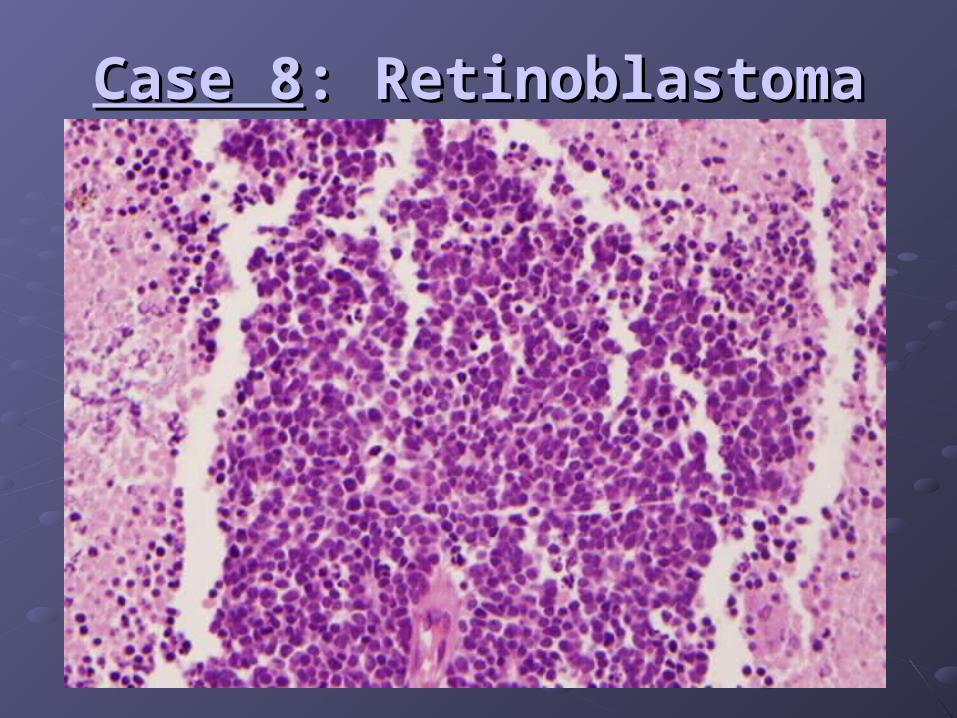
Case 8Case 8: Retinoblastoma: Retinoblastoma

Case 8Case 8: Retinoblastoma: Retinoblastoma
PAS

Case 8Case 8: Retinoblastoma: Retinoblastoma
Deletion of both RB1 tumor suppressor genesDeletion of both RB1 tumor suppressor genesHereditary form 40%Hereditary form 40% Germ cell line mutation; loss of one gene at Germ cell line mutation; loss of one gene at
embryogenesisembryogenesis Usually presents bilaterally and earlyUsually presents bilaterally and early Risk of second malignancyRisk of second malignancy
Non-hereditary form 60%Non-hereditary form 60% Somatic mutation at level of retinal cellSomatic mutation at level of retinal cell Usually unilateral and presents later in childhoodUsually unilateral and presents later in childhood

Case #9Case #9
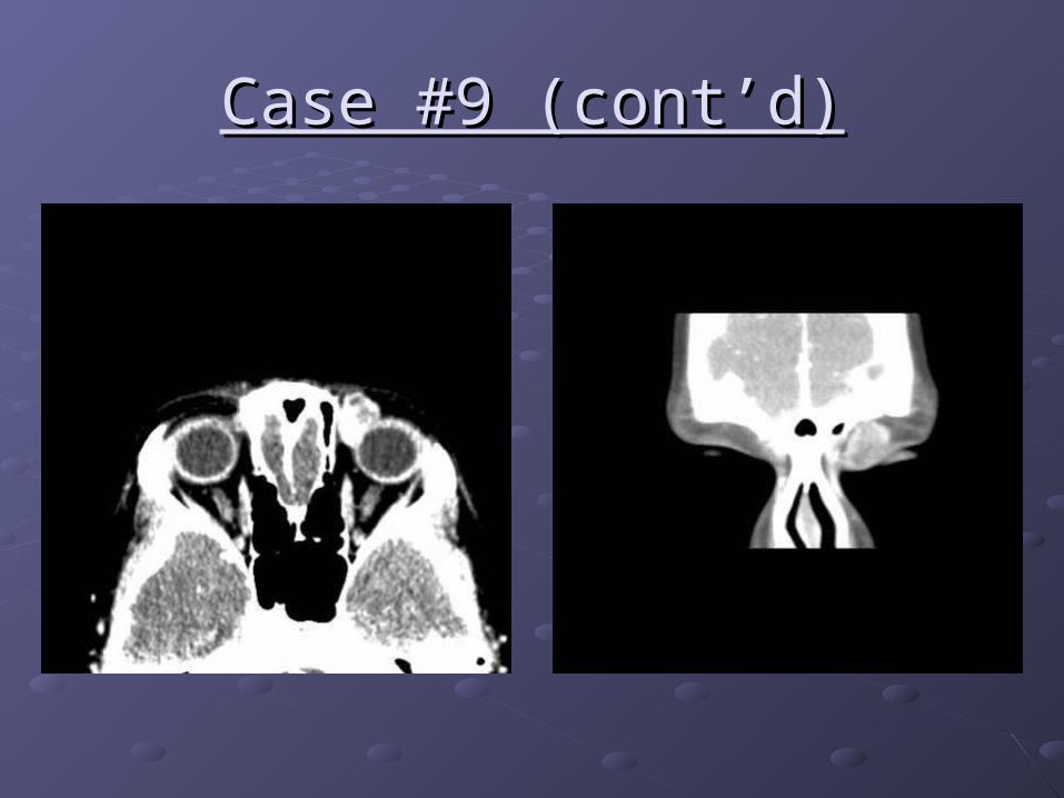
Case #9 (cont’d)Case #9 (cont’d)

Case #9 (cont’d)Case #9 (cont’d)
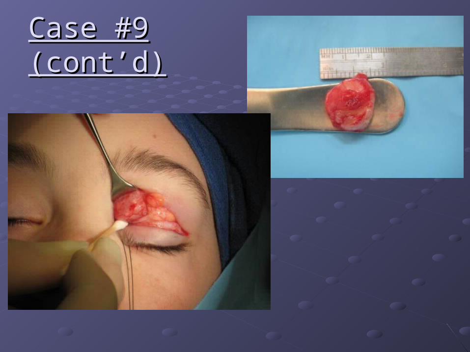
Case #9 (cont’d)Case #9 (cont’d)

Case 9Case 9: Embryonal rhabdomyosarcoma: Embryonal rhabdomyosarcoma

Case 9Case 9: Embryonal rhabdomyosarcoma: Embryonal rhabdomyosarcoma

Case 9Case 9: embryonal rhabdomyosarcoma: embryonal rhabdomyosarcoma
desmin myoD1 myogenin

Case #10Case #10

Case #10 (cont’d)Case #10 (cont’d)

Case #10 (cont’d)Case #10 (cont’d)
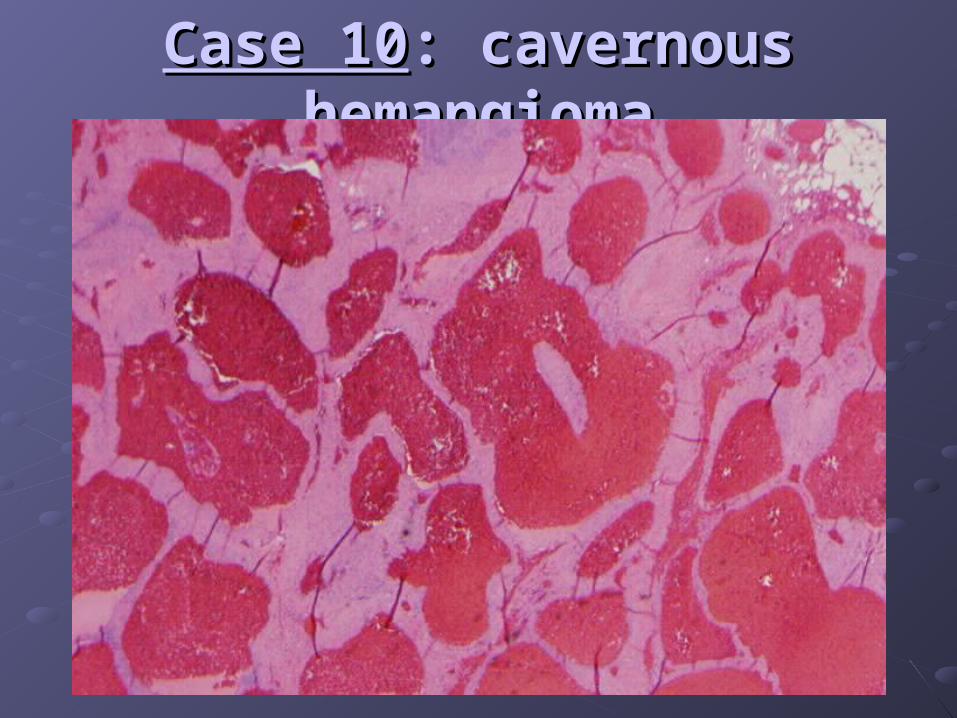
Case 10Case 10: cavernous hemangioma: cavernous hemangioma
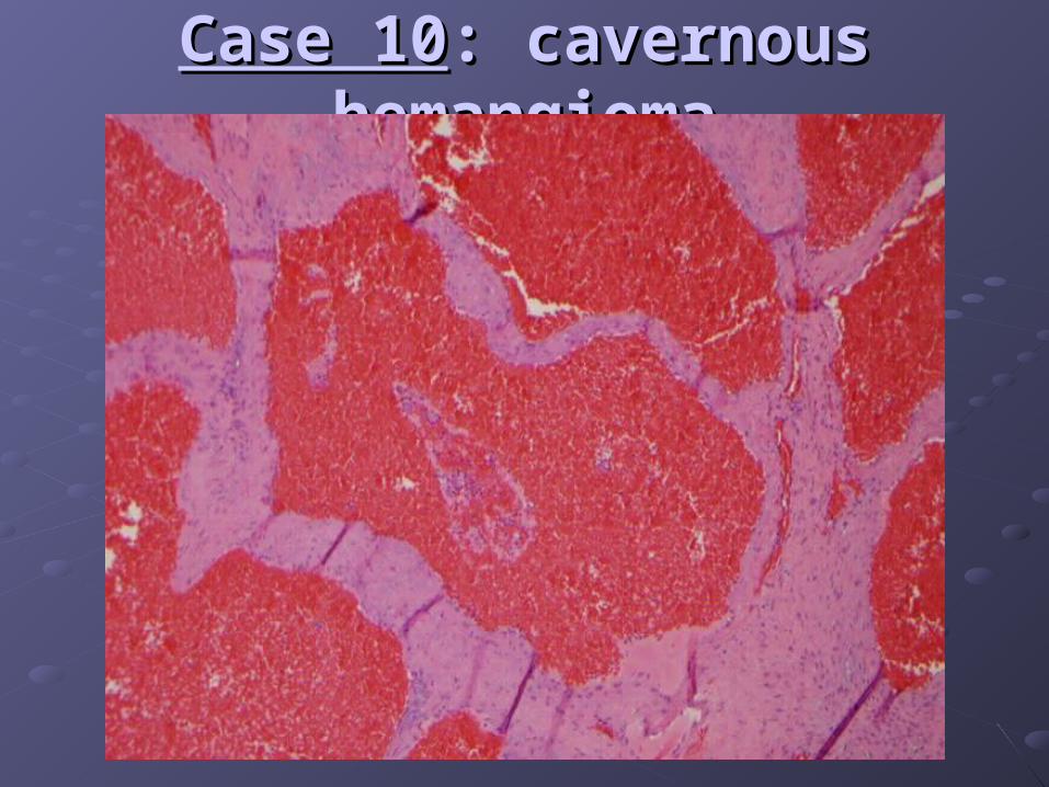
Case 10Case 10: cavernous hemangioma: cavernous hemangioma