of phytoplankton pigments using temperature-controlled ... · extraction solvents, acetone and...
TRANSCRIPT

Vol. 114: 303-313, 1994 MARINE ECOLOGY PROGRESS SERIES
Mar. Ecol. Prog. Ser. Published November 17
Improved separations of phytoplankton pigments using temperature-controlled high performance
liquid chromatography
Laurie Van ~eukelem', Alan J. L e w i t u s l . * , Todd M. ~ana ' , Neal E. craft2
'Horn Point Environmental Laboratory, PO Box 775. Cambridge. Maryland 21613. USA 'Southern Testing and Research Labs, Inc., 3709 Airport Dr., Wilson, North Carolina 27893. USA
ABSTRACT: Varying the temperature of a polymeric C18 high performance liquid chromatography [HPLC) column caused significant changes in elution profiles of carotenoid and chlorophyll (chl) pig- ment mixtures. High temperature operation (60°C) was optimal for carotenoid separations, including the separation of zeaxanthin from lutein. Chlorophyll and chlorophyll derivative separations were opti- mal at a column temperature between 10 and 30°C. A protocol is presented that achieves quantifiable resolution of all taxonomically important pigments tested by running the analysis at 2 temperatures: 10 and 60°C. Of particular significance was the ability to separate divinyl chl a from monovinyl chl a, chl c, from chl c,, and partial separation of chl c, and MG 2,4-divinyl phaeoporphorin a, monomethyl ester. An example is given of the utility in separating chl c pigments, and lutein and zeaxanthin in natural phytoplankton samples.
KEY WORDS: Pigments . High performance liquid chromatography (HPLC) . Polymeric column . Tem- perature . Phytoplankton . Divinyl chlorophyll a . Chlorophyll c . Carotenoids . Lutein . Zeaxanthin
INTRODUCTION
As the utility of high performance liquid chromatog- raphy (HPLC) in characterizing phytoplankton pig- ments becomes increasingly recognized, the need to establish HPLC protocols that are efficient and broadly applicable becomes more immediate. Advances in pig- ment separations by HPLC have centered on reversed phase protocols, largely because the commonly used extraction solvents, acetone and methanol, are incom- patible with the non-water-miscible mobile phase used in normal phase HPLC. Recently, significant advances have been made in the phytoplankton pigment separa- tion capability of reversed phase techniques which involve modifications of column type and/or mobile phase composition (e.g. Kleppel et al. 1988, Kohata et
'Present address: Baruch Marine Field Laboratory/USC, PO Box 1630, Georgetown, South Carolina 29442, USA
al. 1991, Wright et al. 1991, Kraay et al. 1992, Van Heukelem et al. 1992, Goericke & Repeta 1993). Despite these advances, no one protocol has been developed that can resolve all of the chlorophylls and carotenoids of chemotaxonomic significance to phyto- plankton identification, as defined by Wright et al. (1991) and Millie et al. (1993).
One parameter rarely considered a variable in phytoplankton pigment HPLC analysis is column tem- perature. Although the importance of maintaining isothermal conditions for optimizing precision in HPLC is recognized, the role of temperature in separation selectivity for phytoplankton pigments has been largely unevaluated. The reason for this may be 2-fold. Firstly, column compartments capable of precisely regulating temperature (i.e. with combined heating and cooling capabilities) are not standard items with most HPLC systems used in phytoplankton pigment research. Secondly, separation selectivity is weakly affected by temperature variation above 25°C when
0 Inter-Research 1994 Resale of full article not permitted

304 Mar. Ecol. Prog. S er. 114: 303-313. 1994
the popular monomeric CIR bonded phase columns are used (Sander & Wise 1990). On the other hand, Sander & Wise (1990) demonstrated that temperature could affect the separation selectivity of a polymeric Cl* col- umn between temperatures of -20 and 80°C. Craft et al. (1992) subsequently demonstrated the utility of polymeric column temperature control in manipulating separation selectivity of blood serum and food carotenoids. Therefore, a polymeric C18 column used in combination with an HPLC system that can pre- cisely regulate the column temperature at subambient and/or above-ambient temperatures provides flexibil- ity in manipulating separation selectivity independent of modifications in mobile phase composition or col- umn type.
In a previous report (Van Heukelem et al. 1992), we demonstrated that the separation of several key pig- ments could be markedly improved by the relatively simple change from a monomeric to a polymeric CI8 bonded phase column. However, the protocol, which used a setpoint temperature of 40°C, could not quan- tifiably resolve some important pigment pairs, e.g. chlorophyllide a and chl c3, diatoxanthin and lutein, monovinyl chl a and divinyl chl a. Here, we evaluate the effect of column temperature on the separation selectivity of a Vydac 201TP polymeric column for these and several other phytoplankton pigments of chemotaxonomic importance. We demonstrate that resolution of all pigments tested can be achieved by alternating between 2 column temperatures; 10 and 60 "C. This is the first method capable of resolving all of the following pigment pairs using the same column and mobile phase; chlorophylls cl and c?, lutein and zeaxanthin, and monovinyl chl a and divinyl chl a. It is also the first time that monovinyl chl a and divinyl chl a have been separated using a CI8 bonded reversed phase column.
MATERIALS AND METHODS
Sources of reference pigments. Reference pigments were extracted from algal monocultures obtained from the Provasoli-Guillard Center for Culture of Marine Phytoplankton, BigeIow Laboratory for Ocean Sci- ences ('CCMP'), or the Horn Polnt Environmental Lab- oratory, University of Maryland ('HP') culture collec- tion. The phytoplankton species included DunaLiella tertiolecta CCMP (Chlorophyceae), Emiliania huxleyi CCMP373 (Prymnesiophyceae], Pelagococcus sub- vindis CCMP1429 (Chrysophyceae), Pycnococcus provasolii CCMP1203 (Prasinophyceae), Pyrenomonas salina (formerly Chroomonas salina) CCMP3C (Cryp- tophyceae), Gyrodinium uncatenum HP8901 (Dino- phyceae), Phaeodactylum sp. HP9101 (Bacillario-
phyceae), Prochlorococcus marinus CCMP1375 (Pro- chlorophyceae), and Tetraselmis suecica (Prasinophy- ceae). The first 5 species above were chosen based on the recommendations of Wright et al. (1991); see their Table 1. The latter 4 species were selected because they contained the remaining pigments of diagnostic importance, and monocultures of these species were being maintained at Horn Point Environmental Labo- ratory at the time of the study. Astaxanthin was extracted from shrimp carapace obtained from a local grocery store. Canthaxanthin (Hoffman La Roche) was donated by Bruce Malone of Perdue Incorporated, Salisbury, MD, USA.
Field sampling. Water samples were obtained from a freshwater Florida lake (Lake Annie, Highlands County) on March 9, 1993. The samples were collected from the euphotic layer using Nislun bottles. For HPLC pigment analysis, aliquots (1.4 to 2.7 1) were filtered through a Whatman GF/C glass fiber filter or pre- filtered in series through 5 pm and 2 pm Nuclepore fil- ters, and then through a GF/F glass fiber filter. The glass fiber filters were folded in half, wrapped in aluminum foil, and stored first in liquid nitrogen, then in a freezer at -80°C. The sample was extracted (see below) and analyzed at column temperatures of 10, 30, and 60 "C. Aliquots were also taken for phytoplankton species identification and enumeration by light and epifluorescent microscopy. Some samples were fixed with either Bouin's or acid Lugol's solution, concen- trated by settling chamber, and examined by light microscopy. Other samples were stained with primulin (Caron 1983), filtered through an Irgalan black-stained 3.0 pm Nuclepore filter, and examined by epifluores- cence microscopy. The 3.0 pm Nuclepore filtrate was filtered through an Irgalan black-stained 0.2 pm Nuclepore filter in order to identify and quantify pico- phytoplankton.
Pigment extraction. Samples from algal monocul- tures were filtered onto either Whatman GF/F or GF/C filters, folded, wrapped in aluminum foil, and stored at -80°C. Filters were torn into pieces and ground in cold 100% acetone in a dimly lit room, after which the extract was held in a refrigerator (4 "C) for 20 min prior to filtration. Homogenates were clarified using an HPLC syringe cartridge filter (GeIman Acrodisc 13 CR PTFE). The algal extracts were transferred to amber crimp-cap vials (2 ml), and placed in the autosampler compartment held at 2 to 3 "C until injection. Astaxan- thln was prepared by grinding raw shrimp carapace in 100% acetone (G. Kleppel pers, comm.). To prepare the chlorophyllide-rich extract, a dense culture of Phaeodactylum sp. was collected on a GF/C filter and ground in cold acetone : water (50: 50), following Jef- frey & Hallegraeff (1987). The extract was then acidi- fied with 10% HCl to produce acidic chlorophyll

Van Heukelem et al.: Improved method for phytoplankton pigment separation 305
degradation products. The acid was neutralized by the addition of 1.0 M ammonium acetate during the injec- tion cycle (R. Goericke pers. comm.).
High performance liquid chromatography. The HPLC was a fully automated Hewlett Packard Series 11, 1090 M with binary DR5 solvent delivery system, a built-in diode array detector (DAD) with 8 p1 flow cell, a temperature-controlled autosampler compartment and column oven compartment, and a programmable autoinjector (outfitted with a 250 p1 loop) capable of autoaddition and mixing. The autosampler compart- ment and column oven compartment were cooled with an RTE-100 recirculating refrigerated bath (Neslab). A Hewlett Packard 1046A, time-programmable fluores- cence detector was installed after the DAD. The exci- tation (ex) and emission (em) wavelength setpoints were programmed for target analytes in the injected sample. Peak response was either enhanced or sup- pressed by selecting wavelengths specific to the pig- ments of interest (e.g. 440 nm ex, 650 nm em for chlorophylls c, and c2, or 425 nm ex, 670 nm em for monovinyl chl a and its degradation products). All data were collected with a HP 79994A Pascal-based Chem- Station. The reversed phase C,, column was a Vydac 201TP, 300 A pore size, 5 pm particle size, 4.6 mm ID X
250 mm (Separations Group, Hesparia, California). The binary solvent system of Mantoura & Llewellyn
(1983) was used with the exception that tetrabutyl- ammonium acetate was omitted from Solvent A (Zap- ata et al. 1987). The solvents were formulated as fol- lows: solvent A (methanol:0.5 M ammonium acetate, 80 : 20); solvent B (methanol: acetone, 80 : 20). Pumping
Table 1. Exan~ples of HPLC pumping gradients used at each column temperature. Solvent A was methanol:0.5 M amrno- nium acetate, 80:20; Solvent B was methanol:acetone, 80:20
Temp. Time Flow rate % A % B ("C) (mln) (m1 nun-')
60 0.0 1.0 100 0 5.0 1.0 60 4 0
10.0 1.0 30 7 0 13.0 1 .O 30 70 23.0 1 .O 0 100 24.0 1.5 0 100
30 0.0 1 .O 100 0 10.0 1.5 0 100 17.0 1.5 0 100
10 0.0 1.0 100 0 9.5 1.0 50 50
17.0 1.0 25 75 22.0 1.5 15 85 27.0 2.0 0 100 31.5 2.0 0 100
gradients varied with column temperature, and several gradient protocols were used to achieve the desired resolution (Table 1). Using a programmable auto- injector, the acetone extracts were diluted with ammo- nium acetate immediately prior to injection; Wright et al. (1991) caution that the timing of the dilution and resultant extract polarity will affect hydrophobic pig- ment solubility, and advise that dilutions be conducted immediately prior to injection in order to minimize pre- cipitation. It is possible that the concentration of water in the extract varied between samples (Bidigare 1991), because filter specifications were not consistent throughout the study (25 or 47 mm diameter GF/C or GF/F filters), although 100 % acetone was consistently used as the extraction solvent (to minimize activation of chlorophyllase; Jeffrey & Hallegraeff 1987). The autoinjector was programmed to draw 63 p1 of sample, draw 37 p1 of 1.0 M ammonium acetate, mix, wait 5 min, and inject. All samples, including the ammo- nium acetate, were held in the temperature-controlled autosampler compartment at 2 to 3 'C. HPLC analyses were conducted at temperatures of 10, 15, 20, 25, 30, 35, 40, 50, and 60°C. Column temperature varied by <+0.1 "C from the setpoint.
Pigment identification. The identification of pig- ments in algal cultures was based on a comparison of our observed in-line DAD spectra (350 to 600 nm) and %III/II ratios' (Ke et al. 1970) with published values (Kohata et al. 1991, Wnght et al. 1991) (Table 2). Selected pigments were individually fraction- collected, transferred to 100 % acetone, and character- ized in terms of absorbance spectra (350 to 750 nm, Hitachi 3110 scanning spectrophotometer). The %III/I and II/I ratios of these pigments were compared to previously published values (Jeffrey & Wright 1987, Wright & Jeffrey 1987, Kraay et al. 1992).
Resolution. Resolution between pigments was quan- tified based on retention times and peak widths, fol- lowing Kohata et al. (1991) and as described in Van Heukelem et al. (1992).
RESULTS
The effect of column temperature on the resolution of phytoplankton pigments was evaluated in 3 ways.
%III/II (as defined by Ke et al. 1970) is calculated on 3- fingered carotenoid and xanthophyll spectra, where 111 is the absorbance maximum of the peak of highest wavelength and I1 is the absorbance maximum of the middle finger. The mini- mum absorbance value between peaks I1 and I11 (the trough) subtracted from each absorbance peak and the (111-trough)/ (11-trough) value is calculated and expressed as a percentage

306 Mar. Ecol. Prog. Ser. 114: 303-313, 1994
Table 2. Pigments from standard cultures showing in-line DAD spectra (wavelength range 350 to 600 nm) visible absorption max- ima and peak ratios in HPLC eluant and 100% acetone, and retention times (rnin) from coinjections of selected algal cultures at 10 or 60°C. Range of observed absorption maxima IS presented In the cases where spectra varied among injections or specles. Wavelengths of absorption shoulders are given in parentheses Peak ratios are expressed in decimal form as the ratio of Soret band to red region maxima (for chlorophylls) or in percentage form as the ratio of tertiary to secondary (III/II) absorbance peaks
(for carotenoids), following Ke et al. (1970). NA: not available
No. Pigment
1 C h l q 2 Chloropbyllide a 3 Chl cl
6 Peridinin 7 19'-butanoyloxyfucox 8 Fucoxanthln 9 19'-hexanoyloxyfucox.
10 Neoxanthin 11 Prasinoxanthin 12 Violaxanthln 13 Unknown carotenoid 14 Astaxanthin-like 14a Astaxanthin 15 Diadin.oxanthin 16 Alloxanthln 17 Unknown carotenoid 18 Diatoxanthin 19 Lutein 20 Zeaxanthln 21 Canthaxanthin 22 Chl b 23 Divinyl chl b 22 Crocoxanthin 25 Chl a allomer 26 Chl a 27 Divinyl chl a 28 Chl a-like 29 Non-polar chl c-like 30 Non-polar chl c-like 31 a-Carotene 32 p-Carotene 33 Phaeophorbide a
E, F, I D H K All but D D K A A D. H, L B M
Eluant - Absorption maxima (nm)
457, 591, NA 431, NA, NA 440-441,583, NA
439, NA, NA 439 445-447. 583
471 445, 467 451, (465) 447, 469 413, 435-437, 465 455 415, 439,469 417, 441, 469 475 473-479 (4251, 445, 475 (4311, 451, 481 (425), 445, 475 (4311, 451. 479 (4241, 445-447,473 (429), 451, 477 477 467 477 (425), 445, 475 425, NA, NA 431, NA, NA 441, NA, NA 431 457, 587 457, 587 (422), 445, 473-475 (430), 451, 477 (407), 507, 537
Peak ratio
Acetone - Absorption Peak maxima (nm) ratio
Retention time (mm)
dSources: (A) Ern~liania huxleyi, (B) Gyrodinium uncatenum, (C) Phaeodactylum sp., (D) Prochlorococcus marinus, (E) DunalieUa tertiolecta, (F) Pycnococcus provasolii. (G) Pelagococcus subvirid~s. (H) Pyrenornonas salina, (I ) Tetraselmis succica, (J) shrimp carapace, (K) Phaeodactylurn extracted In 50.50 acetone:water for chlorophyllide a, (L) Pluka Chemika- BioChem.ika, (M) source 'K' acidified with 10% HCI for phaeophorbide a
First, resolution of pigments from algal monocultures was tested at several temperatures. From these results, temperatures of 10, 30, and 60°C were selected for fur- ther analyses because the separation selectivity was widely disparate at these temperatures, and separation of closely eluting pigments of diagnostic importance could be achieved at each temperature. Secondly, monoculture extracts were combined such that pig-
ments were CO-injected in similar concentrations. HPLC analysis on various combinations of pigment mixtures was conducted at 10, 30, or 60 "C, and resolu- tion of pigments was determined from the chromato- grams. Finally, HPLC analyses at 10, 30, and 60°C were conducted on lake samples in order to examine the effectiveness of temperature manipulations in resolving pigments from natural waters.
Van Heukelem et al .- Improved method for phytoplankton pigment separation 307
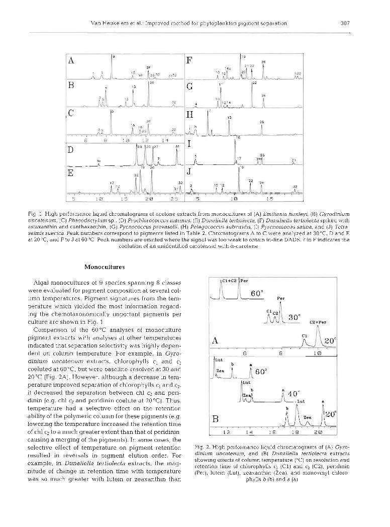
Fig. 1 High performance liquid chromatograms of acetone extracts from monocultures of (A) Emillanla huxleyi, (B) Gyrodlnium uncatenum, (C) Phaeodactylum sp., (D) Prochlorococcus marinus, (E) Dunaliella tertlolecta, (F) Dunaliella tertiolecta spiked with astaxanthin and canthaxanthin, (G) Pycnococcus provasolii, (H) Pelagococcus subv~ridis, ( I ) Pyrenonlonas sahna, and (J) Teti-a- selrnis suecjca Peak numbers correspond to pigments listed in Table 2. Chromatograms A to C were analyzed at 30°C, D and E at 20°C, and F to J at 60°C. Peak numbers are omitted where the signal was too weak to obtaln in-llne DADS ? in F indicates the
coelution of an unidentified carotenold wlth a-carotene
Monocultures
Algal monocultures of 9 species spanning 8 classes were evaluated for plgment composition at several col- umn temperatures. Pigment signatures from the tem- perature which yielded the most information regard- ing the chemotaxonomically important pigments per culture are shown in Flg. 1
Comparison of the 60°C analyses of monoculture pigment extracts with analyses at other temperatures indicated that separation selectivity was highly depen- dent on column temperature. For example, in Gyro- dinium uncatenum extracts, chlorophylls c, and c2 coeluted at 60°C, but were baseline-resolved at 30 and 20 "C (Fig. 2A). However, although a decrease in tem- perature improved separation of chlorophylls c, and c2, it decreased the separation between chl c2 and peri- dinin (e.g. chl c2 and peridinin coelute at 20 "C). Thus, temperature had a selective effect on the retention ability of the polymeric column for these pigments (e.g. lowering the temperature increased the retention time of chl c:, to a much greater extent than that of peridinin, causing a merging of the pigments). In some cases, the selective effect of temperature on pigment retention resulted in reversals in pigment elution order. For example, in Dunaliella tertiolecta extracts, the mag- nitude of change in retention time with temperature was so much greater with lutein or zeaxanthin than
1 J Per
Fig. 2. High performance liquid chromatograms of (A) Gyro- dlnium uncatenum, and (B) Dunaliella tertiolecta extracts showing effects of column temperature ("C) on resolution and retention time of chlorophylls c, ( C l ) and c2 (C2), peridinin (Per), lutein (Lut), zeaxanthin (Zea), and monovinyl chloro-
phylls b (b) and a (a)

308 Mar. Ecol. Prog. Ser 114: 303-313, 1994
C 5
22.23
Fluorescence 440 ex, 650 e m
l o o 2
P. L B 10 1 2 14 16 I B 2 0 2 2 2 4
Fig. 3. High performance liquid chromatograms of acetone-extracted pigment mixtures injected at 60 or 10°C. Peak numbers refer to pigments listed in Table 2. (A) Absorbance (452 nm) signal at 60°C of combined extracts from Emiliania huxleyi, Pycno- coccus provasolii, Tetraselmis suecica, Gyrodinium uncatenum, Pelagococcus subviridis, Phaeodactylum sp., Pyrenomonas salina, and Prochlorococcus marinus. (B) Fluorescence (425 nm excitation, 670 nm emission) signal at 60 'C of (1) Phaeodactylum sp. extracted in 50:50 acetone:water to induce production of chlorophyllide a, (2) the above Phaeodactylurn sp. extract acidified for phaeophorbide a production, and (3) Isochrysissp. extract containing pigments common to both of the above extracts. (C) Flu- orescence (440 nm excitation, 650 nm emission) signal at 10°C of combined extracts from Emiliania huxleyi, Pycnococcusprova- solii, Gyrodinium uncatenum, and Prochlorococcus marinus. Inset shows the partial separation of monovinyl chl b and divinyl
chl b when both pigments are present in similar amounts
with monovinyl chl b that the elution order of the carotenoids and monovinyl chl b were reversed between 40 and 20 "C (Fig. 2B).
Mixed algal extracts
In HPLC analyses of mixed algal extracts run at 60 "C, the resolution between all chemotaxonomically important carotenoids was 21.0 (Fig. 3A, Table 31, indi- cating that, when CO-occurring, these pigments are identifiable and quantifiable at this temperature. Included are diatoxanthin and lutein, which coeluted at 40°C (Van Heukelem et al. 1992). It should be noted, however, that an unidentified carotenoid coeluted with a-carotene in extracts of Tetraselmis sueica and Dunaliella tertiolecta at 60°C (Fig. IF), but not at 30°C (data not shown). Phaeophorbide a can be resolved at 60°C when fluorescence detection is used (Fig. 3B). Limitations of the chlorophyll analysis at 60°C include the inability to separate (1) chl c,,, chl c2, and MG 2,4- divinyl phaeoporphorin a5 monomethyl ester (MG 2,4D), (2) monovinyl chl b and divinyl chl b, or (3) monovinyl chl a and divinyl chl a. Also, chl c3 coeluted
with an unidentified carotenoid (from Gyrodinium uncatenum) in the mixed extract at 60 "C.
Three chlorophyllous pigment groups that were not completely separated at 60°C were baseline or quan- tifiably resolved at 10°C: (1) chl c3 and chlorophyllide a, (2) chl c, and chl c2, and (3) monovinyl chl a and divinyl chl a (Figs. 3C & 4 , Table 3). In addition, 2 groups that coeluted at 60°C were partially resolved at 10°C: (1) monovinyl chl b and divinyl chl b, and (2) MG 2,4D and chl c2 (Figs. 3C plus inset & 4B, Table 3) . Divinyl chl a is resolvable at this temperature even when present in disproportionately low concen- trations (e.g. resolution between monovinyl chl a and divinyl chl a in mixed algal extracts was 1.0; Figs. 3C & 4A, Table 3). Furthermore, although several caro- tenoids coeluted at 10 "C, in-line DAD spectra revealed that monovinyl chl a and divinyl chl a were resolved and not effected by the presence of other pigments (data not shown). Monovinyl chl b and d~vinyl chl b were partially resolved at 10 "C, and this resolution depended on the presence of proportionately similar pigment concentrations (Fig. 3C plus inset).
The partial resolution of chl c2 and MG 2,4D at 10 "C is limited by the potential interference of early-eluting

Van Heukelem et al.: Improved method for phytoplankton pigment separation 309
Table 3. Effect of column temperature on resolution of various pigment pairs. Resolution (R) was measured based on peak height, and is expressed as no resolution (-; R = 0), partial resolution (+; 0 R < 1.0), quantifiable resolution (++; 1.0 < R < 1.5), or base-
line resolution (+++, R > 1.5). 19'-but: 19'- butanoyloxyfucoxanthin; 19'-hex: 19'-hexanoyloxyfucoxanthin
Pigment pair Column temperature ("C)
10 15 20 25 30 35 40
Chl c2/peridinin Chl cz/fucoxanthin Peridinin/l9'-but Fucoxanthln/19'-hex 19'-hex/neoxanthin Neoxanthin/prasinoxanthin Alloxanthin/lutein Diatoxanthin/lutein Luteinheaxanthin Lutein/chl b Zeaxanthin/chl b a-Carotene/p-carotene Crocoxanthin/chl a Chl c3/chlorophylhde a Chl c,/chl c2 MG 2,4D/phaeophorbide a MG 2,4D/chl c2 Chl b/divinyl chl b Chl a/divinyl chl a
Fluorescence
Fluorescence C1
Fig. 4. High performance liquid chromatograms with fluores- cence detection (440 nm excitation, 650 nm emission) of com- bined extracts from Prochlorococcus marinus, Gyrodinium uncatenum, Emiliania huxleyi, and Pycnococcus provasolij showing effects of column temperature ('C) on separation of (A) monovinyl chl a (a) and divinyl chl a (divinyl a ) , and (B) MG 2,4D (MG) and chlorophylls c, ( C l ) , c2 (C2), and c, (C3)
carotenoids with chl c2 (e.g. fucoxanthin or peridinin; Table 3). In the presence of these carotenoids, fluoro- metric detection must be used to selectively detect chl c2 and MG 2,4D at temperatures 525°C (e.g. Figs. 3C & 4B). However, an additional problem is the potential coelution of phaeophorbide a and MG 2,4D at 10 "C (Table 3), and it is necessary to use fluorescence detector wavelength suppression to discriminate be- tween these pigments (see 'Natural samples' section).
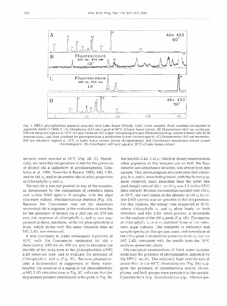
Natural samples
The utility of column temperature adjustments in determining the pigment composition of natural phyto- plankton populations is illustrated in Fig. 5. The pres- ence of taxonomically important carotenoids (e.g. zeaxanthin, fucoxanthin, violaxanthin, lutein, diadi- noxanthin, and neoxanthin) and monovinyl chl b are indicated in the 60°C chromatogram (Fig. 5A). Peak identities were confirmed by pooling extracts of the same size fraction, concentrating by solid phase extraction, reinjecting at 60 "C, and comparing in-line DAD spectra and retention times with pigments from algal reference cultures. Analysis at this temperature could not discriminate between chl c,, chl c2, and MG 2,4D.
The chlorophyllous pigments in Lake Annie samples were further detailed at lower temperatures. First, all
310 Mar. Ecol. Prog. Ser. 114: 303-313, 1994
20 Abs 452
6 0 ' $ 5
32
26 Fluorescence
425 ex. 670 e m
c Fluorescence 440 ex. 650 em
1 o 0 -
Fig. 5. HPLC phytoplankton pigment analyses from Lake Annie (Florida, USA) water samples. Peak numbers correspond to pigments listed in Table 2. (A) Absorbance (452 nrn) signal at 60°C of Lake Annle extract. (B) Fluorescence (425 nrn excitation, 670 nrn emission) signals at 10°C of Lake Annie extract (upper chrornatogram) and Phaeodactylum sp. extract treated with 50:50 acetone:water, and then acidified for phaeophorb~de a production (lower chrornatogram). (C) Fluorescence (440 nrn excltation, 650 nrn emission) signals at 10°C of Lake Annie extract (lower chrornatogram) and Gyrodinjum uncafenum extract (upper
chromatograrn). ( D ) Absorbance (452 nm) signal at 30°C of Lake Annie extract
extracts were injected a t 10°C (Fig. 5B. C). Specifi- cally, we used this temperature to test for the presence of divinyl chl a (indicative of prochlorophytes; Chis- holm et al. 1988, Goericke & Repeta 1992), MG 2,4D, and/or chl c,, and to determine the relative proportion of chlorophylls c, and c2.
Divinyl chl a was not present in any of the samples, as determined by the comparison of retention times and in-line DAD spectra of samples with the algal reference culture, Prochlorococcus marinus (Fig. 5 B ) . Because the fluorometer was set for maximum monovinyl chl a response in the evaluation of samples for the presence of divinyl chl a (425 nm ex, 670 nm em), the response of chlorophylls cl and c2 was sup- pressed in these injections, while the phaeophorbide a peak, which elutes with the same retention time as MG 2,4D, was enhanced.
It was necessary to make subsequent injections at 10°C with the fluorometer optimized for chl c detectability (440 nm ex, 650 nm em) to elucidate the identity of the peak seen at the phaeophorbide aIMG 2,4D retention time, and to evaluate the presence of chlorophylls c, and c2 (Fig. 5C). Because phaeophor- bide a fluorescence is suppressed at these wave- lengths, the absence of a signal a t the phaeophorbide a/MG 2,4D retention time in Fig. 5C indicates that the degradation product contributed to the peak in Fig. 5B.
but not MG 2,4D. Chl c3, which is clearly resolved from other pigments at this temperature on both the fluo- rometer and absorbance detector, was absent from this sample. This chromatogram also indicated that chloro- phylls cl and c2 were both present, with the former pig- ment relatively more abundant than the latter (the peak height ratio of chl c, to chl c2 was 3.2 in this GF/C filter extract). Because fucoxanthin coeluted with chl c2 at 10°C, the verification of the identity of chl c2 by in- line DAD spectra was not possible at this temperature. For this purpose, the extract was reinjected at 3 0 ° C where chlorophylls c, and c2 elute freely on both detectors, and MG 2,4D, when present, is detectable on the upslope of the chl c2 peak (Fig. 5D). The spectra of chlorophylls c, and c2 matched those of the refer- ence algal cultures. The similarity of reference and sample spectra on the upslope, apex, and downslope of the chl c2 peak indicated the presence of chl c2, but not M G 2,4D, consistent with the results from the 10°C analyses presented above.
Microscopical examinations of fixed water samples confirmed the presence of phytoplankton indicated by the HPLC results. The relatively high contribution of zeaxanthin in the 60°C chromatogram (Fig. 5A) sug- gests the presence of cyanobacteria and/or chloro- phytes, and both groups were prevalent in the sample. Cyanobacteria (e.g. Synechococcus spp., Merismope-

Van Heukelem et al.: Improved method for phytoplankton pigment separation 311
dia spp.) were nlalor constituents of the phytoplankton, comprising 28% of the total phototrophic biomass of the sample, while chlorophytes (e.g. Selenastrurn sp., Staurastrum spp.) were also predominant components. The presence of chlorophytes also is consistent with the relatively high peaks in lutein and monovinyl chl b.
The relatively strong fucoxanthin signal in the 60°C chromatogram could indicate a number of algal groups. Based on microscopical evidence and the chro- matograms at 30 and 10°C, we suggest that dinoflagel- lates and chrysophytes are 2 potential contributors to this peak, but not diatoms, which were minor compo- nents of the phytoplankton population. Even though the dominant phototrophic microplankters at this depth were Peridinium spp., no peridinin was de- tected, suggesting that these dinoflagellates contnbu- ted to the fucoxanthin signal. Of the 3 Peridinium spe- cies examined for pigment composition by Jeffrey et al. (1975), 2 contained fucoxanthin instead of peridinin. Interestingly, these same species contained chl c, as well as chl c2 (Jeffrey et al. 1975), suggesting that Peri- dinium spp, may be contributing to both chl c types de- tected in our 30 and 10°C HPLC analyses (Fig. 5B to D) .
Other potential contributors to the fucoxanthin sig- nal are chrysophytes. Pico- and small nano-sized Synura sp. were identified in the sample, and colonies of these cells made up 5 % of the phototrophic biomass (it was impossible to differentiate single cells of these species from other flagellates for quantitative pur- poses). These chrysophytes (or synurophytes; Ander- sen 1987) are unusual in that they contain chl c,, but not chl c2 (Andersen & Mulkey 1983), and thus may account for the relatively high concentration of the for- mer pigment in our sample (Fig. 5 B to D). Further evi- dence that small chrysophytes were important here comes from the observation that fucoxanthin occurred in relatively high proportions in samples pre-filtered through 5.0 and 2.0 pm Nuclepores, and then filtered onto a GF/F glass fiber filter (unpubl, data). Also, the proportion of chl c, to chl c2 was over twice as great in this (< 2 pm) filter extract as in the GF/C (whole water) filter extract (i.e. the C , : c2 peak height ratio was 6.6 in the former and 3.2 in the latter extract; Fig. 5C, unpubl. data). Altogether, the microscopical observations and HPLC analyses suggest that the unusually high ratio of chl c, to chl c2 in Lake Annie was due to a relatively high contribution of pico- and small nano-sized Syn ura sp.
DISCUSSION
Several recent improvements in the HPLC separa- tion of taxonomically important phytoplankton pig- ments have been achieved through manipulations in
column type and mobile phase con~position. Wright et al. (1991) resolved over 50 pigments using a ternary gradient system, while Kraay et al. (1992) used a dif- ferent CI8 column to achieve baseline resolution of chlorophylls c, , c2, and c3. Each method used a mobile phase containing methanol, acetonitrile, and ethyl acetate. Van Heukelem et al. (1992) used a polymeri- cally synthesized CI8 column with a modified Man- toura & Llewellyn (1983) solvent system to achieve improved carotenoid resolution. Goericke & Repeta (1993) demonstrated baseline resolution of chl a and divinyl chl a using a C8 column. To our knowledge, however, an HPLC method that allows quantifiable resolution between all major phytoplankton signature pigments (outlined in Wright et al. 1991 and Millie et al. 1993) has not been reported. In this study, we describe a protocol capable of quantitatively resolving all of the major signature pigments tested to date, using the same column and mobile phase throughout, but with variations in column temperature.
We observed that a resolution of 1.0 or greater is pos- sible for most pigments examined, even when they are combined in a complex mixture and in disproportion- ate concentrations, as they might appear in a hetero- geneous natural water sample. With a resolution of 1.0, quantitative accuracy of 3 % can be achieved when: (1) pigments differ in concentration by a factor 5103, and peak height is used to generate the calibration curve, or (2) pigment levels differ by a factor 1 9 , and peak area is used (Snyder & Kirkland 1979). Where resolution is <1.0 (e.g. MG 2,4D and chl c2 or divinyl chl b and monovinyl chl b), quantitative accuracy is compromised or impossible, and the partial resolution is useful only for commenting on the presence or absence of each component. Where resolution is 21.5, the integration of the peak pair is considered baseline- to-baseline (Poole & Schuette 1984), and quantitative accuracy is not affected by relative pigment concentra- tions (Snyder & Kirkland 1979).
We have demonstrated that several diagnostically important pigments can be quantifiably resolved using 2 successive injections at 60 and 10°C, without changes in mobile phase or column type. We stress, however, that some key pigments were not evaluated (e.g. see Table 2 in Millie et al. 1993), and other tem- peratures may prove advantageous for resolving these pigments. Summarily, 60°C would suffice for quantita- tive resolution of all identified carotenoids tested here, monovinyl chlorophylls a and b, chlorophyllide a, and phaeophorbide a. If the presence of prochlorophytes is in question, the 10°C analysis would be required for the separation of monovinyl chl a and divinyl chl a. Good separation of the chlorophylls c, and c2 also could be obtained at 10°C by selective detection with fluorescence wavelengths set for phaeophorbide a

Mar. Ecol. Prog. Ser. 114: 303-313, 1994
suppression, while absorbance detection of chl c2 would be useable at this temperature only if peridinin and fucoxanthin were absent. If the presence of prochlorophytes was not at issue, or the 60 "C analysis suggested that they were absent (e.g. if no zeaxanthin or a-carotene was detected), then the 30 "C analysis would be an alternative to the 10°C analysis for char- acterizing chlorophylls c,, c2, and c3.
We did not thoroughly examine the effects of differ- ent pumping gradients on the relationship between k' and resolution, and it is possible that further pumping gradient manipulations may decrease k' [k' = (t,- to) / to ; where t, is retention time of retained compound and to is retention time of unretained compound] and in- crease peak height without compromising resolution. We observed that column temperature affected peak height and peak width; e.g. broader, shorter peaks were associated with the lower temperature injections. This, coupled with a potential decrease in solubility at the lower temperatures, could diminish detectability relative to an injection at a higher temperature. There- fore, we suggest injecting at the highest temperature which will provide the desired resolution. Operating at temperatures above 60°C, however, is not recom- mended (Vydac Separations Group column care and maintenance guide).
The HPLC protocol described here requires precise temperature control, a capability not universally avail- able on HPLC systems. In fact, without temperature control, the advantage of polymeric over monomeric columns in improving resolution is lost (e.g. we observed severe deterioration in the resolution of sev- eral key pigment pairs at or near ambient tempera- tures, including chl c2 and peridinin, neoxanthin and prasinoxanthin, alloxanthin and lutein, and monovinyl chl band lutein; Table 3). Although our HPLC was pur- chased with cooling capillaries already installed in the oven compartment, HPLC systems without this equip- ment can be modified for precise column temperature control. Sander & Craft (1990) describe a device which can both heat and cool the column with precision only slightly less than what we experienced with our system (rt 0.2 "C). Given its ease of application and its potential for enhancing flexibility in pigment resolution and selectivity, temperature control capability may provide a more efficient alternative to manipulations of column type and/or mobile phase composition as a means of identifying and quantifying diverse suites of phyto- plankton pigments.
Acknowledgements. We thank Garry Baptist and Lisa West- Johnsrud for providing selected phytoplankton cultures, Deb- orah Kennedy for technical assistance and Robert Shack- elford of Hewlett Packard for assistance wlth instrumentation We are grateful to the staff of Archbold Biological Station (Lake Placid, Florida) for equipmen.t and facdities, Margaret
Hobson for technical assistance, and Robert Ulanowicz for support associated with Lake Annie sampling. Support for this research came from the National Science Foundation grants OCE-9012669 to T.M.K. and BSR-8814272 to Robert Ulanowicz, and from the Analytical Services Laboratory at HPEL. This is contribution number 2545 from the Center for Environmental and Estuarine Studies, University of Maryland System.
LITERATURE CITED
Andersen, R. A. (1987). Synurophyceae classis nov., a new class of algae. Am. J . Bot. 74: 337-353
Andersen, R. A., Mulkey, T. J . (1983). The occurrence of chlorophylls c, and c2 in the Chrysophyceae. J . Phycol. 19: 289-294
Bidigare, R. R. (1991). Analysis of algal chlorophylls and carotenoids. In: Hurd, D. C., Spencer, D. W. (eds.) Marine particles: analysis and characterization, Geophysical Monograph 63. American Geophysical Union, Washing- ton, DC, p. 119-123
Caron, D. A. (1983). Technique for enumeration of het- erotrophic and phototrophic nanoplankton, using epifluo- rescence microscopy, and comparison with other proce- dures. Appl. environ. Microbial. 46: 491-498
Chisholm, S. W., Olson, R. J., Zettler, E. R., Goericke, R.. Waterbury, J. B., Welschmeyer, N. A. (1988). A novel, free- living prochlorophyte abundant in the ocean euphotic zone. Nature 334: 340-343
Craft, N. E., Wise, S. A., Soares. J . H. Jr (1992). Optimization of an isocratic high-perfomance liquid chromatographic separation of carotenoids. J. Chromatogr. 589: 171-176
Goericke, R., Repeta, D. J. (1992). The pigments of Prochloro- coccus marinus: the presence of divinyl chlorophyll a and b in a marine procaryote. Limnol. Oceanogr. 37: 425-433
Goericke, R . , Repeta, D. J. (1993). Chlorophylls a and b and &vinyl chlorophylls a and bin the open subtropical North Atlantic Ocean. Mar. Ecol. Prog. Ser. 101: 307-313
Jeffrey, S. W., Hallegraeff, G. M. (1987) Chlorophyllase dis- tribution in 10 classes of phytoplankton - a problem for chlorophyU analysis. Mar. Ecol. Prog. Ser. 35. 293-304
Jeffrey, S. W., Sielicki, M., Haxo, F. T. (1975). Chloroplast pig- ment patterns in dinoflagellates. J . Phycol. 11. 374-384
Jeffrey, S. W., Wright, S. W. (1987). A new spectrally distlnct component in preparations of chlorophyll c from the microalga Emiliania huxleyi (Prymnes~ophyceae). Biochim. Biophys. Acta 894: 180-188
Ke, B., Imsgard, F., Kjrasen, H., Liaaen-Jensen, S. (1970). Elec- tronic spectra of carotenoids at 77 "K. Biochim. Biophys. Acta 210: 139-152
Kleppel, G. S., Frazel, D., Pieper, R. E., Holliday, D. V. (1988). Natural diets of zooplankton off southern California. Mar. Ecol. Prog. Ser. 49: 231-241
Kohata, K., Watanabe, M., Yamanaka, K. (1991). Highly sen- sitive determination of photosynthetic pigments in marine in situ samples by high-performance liquid chromatogra- phy. J. Chromatogr. 558: 131-40
Kraay, G. W., Zapata, M,, Veldhuis, M. J. W. (1992). Separa- tion of chlorophylls c,, c2, and c3 of marine phytoplankton by reversed-phase-C,,-high-performance liquid chro- matography. J. Phycol. 28: 708-712
Mantoura, R. F. C. , Llewellyn, C. A. (1983). The rapid deter- mination of algal chlorophyll and carotenoid pigments and their breakdown products in natural waters by reverse- phase high performance liquid chromatography. Analyt. Chim. Acta 151: 297-314

Van Heukelem e t al.: Improved method for phytoplankton pigment separation 313
Millie, D. F., Paerl, H. W., Hurley, J. P (1993). Microalgal pig- ment assessments using high-performance liquid chro- matography: a synopsis of organismal and ecological applications. Can. J. Fish. Aquat. Sci. 50. 2513-2527
Poole, C. F., Schuette, S . A. (1984). Contemporary practice of chromatography. Elsevier Science Publishing, New York
Sander, L. C., Craft, N. E. (1990). Device for subambient tem- perature control in liquid chromatography. Analyt. Chem. 62: 1545-1547
Sander, L. C., Wise, S. A. (1990). Evaluation of shape selectiv- ity in liquid chromatography. LG-GC 8: 378-90
Snyder, L. R., Kirkland, J. J. (1979). Introduction to modern liquid chromatography, 2nd edn. John Wiley & Sons, Inc., New York
Van Heukelem, L. , Lewitus, A. J . , Kana, T. M , , Craft, N. E.
This article waspresented by 0. Holm-Hansen [Senior Editorial Advisor), La Jolla, California, USA
(1992) High-performance liquid chromatography of phytoplankton pigments using a polymeric reversed- phase C18 column J. Phycol. 28: 867-872
Wright, S W., Jeffrey, S. W. (1987). Fucoxanthin pigment markers of marine phytoplankton analysed by HPLC and HPTLC Mar Ecol. Prog. Ser. 38: 259-266
Wright, S. W., Jeffrey, S. W., Mantoura, R. F. C., Llewellyn, C. A., Bjsrnland, T., Repeta, D., Welschmeyer, N. (1991). Improved HPLC method for the analysis of chlorophylls and carotenoids from marine phytoplankton. Mar. Ecol. Prog. Ser. 77- 183-96
Zapata, M., Ayala, A. M., Franco, J. M., Garrido, J. L. (1987). Separation of chlorophylls and their degradation products in marine phytoplankton by reversed-phase high per- formance liquid chromatography. Chromatographia 23: 26-30
Manuscript first received: July 7, 1993 Revised version accepted: July 28, 1994