ohta s, misawa a, lefebvre v, okano h, kawakami y, toda m. sox6
TRANSCRIPT
Sox6 Up-Regulation by Macrophage Migration InhibitoryFactor Promotes Survival and Maintenance of MouseNeural Stem/Progenitor CellsShigeki Ohta1¤, Aya Misawa1, Veronique Lefebvre2, Hideyuki Okano3, Yutaka Kawakami1*.,
Masahiro Toda4*.
1Division of Cellular Signaling, Institute for Advanced Medical Research, Keio University School of Medicine, Shinjuku-ku, Tokyo, Japan, 2Department of Cellular and
Molecular Medicine (NC10) Cleveland Clinic Lerner Research Institute, Cleveland, Ohio, United States of America, 3Department of Physiology, Keio University School of
Medicine, Shinjuku-ku, Tokyo, Japan, 4Department of Neurosurgery, Keio University School of Medicine, Shinjuku-ku, Tokyo, Japan
Abstract
Macrophage migration inhibitory factor (MIF) has important roles in supporting the proliferation and/or survival of murineneural stem/progenitor cells (NSPCs), but downstream effectors remain unknown. We show here that MIF robustly increasesthe expression of Sox6 in NSPCs in vitro. During neural development, Sox6 is expressed in the ventricular zone of theganglionic eminence (GE) of mouse brains at embryonic day 14.5 (E14.5), cultured NSPCs from E14.5 GE, and NSPCs in thesubventricular zone (SVZ) around the lateral ventricle (LV) of the adult mouse forebrain. Retroviral overexpression of Sox6 inNSPCs increases the number of primary and secondary neurospheres and inhibits cell differentiation. This effect isaccompanied with increased expression of Hes1 and Bcl-2 and Akt phosphorylation, thus suggesting a role for Sox6 inpromoting cell survival and/or self-renewal ability. Constitutive activation of the transcription factor Stat3 results in up-regulation of Sox6 expression and chromatin immunoprecipitation analysis showed that MIF increases Stat3 binding to theSox6 promoter in NSPCs, indicating that Stat3 stimulates Sox6 expression downstream of MIF. Finally, the ability of MIF toincrease the number of primary and secondary neurospheres is inhibited by Sox6 gene silencing. Collectively, our dataidentify Sox6 as an important downstream effector of MIF signaling in stemness maintenance of NSPCs.
Citation: Ohta S, Misawa A, Lefebvre V, Okano H, Kawakami Y, et al. (2013) Sox6 Up-Regulation by Macrophage Migration Inhibitory Factor Promotes Survival andMaintenance of Mouse Neural Stem/Progenitor Cells. PLoS ONE 8(9): e74315. doi:10.1371/journal.pone.0074315
Editor: Wei Yan, University of Nevada School of Medicine, United States of America
Received April 30, 2013; Accepted July 30, 2013; Published September 16, 2013
Copyright: � 2013 Ohta et al. This is an open-access article distributed under the terms of the Creative Commons Attribution License, which permitsunrestricted use, distribution, and reproduction in any medium, provided the original author and source are credited.
Funding: This work was supported by grants from the Ministry of Education, Culture, Sports, Science and Technology (MEXT), Japan, a Grant-in-Aid for the GlobalCOE program to Keio University. The funders had no role in study design, data collection and analysis, decision to publish, or preparation of the manuscript.
Competing Interests: H.O. is a scientific consultant for San Bio, Inc. Eisai Co Ltd. and Daiichi Sankyo Co Ltd. Other authors declare they have no competingfinancial interests. This does not alter the authors’ adherence to all the PLOS ONE policies on sharing data and materials.
* E-mail: [email protected] (YK); [email protected] (MT)
¤ Current address: Department of Physiology, Keio University School of Medicine, 35 Shinanomachi, Shinjuku-ku, Tokyo 160–8582, Japan
. These authors contributed equally to this work.
Introduction
The Sox protein family of transcription factors has been
identified as a major group of developmental regulators in
vertebrates and invertebrates [1]. Sox transcription factors induce
or suppress progenitor cell properties, such as proliferation and
multipotentiality, or initiate differentiation programs by activating
the expression of cell type-specific genes. The Sox family is
comprised of 20 genes, classified into 8 groups (A to H), which
encode transcription factors with a high-mobility-group (HMG)
box DNA-binding domain highly similar to that of the sex-
determining region (Sry) protein [2]. Sox2, which is a SoxB
protein, is a mandatory maintenance factor in neural/stem
progenitor cells (NSPCs) in fetal and adult mouse brains
[3],[4],[5]. However, the detailed function of most Sox genes in
the developing nervous system and in NSPCs remains to be
elucidated.
Sox6 belongs to the SoxD family, along with Sox5 and Sox13.
SoxD proteins harbor two highly conserved functional domains:
the family-specific HMG box DNA-binding domain and a group-
specific coiled-coil domain that mediates homodimerization [6].
They have no known transactivation or transrepression domain,
but participate in transcriptional activation and repression by
utilizing various cofactors to modulate cell proliferation, survival,
differentiation, and terminal maturation in a number of
mesoderm-, ectoderm-, and endoderm-derived cell lineages [7].
Sox6 contributes to erythropoiesis and chondrogenesis and Sox6
knockout mice die soon after birth, presumably from cardiac
malformation [8], [9]. In addition, Sox6 inhibits terminal
differentiation of oligodendrocytes [10] and contributes to the
specification of diverse types of neurons in vitro and in vivo [11],
[12], [13] and favors the differentiation of rat NSPCs in vitro
towards the astrocyte rather than neuronal lineage [14]. However,
the mechanisms underlying Sox6 expression and the exact
functions of Sox6 in NSPCs remain underexplored.
We previously reported that SOX6 is highly expressed in human
gliomas [15] and that Macrophage migration inhibitory factor
(MIF) supports the proliferation and/or survival of murine NSPCs
in vitro [16]. Here we show that Sox6 is expressed in the ventricular
zone of the ganglionic eminence (GE) of E14.5 mouse fetal brains,
PLOS ONE | www.plosone.org 1 September 2013 | Volume 8 | Issue 9 | e74315
and in the subventricular zone (SVZ) around the lateral ventricle
(LV) of mouse adult forebrains, where NSPCs are located. We also
show that Sox6 expression is increased by MIF in NSPCs in vitro,
and that this effect is mediated by the transcription factor Stat3.
Further, we demonstrate an important role for Sox6 in supporting
the viability and self-renewal ability of NSPCs under the control of
MIF.
Materials and Methods
AnimalsAll interventions and animal care procedures were performed in
accordance with the Laboratory Animal Welfare Act, the Guide
for the Care and Use of Laboratory Animals (National Institutes of
Health, USA), and the Guidelines and Policies for Animal Surgery
provided by the Animal Study Committee of the Keio University
and were approved by the Animal Study Committee of Keio
University (IRB approval number 12017-0). Pregnant C57BL/6J
mice were purchased from Sankyo Labo Service (Tokyo, Japan).
Sox6 knockout mice [9] were maintained on a 129 background.
Neurosphere CultureNSPCs were isolated from mouse E14.5 GEs and the cells were
cultured as neurospheres [17] at a cell density of 50 cells/ml inneurosphere culture medium (NSP medium) consisting of
neurobasal medium (Invitrogen, Carlsbad, CA, www.invitrogen.
com) supplemented with B27 (Invitrogen), human recombinant
(hr) EGF (20 ng/ml; Peprotech, Rocky Hill, NJ, www.peprotech.
com), and hrFGF2 (10 ng/ml; Peprotech). Neurosphere formation
assays were performed as described previously [16]. In the
experiments using NSPCs, mouse recombinant MIF (R&D
systems, Mineapolis, MN, www.rndsystems.com) and MIF inhib-
itor ISO-1 (Calbiochem, La Jolla, CA, www.merckmillipore.com)
were used. Human NSPCs were cultured as reported by Hattori Y
et al., [18]. The study using human NSPCs was carried out in
accordance with the principles of the Helsinki Declaration, and the
Japan Society of Obstetrics and Gynecology. Approval to use
human fetal neural tissues was obtained from the ethical
committees of both Osaka National Hospital and Keio University.
Written informed consent was obtained from all parents through
routine legal terminations performed at Osaka National Hospital.
Human glioma cells (SF126) and human fibroblast cells (TIG-118)
were obtained from the Japanese Collection of Research
Bioresources (JCRB, Osaka, Japan, www.cellbank.nibio.go.jp),
and human glioma cells (U87MG) were obtained from the
American Type Culture Collection (ATCC, Manassas, VA, www.
atcc.org).
RNA Extraction and Quantitative (q) RT-PCRTotal RNA was isolated from tissues or cultured cells using
TRIZOL (Invitrogen). Total RNA (0.5 m g) was subjected to the
cDNA Synthesis using ReverTra AceH qPCR RTMaster Mix with
gDNA Remover (Toyobo, Osaka, Japan, www.toyobo.co.jp).
Quantitative RT-PCR analysis was performed with a FastStart
Universal SYBR Green Master (Roche, Tokyo, www.app.roche-
biochem.jp), using the ABI prism 7900 HT Sequence Detection
System (Applied Biosystems, Life Technologies, Carlsbad, www.
appliedbiosystems.com). The PCR conditions were as follows: 1
cycle of 5 min at 95uC, followed by 40 cycles of 95uC for 30 sec,
60uC for 60 sec, and 72uC for 30 sec. PCR reactions were
performed in triplicate. Relative gene expression levels were
determined using the DDCt-method. GAPDH mRNA levels were
used as the internal normalization control. The primer sequences
are listed in Table.S1 and the sequence for Hes1, Hes3, and
GAPDH primers were described in the previous study [16].
Retrovirus and Lentivirus ProductionThe mouse Sox6 cDNA (IRAKp961F19144Q, RZPD, Berlin,
Germany, www.rzpd.de) was subcloned into the pMX vector [19]
and pMX-Stat3-C was obtained from Addgene (Cambridge, MA,
www.addgene.org). To construct short hairpin RNA (shRNA)-
expressing retroviral vectors, oligonucleotides targeting the coding
sequence of Sox6 gene (CAGCCCUGUAACUCAAGUU) and
luciferase (Clontech, Mountain View, CA, www.clontech.com)
were inserted into pSIREN vector (Clontech). A self-inactivating
vector plasmid containing DNA fragments of human SOX6
promoter (2425,+34) and Venus cDNA (gifted from Dr.
Miyawaki A) were constructed based on the modified CS-CDF-
CG-PRE vector (gifted from Dr. Miyoshi H) as described in a
previous report [20]. Recombinant lentiviruses were produced by
the shSox6 lentivirus vector (Validated clone,
TRCN00000085945, Sigma, St. Louis, MO, www.sigmaaldrich.
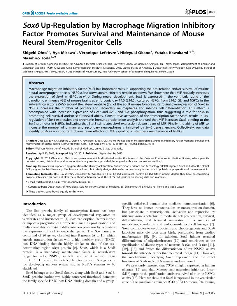
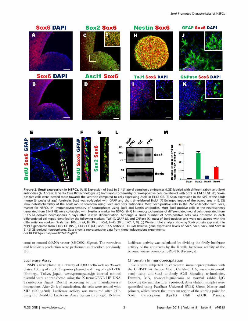
Figure 1. Sox6 functions downstream of MIF in NSPCs. (A) MIF treatment for 24 h increases Sox6 gene expression. (B) MIF antagonist (ISO-1)treatment for 24 h decreased Sox6 gene expression in NSPCs. (C) Changes in Sox gene expression levels in NSPCs following MIF treatment (400 ng/ml) for 24 h. Data are derived from three independent experiments. Error bars indicate S.D. values; *P,0.05, **P,0.01 versus control; Student’s t-test.doi:10.1371/journal.pone.0074315.g001
Sox6 Promotes Characteristics of NSPCs
PLOS ONE | www.plosone.org 2 September 2013 | Volume 8 | Issue 9 | e74315
com) or control shRNA vector (SHC002, Sigma). The retrovirus
and lentivirus production were performed as described previously
[16].
Luciferase AssayNSPCs were plated at a density of 5,000 cells/well on 96-well
plates. 100 ng of a pGL3 reporter plasmid and 1 ng of a pRL-TK
(Promega, Tokyo, Japan, www.promega.co.jp) internal control
plasmid were co-transfected using the X-tremeGENE HP DNA
Transfection Agent (Roche) according to the manufacturer’s
instructions. After 24 h of transfection, the cells were treated with
MIF (400 ng/ml). Luciferase activity was measured after 24 h
using the Dual-Glo Luciferase Assay System (Promega). Relative
luciferase activity was calculated by dividing the firefly luciferase
activity of the constructs by the Renilla luciferase activity of the
tyrosine kinase promoter, pRL-TK (Promega).
Chromatin ImmunoprecipitationCells were subjected to chromatin immuneprecipitation with
the ChIP-IT kit (Active Motif, Carlsbad, CA, www.activemotif.
com) using anti-Stat3 antibody (Cell Signaling technologies,
Danvers, MA, www.cellsignal.com) or normal rabbit IgG
following the manufacturer’s protocol. After elution, samples were
quantified using FastStart Universal SYBR Green Master and
primers, which targets the upstream region of the starting point for
Sox6 transcription (EpiTct ChIP qPCR Primers,
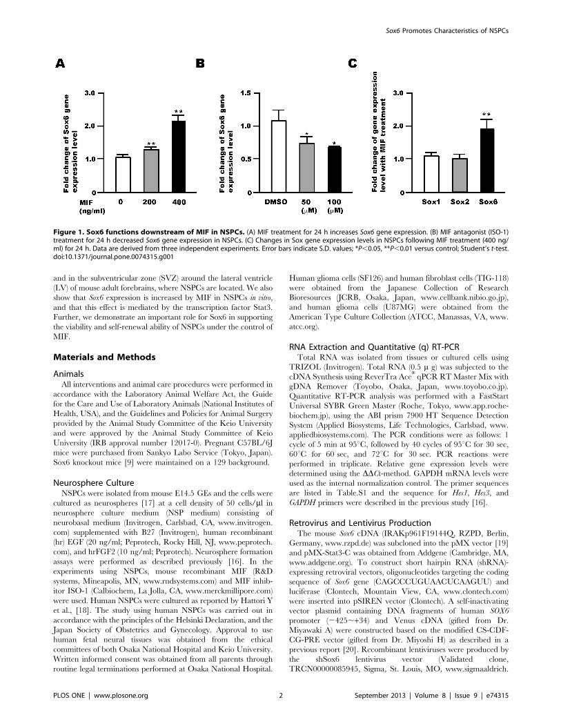
Figure 2. Sox6 expression in NSPCs. (A, B) Expression of Sox6 in E14.5 lateral ganglionic eminences (LGE) labeled with different rabbit anti-Sox6antibodies (A, Abcam; B, Santa Cruz Biotechnology). (C) Immunohistochemistry of Sox6-positive cells co-labeled with Sox2 in E14.5 LGE. (D) Sox6-positive cells were located more towards the ventricle compared to cells expressing Ascl1 in E14.5 GE. (E) Sox6 expression in the SVZ of the adultmouse (6 weeks of age) forebrain. Sox6 was co-labeled with GFAP and short time-labeled BrdU. (F) Enlarged image of the boxed area in E. (G)Immunohistochemistry of the adult mouse forebrain using Sox6 and Sox2 antibodies. Most Sox6-positive cells in the SVZ co-labeled with Sox2,marker for NSPCs. (H) Immunocytochemistry of neurospheres using Sox6 and Nestin antibodies. Most Sox6-positive cells in the neurospheresgenerated from E14.5 GE were co-labeled with Nestin, a marker for NSPCs. (I–K) Immunocytochemistry of differentiated neural cells generated fromE14.5 GE-derived neurospheres 5 days after in vitro differentiation. Although a small number of Sox6-positive cells was observed in eachdifferentiated cell types identified by the following markers: TuJ1(I), GFAP (J), and CNPase (K), most of Sox6-positive cells were not stained with thedifferentiation markers. Scale bar: 100 mm (A, B), 50 mm (C–E, H–K), 20 mm (C’, F, G). (L) Western blot analysis showing Sox6 protein expression inNSPCs generated from E14.5 GE (NSP), E14.5 GE (GE), and E14.5 cortex (CTX). (M) Relative gene expression levels of Sox1, Sox2, Sox5, and Sox6 inE14.5 GE-derived neurospheres. Data show a representative data from three independent experiments.doi:10.1371/journal.pone.0074315.g002
Sox6 Promotes Characteristics of NSPCs
PLOS ONE | www.plosone.org 3 September 2013 | Volume 8 | Issue 9 | e74315
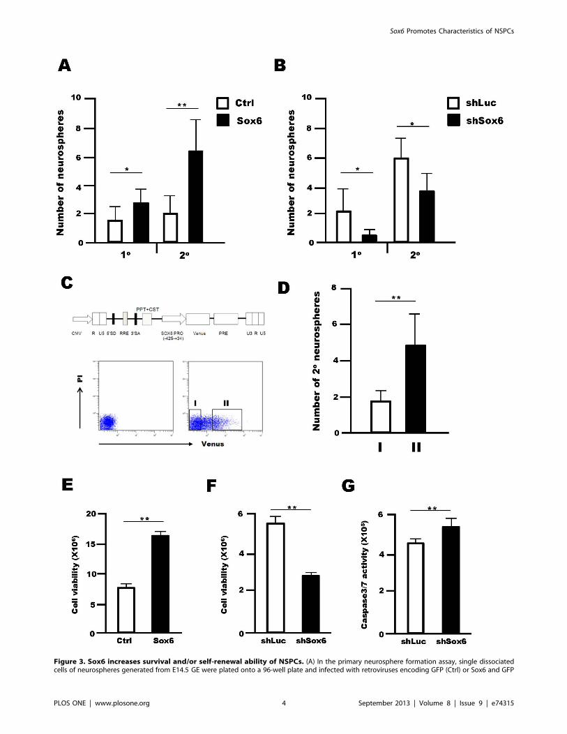
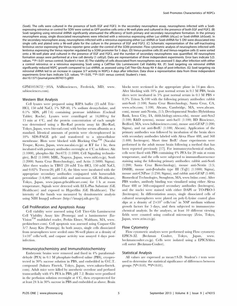
Figure 3. Sox6 increases survival and/or self-renewal ability of NSPCs. (A) In the primary neurosphere formation assay, single dissociatedcells of neurospheres generated from E14.5 GE were plated onto a 96-well plate and infected with retroviruses encoding GFP (Ctrl) or Sox6 and GFP
Sox6 Promotes Characteristics of NSPCs
PLOS ONE | www.plosone.org 4 September 2013 | Volume 8 | Issue 9 | e74315
GPM1053672(2)03A, SABiosciences, Frederick, MD, www.
sabiosciences.com).
Western Blot AnalysisCell lysates were prepared using RIPA buffer (25 mM Tris–
HCl, 150 mM NaCl, 1% NP-40, 1% sodium deoxycholate, and
0.1% SDS, pH 7.6) containing protease inhibitors (Cocktail
Tablet; Roche). Lysates were centrifuged at 14,0006g for
15 min at 4uC, and the protein concentration of each sample
was determined using a Bio-Rad protein assay kit (Bio-Rad,
Tokyo, Japan, www.bio-rad.com) with bovine serum albumin as a
standard. Identical amounts of protein were electrophoresed in
10% SDS-PAGE gels and transferred to a nitrocellulose
membrane. Blots were blocked with Blocking OneTM (Nacalai
Tesque, Kyoto, Japan, www.nacalai.co.jp) at RT for 1 hr, then
incubated with primary antibodies overnight at 4uC as follows: Akt
(1:1000), phospho-Akt (Ser473) (1:1000; Cell Signaling technolo-
gies), Bcl2 (1:1000; MBL, Nagoya, Japan, www.mbl.co.jp), Sox6
(1:2000; Santa Cruz Biotechnology), and Actin (1:5000; Sigma).
After three washes in TBST (20 mM Tris–HCl, 150 mM NaCl,
and 0.02% Tween-20, pH 7.4), the blots were incubated with the
appropriate secondary antibodies conjugated with horseradish
peroxidase (1:4,000, anti-rabbit and anti-mouse; GE Healthcare,
Tokyo, Japan, www.japan.gehealthcare.com) for 1 h at room
temperature. Signals were detected with ECL-Plus Substrate (GE
Healthcare) and exposed to Hyperfilm (GE Healthcare). The
intensity of the bands was measured by densitometric analysis
using NIH ImageJ software (http://imagej.nih.gov/ij).
Cell Proliferation and Apoptosis AssayCell viability were assessed using Cell Titer-Glo Luminescent
Cell Viability Assay kits (Promega) and a luminometer (En-
VisionTM multilabel reader, Perkin Elmer, Waltham, MA, www.
perkinelmer.com). Cell apoptosis was assessed using Caspase-Glo
3/7 Assay Kits (Promega). In both assays, single cells dissociated
from neurospheres were seeded onto 96-well plates at a density of
56103 cells/well, and caspase activity was assayed 4 days post-
infection.
Immunocytochemistry and ImmunohistochemistryEmbryonic brains were removed and fixed in 4% paraformal-
dehyde (PFA) in 0.1 M phosphate-buffered saline (PBS), cryopro-
tected in 30% sucrose solution in PBS, and embedded in O.C.T.
compound (Sakura Finetek, Tokyo, Japan, www.sakura-finetek.
com). Adult mice were killed by anesthetic overdose and perfused
transcardially with 4% PFA in PBS, pH 7.2. Brains were postfixed
in the perfusion solution overnight at 4uC, then cryoprotected for
at least 24 h in 30% sucrose in PBS and embedded as above. Brain
blocks were sectioned in the appropriate plane in 14 mm slices.
After blocking with 10% goat normal serum in 0.1 M PBS, brain
slices were incubated in 5% goat normal serum in 0.1 M PBS +0.3% Triton X-100 with the following primary antibodies: rabbit
anti-Sox6 (1:100; Santa Cruz Biotechnology, Santa Cruz, CA,
www.scbt.com; 1:100, Abcam, Cambridge, MA, www.abcam.
com), mouse anti-Nestin, (1:5; Developmental Studies Hybridoma
Bank, Iowa City, IA, dshh.biology.uiowa.edu), mouse anti-Sox2
(1:100; R&D systems), mouse anti-Ascl1 (1:100; BD Bioscience,
Bedford, MA, www.bdbiosciences.com), mouse anti-GFAP (1:200;
Sigma), and rat anti-BrdU (1:100; Abcam). Application of the
primary antibodies was followed by incubation of the brain slices
with secondary antibodies labeled with Alexa Fluor 488, and 568
(1:400; Invitrogen). Short time BrdU chase experiments were
performed in the adult mouse brain following a method that has
been reported previously [17]. For immunocytochemical studies,
cells were fixed with PBS containing 4% PFA for 20 min at room
temperature, and the cells were subjected to immunofluorescence
staining using the following primary antibodies: rabbit anti-Sox6
(1:200; Santa Cruz Biotechnology), mouse anti-Nestin (1:5;
DSHB), mouse anti-b-tubulin type III (TuJ1) (1:1000; Sigma),
mouse anti-CNPase (1:250; Sigma), and rabbit anti-GFAP (1:400;
Biomedical Technologies, Stoughton, MA, www.btiinc.com). After
PBS washes, antibody binding was visualized using either Alexa
Fluor 488 or 568-conjugated secondary antibodies (Invitrogen),
and the nuclei were stained with either DAPI or TO-PRO-3
(Invitrogen). In differentiation assays, single dissociated cells of
cultured neurospheres were plated on poly-L-lysine coated glass
slips at a density of 26105 cells/cm2 in NSP medium without
growth factors for 5 days, and then subjected to immunocyto-
chemical analysis. In the analyses, at least 10 different viewing
fields were counted using confocal microscopy (Zeiss, Tokyo,
Japan, www.zeiss.co.jp).
Flow CytometryFlow cytometric analyses were performed using Flow cytometry
EPICS-XL (Beckman Coulter, Tokyo, Japan, www.
beckmancoulter.co.jp). Cells were isolated using a EPICSAltra
cell sorter (Beckman-Coulter).
Statistical AnalysisAll values are expressed as mean6S.D. Student’s t tests were
used to determine the statistical significance of differences between
groups (*P,0.05, **P,0.01).
(Sox6). The cells were cultured in the presence of both EGF and FGF2. In the secondary neurosphere assay, neurospheres infected with a Sox6-expressing retrovirus or control for 5DIV were sorted as GFP-positive cells onto a 96-well plate and cultured in the presence of both EGF and FGF2. (B)Sox6 targeting using retroviral shRNA significantly attenuated the efficiency of both primary and secondary neurosphere formation. In the primaryneurosphere assay, single dissociated neurospheres were infected with a retrovirus expressing either Luc-shRNA (shLuc) or Sox6-shRNA (shSox6). Inthe secondary neurosphere assay, neurospheres infected with a retrovirus expressing either Luc-shRNA or Sox6-shRNA for 5 DIV were dissociated intosingle cells and plated onto a 96-well plate for the culture in the presence of EGF and FGF2. (C) Schematic representation of the self-inactivatinglentivirus vector expressing the Venus reporter gene under the control of the SOX6 promoter. Flow cytometric analysis of neurospheres infected withlentivirus expressing the Venus reporter regulated by a SOX6 promoter for 5 days. (D) Venus-positive cells (II) and Venus-negative cells (I) were sortedonto a 96-well plate and cultured in the presence of EGF and FGF2, and the number of secondary neurospheres was quantified. All neurosphereformation assays were performed at a low cell density (1 cell/ml). Data are representative of three independent experiments. Error bars indicate S.D.values. **P,0.01 versus control; Student’s t-test. (E) The viability of cells dissociated from neurospheres was assessed 5 days after infection with eithera control retrovirus or a retrovirus expressing Sox6 using a CellTiter Glo Luminescent Cell Viability Kit. (F) Sox6 targeting via retroviral shRNAsignificantly reduced NSPC growth compared to Luc-shRNA, as assessed using Cell Titer-Glo Assay Kit 4 days of post-infection. (G) Sox6 targeting viaretroviral shRNA led to an increase in caspase 3/7 activity in NSPCs 4 days after infection. Data show a representative data from three independentexperiments Error bars indicate S.D. values; *P,0.05, **P,0.01 versus control; Student’s t-test.doi:10.1371/journal.pone.0074315.g003
Sox6 Promotes Characteristics of NSPCs
PLOS ONE | www.plosone.org 5 September 2013 | Volume 8 | Issue 9 | e74315
Results
Sox6 is a Downstream Target of MIF Signaling in NSPCsChanges in the expression levels of the Sox genes upon
treatment of NSPCs with MIF were examined by qRT-PCR. MIF
treatment increased the RNA level of Sox6 in NSPCs (Fig. 1A). In
addition, cell treatment with ISO-1, a MIF inhibitor, led to a
decrease in Sox6 RNA level in a dose-dependent manner (Fig. 1B).
Interestingly, MIF treatment increased the RNA level of Sox6, but
not that of Sox1 and Sox2 (Fig. 1C). Together, these results suggest
that the Sox6 gene is a downstream target of MIF signaling in
NSPCs.
Sox6 is Expressed in Mouse Embryonic Brain NSPCsThe expression of the Sox6 protein in the mouse embryonic
brain at E14.5 was examined by immunofluorescence. Sox6 was
expressed in the ventricular zone of the cortex and the dorsal part
of the lateral ganglionic eminence (LGE), as previously shown [12]
(Fig. 2A and B). The Sox6-positive cells also expressed Sox2, a
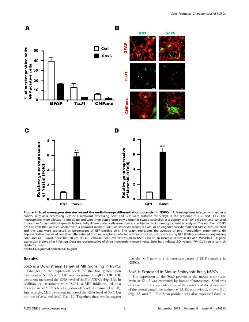
Figure 4. Sox6 overexpression decreased the multi-lineage differentiation potential in NSPCs. (A) Neurospheres infected with either acontrol retrovirus expressing GFP or a retrovirus expressing Sox6 and GFP were cultured for 5 days in the presence of EGF and FGF2. Theneurospheres were allowed to dissociate and were then plated onto poly-L-ornithin-coated coverslips at a density of 26105 cells/cm2 and culturedfor another 5 days without growth factors. Fully differentiated cells were fixed and subjected to immunocytochemical analyses. The number of GFP-positive cells that were co-labeled with a neuronal marker (TuJ1), an astrocyte marker (GFAP), or an oligodendrocyte marker (CNPase) was countedand the data were expressed as percentages of GFP-positive cells. The graph represents the average of two independent experiments. (B)Representative images of cells that differentiated from neurospheres infected with a control retrovirus expressing GFP (Ctrl) or a retrovirus expressingSox6 and GFP (Sox6). Scale bar: 20 mm. (C, D) Retroviral Sox6 overexpression in NSPCs led to an increase in Nestin (C) and Musashi-1 (D) geneexpression 5 days after infection. Data are representative of three independent experiments. Error bars indicate S.D. values; **P,0.01 versus control;Student’s t-test.doi:10.1371/journal.pone.0074315.g004
Sox6 Promotes Characteristics of NSPCs
PLOS ONE | www.plosone.org 6 September 2013 | Volume 8 | Issue 9 | e74315
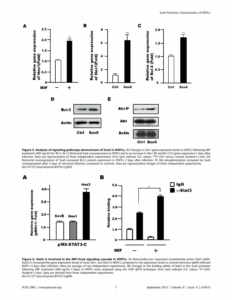
Figure 5. Analysis of signaling pathways downstream of Sox6 in NSPCs. (A) Changes in Hes1 gene expression levels in NSPCs following MIFtreatment (400 ng/ml) for 48 h. (B, C) Retroviral Sox6 overexpression in NSPCs led to an increase in Hes1 (B) and Bcl-2 (C) gene expression 5 days afterinfection. Data are representative of three independent experiments. Error bars indicate S.D. values; **P,0.01 versus control; Student’s t-test. (D)Retroviral overexpression of Sox6 increased Bcl-2 protein expression in NSPCs 2 days after infection. (E) Akt phosphorylation increased by Sox6overexpression after 5 days of retroviral infection compared to controls. Data are representative images of three independent experiments.doi:10.1371/journal.pone.0074315.g005
Figure 6. Stat3 is involved in the MIF-Sox6 signaling cascade in NSPCs. (A) Retrovirally-over expressed constitutively active Stat3 (pMX-Stat3-C) increased the gene expression levels of Sox6, Hes1, and Hes3 in NSPCs compared to the expression levels in control retrovirus (pMX)-infectedNSPCs 4 days after infection. Data are average of two independent experiments. (B) Changes in the binding ability of Stat3 to the Sox6 promoterfollowing MIF treatment (400 ng/ml, 5 days) in NSPCs were analyzed using the ChIP qPCR technique. Error bars indicate S.D. values; *P,0.05;Student’s t-test. Data are derived from three independent experiments.doi:10.1371/journal.pone.0074315.g006
Sox6 Promotes Characteristics of NSPCs
PLOS ONE | www.plosone.org 7 September 2013 | Volume 8 | Issue 9 | e74315
known marker of NPSCs (Fig. 2C), and these cells were located
more towards the ventricle compared to cells expressing Ascl1, a
marker of neural progenitor cells, in the E14.5 GE (Fig. 2D). In
the adult mouse forebrain, Sox6 was expressed in the subven-
tricular zone (SVZ) (Fig. 2E). Some Sox6-expressing cells were
positive for BrdU after short-term labeling, indicating that they
were actively proliferating, and they were also positive for Sox2,
proving that they corresponded to NSPCs (Fig. 2, E–G). In the
sub-granular zone (SGZ) of the adult mouse hippocampus, Sox6
expression coincided with BrdU labeling expression (data not
shown), indicating that Sox6 was expressed in NSPCs in the adult
as it is in the embryo. Next, we generated neurospheres from
E14.5 GEs and observed co-expression of Sox6 with Nestin, a
marker of NSPCs (Fig. 2H). To examine the expression of Sox6 in
differentiated cells, neurospheres were cultured without growth
factors for 5 days in vitro (DIV). Besides a few exceptions, Sox6
expression was detected only in cells negative for differentiation
markers, namely TuJ1 (neurons), GFAP (glia), and CNPase
(oligodendrocytes) (Fig. 2, I–K). This result is consistent with a
report showing that Sox6 is highly expressed in oligodendrocyte
progenitor cells (OPCs) but not in post-mitotic differentiating
oligodendrocytes [10]. Sox6 protein expression in NSPCs from
14.5 GE was also confirmed by Western blot (Fig. 2L). Finally, we
compared the expression level of Sox6 to that of other Sox genes by
qRT-PCR in NSPCs derived from E14.5 GE. The data clearly
indicated that NSPCs contained higher levels of RNA for Sox2 and
Sox6 than for their respective close relatives, Sox1 and Sox5
(Fig. 2M).
Sox6 Supports Cell Survival and the Self-renewal Abilityof NSPCsTo identify the function of Sox6 in NSPCs, we first performed
neurosphere-forming assays, a commonly used method to measure
NSPC self-renewal. Retrovirus-mediated Sox6 overexpression led
to a 1.760.6 fold increase in the number of primary neurospheres,
and 3.261.1 fold increase in the number of secondary neuro-
spheres (Fig. 3A). In contrast, Sox6 silencing by retroviral
expression of shRNA-Sox6 in NSPCs attenuated the formation
of primary and secondary neurospheres by 0.2360.25 fold and
0.6260.22 fold, respectively (Fig. 3B). This result was also
supported in experiments using Sox6-null mice (Fig. S1). Changes
in Sox6 protein levels in NSPCs by Sox6 overexpression and gene
silencing were assessed by Western blot analysis (Fig. S2).
Moreover, we performed the neurosphere-forming assay using a
lentivirus-based Sox6 reporter system. A reporter plasmid was
constructed using a self-inactivating lentivirus vector harboring the
Venus reporter gene under the control of the human Sox6
promoter region, which contains elements highly conserved in
humans and mice in terms of tissue-specific expression [21]
(Fig. 3C). Venus-positive cells (II) and Venus-negative cells (I) were
sorted onto a 96-well plate and then cultured in the presence of
EGF and FGF2, and the number of secondary neurospheres was
assessed (Fig. 3D). The specificity of the reporter assay was
confirmed in human cells and mouse NSPCs by showing a strong
correlation between Venus activity and Sox6 protein level (Fig.
S3). In this system, Venus-positive NSPCs formed 2.861.0 fold
more secondary neurospheres than Venus-negative cells (Fig. 3D).
Collectively, these data indicated that Sox6 supports the self-
renewal ability of NSPCs in vitro. We then asked whether the Sox6
promoter featured MIF responsive regions. The promoter contains
two regions, called A-box (79 bp) and B-box (48 bp), which are 85
and 96% identical, respectively, in the rat, mouse, and human
genomes [21]. NSPCs were transfected with luciferase reporters
containing either a promoter fragment (517 bp) or tandem repeats
of the A-box (4xA-box) or B-box (4xB-box) and were then treated
with or without MIF (Fig. S4). The construct containing the A-box
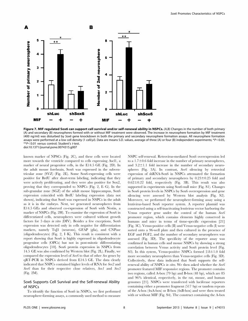
Figure 7. MIF regulated Sox6 can support cell survival and/or self-renewal ability in NSPCs. (A,B) Changes in the number of both primary(A) and secondary (B) neurospheres formed with or without MIF treatment were observed. The increase in neurosphere formation by MIF treatment(400 ng/ml) was disturbed by Sox6 gene knockdown in both the primary and secondary neurosphere formation assays. All neurosphere formationassays were performed at a low cell density (1 cell/ml). Data are means S.D. values, average of three (A) or four (B) independent experiments; *P,0.05,**P,0.01 versus control; Student’s t-test.doi:10.1371/journal.pone.0074315.g007
Sox6 Promotes Characteristics of NSPCs
PLOS ONE | www.plosone.org 8 September 2013 | Volume 8 | Issue 9 | e74315
tandem repeats showed strong transcriptional activity, and this
activity was increased 3.460.8 fold by MIF treatment, suggesting
that the A-box contains MIF-responsive elements (Fig. S4). We
also examined changes in NSPC viability upon Sox6 overexpres-
sion and silencing. Retrovirus-mediated overexpression of Sox6 in
NSPCs increased cell viability by 2.160.12 fold 5 days after
infection (Fig. 3E), whereas retrovirus-mediated silencing of Sox6
decreased cell viability by half (0.5060.02 fold) 4 days after
infection (Fig. 3F). Sox6 knockdown also led to increased
caspase3/7 activity 4 days after infection, confirming that Sox6
acts as a survival factor in NSPCs (Fig. 3G, Fig. S5).
Sox6 is a Maintenance Factor in NSPCsChanges in the multi-lineage differentiation potential of NSPCs
were examined upon Sox6 overexpression. NSPCs infected with a
Sox6-expressing retrovirus were cultured for 5 days, and then
replated and cultured for 5 additional days in the absence of
growth factors. Then, the number of cells differentiated into each
neuronal cell type was assessed by immunocytochemistry. In this
assay, only GFP-positive cells, i.e., infected by the retrovirus, were
examined. Sox6 overexpression led to a decrease in the number of
cells differentiated into neurons (TuJ1), astrocytes (GFAP), and
oligodendrocytes (CNPase) (Fig. 4, A and B). In addition, the
expression of Nestin and Musashi-1, which are known as NSPC
markers [22],[23], was also upregulated by Sox6 overexpression
(Fig. 4, C and D). Thus, Sox6 functions as a maintenance factor
for NSPCs in vitro. Consistent with this experiment, the numbers
of neurons, astrocytes and oligodendrocytes were increased in
NSPC cultures from Sox6-null mice (Fig. S6).
Downstream Signal of Sox6 in NSPCsHes1 is a transcription factor acting downstream of Notch
signaling and well known for its ability to maintain the stemness of
NSPCs [24]. Interestingly, Hes1 expression was increased upon
treatment of NSPCs with MIF (Fig. 5A) as well as upon
overexpression of Sox6 (Fig. 5B). Furthermore, expression of the
Bcl-2 gene, which is important for cell survival and is regulated by
MIF in NSPCs [16], also increased with Sox6 overexpression
(Fig. 5C). This increase was confirmed at the protein level for Bcl-2
(Fig. 5D) and found to occur concomitantly with an increase in Akt
phosphorylation (1.560.11 fold) (Fig. 5E).
MIF-regulated Stat3 Controls Sox6 Gene Expression inNSPCsWe previously demonstrated that MIF treatment results in
activation of the transcription factor Stat3 [16]. Hes3 expression,
which is activated by Stat3-pSer727 in NSPCs [25], was also
shown to be upregulated by MIF [16]. Thus, we examined the
changes in gene expression of Sox6, Hes1, and Hes3 in NSPCs upon
overexpression of constitutively active Stat3 in NSPCs, and found
that the expression level of all genes was increased (Fig. 6A).
Moreover, ChIP analysis showed an increase in Stat3 binding to
the Sox6 promoter following MIF treatment of NSPCs (Fig. 6B).
These data thus suggest that Sox6 is upregulated by MIF signaling
in NSPCs through binding of Stat3 to its promoter.
MIF-mediated Up-regulation of Sox6 Supports the Self-renewal Ability of NSPCsTo examine whether Sox6 plays a role in neurosphere
formation in response to MIF, we performed neurosphere
formation assays using cells treated with a retrovirus expressing
Sox6-shRNA. While MIF treatment of control cells resulted in a
larger number of primary (Fig. 7A) and secondary neurospheres
(Fig. 7B), this effect was blunted by knockdown of Sox6, indicating
that Sox6 is a downstream effector of MIF signaling in NSPCs.
The same result was observed in a cell growth assay using
lentivirally expressed Sox6 shRNA in NSPCs (Fig. S7).
Discussion
In the present study, we newly identified Sox6, which is
expressed in the GE of the fetal mouse brain, as a maintenance
factor for NSPCs. Moreover, we showed that Sox6 gene expression
is upregulated by Stat3 downstream of MIF, a signaling molecule
that supports the proliferation and/or survival of murine NSPCs.
To determine whether MIF can regulate Sox genes to maintain
NSPC stemness, we analyzed change in gene expression levels of
Sox1, Sox2 and Sox6 in response to MIF treatment in NSPCs.
Sox2 is known as a maintenance factor for NSPC stemness in fetal
and adult mouse brains [3]. Interestingly, in this assay system, MIF
treatment increased Sox6, but not Sox1 or Sox2 gene expression.
Consistent with this finding, treatment of NSPCs with ISO-1, a
MIF inhibitor, decreased Sox6 gene expression, suggesting that
Sox6 is a downstream molecule of MIF in NSPCs. However, it is
possible that MIF may modulate Sox1 or Sox2 gene expression in
other assay conditions and in NSPCs derived from different tissue
types, including the cortex and spinal cord. Analysis by luciferase
assay using a Sox6 promoter also showed that Sox6 gene
transcription is upregulated by MIF in NSPCs, suggesting that
Sox6 is a direct target of MIF signaling in this system.
We demonstrated that Sox6 overexpression can increase the
survival and self-renewal ability of NSPC, and that Sox6 gene
silencing in NSPCs has opposite effects. We observed an increase
in Bcl2 expression and Akt phosphorylation in response to MIF
treatment, both of which play an important role in NSPC cell
survival [16], further suggesting that Sox6 is a survival factor
acting downstream of MIF in NSPCs. In contrast, a previous
report showed that SOX6 overexpression decreased cell prolifer-
ation in INS-1E insulinoma cells and NIH3T3 cells [26],
indicating the possibility that Sox6 may regulate cell proliferation
in a cell type-dependent manner. Furthermore, it is known that
Sox proteins function in concert with partner proteins in a cell
type-specific manners [27]. Thus, the identification of Sox6
partner proteins in NSPCs is a topic for future studies that would
allow for an understanding of Sox6 protein mode of action.
We showed here that Sox6 is a maintenance factor for NSPCs
in neurosphere-forming and differentiation assays in vitro, and
observed up-regulation of Hes1 gene expression upon Sox6
overexpression, as also seen upon MIF treatment. Hes1 is known
as an important factor for NSPC maintenance [24]. Although
Hes3 was also found to be up-regulated by MIF [16], Hes1 has
Sox6 binding elements in its promoter, while Hes3 does not. Thus,
Sox6 may possibly activate Hes1 with other transcription factors,
as seen in chondrocytes [28], while Hes3 may be regulated directly
by Stat3 without the intervention of Sox6. In previous reports,
Sox6 was shown to support neurogenesis and gliogenesis and to
inhibit terminal differentiation of oligodendrocytes [10], [11],
[14]. Scheel et al have reported that Sox6 overexpression induces
astrocytic differentiation from rat hippocampal NSPCs [14]. Thus,
it may be important to examine Sox6 function in NSPCs derived
from different species and tissue types (e.g. adult SVZ of forebrain,
adult hippocampus, fetal cortex), especially if different NSPC
culture methods are used (neurosphere floating culture VS
adherent culture). Additionally, it would also be interesting to
examine the regulation of Sox6 function by MIF in different types
of tissue stem cells, including hair follicle stem cells [29], [30].
Moreover, Sox6 was reported to regulate the differentiation of
Sox6 Promotes Characteristics of NSPCs
PLOS ONE | www.plosone.org 9 September 2013 | Volume 8 | Issue 9 | e74315
neural progenitor cells into different neural cell types in the mouse
embryonic brain in vivo [12], [13]. In those in vivo studies, the
expression patterns of many transcription factors changed in the
mouse embryonic GE in response to the loss of Sox6, showing that
Sox6 may play a role in controlling the maintenance of stemness of
NSPCs via or together with several transcription factors.
In our previous report [16] we found that although MIF
induced the self-renewal ability of NSPCs, it did not change the
cell fate of NSPCs in vitro, as there was no significant difference in
the cell differentiation potential of NSPCs upon MIF treatment.
However, in the present study, Sox6 overexpression in NSPCs
in vitro resulted in fewer differentiated cells belonging to three
lineages. As Sox6 is just one of multiple genes activated by MIF,
changes in the cell fate of NSPCs by Sox6 overexpression do not
necessarily mirror changes induced by MIF treatment. Strong
Sox6 activity may have been responsible for the decrease in
differentiation markers in this system, which was not achieved in a
previous study which examined the effects of MIF, a factor that lies
upstream of Sox6.
The expression of SOX genes has been reported in many
tumors [31]. We reported high expression of SOX genes in human
gliomas in a previous report [15]. To date, high expression of
SOX6 in gliomas has been confirmed by in silico gene expression
databases, including Oncomine (www.oncomine.org). We have
observed higher levels of SOX6 expression in glioma-initiating cells
generated from glioma specimens compared to neural stem cells
(Ohta et al., unpublished data). Thus, it may be important to
analyze the detailed function of Sox6 in gliomas and glioma-
initiating cells in the future.
In this study, we focused on Sox6 function in NSPCs derived
from the GE of mouse embryonic brains in vitro, showing a new
function of Sox6 as a maintenance factor of NSPCs stemness.
Functional analyses of Sox6 in adult mouse NSPCs, and especially
in human gliomas and glioma-initiating cells, will pave the way for
evaluating Sox6 as a therapeutic target for many brain diseases.
Supporting Information
Figure S1 Self-renewal ability of Sox6 mutant NSPCs. Inthe primary neurosphere formation assay, single dissociated cells
from E14.5 GEs of mouse fetal brains taken from of Sox6 mutant
and littermate controls were seeded onto a 96-well plate at a cell
density of 10 cells/ml in the presence of EGF and FGF2. WT
(n= 9), KO (n= 6). In the secondary neurosphere assay, primary
neurospheres were dissociated into single cells and seeded onto a
96-well plate at a cell density of 10 cells/ml in the presence of EGF
and FGF2. WT (n= 8), KO (n= 9). Error bars indicate S.D.
values; **P,0.01 versus control; Student’s t-test.
(TIF)
Figure S2 Expression of Sox6 in the gain and loss offunction experiments in NSPCs. (A) Western blot analysis
shows Sox6 protein expression in NSPCs infected with retrovirus
expressing GFP alone (Ctrl), or GFP and Sox6 (Sox6) 5 days after
infection. (B) Retroviral Sox6-shRNA expression significantly
reduced Sox6 protein expression in NSPCs 5 days after infection.
(TIF)
Figure S3 Expression pattern analysis of SOX6 promot-er-derived Venus expression cells in glioma cells andNSPCs. (A) FACS analysis of Venus reporter expression under
the control of the human SOX6 promoter in human dermal cells
(TIG118), human glioma cells (SF126, U87MG), and human
NSPCs (NSP). (B) SOX6 gene expression levels in TIG118, SF126,
U87MG, and human NSPCs. (C) Immunostaining of Sox6 in
mouse NSPCs infected with a lentivirus expressing the Venus
reporter under the control of Sox6 showing co-localization of
Sox6-positive cells and Venus- positive cells. Scale bar; 100 mm,
20 mm (enlarged image). Data are derived from three independent
experiments.
(TIF)
Figure S4 Identification of MIF-responsive elements inthe SOX6 promoter. (A) Schematic representation of A-box
and B-box location in human SOX6 gene promoter [21]. (B)
Luciferase-reporter analysis of a region from the SOX6 promoter
(2517), and A-box and B-box tandem repeats in NSPCs, either
with or without MIF treatment, 48 h after transfection. Relative
luciferase activity was calculated by dividing the firefly luciferase
activity of the constructs by the Renilla luciferase activity of the
tyrosine kinase promoter, pRL-TK. Data show a representative
data from three independent experiments. Error bars indicate S.D.
values; *P,0.05, **P,0.01 versus control; Student’s t-test.
(TIF)
Figure S5 Sox6 supports cell survival in NSPCs. (A) Sox6targeting using lentiviral shRNA significantly reduced NSPC
growth compared to control shRNA, as assessed using a Cell
Titer-Glo Assay Kit 4 days after infection. (B) Sox6 knockdown by
lentvirally-expressed shRNA led to an increase in caspase 3/7
activity in NSPCs 4 days after infection. (C) Sox6 gene expression
in NSPCs infected with control lentivirus or lentivirus expressing
Sox6-shRNA 4 days after infection. Data are derived from three
independent experiments. Error bars indicate S.D. values;
*P,0.05, **P,0.01 versus control; Student’s t-test.
(TIF)
Figure S6 Differentiation potential of NSPCs derivedfrom Sox6 knockout mice. (A) Secondary neurospheres of
Sox6 mutant and wild type were dissociated and cultured for
5DIV in the absence of growth factors. The differentiated cells
were labeled with a neuronal marker (TuJ1), an astrocyte marker
(GFAP), or an oligodendrocyte marker (CNPase) and counted.
Data are averages of five independent experiments. Error bars
indicate S.D. values; *P,0.05, **P,0.01 versus control; Student’s
t-test. (B) Representative images of cells differentiated from Sox6
mutant and wild type neurospheres. Scale bar: 50 mm.
(TIF)
Figure S7 MIF regulated Sox6 can support cell survivaland/or proliferative ability in NSPCs. Changes in cell
number with or without MIF treatment (400 ng/ml) in NSPCs
were observed using a CellTiter Glo Luminescent Cell kit. The
increase in cell viability by MIF treatment was inhibited by
lentiviral Sox6 gene knockdown 4 days after infection (n= 3).
Error bars indicate S.D. values; *P,0.05, **P,0.01 versus
control; Student’s t-test from three independent experiments.
(TIF)
Table S1 Primer sequence.
(DOC)
Acknowledgments
The authors would like to acknowledge Dr. T. Kitamura (The University
of Tokyo) for providing the pMX vector, Dr. H. Miyoshi (RIKEN) for
providing the CS-CDF-CG-PRE vector, Dr. Y. Kanemura (Osaka
National Hospital) for providing the human NSP, Dr. A. Miyawaki for
providing the Venus cDNA, Dr. UI. Chung for providing the Sox6 (2517),
4xA-box, and 4xB-box constructs for the luciferase assay, and Ms. S.
Teramoto (Keio University) for her technical assistance.
Sox6 Promotes Characteristics of NSPCs
PLOS ONE | www.plosone.org 10 September 2013 | Volume 8 | Issue 9 | e74315
Author Contributions
Conceived and designed the experiments: SO HO YKMT. Performed the
experiments: SO AM. Analyzed the data: SO. Contributed reagents/
materials/analysis tools: SO AM VL. Wrote the paper: SO AM VL HO
MT.
References
1. Bowles J, Schepers G, Koopman P (2009) Phylogeny of the SOX family of
developmental transcription factors based on sequence and structural indicators.
Dev Biol 227: 239–255.
2. Chew LJ, Gallo V (2009) The Yin and Yang of Sox proteins: Activation and
repression in development and disease. J Neurosci Res 87: 3277–3287.
3. Pevny LH, Nicolis SK (2010) Sox2 roles in neural stem cells. Int J Biochem Cell
Biol 42: 421–424.
4. Sarkar A, Hochedlinger K (2013) The Sox family of transcription factors:
versatile regulators of stem and progenitor cells. Cell Stem Cell 12: 15–30.
5. Wegner M, Slot C (2005) From stem cells to neurons and glia: a Soxist’s view of
neural development. Trends Neurosci 28: 583–588.
6. Lefebvre V (2010) The SoxD transcription factors-Sox5, Sox6, and Sox13-are
key cell fate modulators. Int J Biochem Cell Biol 42: 429–432.
7. Hagiwara N (2011) Sox6, jack of all trades: a versatile regulatory protein in
vertebrate development. Dev Dyn 240: 1311–1321.
8. Hagiwara N, Klewer SE, Samson RA, Erickson DT, Lyon MF, et al. (2000)
Sox6 is a candidate gene for p100H myopathy, heart block, and sudden neonatal
death. Proc Natl Acad Sci U S A 97: 4180–4185.
9. Smits P, Li P, Mandel J, Zhang Z, Deng JM, et al. (2001) The transcription
factors L-Sox5 and Sox6 are essential for cartilage formation. Dev Cell 1: 277–
290.
10. Stolt CC, Schlierf A, Lommes P, Hillgartner S, Werner T, et al. (2006) SoxD
proteins influence multiple stages of oligodendrocyte development and modulate
SoxE protein function. Dev Cell 11: 697–709.
11. Hamada-Kanazawa M, Ishikawa K, Nomoto K, Uozumi T, Kawai Y, et al.
(2004) Sox6 overexpression causes cellular aggregation and the neuronal
differentiation of P19 embryonic carcinoma cells in the absence of retinoic acid.
FEBS Lett 560: 192–198.
12. Azim E, Jabaudon D, Fame RM, Macklis JD (2009) SOX6 controls dorsal
progenitor identity and interneuron diversity during neocortical development.
Nat Neurosci 12: 1238–1247.
13. Batista-Brito R, Rossignol E, Hjerling-Leffler J, Denaxa M, Wegner M, et al.
(2009) The cell-intrinsic requirement of Sox6 for cortical interneuron
development. Neuron 63: 466–481.
14. Scheel JR, Ray J, Gage FH, Barlow C (2005) Quantitative analysis of gene
expression in living adult neural stem cells by gene trapping. Nat Methods 2:
363–370.
15. Ueda R, Iizuka Y, Yoshida K, Kawase T, Kawakami Y, et al. (2004)
Identification of a human glioma antigen, SOX6, recognized by patients’ sera.
Oncogene 23: 1420–1427.
16. Ohta S, Misawa A, Fukaya R, Inoue S, Kanemura Y, et al. (2012) Macrophage
migration inhibitory factor (MIF) promotes cell survival and proliferation of
neural stem/progenitor cells. J Cell Sci 125: 3210–3220.
17. Ohta S, Gregg C, Weiss S (2006) Pituitary adenylate cyclase-activating
polypeptide regulates forebrain neural stem cells and neurogenesis in vitro andin vivo. J Neurosci Res 84: 1177–1186.
18. Hattori Y, Ohta S, Hamada K, Yamada-Okabe H, Kanemura Y, et al. (2007)Identification of a neuron-specific human gene, KIAA1110, that is a guanine
nucleotide exchange factor for ARF1. Biochem Biophys Res Commun 364:
737–742.19. Kitamura T (1998) New experimental approaches in retrovirus-mediated
expression screening. Int J Hematol 67: 351–359.20. Miyagi S, Saito T, Mizutani K, Masuyama N, Gotoh Y, et al. (2004) The Sox-2
regulatory regions display their activities in two distinct types of multipotent stem
cells. Mol Cell Biol 24: 4207–4220.21. Ikeda T, Saito T, Ushita M, Yano F, Kan A, et al. (2007) Identification and
characterization of the human SOX6 promoter. Biochem Biophys ResCommun 357: 383–390.
22. Zimmerman L, Parr B, Lendahl U, Cunningham M, McKay R, et al. (1994)Independent regulatory elements in the nestin gene direct transgene expression
to neural stem cells or muscle precursors. Neuron 12: 11–24.
23. Sakakibara S, Imai T, Hamaguchi K, Okabe M, Aruga J, et al. (1996) Mouse-Musashi-1, a neural RNA-binding protein highly enriched in the mammalian
CNS stem cell. Dev Biol 176: 230–242.24. Kageyama R, Ohtsuka T, Shimojo H, Imayoshi I (2009) Dynamic regulation of
Notch signaling in neural progenitor cells. Curr Opin Cell Biol 21: 733–740.
25. Androutsellis-Theotokis A, Leker RR, Soldner F, Hoeppner DJ, Ravin R, et al.(2006) Notch signalling regulates stem cell numbers in vitro and in vivo. Nature
442: 823–826.26. Iguchi H, Urashima Y, Inagaki Y, Ikeda Y, Okamura M, et al. (2007) SOX6
suppresses cyclin D1 promoter activity by interacting with beta-catenin and
histone deacetylase 1, and its down-regulation induces pancreatic beta-cellproliferation. J Biol Chem 282: 19052–19061.
27. Wilson M, Koopman P (2002) Matching SOX: partner proteins and co-factorsof the SOX family of transcriptional regulators. Curr Opin Genet Dev 12: 441–
446.28. Lefebvre V, Li P, de Crombrugghe B (1998) A new long form of Sox5 (L-Sox5),
Sox6 and Sox9 are coexpressed in chondrogenesis and cooperatively activate the
type II collagen gene. EMBO J 17: 5718–5733.29. Li L, Mignone J, Yang M, Matic M, Penman S, et al. (2003) Nestin expression in
hair follicle sheath progenitor cells. Proc Natl Acad Sci U S A 100: 9958–9961.30. Liu F, Uchugonova A, Kimura H, Zhang C, Zhao M, et al. The bulge area is
the major hair follicle source of nestin-expressing pluripotent stem cells which
can repair the spinal cord compared to the dermal papilla. Cell Cycle10: 830–839.
31. Dong C, Wilhelm D, Koopman P (2004) Sox genes and cancer. CytogenetGenome Res 105: 442–447.
Sox6 Promotes Characteristics of NSPCs
PLOS ONE | www.plosone.org 11 September 2013 | Volume 8 | Issue 9 | e74315