on october 6, 2016 downloaded from
TRANSCRIPT
T H E M A J O R H I S T O C O M P A T I B I L I T Y C O M P L E X - R E S T R I C T E D
A N T I G E N R E C E P T O R ON T CELLS
IV. An Anti idiotypic An t ibody Predicts Both Ant igen and I-specificity*
BY PHILIPPA MARRACK, RICHARD SHIMONKEVITZ, CHARLES HANNUM, KATHRYN HASKINS, AND JOHN KAPPLER*
From the Department of Medicine, National Jewish Hospital and Research Center, Denver, Colorado 80206
A number of recent papers have described the production of antisera and monoclonal antibodies against cloned antigen-specific, major histocompatibility complex-restricted (Ag/MHC-specific) ~ or ailo-reactive T cells. In each case the antibodies were assayed for activity against Ag/MHC receptors on these T cells by their ability to substitute for Ag/MHC, or inhibit Ag/MHC reaction (1-8). Interestingly, in every case so far in which antibodies with these activities have been described, they have proved to be clone-specific in their activity, suggesting that they recognize the idiotypic portion of the Ag/MHC receptors on target cells. This, of course, has been an important point in proving that the antibodies in question were indeed directed at the Ag/MHC receptor on the T cell, and not some other, perhaps associated, molecule such as lymphocyte functional antigen-l, T3, L3T4, Lyt-2, or T8 (9-13).
In several cases the target molecules of these antibodies have been identified by immunoprecipitation (4-7, 14). They are 80-90-kdalton disulfide-bonded dimers of two dissimilar glycopeptide chains of molecular weights varying from 47 kdaltons to 39 kdaitons, depending on the T cell and species from which they were derived. Both chains vary in amino acid sequence between clones (7).
The experiments presented in this paper were designed to bolster the idea that the T cell clone-specific antibodies described so far do indeed bind to the Ag/MHC receptor on T cells. A clone-specific monoclonal antibody, KJ1-26.1, isolated on the basis of its reactivity with a chicken ovalbumin (cOVA)/I-A d- reactive T cell hybridoma, D0-1 1.10, was tested for its ability to react with 397 independent T cell hybridomas derived from BALB/c T cells primed with cOVA. Of these 397 T cell hybridomas only one, 7D0-286, bound the monoclonal antibody KJ1-26.1 specifically. 7D0-286 proved to share the unusual Ag/I fine
* Supported by U.S. Public Health Service research grant AI-18785, training grant AI-07035, and National Research Service Institutional Award T32 AI-00048, and by research grant IM-49 from the American Cancer Society and postdoctoral fellowship, number 2247, from the American Cancer Society.
*This work was done during the tenure by J. K. of Faculty Research Award 218 from the American Cancer Society.
t Abbreviations used in this paper: Ag, antigen; c, chicken; d, duck; g, goose; IL-2, interleukin-2;jf, jungle fowl; MHC, major histocompatibility complex; nd, not done; OD, optical density; OVA, ovalbumin; PAGE, polyacrylamide gel electrophoresis; ph, pheasant; p143, tryptic peptide 316-332 of chicken ovalbumin; q, quail; SDS, sodium dodecyl sulfate; SN, supernatant; t, turkey; w, widgeon.
J. Exp. MEIg. © The Rockefeller University Press • 0022-1007/83/11/1635/12 $1.00 1635 Volume 158 November 1983 1635-1646
on February 15, 2018
jem.rupress.org
Dow
nloaded from
1636 AN ANTI-T CELL IDIOTYPE DEFINES ANTIGEN AND I SPECIFICITY
specificity of D0-11.10 both for Ag and I. Thus , reactivity with KJ1-26.1 predicted the fine specificity o f T cell hybr idoma both for Ag and I. This result strongly suggests that the monoclonal antibody, KJI-26.1, binds an idiotypic de te rminant on the A g / M H C receptors o f D0-11.10 and 7D0-286.
Materials and Methods Animals. BALB/c By (BALB/c), C57BL/10 (B10), B10.BR, B10.M, B10.Q,
B10.S(7R), and CBA/J animals were either bred in our colony from breeding triplets purchased from The Jackson Laboratory, Bar Harbor, ME, or were the gifts of Drs. Chella David (The Mayo Clinic, Rochester, MN) and Donald Shreffler (Washington University, St Louis, MO).
Antigens and Other Reagents. Chicken (c) and turkey (t) ovalbumin were purchases from Sigma Chemical Co. St. Louis, MO.
Jungle fowl Of), duck (d), pigeon (p), and widgeon (w) OVA were prepared by standard methods (15) from eggs provided by the inmates of the Denver Zoo courtesy of Ed Schmitt, the curator. Bobwhite quail (q) OVA was prepared by similar methods from eggs obtained from the GQF Manufacturing Co. Savannah, GA. Reagents for ELISA, p- nitrophenyl phosphate, and alkaline phosphatase-coupled rabbit anti-mouse immunoglob- ulin, were obtained from the Sigma Chemical Co.
T Cell Hybridomas. T cell hybridomas were produced as previously described by the fusion of Ag/MHC-reactive T cell blasts to the AKR T cell thymoma, BW5147 (15, 16). The T cell hybridomas DO- 11.10 and 7D0-286.2, for example, were produced by priming BALB/c mice in the base of the tail with cOVA. 7 d later, cells from draining lymph nodes were harvested and cultured for 4 d with Ag followed by 3 d incubation with interleukin 2 (IL-2). Live cells were isolated and hybridomas prepared by standard techniques. DO-11.10 was picked and cloned on the basis of its reactivity with cOVA/H- 2 d (3).7D0-286.2 was picked and cloned on the basis of its reactivity with a monoclonal anti-Ag/MHC receptor antibody prepared against DO-11.10 (see below). DO-11,10 was a fusion product of female BALB/c mice, 7D0-286.2 was derived from male BALB/c donors.
Monoclonal Antibodies. Monocional antibodies against Ag]MHC receptors on T cell hybridomas were prepared as previously described (5). Two were used in the experiments described in this paper. KJ1-26.1, raised against and specific for the Ag/MHC receptor on D0-11.10 (5), and KJ12-98.15, raised against and specific for the receptor on 3DT- 52.5 (7), a T cell hybridoma which recognizes D d (17). The anti-Thy-1 monoclonal antibody T24/40.8 (18) was used as a control in ELISA. The following monocional antibodies were used to map I-region specificities of T cell hybridomas, MK-D6, specific
b d d k for I-A d'p'q (16), M5/114, specific for I-A • , I-E ' (19), 8KP, specific for I-A b'k (amongst many) but not I-A n (20), 14-4-4, specific for I-E d'k (21), MK-S4 specific for I-A S (16), and 11-5.2 specific for I-A k (22).
Assay for Ag]MHC Recognition. Receptors for Ag/MHC on T cell hybridomas were assayed by standard methods (16). Briefly, T cell hybridomas were incubated in micro- culture wells for 24 h with Ag (usually at 1 mg/ml) and Ag-presenting cells. These last were B cell lymphomas or hybridomas at 10h/well or 4,000 rad-irradiated spleen cells at 106/well. After incubation the supernatants (SN) of these cultures were assayed for IL-2, produced by the T cell hybridomas in response to Ag/MHC. IL-2 concentrations were estimated by serially diluting the SN and measuring their ability to support the growth and survival of the IL-2-dependent T cell line HT-2 (the kind gift of Dr. J. Watson, Auckland, New Zealand). The first dilution in a serial titration that yielded <90% viable HT-2 cells 24 h later was said to contain 1 U of IL-2. 10 U/ml IL-2 was the minimum detectable by this assay.
Karyotyping. T hybridoma cells were prepared for karyotyping by standard methods. They were incubated with 0.2 tzg/ml colchicine for 30 min at 37°C. After pelleting, they were incubated in 0.075 M potassium chloride for 10 min at 37°C, spun down, and resuspended in fixative (3 parts methanol/1 part glacial acetic acid). After 20 min fixation
on February 15, 2018
jem.rupress.org
Dow
nloaded from

MARRACK ET AL. 1637
at room temperature the cells were thoroughly washed in fixative before being dropped from a height onto ethanol-washed, water-covered slides. Banding was done after heating the slides at 95°C for 10 rain by incubating the slides for 10-90 s in tryspin (23), followed by washing and Giemsa staining. Chromosome markers were assigned by the rules of Nesbitt and Francke (24).
ELISA. T hybridoma cells were assayed for the presence of cell surface antigens by ELISA. The procedures used have already been described (25). Briefly 3 × 10 ~ T hybridoma cells were incubated in 100 #1 of the neat culture SN of an anti-Thy-1 secreting hybridoma (18), or a 1:250 dilution of ascites produced by the anti-T cell idiotype-secreting B cell hybridomas KJ1-26.1 or KJ12-98.15. The cells were then thor- oughly washed before incubation with 1:500 alkaline phosphatase coupled rabbit anti- mouse immunoglobulin (Sigma Chemical Co.). After thorough washing the cells were incubated at 37°C with p-nitro phenyl phosphate in pH 10.4 buffer (26). Optical densities (OD) were then read using an ELISA reader (Bio-Tek Instruments, Inc., Burlington, VT).
Immunoprecipitation. Surface membrane proteins on T cell hybridomas were labelled with 12~I using Iodo-gen (Pierce Chemical Go., Rockford, IL) (5). Cell lysates were prepared with NP-40 (5) and immunoprecipitated with monoclonal antibody coupled to Sepharose beads (Pharmacia, Uppsala, Sweden) (27). Immunoprecipitates were washed thoroughly before solubilization by boiling in electrophoresis loading buffer (28). Some samples were also reduced by treatment with 5% 2-mercaptoethanol during the boiling. Reduced and unreduced samples were electrophoresed on 10% polyacrylamide gels (PAGE) containing SDS (28).
Results Production of a T Cell Hybridoma That Cross-reacts Idiotypically with DO-11.10. We
have recently reported (5) the properties of a monoclonal antibody, KJ1-26.1, which appears to bind all or part of the receptor for cOVA/I-A d on the T cell hybridoma D0-11.10. In our original studies this antibody was shown to bind D0-11.10 and cOVA/I-Ad-responsive subclones of this hybridoma, but the anti- body did not bind any other T cell hybridoma screened, nor to subclones of DO- 11.10 that had lost the ability to respond to cOVA/I-A d. We were curious to find out, however, how frequent the idiotype recognized by KJ1-26.1 was on BALB/c T cells, so for this and other reasons we decided to screen a number of independent T cell hybridomas for reactivity with KJ1-26.1 or KJ12-98, a monoclonal antibody specific for the receptor for D d on the T cell hybridoma 3DT-52.5 (7). Since D0-11.10 was derived from BALB/c T cell blasts specific for cOVA we reasoned that we would be most likely to find a KJ1-26.1 idiotype- bearing cell amongst this population. Hybridomas prepared from these T cell blasts were therefore screened. In the vast majority of cases the screening used ELISA to detect KJ1-26.1-binding T cell hybridomas. 307 independent T cell hybridomas prepared in four different fusions were thus tested; some sample results are shown in Table I. All the uncloned T cell hybridomas we tested reacted well with the anti-Thy-1 antibody.
A few hybridomas reacted to a small degree with either KJ1-26.1 or KJ12-08. For example of the hybrids shown in Table I, 7D0-248 had this property. This hybrid, and others like it, were reassayed and showed no binding to either KJ 1- 26.1 or KJ12-O8 in subsequent assays. The small positive reading on first assay was probably due to contamination of the hybridoma cells with dead cells or spleen cells from the initial fusion well when they were used in the first assay. A few hybridomas gave very large ELISA readings regardless of the primary
on February 15, 2018
jem.rupress.org
Dow
nloaded from

1638 AN ANTI-T CELL IDIOTYPE DEFINES ANTIGEN AND 1 SPECIFICITY
TABLE I
Sample Screening of T Hybridomas for Reactivity with KJ I-26.1
T cell hybridoma ELISA OD x 1,000 after incubation with
KJ1-26.1 KJ12-98 T24/40.8
7D0-185 777 874 966 7D0-248 63 33 287 7D0-250 17 6 202 7D0-251 14 2 157 7D0-272 21 23 122 7D0-277 -7 -10 89 7D0-282 10 2 298 7D0-286 355 103 314 7D0-290 1 10 283 D0-11.10 212 3 291
antibody added to the assay wells. In Table I, 7D0-185 is an example of this. This hybridoma, and others like it, continued to give high ELISA readings in subsequent assays, i.e., they appeared to bind nonspecifically to either, or both of, the primary or secondary antibody reagents used in the ELISA. Hybrids of this type showed no particularly unusual reactivity with OVA/H-2 d in IL-2 production assays and these reactions were not inhibited or stimulated by KJ1- 26.1 or KJ12-98.15. These hybrids were therefore not studied further.
One hybridoma, 7D0-286, reacted as strongly with KJ 1-26.1 as with anti-Thy- 1 (Table I). This reaction was consistent in three independent assays. The hybridoma also reacted, less strongly, with KJ12-98.15. The reaction with KJ12- 98.15 was variable and disappeared after prolonged culture and cloning of the line, moreover it did not appear to be specific since KJ 12-98.15 did not precipi- tate anything from 7D0-286 (see Fig. 2), nor did it interfere with or mimic any reaction of the cell (data not shown).
Because 7D0-286 reacted strongly and consistently with KJ 1-26.1 it was chosen for further examination.
7D0-286 Has a Rare Specificity for Ag/MHC, Shared with DO-11. I0. Preliminary assays showed that 7D0-286 responded well to cOVA/I-A d by producing IL-2, and that this response, like that of D0-11.10, could be inhibited by KJ1-26.1. 7D0-286 was therefore cloned at limiting dilution, all seven clones tested re- sponded to cOVA/I-A d, in every case this response was inhibited by KJ1-26.1. Clone 7D0-286.2 was chosen for further study.
The T cell hybridoma, D0-11.10, has a rare fine specificity for cOVA, as measured by its ability to respond to a variety of bird OVA in the presence of H-2d-bearing antigen-presenting cells, and by its ability to respond to six inde- pendent H-2 hapiotypes in the presence or absence of cOVA. In previous experiments (not shown here) we have compared the fine specificities of D0- 11.10 and 207 other independent T cell hybridomas, randomly selected from four different fusions of cOVA-specific BALB/c T cell blasts to BW5147, by these criteria. None of the randomly selected hybridomas had the same cross- reactivity pattern as D0-11.10. We reasoned that if KJ1-26.1 were specific for an idiotypic determinant on the Ag/MHC receptor of D0-11.10. then 7D0-286.2
on February 15, 2018
jem.rupress.org
Dow
nloaded from
MARRACK ET AL. 1 6 3 9
should show the same fine specificity for Ag/MHC as D0-11.10. The two T cell hybridomas, and two control cOVA/I-Ad-specific BALB/c-derived T cell hybri- domas, 3D0-18.3 and 3D0-54.8 (15) were therefore tested for IL-2 production in the presence of various stimuli. Our results are shown in Tables II-V.
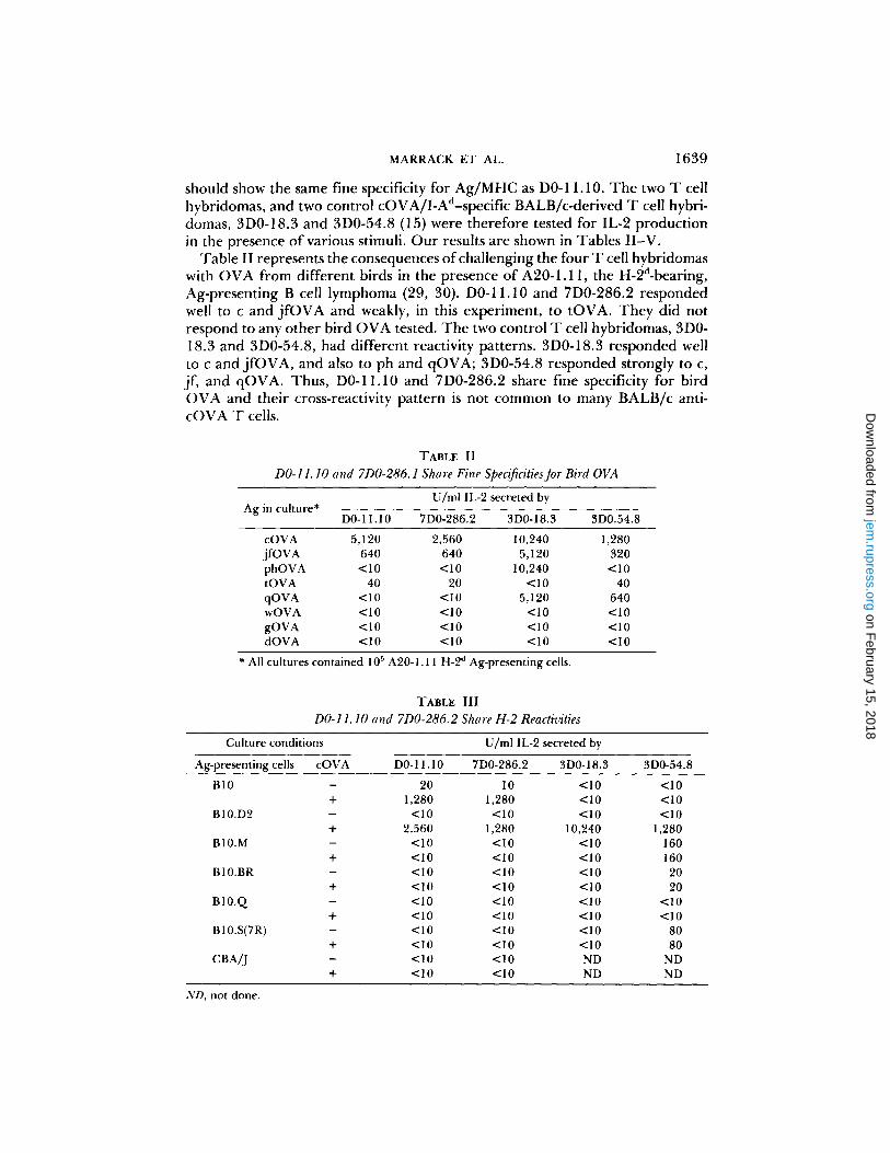
Table II represents the consequences of challenging the four T cell hybridomas with OVA from different birds in the presence of A20-1.11, the H-2d-bearing, Ag-presenting B cell lymphoma (29, 30). D0-11.10 and 7D0-286.2 responded well to c and j fOVA and weakly, in this experiment, to tOVA. They did not respond to any other bird OVA tested. The two control T cell hybridomas, 3D0- 18.3 and 3D0-54.8, had different reactivity patterns. 3D0-18.3 responded well to c and jfOVA, and also to ph and qOVA; 3D0-54.8 responded strongly to c, if, and qOVA. Thus, D0-11.10 and 7D0-286.2 share fine specificity for bird OVA and their cross-reactivity pattern is not common to many BALB/c anti- cOVA T cells.
TABLE II
DO-11.10 and 7D0-286.1 Share Fine Specificities for Bird OVA
Ag in culture* U/ml IL-2 secreted by
D0-11.10 7D0-286.2 3D0-18.3 3D0.54.8
cOVA 5,120 2,560 10,240 1,280 i f OVA 640 640 5,120 320 phOVA <10 <10 10,240 <10 tOVA 40 20 <10 40 qOVA <10 <10 5,120 640 wOVA <10 <10 <10 <10 gOVA <10 <10 <10 <10 dOVA <10 <10 <10 < I 0
* All cultures contained 105 A20-1.11 H-2 d Ag-presenting cells.
TABLE III
DO-11.10 and 7D0-286.2 Share H-2 Reactivities
Culture conditions
Ag-presenting cells cOVA DO- 11.10
U/rnl IL-2 secreted by
7D0-286.2 3D0-18.3 3D0-54.8
B10 - 20 l0 <10 <10 + 1,280 1,280 <10 <10
B10.D2 - <10 <10 <10 <10 + 2,560 1,280 10,240 1,280
BI0.M - <10 < I 0 <10 160 + < I 0 <10 <10 160
B10.BR - <10 <10 <10 20 + <10 <10 <10 20
B10.Q - <10 <10 <10 <10 + <10 <10 <10 <10
B10.S(7R) - <10 <10 <10 80 + <10 <10 <10 80
CBA/J - <10 <10 ND ND + <10 <10 ND ND
ND, not done.
on February 15, 2018
jem.rupress.org
Dow
nloaded from
1640 AN ANTI-T CELL IDIOTYPE DEFINES ANTIGEN AND I SPECIFICITY
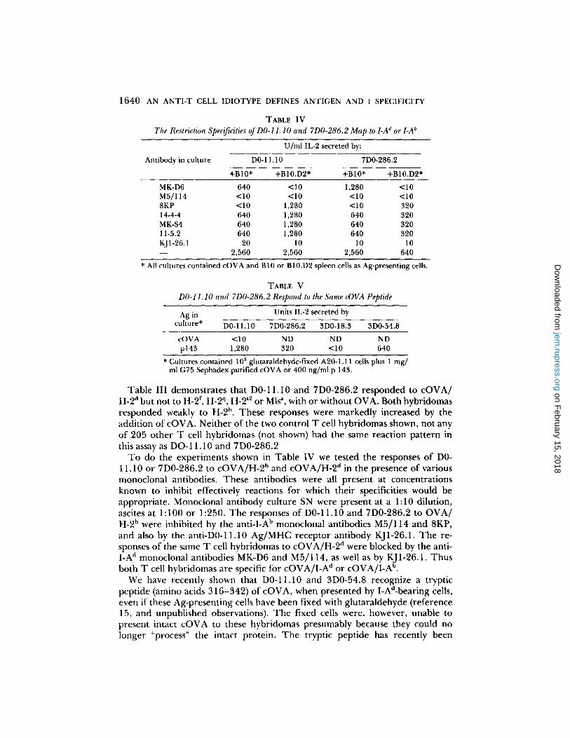
TABLE IV
The Restriction Specificities of DO-11.10 and 7D0-286.2 Map to LA d or LA b
U/ml IL-2 secreted by:
Antibody in culture DO- 11.10 7D0-286.2
+B10* +B10.D2* +B10* +B10.D2*
MK-D6 640 <10 1,280 <10 M5/114 <10 <10 <10 <10 8KP <10 1,280 <10 320 14-4-4 640 1,280 640 320 MK-S4 640 1,280 640 320 11-5.2 640 1,280 640 320 KJ1-26.1 20 10 10 10 - - 2,560 2,560 2,560 640
* All cultures contained cOVA and B10 or B10.D2 spleen cells as Ag-presenting cells.
TABLE V
D0-11.10 and 7D0-286.2 Respond to the Same cOVA Peptide
Ag in Units IL-2 secreted by
culture* D0-11.10 7D0-286.2 3D0-18.3 3D0-54.8
cOVA <10 ND ND ND p143 1,280 320 <10 640
* Cultures contained 105 glutaraldehyde-fixed A20-1.11 cells plus 1 mg/ ml G75 Sephadex purified cOVA or 400 ng/ml p 143.
Table III demonstrates that D0-11.10 and 7D0-286.2 responded to cOVA/ H-2 d but not to H-2 r, H-2 q, H-2 t2 or Mls a, with or without OVA. Both hybridomas responded weakly to H-2 b. These responses were markedly increased by the addition of cOVA. Neither of the two control T cell hybridomas shown, not any of 205 other T cell hybridomas (not shown) had the same reaction pattern in this assay as DO-11.10 and 7D0-286.2
To do the experiments shown in Table IV we tested the responses of D0- 11.10 or 7D0-286.2 to cOVA/H-2 b and cOVA/H-2 a in the presence of various monoclonal antibodies. These antibodies were all present at concentrations known to inhibit effectively reactions for which their specificities would be appropriate. Monocional antibody culture SN were present at a 1:10 dilution, ascites at 1:100 or 1:250. The responses of D0-11.10 and 7D0-286.2 to OVA/ H-2 b were inhibited by the anti-I-A b monoclonal antibodies M5/114 and 8KP, and also by the anti-D0-11.10 Ag/MHC receptor antibody KJ1-26.1. The re- sponses of the same T cell hybridomas to cOVA/H-2 d were blocked by the anti- I-A d monoclonal antibodies MK-D6 and M5/114, as well as by KJ1-26.1. Thus both T cell hybridomas are specific for cOVA/I-A d or cOVA/I-A b.
We have recently shown that D0-11.10 and 3D0-54.8 recognize a tryptic peptide (amino acids 316-342) of cOVA, when presented by I-Ad-bearing cells, even if these Ag-presenting cells have been fixed with glutaraldehyde (reference 15, and unpublished observations). The fixed cells were, however, unable to present intact cOVA to these hybridomas presumably because they could no longer "process" the intact protein. The tryptic peptide has recently been
on February 15, 2018
jem.rupress.org
Dow
nloaded from

MARRACK ET AL. 1641
synthesized for us (with the addition of a C terminal tyrosine) by Dr. Gary Matsueda, Massachusetts General Hospital, Boston, MA. The synthetic peptide is called p143. D0-11.10 and 3D0-54.8 respond well to p143, presented on live or fixed I-Ad-bearing cells. 7D0-286.2 was therefore tested for the same activity. As shown in Table V fixed A20-1.11 cells presented p143 to D0-11.10, 3D0- 54.8, and 7D0-286.2, but not to 3D0-18.3, which is known to recognize a different peptide derivative of cOVA (15). Thus, D0-11.10 and 7D0-286.2 recognize the same peptide fragment of cOVA.
7D0-286.2 Is Not a Reisolate of DO-ll. 10. The experiments described above show that the Ag and H-2 specificities of 7 D0-286.2 and DO- 11.10 are both rare, and indistinguishable. Since D0-11.10 is almost always being cultured by one member or another of our laboratory it was possible that 7D0-286.2 was not an independent T cell hybridoma, but simply DO- 11.10 that had accidently contam- inated the 7D0 fusion plates. One cell would have been sufficient to give us the results we have reported so far. Even though every precaution was taken to ensure that this would not occur, and even though contamination of this type has not previously been observed in our laboratories, we felt we should demon- strate directly that this had not happened.
It is our experience that T cell hybridomas, even though they are products of the fusion of a normal mouse T cell (containing 40 chromosomes) to the same tumor cell parent, BW5147, contain variable numbers of normal and abnormal mouse chromosomes (31). Most of the abnormal chromosomes seem to be derived from BW5147, which contains a number of characteristic chromosomes of this type (32). Others appear to form soon after the fusion of the normal T cell parent and BW5147, by fusion or rearrangement of chromosomes in the hybrid. The creation of the abnormal chromosomes seems to be an early rather than a late event in the life history of hybridomas, since once the hybridoma has been cloned marker chromosomes remain constant. Subcloning the hybridoma may lead to the production of subclones that have lost particular abnormal chromo- somes, but no new ones appear (unpublished observations).
D0-11.10 was derived from a female BALB/c mouse; BW5147 does not contain a Y chromosome; the normal T cell parent of 7D0-286.2, on the other hand, was male. We therefore karyotyped D0-11.10 and 7D0-286.2 with the hope that the presence or absence of a Y chromosome, as well as clone-specific distribution of chromosomes would allow us to distinguish unequivocally the two hybridomas. Sample spreads are shown in Fig. 1.
Even in these cloned T cell hybridomas, chromosome numbers did vary somewhat from spread to spread. Nevertheless, D0-11.10 and 7D0-286.2 did differ chracteristically. For example all spreads of 7D0-286.2 contained a Y chromosome (in Fig. 1 an arrow points to this chromosome); this was never observed in a karyotype of D0-11.10. 7D0-286.2 cells consistently contained 2 chromosomes 4, 2 or 3 chromosomes 12, and one or zero chromosomes 18. D0- 11.10 spreads always contained one normal chromosome 4, 4 normal chromo- somes 12, and 2 or 3 chromosomes 18. These data demonstrate unequivocally that 7D0.286.2 is not a reisolate of D0-11.10.
Immunoprecipitation of 7D0-286.2. D0-11.10 and 7D0-286.2 were surface la- beled with 125I and lysed, and immunoprecipitates prepared with either KJ1-26.1
on February 15, 2018
jem.rupress.org
Dow
nloaded from

1642 AN ANT1-T CELL IDIOTYPE DEFINES ANTIGEN AND I SPECIFICITY
FICURE 1. Chromosomes of D0-11.10 and 7D0-286.2. D0-11.10 and 7D0-286.2 were pre- pared for karyotyping as described in Materials and Methods. An arrow points to the Y chromosome in 7D0-286.2.
FIGURE 2. lmmunoprecipitation of D0-11.10 or 7D0-286.2 by KJ1-26.1 and KJ12-98.15. Cell lysates from J 25I-surface-labeled T cell hybridomas were incubated with KJ 1-26.1 or KJ 12- 98.12 coupled to sepharose beads. Samples were analyzed by SDS-PAGE on 10% gels with or without reduction by 2-mercaptoethanol.
o r KJ12-98 .15 . Samp le s we re r u n n o n r e d u c e d o r r e d u c e d on S D S - P A G E . As shown in Fig. 2 a n d p r e v i o u s l y (5), KJ1-26.1 p r e c i p i t a t e d an 8 5 - k d a l t o n m o l e c u l e f r o m D 0 - 1 1 . 1 0 tha t a f t e r r e d u c t i o n r a n as a d i f fuse b a n d o f ~ 4 3 kda l tons . KJ1- 26.1 p r e c i p i t a t e d a s imi la r m o l e c u l e f r o m 7D0-286 .2 . KJ12 -98 .15 d i d no t p re - c ip i t a t e any d e t e c t a b l e m a t e r i a l f r o m 7D0-286 .2 . T h e r e f o r e , t he gross b io log ica l
on February 15, 2018
jem.rupress.org
Dow
nloaded from
MARRACK ET AL. 1 6 4 3
properties of the Ag/MHC receptor on D0-11.10 and 7D0-286.2 appear to be similar.
Discussion In this paper we show that a T cell hybridoma, 7D0-286.2, selected because it
binds an antibody, KJ1-26.1, which is thought to bind to all or part of the Ag/ MHC receptor on a different T cell hybridoma, D0-11.10, has the same specificity both for Ag and H-2 as DO-11.10. Both T cell hybridomas respond strongly to cOVA/I-A d, j fOVA/I-A d, and cOVA/I-A b, and weakly to tOVA/I-A d and I-A b alone.
These fine specificities are relatively rare amongst T cell hybrids prepared from BALB/c T cell blasts specific for cOVA, in fact others with this pattern have not been seen amongst 207 tested. In addition none other of 397 inde- pendent T cell hybridomas tested reacted specifically with the anti-receptor antibody KJ1-26.1.
From these results we would like to draw several conclusions. First, and most importantly, we feel that these results show unequivocally that KJ1-26.1 binds to all or part of the receptor for cOVA/I-A d on D0-11.10 and 7D0-286.2, since the presence of the idiotype predicted exactly the specificity of the T cells. Prior to this discovery the evidence that antibodies of this type reacted with Ag/MHC receptors on T ceils rested on the facts that they are clone-specific, that they block responses against and binding to Ag/MHC and that they do not bind to subclones of T cell hybridomas thought to have lost the ability to put Ag/MHC receptors on their surfaces (1-8, 14). Of course, in the case of the long-sought- for T cell Ag/MHC receptors, the more evidence that can be mustered to support the candidacy of a particular molecule the better; a demonstration that the suspect binds to Ag/MHC, for example, would be most welcome. Unfortu- nately, our preliminary attempts to demonstrate this have been unsuccessful, perhaps because of obligatory requirements for other molecules, LFA-1, L3T4, or Lyt-2 (9-13). In lieu of such data we feel that our demonstration that idiotype determines specificity is a useful addition to the literature on this subject.
The fact that reaction with KJ1-26.1 predicted the fine specificity of 7D0- 286.2 both for Ag and MHC places certain restrictions on models for how Ag/ MHC recognition occurs. Certainly, it is compatible with a single receptor model (33), as is the fact that KJ1-26.1, and other antibodies like it, have been shown to precipitate 80-90-kdalton disulphide-bonded heterodimers from cell surfaces which, on reduction yield two polypeptide chains, both of variable sequence of mol wt 39-47 kdaltons (4-7, 14). Moreover, recently a number of other publi- cations have also suggested that a single receptor model may describe the structure of the T cell receptor and its ligands (16, 34, 35). To allow the data in this paper to be compatible with two receptor models (36), we have to postulate that the structure of the Ag and MHC receptors are not independent: one might be derived from the other (37), or they might be constructed from different parts of the same polypeptide chains (38). Constraints of this type have, of course, already been imposed on two receptor theories by the existence of antigen- specific, MHC-iinked Ir genes.
A second point worth noting is that the idiotype recognized by KJ 1-26.1 occurs
on February 15, 2018
jem.rupress.org
Dow
nloaded from

1644 AN ANTI-T CELL IDIOTYPE DEFINES ANTIGEN AND I SPECIFICITY
relatively infrequently. Only 1 of about 400 T cell hybridomas was found to bear it, even when the hybridomas were derived from T cells with the same gross Ag/MHC specificity as the original target of KJ1-26.1. Although it is impossible to generalize from this single example, the result is consistent with the idea that the variability of T cell Ag/MHC receptors is relatively large, at least as large as that of B cells.
Summary In order to prove that a monoclonal antibody, KJ1-26.1, reacted with the
idiotypic portion of the receptor for antigen plus major histocompatibility com- plex product (Ag/MHC) on the T cell hybridoma D0-11.10,397 independent T cell hybridomas prepared from BALB/c T cells primed with ovalbumin were screened by ELISA for reactivity with the antibody. Of these T cell hybridomas one, 7D0-286, and a subclone of this hybridoma, 7D0-286.2, reacted strongly and consistently with KJ 1-26.1. Further analysis revealed that KJ 1-26.1 blocked the reaction of 7D0-286 with Ag/MHC, and that 7D0-286 had the same fine specificity both for Ag and MHC as D0-11.10. None of 207 other BALB/c T cell hybridomas specific for ovalbumin shared these fine specificity patterns.
Thus, reaction with KJ1-26.1 defined the specificity of a T cell hybridoma both for Ag and MHC, suggesting that KJ1-26.1 reacted with the receptors for Ag/MHC on the T cells in question.
KJ1-26.1 precipitated an 85-kdalton molecule from 7D0-286.2 that on reduc- tion ran as a diffuse spot of ~ 4 3 kdaitons. A similar molecule was precipitated from D0-11.10.
The authors would like to thank Dr. Andrei Augustin for advice, Janice White, James Leibson, and Eleanora Kushnir for excellent technical assistance, and Edna Squiilante for preparing this manuscript. Two of them (P. M. and J. K.) would also like to thank Dr. Mel Cohn of The Salk Institute for space in his laboratory whilst they were trying to learn to karyotype mouse chromosomes.
Received for publication 25July 1983.
References 1. Infante, A.J., P. D. Infante, S. Gillis, and C. G. Fathman. 1982. Definition o fT cell
idiotypes using antiidiotype antisera produced by immunization with T cell clones. J. Exp. Med. 155:1100.
2. Thomas, W. R., P. L. Moltram, and J. F. A. P. Miller. 1982. Anti-T cell idiotype activity in serum of mice injected with syngeneic hapten-specific T-cell lines. Proc. Natl. Acad. Sci. USA. 79:6671.
3. White, J., K. M. Haskins, P. Marrack, and J. Kappler. 1983. Use of I-region restricted, antigen-specific T cell hybridomas to produce idiotypically specific anti-receptor antibodies. J. Immunol. 130:1033.
4. Meuer, S. C., K. A. Fitzgerald, R. E. Hussey, J. C. Hodgdon, S. F. Schlossman, and E. L. Reinherz. 1983. Clonotypic structures involved in antigen-specific human T cell function. Relationship to the T3 molecular complex. J. Exp. Med. 157:705.
5. Haskins, K., R. Kubo, J. White, M. Pigeon, J. Kappler, and P. Marrack. 1983. The major histocompatibility complex-restricted antigen receptor on T cells. I. Isolation with a monoclonal antibody.J. Exp. Med. 157:1149.
on February 15, 2018
jem.rupress.org
Dow
nloaded from
MARRACK ET AL. 1645
6. Meuer, S. C., O. Acuto, R. E. Hussey, J. C. Hodgdon, K. A. Fitzgerald, S. F. Schiossman, and E. L. Reinherz. 1983. Evidence for the T3-associated 90 K hetero- dimer as the T cell antigen receptor. Nature (Lond.). 303:808.
7. Kappler, J., R. Kubo, K. Haskins, J. White, and P. Marrack. The murine T cell receptor. Comparison of major histocompatibility complex-restricted receptors from two different T cell hybridomas. Cell. In press.
8. Lancki, D. W., M. I. Lorber, M. R. Loken, and F. W. Fitch. A clone-specific monoclonal antibody that inhibits cytolysis of a cytolytic T cell clone. J. Exp. ivied. 157:921.
9. Kurzinger, K., T. Reynolds, R. N. Germain, D. Davignon, E. Martz, and T. A. Springer. 1981. A novel lymphocyte function-associated antigen (LFA-1): cellular distribution, quantitative expression, and structure.J. Immunol. 127:596.
10. Meuer, S. C., R. E. Hussey, J. C. Hodgdon, T. Herend, S. F. Schlossman, and E. L. Reinherz. 1982. Surface structures involved in target recognition by human cytotoxic T lymphocytes. Science (Wash. DC). 218:471.
11. Engleman, E. G., C. G. Benike, E. Glickman, and R. L. Evans. 1981. Antibodies to membrane structures that distinguish suppressor/cytotoxic and helper T lymphocyte subpopulations block the mixed lymphocyte reaction in man.J. Exp. Med. 154:193.
12. Sarmiento, M., D. P. Dialynas, D. W. Lancki, K. A. Wall, M. I. Lorber, M. R. Loken, and F. W. Fitch. 1982. Cloned T lymphocytes and monoclonal antibodies as probes for cell surface molecules active in T cell-mediated cytolysis. Immunol. Rev. 68:135.
13. Nakayama, E., H. Shiku, E. Stockert, H. F. Oettgen, and L. H. Old. 1979. Cytotoxic T cells: Lyt phenotype and blocking of killing activity by Lyt antisera. Proc. Natl. Acad. Sci. USA. 76:1977.
14. Allison, J. P., B. W. Mclntyre, and D. Bloch. 1982. Tumor-specific antigen and murine T-lymphoma defined with monoclonal antibody.J. Immunol. 129:2293.
15. Shimonkevitz, R., J. Kappler, P. Marrack, and H. Grey. 1982. Antigen recognition by H-2-restricted T cells. I. Cell-free antigen processing. J. Exp. Med. 157:303.
16. Kappler, J. W., B. Skidmore, J. White, and P. Marrack. 1981. Antigen-inducible, H- 2-restricted interleukin-2 producing T cell hybridomas. Lack of independent antigen and H-2 recognition. J. Exp. Med. 153:1198.
17. Endres, R. O., P. Marrack, and J. Kappler. 1983. An IL-2 secreting T hybridoma that responds to a self-class I histocompatibility antigen in the H-2D region. J. Immunol. In press.
18. Dennert, G., R. Hyman, J. Leslie, and I. S. Trowbridge. 1980. Effects of cytotoxic monoclonal antibodies specific for T200 glycoproteins on functional lymphoid cell populations. Cell. hnmunol. 53:350.
19. Bhattacharya, A., M. E. Dorf, and T. A. Springer. 1981. A shared alloantigenic determinant on Ia antigens encoded by the I-A and I-A subregions: evidence for I- region duplication.J. Immunol. 127:2488.
20. Conrad, P.J., E. A. Lerner, D. B. Murphy, P. P.Jones, and C. A.Janeway,Jr. 1982. Differential expression of Ia glycoprotein complexes in F~ hybrid mice detected with alloreactive cloned T cell lines. J. Immunol. 129:2616.
21. Ozato, K., N. Mayer, and D. H. Sachs. 1980. Hybridoma cell lines secreting antibodies to mouse and Ia antigens.J. Immunol. 124:533.
22. Oi, v. T., P. P.Jones,J. W. Goding, L. A. Herzenberg, and L. A. Herzenberg. 1978. Properties of monoclonal antibodies to mouse Ig allotypes, H-2 and Ia antigens. Curt. Top. Microbiol. hnmunol. 81 : 115.
23. Francke, U., and N. Oliver. 1978. Quantitative analysis of high-resolution trypsin- Giemsa bands of human prometaphase chromsomes. Hum. Genet. 45:137.
24. Nesbitt, M. N., and U. Francke. 1973. A system of nomenclature for band patterns
on February 15, 2018
jem.rupress.org
Dow
nloaded from
1646 AN ANTI-T CELL IDIOTYPE DEFINES ANTIGEN AND I SPECIFICITY
of mouse chromosomes. Chromosoma. 41:145. 25. Marrack, P., R. Endres, R. Shimonkevitz, A. Zlomik, D. Dialynas, F. Fitch, and J.
Kappler. The major histocompatibility complex-restricted antigen receptor on T cells. II. Role of the L3T4 product.J. Exp. Med. 158:1077.
26. Sigma Chemical Company catalogue. 1983. St. Louis, MO. p. 438. 27. Pharmacia Fine Chemical Company. 1976. CNBr-activated Sepharose 6MB. User's
Booklet. Piscataway, NJ. 28. Laemmli, U. K. 1970. Cleavage of structural proteins during the assembly of the
head of bacteriophage T4. Nature (Lond.). 227:680. 29. Kim, K.J., C. Kanellopoulos-Langevin, R. M. Merwin, D. H. Sachs, and R. Asofsky.
1979. Establishment and characterization of BALB/c lymphoma lines with B cell properties. J. hnmunol. 122:549.
30. Walker, E., N. L. Warner, R. Chesnut, J. Kappler, and P. Marrack. 1982. Antigen- specific, I-region-restricted interactions in vitro between tumor cell lines and T cell hybridomas. J. hnmunol. 128:2164. Marrack, P., and J. W. Kappler. 1983. Use of somatic cell genetics to study chromo- somes contributing to antigen plus I recognition by T cell hybridomas. J. Exp. Med. 157:404. Robertson, S. M., V. G. Dev, D. Jeske, A. Pacifico, J. R. Kettman, and J. D. Capra. 1981. Characterization of T cell hybrids producing proteins binding the arsonate hapten. In Lymphokine Reports. M. Feldmann and M. Schreier, editors. Academic Press, Inc., New York. p. 26. Zinkernagel, R. M., and P. C. Doherty. 1977. Major transplantation antigens, virus and specificity of surveillance T cells. The "altered-selF' hypothesis. Contemp. Top. hnmunobiol. 7:179. Hunig, T. R., and M. J. Bevan. 1982. Antigen recognition by cloned cytotoxic T lymphocytes follows rules predicted by the altered self hypothesis. J. Exp. Med. 155:111. Hedrick, S. M., L. A. Matis, T. T. Hecht, L. E. Samelson, D. L. Longo, E. Heber- Katz, and R. H. Schwartz. 1982. The fine specificity of antigen and Ia determinant recognition by T cell hybridoma clones specific for pigeon cytochrome c. Cell. 30:141. Blanden, R. V., and G. L. Ada. 1978. A dual recognition model for cytotoxic T cells based on thymic selection of precursors with low affinity for self-H-2 antigens. Scand. J. Immunol. 7:181. von Boehmer, H., W. Haas, and N. K.Jerne. 1978. Major histocompatibility complex- linked immune-responsiveness is acquired by lymphocytes of low responder mice differentiating in thymus of high responder mice. Proc. Natl. Acad. Sci. USA. 75:2439. Langman, R. E. 1978. The role of the major histocompatibility complex in immunity: a new concept in the functioning of a cell-mediated immune system. Rev. Physiol. Biochem. Pharmacol. 81:1.
31.
32.
33.
34.
35.
36.
37.
38.
on February 15, 2018
jem.rupress.org
Dow
nloaded from